Embed Size (px)
Citation preview
LETTERdoi:10.1038/nature13315
A single female-specific piRNA is the primarydeterminer of sex in the silkwormTakashi Kiuchi1, Hikaru Koga1*, Munetaka Kawamoto1*, Keisuke Shoji1*, Hiroki Sakai2, Yuji Arai1, Genki Ishihara1,Shinpei Kawaoka1, Sumio Sugano3, Toru Shimada1, Yutaka Suzuki3, Masataka G. Suzuki2 & Susumu Katsuma1
The silkworm Bombyx mori uses a WZ sex determination systemthat is analogous to the one found in birds and some reptiles. In thissystem, males have two Z sex chromosomes, whereas females have Zand W sex chromosomes. The silkworm W chromosome has a dom-inant role in female determination1,2, suggesting the existence of adominant feminizing gene in this chromosome. However, the W chro-mosome is almost fully occupied by transposable element sequences3–5,and no functional protein-coding gene has been identified so far.Female-enriched PIWI-interacting RNAs (piRNAs) are the only knowntranscripts that are produced from the sex-determining region ofthe W chromosome6, but the function(s) of these piRNAs are unknown.Here we show that a W-chromosome-derived, female-specific piRNAis the feminizing factor of B. mori. This piRNA is produced from apiRNA precursor which we named Fem. Fem sequences were arrangedin tandem in the sex-determining region of the W chromosome. Inhi-bition of Fem-derived piRNA-mediated signalling in female embryosled to the production of the male-specific splice variants of B. moridoublesex (Bmdsx), a gene which acts at the downstream end of thesex differentiation cascade7,8. A target gene of Fem-derived piRNAwas identified on the Z chromosome of B. mori. This gene, which wenamed Masc, encoded a CCCH-type zinc finger protein. We showthat the silencing of Masc messenger RNA by Fem piRNA is requiredfor the production of female-specific isoforms of Bmdsx in femaleembryos, and that Masc protein controls both dosage compensationand masculinization in male embryos. Our study characterizes asingle small RNA that is responsible for primary sex determinationin the WZ sex determination system.
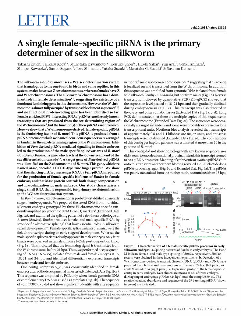
In Bombyx mori, sex determination is probably established at an earlystage of embryogenesis. We prepared the sexed RNA from individualsilkworm embryos genotyped by three W chromosome-specific ran-domly amplified polymorphic DNA (RAPD) markers4 (Extended DataFig. 1a), and examined the splicing pattern of a doublesex orthologue ofB. mori (Bmdsx). Bmdsx produces female- and male-specific RNAs bysex-specific alternative splicing9 that have essential roles in silkwormsexual development7,8. Female-specific splice variants of Bmdsx were thedefault transcripts during an early stage of development. Whereas themale-specific splice variants clearly appeared in male embryos, only faintbands were observed in females, from 21–24 h post-oviposition (hpo)(Fig. 1a). This indicated that the feminizing signal is transmitted fromthe W chromosome before 21 hpo. Thus, we performed deep sequenc-ing of RNAs (RNA-seq) isolated from male and female embryos at 15,18, 21 and 24 hpo, and identified differentially expressed transcriptsbetween male and female embryos.
One contig, comp73859_c0, was consistently identified in femaleembryos at all of the developmental times tested (Extended Data Fig. 1b, c).This sequence was amplified by PCR only when female genomic DNAor complementary DNA was used as a template (Fig. 1b). The sequenceof comp73859_c0 did not show significant identity with any sequence
in the draft male silkworm genome sequence10, suggesting that this contigis localized on and transcribed from the W chromosome. In addition,this sequence was amplified from genomic DNA isolated from femalewild silkmoth Bombyx mandarina, but not from males (Fig. 1b). Reversetranscription followed by quantitative PCR (RT–qPCR) showed thatthe expression level peaked at 18–21 hpo, and then gradually declinedduring embryogenesis (Fig. 1c). This transcript was also detected inthe ovary and other somatic tissues (Extended Data Fig. 2a, b, d). LongPCR demonstrated that there are multiple copies of this sequence onthe W chromosome (Extended Data Fig. 2c). The sequences were occa-sionally arranged in tandem and some were probably expressed as longtranscriptional units. Northern blot analysis revealed that transcriptsof approximately 0.8 and 1.4 kilobase are major units, and antisensetranscripts were not detected (Extended Data Fig. 2d). The copy numberof this contig per haploid genome was estimated at more than 30 in thegenome of B. mori.
This contig did not show homology with any known sequence, nordid it seem to encode a functional protein. Instead, this transcript seemedto be a piRNA precursor. Mapping of embryonic or ovarian piRNAs6,11,12
onto this transcript and northern blotting revealed a 29-nucleotide-longpiRNA-producing region (Fig. 1d and Extended Data Fig. 3a). This piRNAwas poorly transmitted from the mother moth, accumulated from 15 hpo,
*These authors contributed equally to this work.
1Department of Agricultural and Environmental Biology, Graduate School of Agricultural and Life Sciences, The University of Tokyo, 1-1-1 Yayoi, Bunkyo-ku, Tokyo 113-8657, Japan. 2Department ofIntegrated Biosciences, Graduate School of Frontier Sciences, The University of Tokyo, 5-1-5 Kashiwanoha, Kashiwa, Chiba 277-8562, Japan. 3Department of Medical Genome Sciences, Graduate School ofFrontier Sciences, The University of Tokyo, 4-6-1 Shirokanedai, Minato-ku, Tokyo 108-8639, Japan.
]
]
F
M
a
gDNA cDNA
b
c d
comp73859_c0
1.0
0.5
kb
1F2R
B. mori B. mandarina
(hpo)1.06
0.50
kb
767 bases
gDNA
0
0.5
1.0
1.5
2.0
2.5
3.0
15 18 21 24 27 15 18 21 24 27
mR
NA
level
(co
ntig
/rp
49)
hpo
0
Sense
piRNA
Read
s p
er
mill
ion
200
100
0
668 bp
UACCUCUUUUUGUCAAUUCAUAAAGUCAU
Antisense
15 18 21 24 27
Figure 1 | Characterization of a female-specific piRNA precursor in earlysilkworm embryos. a, Splicing patterns of Bmdsx in early embryos. The F andM indicate female- and male-type splicing of Bmdsx, respectively. Similarresults were obtained in three independent experiments. b, Detection of aW chromosome-derived transcript. Genomic DNA (gDNA) and cDNA wereprepared from female and male embryos of B. mori at 24 hpo (left panel) oradult B. mandarina (right panel). c, Expression profile of the female-specificcontig in early embryos. Data shown are means 6 s.d. of three embryos.d, Mapping of embryonic piRNAs (24 hpo) onto the comp73859_c0. Therelative location, abundance and sequence of the 29 base-long piRNA (shownin green) are indicated.
0 0 M O N T H 2 0 1 4 | V O L 0 0 0 | N A T U R E | 1
Macmillan Publishers Limited. All rights reserved©2014
and increased rapidly between 18 and 21 hpo (Extended Data Fig. 3b, c).By screening piRNA libraries that were generated from three B. moristrains that each possess a unique W chromosome structure6, we foundthat this piRNA was produced from the sex-determining region of Wchromosome (Extended Data Fig. 3d).
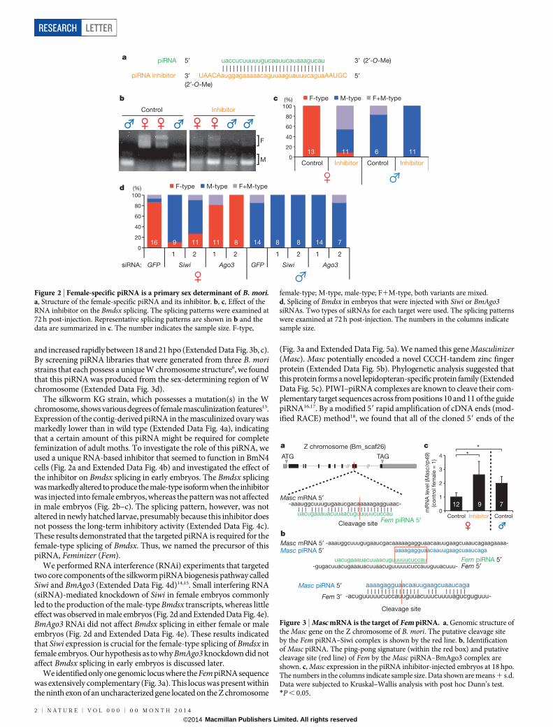
The silkworm KG strain, which possesses a mutation(s) in the Wchromosome, shows various degrees of female masculinization features13.Expression of the contig-derived piRNA in the masculinized ovary wasmarkedly lower than in wild type (Extended Data Fig. 4a), indicatingthat a certain amount of this piRNA might be required for completefeminization of adult moths. To investigate the role of this piRNA, weused a unique RNA-based inhibitor that seemed to function in BmN4cells (Fig. 2a and Extended Data Fig. 4b) and investigated the effect ofthe inhibitor on Bmdsx splicing in early embryos. The Bmdsx splicingwas markedly altered to produce the male-type isoform when the inhibitorwas injected into female embryos, whereas the pattern was not affectedin male embryos (Fig. 2b–c). The splicing pattern, however, was notaltered in newly hatched larvae, presumably because this inhibitor doesnot possess the long-term inhibitory activity (Extended Data Fig. 4c).These results demonstrated that the targeted piRNA is required for thefemale-type splicing of Bmdsx. Thus, we named the precursor of thispiRNA, Feminizer (Fem).
We performed RNA interference (RNAi) experiments that targetedtwo core components of the silkworm piRNA biogenesis pathway calledSiwi and BmAgo3 (Extended Data Fig. 4d)14,15. Small interfering RNA(siRNA)-mediated knockdown of Siwi in female embryos commonlyled to the production of the male-type Bmdsx transcripts, whereas littleeffect was observed in male embryos (Fig. 2d and Extended Data Fig. 4e).BmAgo3 RNAi did not affect Bmdsx splicing in either female or maleembryos (Fig. 2d and Extended Data Fig. 4e). These results indicatedthat Siwi expression is crucial for the female-type splicing of Bmdsx infemale embryos. Our hypothesis as to why BmAgo3 knockdown did notaffect Bmdsx splicing in early embryos is discussed later.
We identified only one genomic locus where the Fem piRNA sequencewas extensively complementary (Fig. 3a). This locus was present withinthe ninth exon of an uncharacterized gene located on the Z chromosome
(Fig. 3a and Extended Data Fig. 5a). We named this gene Masculinizer(Masc). Masc potentially encoded a novel CCCH-tandem zinc fingerprotein (Extended Data Fig. 5b). Phylogenetic analysis suggested thatthis protein forms a novel lepidopteran-specific protein family (ExtendedData Fig. 5c). PIWI–piRNA complexes are known to cleave their com-plementary target sequences across from positions 10 and 11 of the guidepiRNA16,17. By a modified 59 rapid amplification of cDNA ends (mod-ified RACE) method18, we found that all of the cloned 59 ends of the
a piRNA 5′
UAACAauggagaaaaacaguuaaguauuucaguaAAUGC
uaccucuuuuugucaauucauaaagucau
3′
3′
5′(2′-O-Me)
(2′-O-Me)
piRNA inhibitor
d
0
20
40
60
80
100
(%)
GFP siRNA: Siwi Ago3 GFP Siwi Ago3
InhibitorControl
InhibitorControl InhibitorControl
]
]
F
M 0
20
40
60
80
100
F-type M-type F+M-type
F-type M-type F+M-type
(%) c
13
16 11 11 8 89 8 714 14
11 6 11
b
1 2 1 2 1 2 1 2
Figure 2 | Female-specific piRNA is a primary sex determinant of B. mori.a, Structure of the female-specific piRNA and its inhibitor. b, c, Effect of theRNA inhibitor on the Bmdsx splicing. The splicing patterns were examined at72 h post-injection. Representative splicing patterns are shown in b and thedata are summarized in c. The number indicates the sample size. F-type,
female-type; M-type, male-type; F1M-type, both variants are mixed.d, Splicing of Bmdsx in embryos that were injected with Siwi or BmAgo3siRNAs. Two types of siRNAs for each target were used. The splicing patternswere examined at 72 h post-injection. The numbers in the columns indicatesample size.
a
b -aaauggcuuugugaaucgacaaaaagagguaacaauugaagcuaaucagaagaaaa-
aaaagagguaacaauugaagcuaaucaga
uacugaaauacuuaacuguuuuucuccau-gugacuuacugaaauacuuaacuguuuuucuccauuguuacuuu-
aaaagagguaacaauugaagcuaaucaga
-aaauggcuuugugaaucgacaaaaagagguaac-
uacugaaauacuuaacuguuuuucuccauFem piRNA 5′
Fem piRNA 5′Fem 5′
Masc piRNA 5′
Masc piRNA 5′Masc mRNA 5′
Fem 3′
Masc mRNA 5′
-acuguuuuucuccauuguuacuuucuuuuagucguguuu-
Cleavage site
Cleavage site
Z chromosome (Bm_scaf26)
ATG TAG
0
1
2
3
4
mR
NA
level (M
asc/
rp49
)
(co
ntr
ol fe
male
= 1
)
Control Inhibitor Control
12 9 7
c *
*
Figure 3 | Masc mRNA is the target of Fem piRNA. a, Genomic structure ofthe Masc gene on the Z chromosome of B. mori. The putative cleavage siteby the Fem piRNA–Siwi complex is shown by the red line. b, Identificationof Masc piRNA. The ping-pong signature (within the red box) and putativecleavage site (red line) of Fem by the Masc piRNA–BmAgo3 complex areshown. c, Masc expression in the piRNA inhibitor-injected embryos at 18 hpo.The numbers in the columns indicate sample size. Data shown are means 1 s.d.Data were subjected to Kruskal–Wallis analysis with post hoc Dunn’s test.*P , 0.05.
RESEARCH LETTER
2 | N A T U R E | V O L 0 0 0 | 0 0 M O N T H 2 0 1 4
Macmillan Publishers Limited. All rights reserved©2014
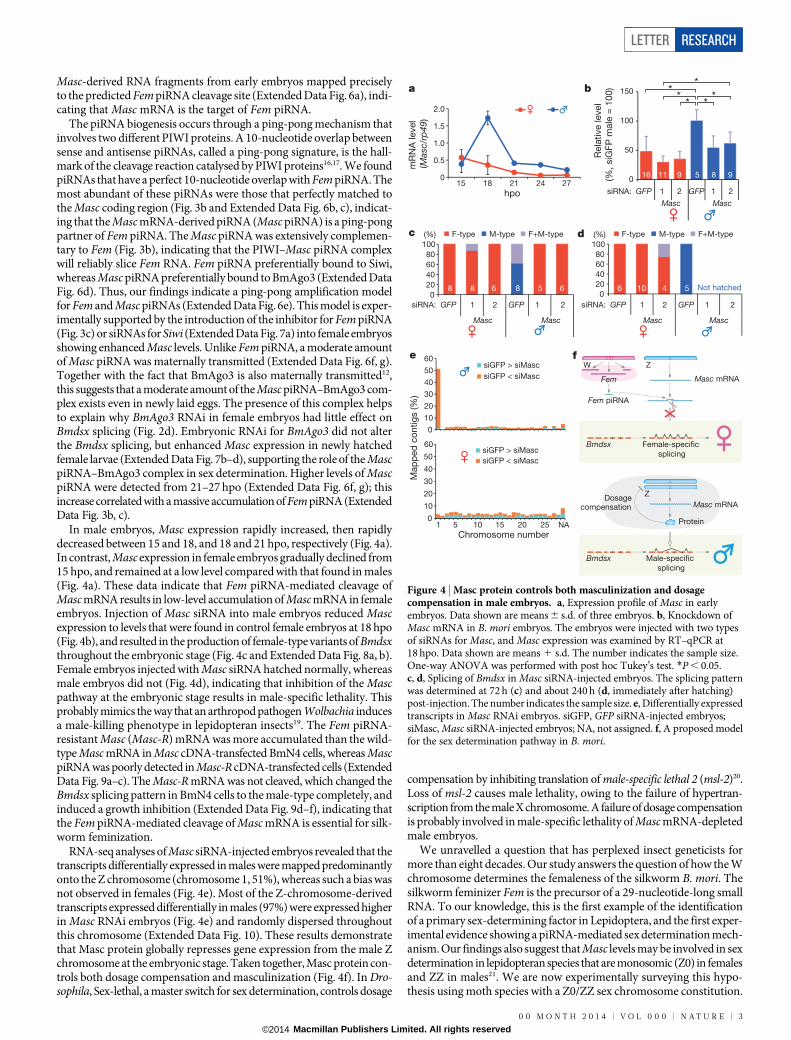
Masc-derived RNA fragments from early embryos mapped preciselyto the predicted Fem piRNA cleavage site (Extended Data Fig. 6a), indi-cating that Masc mRNA is the target of Fem piRNA.
The piRNA biogenesis occurs through a ping-pong mechanism thatinvolves two different PIWI proteins. A 10-nucleotide overlap betweensense and antisense piRNAs, called a ping-pong signature, is the hall-mark of the cleavage reaction catalysed by PIWI proteins16,17. We foundpiRNAs that have a perfect 10-nucleotide overlap with Fem piRNA. Themost abundant of these piRNAs were those that perfectly matched tothe Masc coding region (Fig. 3b and Extended Data Fig. 6b, c), indicat-ing that the Masc mRNA-derived piRNA (Masc piRNA) is a ping-pongpartner of Fem piRNA. The Masc piRNA was extensively complemen-tary to Fem (Fig. 3b), indicating that the PIWI–Masc piRNA complexwill reliably slice Fem RNA. Fem piRNA preferentially bound to Siwi,whereas Masc piRNA preferentially bound to BmAgo3 (Extended DataFig. 6d). Thus, our findings indicate a ping-pong amplification modelfor Fem and Masc piRNAs (Extended Data Fig. 6e). This model is exper-imentally supported by the introduction of the inhibitor for Fem piRNA(Fig. 3c) or siRNAs for Siwi (Extended Data Fig. 7a) into female embryosshowing enhanced Masc levels. Unlike Fem piRNA, a moderate amountof Masc piRNA was maternally transmitted (Extended Data Fig. 6f, g).Together with the fact that BmAgo3 is also maternally transmitted12,this suggests that a moderate amount of the Masc piRNA–BmAgo3 com-plex exists even in newly laid eggs. The presence of this complex helpsto explain why BmAgo3 RNAi in female embryos had little effect onBmdsx splicing (Fig. 2d). Embryonic RNAi for BmAgo3 did not alterthe Bmdsx splicing, but enhanced Masc expression in newly hatchedfemale larvae (Extended Data Fig. 7b–d), supporting the role of the MascpiRNA–BmAgo3 complex in sex determination. Higher levels of MascpiRNA were detected from 21–27 hpo (Extended Data Fig. 6f, g); thisincrease correlated with a massive accumulation of Fem piRNA (ExtendedData Fig. 3b, c).
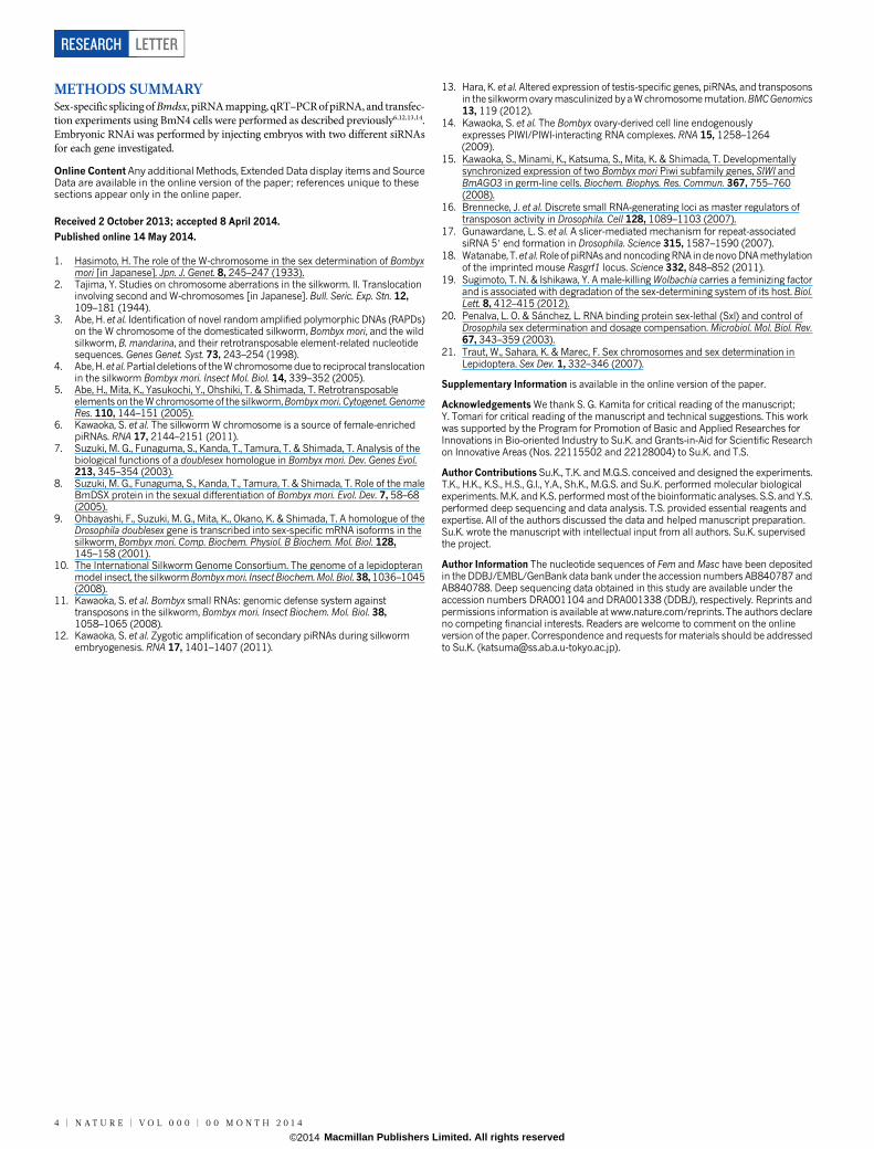
In male embryos, Masc expression rapidly increased, then rapidlydecreased between 15 and 18, and 18 and 21 hpo, respectively (Fig. 4a).In contrast, Masc expression in female embryos gradually declined from15 hpo, and remained at a low level compared with that found in males(Fig. 4a). These data indicate that Fem piRNA-mediated cleavage ofMasc mRNA results in low-level accumulation of Masc mRNA in femaleembryos. Injection of Masc siRNA into male embryos reduced Mascexpression to levels that were found in control female embryos at 18 hpo(Fig. 4b), and resulted in the production of female-type variants of Bmdsxthroughout the embryonic stage (Fig. 4c and Extended Data Fig. 8a, b).Female embryos injected with Masc siRNA hatched normally, whereasmale embryos did not (Fig. 4d), indicating that inhibition of the Mascpathway at the embryonic stage results in male-specific lethality. Thisprobably mimics the way that an arthropod pathogen Wolbachia inducesa male-killing phenotype in lepidopteran insects19. The Fem piRNA-resistant Masc (Masc-R) mRNA was more accumulated than the wild-type Masc mRNA in Masc cDNA-transfected BmN4 cells, whereas MascpiRNA was poorly detected in Masc-R cDNA-transfected cells (ExtendedData Fig. 9a–c). The Masc-R mRNA was not cleaved, which changed theBmdsx splicing pattern in BmN4 cells to the male-type completely, andinduced a growth inhibition (Extended Data Fig. 9d–f), indicating thatthe Fem piRNA-mediated cleavage of Masc mRNA is essential for silk-worm feminization.
RNA-seq analyses of Masc siRNA-injected embryos revealed that thetranscripts differentially expressed in males were mapped predominantlyonto the Z chromosome (chromosome 1, 51%), whereas such a bias wasnot observed in females (Fig. 4e). Most of the Z-chromosome-derivedtranscripts expressed differentially in males (97%) were expressed higherin Masc RNAi embryos (Fig. 4e) and randomly dispersed throughoutthis chromosome (Extended Data Fig. 10). These results demonstratethat Masc protein globally represses gene expression from the male Zchromosome at the embryonic stage. Taken together, Masc protein con-trols both dosage compensation and masculinization (Fig. 4f). In Dro-sophila, Sex-lethal, a master switch for sex determination, controls dosage
compensation by inhibiting translation of male-specific lethal 2 (msl-2)20.Loss of msl-2 causes male lethality, owing to the failure of hypertran-scription from the male X chromosome. A failure of dosage compensationis probably involved in male-specific lethality of Masc mRNA-depletedmale embryos.
We unravelled a question that has perplexed insect geneticists formore than eight decades. Our study answers the question of how the Wchromosome determines the femaleness of the silkworm B. mori. Thesilkworm feminizer Fem is the precursor of a 29-nucleotide-long smallRNA. To our knowledge, this is the first example of the identificationof a primary sex-determining factor in Lepidoptera, and the first exper-imental evidence showing a piRNA-mediated sex determination mech-anism. Our findings also suggest that Masc levels may be involved in sexdetermination in lepidopteran species that are monosomic (Z0) in femalesand ZZ in males21. We are now experimentally surveying this hypo-thesis using moth species with a Z0/ZZ sex chromosome constitution.
0
10
20
30
40
50
60
0
10
20
30
40
50
60siGFP > siMasc
siGFP < siMasc
1 5 10 15 20 25 NA
siGFP > siMasc
siGFP < siMasc
Chromosome number
e
0
0.5
1.0
1.5
2.0
hpo15 18 21 24 27
0
50
100
150
Rela
tive level
(%, siG
FP
male
= 1
00
)
MascMasc
MascMascMascMasc
16 11 9 5 8 9
0
20
40
60
80
100
(%)
siRNA: GFP 1 2
siRNA: GFP 1 2 GFP 1 2siRNA: GFP 1 2 GFP 1 2
GFP 1 2
a b
f
0
20
40
60
80
100
F-type M-type F+M-typeF-type M-type F+M-type (%) d c
Not hatched666 10 4 55888
Map
ped
co
ntig
s (%
)
W Z
Z
Female-specific
splicing
Male-specific
splicing
Masc mRNA
Masc mRNA
Fem piRNA
Fem
Bmdsx
Bmdsx
Dosagecompensation
Protein
mR
NA
level
(Mas
c/rp
49)
* ** *
**
Figure 4 | Masc protein controls both masculinization and dosagecompensation in male embryos. a, Expression profile of Masc in earlyembryos. Data shown are means 6 s.d. of three embryos. b, Knockdown ofMasc mRNA in B. mori embryos. The embryos were injected with two typesof siRNAs for Masc, and Masc expression was examined by RT–qPCR at18 hpo. Data shown are means 1 s.d. The number indicates the sample size.One-way ANOVA was performed with post hoc Tukey’s test. *P , 0.05.c, d, Splicing of Bmdsx in Masc siRNA-injected embryos. The splicing patternwas determined at 72 h (c) and about 240 h (d, immediately after hatching)post-injection. The number indicates the sample size. e, Differentially expressedtranscripts in Masc RNAi embryos. siGFP, GFP siRNA-injected embryos;siMasc, Masc siRNA-injected embryos; NA, not assigned. f, A proposed modelfor the sex determination pathway in B. mori.
LETTER RESEARCH
0 0 M O N T H 2 0 1 4 | V O L 0 0 0 | N A T U R E | 3
Macmillan Publishers Limited. All rights reserved©2014
METHODS SUMMARYSex-specific splicing of Bmdsx, piRNA mapping, qRT–PCR of piRNA, and transfec-tion experiments using BmN4 cells were performed as described previously6,12,13,14.Embryonic RNAi was performed by injecting embryos with two different siRNAsfor each gene investigated.
Online Content Any additional Methods, Extended Data display items and SourceData are available in the online version of the paper; references unique to thesesections appear only in the online paper.
Received 2 October 2013; accepted 8 April 2014.
Published online 14 May 2014.
1. Hasimoto, H. The role of the W-chromosome in the sex determination of Bombyxmori [in Japanese]. Jpn. J. Genet. 8, 245–247 (1933).
2. Tajima, Y. Studies on chromosome aberrations in the silkworm. II. Translocationinvolving second and W-chromosomes [in Japanese]. Bull. Seric. Exp. Stn. 12,109–181 (1944).
3. Abe, H. et al. Identification of novel random amplified polymorphic DNAs (RAPDs)on the W chromosome of the domesticated silkworm, Bombyx mori, and the wildsilkworm, B. mandarina, and their retrotransposable element-related nucleotidesequences. Genes Genet. Syst. 73, 243–254 (1998).
4. Abe, H.et al.Partial deletions of the W chromosome due to reciprocal translocationin the silkworm Bombyx mori. Insect Mol. Biol. 14, 339–352 (2005).
5. Abe, H., Mita, K., Yasukochi, Y., Ohshiki, T. & Shimada, T. Retrotransposableelements on the W chromosome of the silkworm,Bombyx mori. Cytogenet. GenomeRes. 110, 144–151 (2005).
6. Kawaoka, S. et al. The silkworm W chromosome is a source of female-enrichedpiRNAs. RNA 17, 2144–2151 (2011).
7. Suzuki, M. G., Funaguma, S., Kanda, T., Tamura, T. & Shimada, T. Analysis of thebiological functions of a doublesex homologue in Bombyx mori. Dev. Genes Evol.213, 345–354 (2003).
8. Suzuki, M. G., Funaguma, S., Kanda, T., Tamura, T. & Shimada, T. Role of the maleBmDSX protein in the sexual differentiation of Bombyx mori. Evol. Dev. 7, 58–68(2005).
9. Ohbayashi, F., Suzuki, M. G., Mita, K., Okano, K. & Shimada, T. A homologue of theDrosophila doublesex gene is transcribed into sex-specific mRNA isoforms in thesilkworm, Bombyx mori. Comp. Biochem. Physiol. B Biochem. Mol. Biol. 128,145–158 (2001).
10. The International Silkworm Genome Consortium. The genome of a lepidopteranmodel insect, the silkworm Bombyx mori. Insect Biochem. Mol. Biol. 38, 1036–1045(2008).
11. Kawaoka, S. et al. Bombyx small RNAs: genomic defense system againsttransposons in the silkworm, Bombyx mori. Insect Biochem. Mol. Biol. 38,1058–1065 (2008).
12. Kawaoka, S. et al. Zygotic amplification of secondary piRNAs during silkwormembryogenesis. RNA 17, 1401–1407 (2011).
13. Hara, K. et al. Altered expression of testis-specific genes, piRNAs, and transposonsin the silkworm ovary masculinized by a W chromosome mutation. BMCGenomics13, 119 (2012).
14. Kawaoka, S. et al. The Bombyx ovary-derived cell line endogenouslyexpresses PIWI/PIWI-interacting RNA complexes. RNA 15, 1258–1264(2009).
15. Kawaoka, S., Minami, K., Katsuma, S., Mita, K. & Shimada, T. Developmentallysynchronized expression of two Bombyx mori Piwi subfamily genes, SIWI andBmAGO3 in germ-line cells. Biochem. Biophys. Res. Commun. 367, 755–760(2008).
16. Brennecke, J. et al. Discrete small RNA-generating loci as master regulators oftransposon activity in Drosophila. Cell 128, 1089–1103 (2007).
17. Gunawardane, L. S. et al. A slicer-mediated mechanism for repeat-associatedsiRNA 59 end formation in Drosophila. Science 315, 1587–1590 (2007).
18. Watanabe, T. et al. RoleofpiRNAsand noncoding RNA indenovo DNA methylationof the imprinted mouse Rasgrf1 locus. Science 332, 848–852 (2011).
19. Sugimoto, T. N. & Ishikawa, Y. A male-killing Wolbachia carries a feminizing factorand is associated with degradation of the sex-determining system of its host. Biol.Lett. 8, 412–415 (2012).
20. Penalva, L. O. & Sanchez, L. RNA binding protein sex-lethal (Sxl) and control ofDrosophila sex determination and dosage compensation. Microbiol. Mol. Biol. Rev.67, 343–359 (2003).
21. Traut, W., Sahara, K. & Marec, F. Sex chromosomes and sex determination inLepidoptera. Sex Dev. 1, 332–346 (2007).
Supplementary Information is available in the online version of the paper.
Acknowledgements We thank S. G. Kamita for critical reading of the manuscript;Y. Tomari for critical reading of the manuscript and technical suggestions. This workwas supported by the Program for Promotion of Basic and Applied Researches forInnovations in Bio-oriented Industry to Su.K. and Grants-in-Aid for Scientific Researchon Innovative Areas (Nos. 22115502 and 22128004) to Su.K. and T.S.
Author Contributions Su.K., T.K. and M.G.S. conceived and designed the experiments.T.K., H.K., K.S., H.S., G.I., Y.A., Sh.K., M.G.S. and Su.K. performed molecular biologicalexperiments. M.K. and K.S. performed most of the bioinformatic analyses. S.S. and Y.S.performed deep sequencing and data analysis. T.S. provided essential reagents andexpertise. All of the authors discussed the data and helped manuscript preparation.Su.K. wrote the manuscript with intellectual input from all authors. Su.K. supervisedthe project.
Author Information The nucleotide sequences of Fem and Masc have been depositedin the DDBJ/EMBL/GenBank data bank under the accession numbers AB840787 andAB840788. Deep sequencing data obtained in this study are available under theaccession numbers DRA001104 and DRA001338 (DDBJ), respectively. Reprints andpermissions information is available at www.nature.com/reprints. The authors declareno competing financial interests. Readers are welcome to comment on the onlineversion of the paper. Correspondence and requests for materials should be addressedto Su.K. ([email protected]).
RESEARCH LETTER
4 | N A T U R E | V O L 0 0 0 | 0 0 M O N T H 2 0 1 4
Macmillan Publishers Limited. All rights reserved©2014
METHODSInsects and cell lines. Larval B. mori (p50T, N4, and F1 hybrid Kinshu 3 Showa)and B. mandarina were reared as described previously6. The silkworm ovary-derivedBmN4 cells were grown at 27 uC in TC-100 or IPL-41 medium supplemented with10% fetal bovine serum14.Molecular sexing. Total RNA and genomic DNA were prepared simultaneouslyfrom a single embryo using TRIzol reagent (Invitrogen) according to the manu-facturer’s protocol. We previously reported that the polar-body-derived W chromo-some fragment can be detected at the early stage of embryogenesis22. To performaccurate molecular sexing of each embryo, we used three sets of W chromosomeprimers for PCR (Supplementary Table 1) or performed RT–qPCR for Fem.RNA-seq. Libraries for RNA sequencing were generated from 15, 18, 21, 24 hpo ofmolecularly sexed embryos using the TruSeq RNA Sample Preparation kit (Illumina)and were analysed using the Illumina HiSeq 2000 platform with 101-bp paired-endreads (normal embryo samples, 8 data set) or HiSeq 2500 platform with 100-bppaired-end reads (RNAi embryo samples, 4 data set) according to the manufac-turer’s protocol23.Quantifications of Fem copy number. We estimated Fem copy number per hap-loid genome by quantitative PCR as reported previously24. Genomic DNA wasextracted from larval tissues using standard procedures. Siwi was used as a singlecopy control gene on the autosome. qPCR analyses were performed using a KAPASYBR FAST qPCR kit (Kapa Biosystems) and specific primers listed in Supplemen-tary Table 1.RT–PCR. Total RNA was prepared using TRIzol reagent (Invitrogen) accordingto the manufacturer’s protocol and subjected to reverse transcription using avianmyeloblastosis virus (AMV) reverse transcriptase with an oligo-dT primer (TaKaRa).PCR was carried out with KOD FX-neo DNA polymerase (TOYOBO). Sex-specificsplicing of Bmdsx was examined by PCR with primers listed in SupplementaryTable 125. RT–qPCR analyses were performed using a KAPA SYBR FAST qPCRkit (Kapa Biosystems) and specific primers listed in Supplementary Table 1. RT–qPCR of piRNAs was performed as described previously6. In brief, small RNAfractions were enriched with the aid of a mirVana miRNA isolation kit (Ambion)and reverse transcribed using a miScript Reverse Transcription Kit (QIAGEN).qPCR was performed using a miScript PCR System (QIAGEN). The qPCR pro-ducts were verified by cloning and DNA sequencing. let-7, one of the well-knownsilkworm microRNAs, was used as a control. The primers used in this experimentare described in Supplementary Table 1.Embryonic RNAi. The short interfering RNA (siRNA) sequences listed in Sup-plementary Table 1 were designed based on the ORF sequences of the target genesand enhanced green fluorescent protein (GFP, control). Two different siRNAs weredesigned for each gene (that is, Siwi-1 and Siwi-2). Double-stranded siRNAs werepurchased from FASMAC Corp (Japan), dissolved in annealing buffer (100 mMpotassium acetate, 2 mM magnesium acetate, 30 mM HEPES-KOH; pH 7.4), andstored at 280 uC for later use. The B. mori N4 eggs used for siRNA injection wereprepared as described previously26. Injection was performed according to the methoddescribed previously27 using a microinjector (IM 300 Microinjector, Narishige Japan).One to 5 nl of each siRNA solution (50mM for Siwi, 100mM for BmAgo3 andMasc (18 hpo and 72 h post-injection), and 500mM for Masc (144, 216 and about240 h post-injection)) was injected into each egg within 4–8 h after oviposition.The injected embryos were incubated at 25 uC in a humidified Petri dish. At 72 hpost-injection, the expression level of the target gene was quantified by RT–qPCR,and samples whose target mRNA level (Siwi and BmAgo3) was knocked downby at least 80% was used for further analysis. Masc expression levels in embryosthat were injected with siRNA were analysed at 18 hpo. Randomization and blind-ing were not applied to determine how embryo samples were allocated to experi-mental groups, because it is not possible to visually distinguish female and maleembryos of silkworm N4 strain. The expression levels of rp49 were used to nor-malize transcript levels. Primers used for RT–qPCR are listed in SupplementaryTable 1.Injection of the piRNA inhibitor into embryos. We designed a unique RNA-based inhibitor by modification of a previously described strategy28 (Fig. 2a). We firsttested the efficacy of our inhibitor using BmN4 cell line, a silkworm-ovary-derived,W-chromosome-harbouring cell line. BmN4 cells express the corresponding piRNAprecursors (Extended Data Fig. 2b, d) as well as female-type Bmdsx transcripts(Extended Data Fig. 4b), and possess a complete piRNA pathway14. The male-typesplice variant of Bmdsx was enhanced in BmN4 cells when transfected with theinhibitor (Extended Data Fig. 4b), indicating that our RNA inhibitor functioned toinhibit the piRNA-mediated signalling cascade.
One to five nl of a 1 mM RNA solution (anti-Fem piRNA or anti-GFP piRNA, Sup-plementary Table 1) was injected into the B. mori N4 strain eggs within 4–8 h afteroviposition as described above. Masc expression levels in embryos that were injectedwith the inhibitor of Fem piRNA were analysed at 18 hpo.
RNA transfection in BmN4 cells. BmN4 cells (2.5 3 105 cells per 60-mm diameterdish) were transfected with single-stranded RNAs (250 pmol per dish, SupplementaryTable 1) using X-tremeGENE HP (Roche)29. Following incubation for 12 h, theculture medium was removed and fresh medium was added. Cells were collected at48 h after transfection, and total RNA was isolated. For transfection experimentsusing BmN4 cells, at least three independent experiments were performed.Transient expression of Masc mRNA in BmN4 cells. The Fem piRNA-resistantMasc (Masc-R) cDNA was constructed by using PrimeSTAR Mutagenesis BasalKit (TaKaRa). Five nucleotide mutations that do not result in amino acid sub-stitutions for the Masc protein were introduced (Extended Data Fig. 9a). Masc orMasc-R cDNA was cloned into the pIZ/V5-His vector (Invitrogen). BmN4 cells(2.5 3 105 cells per 35-mm diameter dish) were transfected with plasmid DNAs(0.5mg) using FuGENE HD (Promega)29. Cells were collected at 72 h after trans-fection. mRNA was prepared using Micro-FastTrack 2.0 Kit (Invitrogen) and sub-jected to RT–qPCR. Masc mRNA level was normalized to that of rp49. Masc piRNAwas also quantified by RT–qPCR as described above.Generation of BmN4 cells stably expressing Masc proteins. BmN4 cells stablyexpressing empty vector (pIZ/V5-His), Masc or Masc-R were generated as describedpreviously14. Three days after transfection, zeocin (final concentration, 500mg ml21)was added to the medium. Six days after drug selection, the splicing patterns ofBmdsx were examined by RT–PCR.Northern blot analysis. Total RNA was separated by electrophoresis, transferredto a nylon membrane, and probed with strand-specific oligonucleotide probes asdescribed previously30 with some modifications. Small RNA fractions for piRNAdetection were prepared from early embryos whose diapause was artificially ter-minated. The probe sequences are listed in Supplementary Table 1.Modified RACE. The Masc mRNA-derived RNA fragments were determined by amodified RACE procedure as described previously18. To detect the cleaved frag-ments from exogenously introduced Masc, we used the primers designed on thepIZ/V5-His vector (Extended Data Fig. 9d, Supplementary Table 1).RNA-seq analysis. De novo assembly of RNA-seq data from 8 data sets (15, 18, 21,24 hpo of each sex, 303,483,056 reads in total) was performed using Trinity31, and221,677 contigs (170,255 kinds of transcripts) were produced. Transcript abun-dance in each contig was quantified by RSEM32. Differentially expressed transcripts(adjusted P value , 0.05) between female and male embryos were identified bythe R/Bioconductor package, DESeq33. Contigs with more than 10 transcripts permillion at any data set were selected and 157 contigs were used for further analysis.Fem contig was the only transcript showing significantly statistical scores betweenfemale and male at all time points examined (adjusted P values were 1.89 3 1026
at 15 hpo, 1.42 3 10228 at 18 hpo, 1.92 3 1027 at 21 hpo, and 3.24 3 10283 at 24 hpo).The R-code for this analysis is available as Supplementary Information.
Analysis of RNA-seq data from Masc RNAi experiments (GFP and Masc RNAiembryos of each sex, 72 h post-injection, 4 data sets) was performed as describedabove. We selected 585 and 608 differentially expressed transcripts (P value , 0.05,GFP siRNA versus Masc siRNA-1) in male and female, respectively. The chromo-some on which each transcript is localized was identified by mapping the contigs tothe silkworm genome scaffolds.
Raw RNA-seq data from control and Masc RNAi embryos were also mapped tothe silkworm genome scaffolds by Bowtie34 without mismatches. The coverage ateach nucleotide position was estimated by coverageBed (included in BEDtools). Thetotal mapped reads in each RNA-seq library to the scaffolds were used for normal-ization. The average coverage across each 1-kb window was determined and com-pared between the two RNA-seq libraries. The genome regions where the averagecoverage was more than 10 in either library were selected, grouped into three cate-gories (siGFP/siMasc . 2, 0.5 # siGFP/siMasc # 2, and siGFP/siMasc , 0.5) andvisualized as Extended Data Fig. 10.piRNA mapping. piRNA mapping was performed allowing two mismatches byBowtie as described previously6. The total mapped reads in each piRNA library6,11–14
to B. mori repetitive sequences (121 annotated transposons and 1,690 ReAS clones)were used for normalization.
To determine the genomic locus from which Fem piRNA is produced, we usedpiRNA libraries prepared from three B. mori strains that each possess a unique Wchromosome structure6 (Extended Data Fig. 3d). The sex-limited yellow (LY) strain35
has a W chromosome that is approximately 90% shorter than the W chromosomeof wild-type B. mori. This extensively truncated W chromosome, however, retainsthe ability to determine femaleness35, indicating that this W fragment contains theputative sex-determining region. Of 12 RAPD markers identified in the normal Wchromosomes, the LY W chromosome contained only one (W-Rikishi). The sex-determining region can be defined as the region where the W-Rikishi markerexists35. The DfZ-DfW strain (‘without Fem’, WF) on the other hand has a trun-cated W chromosome (approximately 75% shorter than the wild-type chromo-some) that is attached to a Z chromosome36. This W chromosome fragment is notsufficient for determining femaleness, and indicates that it does not contain the
LETTER RESEARCH
Macmillan Publishers Limited. All rights reserved©2014
sex-determining region36. The Mandarina W (MW) strain of B. mori has a W chro-mosome that originates from B. mandarina6. When B. mandarina is crossed withB. mori, fertile hybrids are produced, indicating that the W chromosome of B.mandarina can determine the femaleness of B. mori, and implying that both speciesuse the same sex-determination system. Examining abundance of Fem piRNA ineach piRNA library6,11,12,14 revealed that Fem piRNA was expressed in the ovaries ofwild-type B. mori but not in the testes (Extended Data Fig. 3d). In ovaries from theLY and MW strains, this piRNA was expressed at 9% and 26%, respectively, of thelevel found in wild-type B. mori. Testes from the WF strain expressed an extremelylow level (0.4% of the wild-type) of this piRNA even though the Z chromosomeof this strain retains one-fourth of the W chromosome (Extended Data Fig. 3d).These results indicated that the sex-determining region of W chromosome pro-duces Fem piRNA.Target search for Fem piRNA. Base pairing of 11 or 12 nucleotides (nucleotides2–12 or 2–13) at the 59 end of a target sequence of the piRNA is required for effi-cient target cleavage by the mouse Piwi protein homologue Miwi37. To identify apotential target of Fem piRNA, we searched for genomic sequences of B. mori thatwere completely identical to nucleotides 2–12 of the 59-end Fem piRNA. From thissearch we identified three candidate loci, among which Masc showed the lowestE value of 0.008, whereas the other two candidate loci showed E values that were.0.1. Bioinformatic analysis using the silkworm transcriptome and genome data-bases revealed that these two loci were not located within a predicted protein-codinggene or transcriptional unit. The Masc locus was thus predicted as the primary targetof Fem piRNAs.Phylogenetic analysis. The amino acid sequences of proteins in the NCBI data-base that showed significant homology (E value of , 1 3 1029) to residues 51–122of Masc were identified using the BLAST program. A neighbour-joining tree wasconstructed using 39 sequences and the reliability of the tree was tested by boot-strap analysis with 1,000 replications.Statistical analysis. The sample size in each experiment was adjusted dependingon the initial experimental results. Data distribution and normality were assessedby Prism 5 software (Graphpad). The data for Fem piRNA inhibitor (Fig. 3c) andSiwi RNAi (Extended Data Fig. 7a) experiments were subjected to Kruskal–Wallisanalysis with post hoc Dunn’s test. For Masc RNAi (Fig. 4b) experiment, one-wayanalyses of variance (ANOVA) was performed with post hoc Tukey’s test. The
data for BmAgo3 RNAi (Extended Data Fig. 7b, d) experiments were subjected toMann–Whitney test.
22. Sakai, H., Yokoyama, T., Abe, H., Fujii, T. & Suzuki, M. G. Appearance ofdifferentiated cells derived from polar body nuclei in the silkworm, Bombyx mori.Front. Physiol. 4, 235 (2013).
23. Sato, Y. et al. Integrated molecular analysis of clear-cell renal cell carcinoma.Nature Genet. 45, 860–867 (2013).
24. Sakudoh, T. et al. Diversity in copy number and structure of a silkwormmorphogenetic gene as a result of domestication. Genetics 187, 965–976 (2011).
25. Suzuki, M. G. et al. Establishment of a novel in vivo sex-specific splicing assaysystem to identifya trans-acting factor thatnegatively regulates splicing ofBombyxmori dsx female exons. Mol. Cell. Biol. 28, 333–343 (2008).
26. Wang, L. et al. Mutation of a novel ABC transporter gene is responsible for thefailure to incorporate uric acid in the epidermis of ok mutants of the silkworm,Bombyx mori. Insect Biochem. Mol. Biol. 43, 562–571 (2013).
27. Yamaguchi, J., Mizoguchi, T. & Fujiwara, H. siRNAs induce efficient RNAi responsein Bombyx mori embryos. PLoS ONE 6, e25469 (2011).
28. Hutvagner, G., Simard, M. J., Mello, C. C. & Zamore, P. D. Sequence-specificinhibition of small RNA function. PLoS Biol. 2, e98 (2004).
29. Shoji, K.et al. Characterizationof a novel chromodomain-containing gene fromthesilkworm, Bombyx mori. Gene 527, 649–654 (2013).
30. Katsuma, S. et al. Novel macula-like virus identified in Bombyx mori cultured cells.J. Virol. 79, 5577–5584 (2005).
31. Grabherr, M. G. et al. Full-length transcriptome assembly from RNA-Seq datawithout a reference genome. Nature Biotechnol. 29, 644–652 (2011).
32. Li, B. & Dewey, C. N. RSEM: accurate transcript quantification from RNA-Seq datawith or without a reference genome. BMC Bioinformatics 12, 323 (2011).
33. Anders, S. & Huber, W. Differential expression analysis for sequence count data.Genome Biol. 11, R106 (2010).
34. Langmead, B., Trapnell, C., Pop, M. & Salzberg, S. L. Ultrafast and memory-efficientalignment of short DNA sequences to the human genome. Genome Biol. 10, R25(2009).
35. Abe, H. et al. Identification of the female-determining region of the W chromosomein Bombyx mori. Genetica 133, 269–282 (2008).
36. Fujii, T. et al. The female killing chromosome of the silkworm, Bombyx mori, wasgenerated by translocation between the Z and W chromosomes. Genetica 127,253–265 (2006).
37. Reuter, M. et al. Miwi catalysis is required for piRNA amplification-independentLINE1 transposon silencing. Nature 480, 264–267 (2011).
RESEARCH LETTER
Macmillan Publishers Limited. All rights reserved©2014
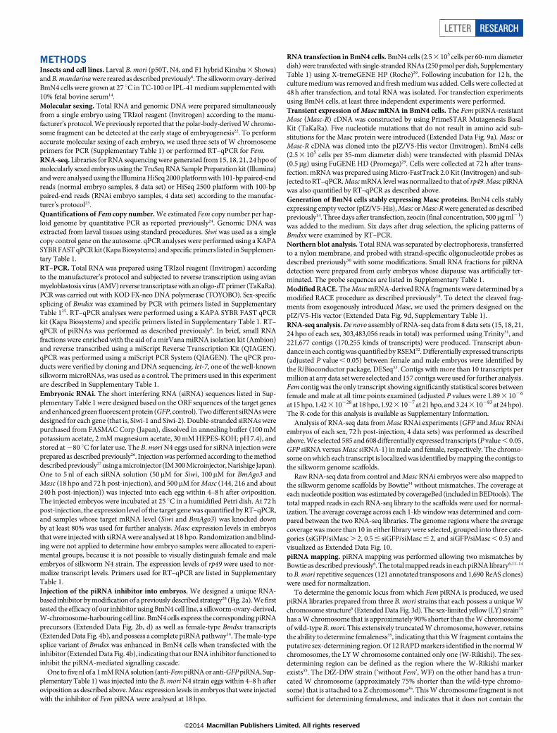
Extended Data Figure 1 | Molecular sexing and comparative transcriptomeanalysis of embryonic B. mori. a, Molecular sexing of individual embryosat 21 hpo. Musashi, Sasuke and Bonsai are W chromosome RAPD markers.‘Chr2’ control bands are generated from a primer set that amplifies a sequencewithin the 2nd chromosome of B. mori. b, MA plots of RNA-seq data. Thecomp73859_c0 contig is indicated by red dots and highlighted by arrows. Theaxes show: A (x-axis) 5 (log2(transcripts per million in male) 1 log2(transcripts
per million in female))/2. M (y-axis) 5 log2(transcripts per millionin male) 2 log2(transcripts per million in female). c, Number of thecomp73859_c0-derived transcripts in each RNA-seq library. Note that thecomp73859_c0-derived transcripts detected in male libraries may be derivedfrom incorrectly sexed embryos or RNA produced by polar bodies. Combinedwith RT–qPCR results of Fig. 1c, the expression level of this contig peaksaround 18–21 hpo in the B. mori embryo.
LETTER RESEARCH
Macmillan Publishers Limited. All rights reserved©2014
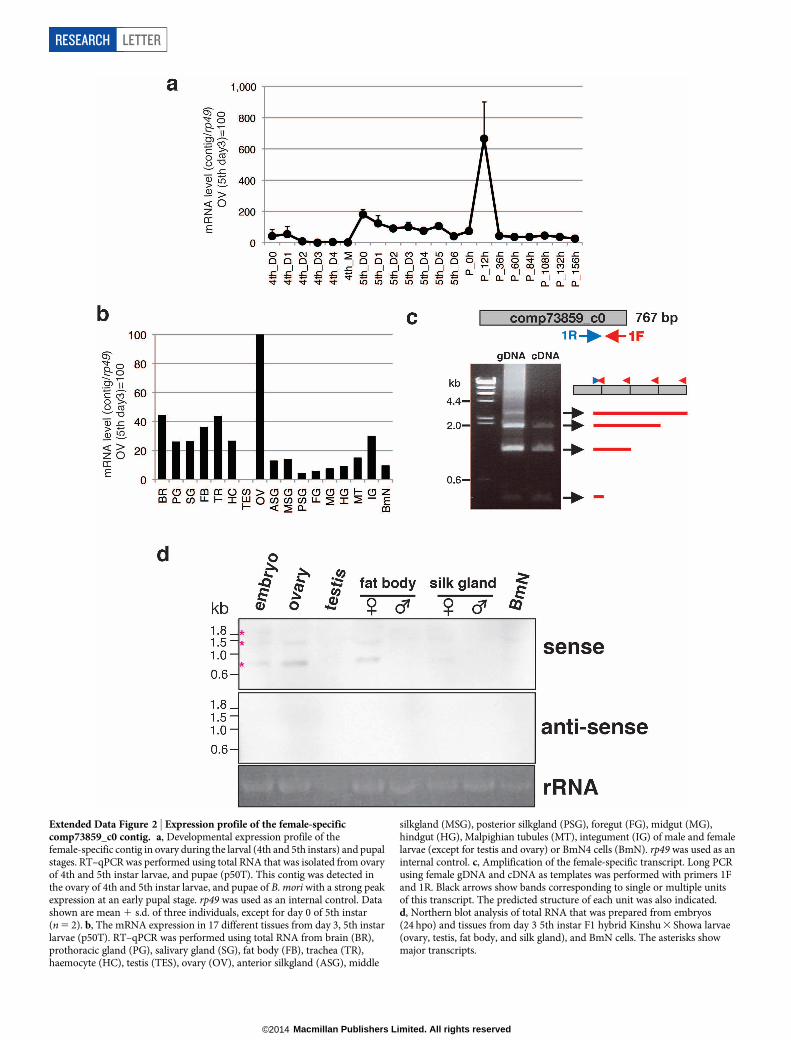
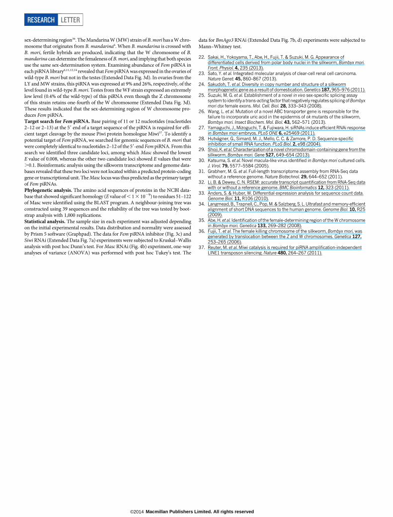
Extended Data Figure 2 | Expression profile of the female-specificcomp73859_c0 contig. a, Developmental expression profile of thefemale-specific contig in ovary during the larval (4th and 5th instars) and pupalstages. RT–qPCR was performed using total RNA that was isolated from ovaryof 4th and 5th instar larvae, and pupae (p50T). This contig was detected inthe ovary of 4th and 5th instar larvae, and pupae of B. mori with a strong peakexpression at an early pupal stage. rp49 was used as an internal control. Datashown are mean 1 s.d. of three individuals, except for day 0 of 5th instar(n 5 2). b, The mRNA expression in 17 different tissues from day 3, 5th instarlarvae (p50T). RT–qPCR was performed using total RNA from brain (BR),prothoracic gland (PG), salivary gland (SG), fat body (FB), trachea (TR),haemocyte (HC), testis (TES), ovary (OV), anterior silkgland (ASG), middle
silkgland (MSG), posterior silkgland (PSG), foregut (FG), midgut (MG),hindgut (HG), Malpighian tubules (MT), integument (IG) of male and femalelarvae (except for testis and ovary) or BmN4 cells (BmN). rp49 was used as aninternal control. c, Amplification of the female-specific transcript. Long PCRusing female gDNA and cDNA as templates was performed with primers 1Fand 1R. Black arrows show bands corresponding to single or multiple unitsof this transcript. The predicted structure of each unit was also indicated.d, Northern blot analysis of total RNA that was prepared from embryos(24 hpo) and tissues from day 3 5th instar F1 hybrid Kinshu 3 Showa larvae(ovary, testis, fat body, and silk gland), and BmN cells. The asterisks showmajor transcripts.
RESEARCH LETTER
Macmillan Publishers Limited. All rights reserved©2014
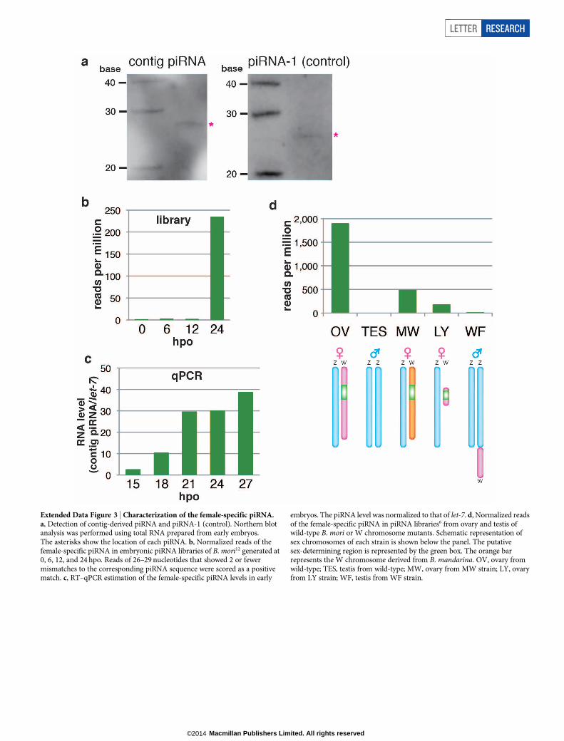
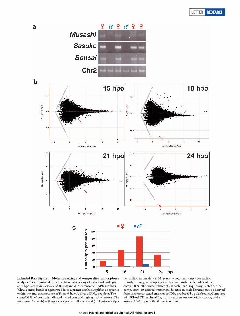
Extended Data Figure 3 | Characterization of the female-specific piRNA.a, Detection of contig-derived piRNA and piRNA-1 (control). Northern blotanalysis was performed using total RNA prepared from early embryos.The asterisks show the location of each piRNA. b, Normalized reads of thefemale-specific piRNA in embryonic piRNA libraries of B. mori12 generated at0, 6, 12, and 24 hpo. Reads of 26–29 nucleotides that showed 2 or fewermismatches to the corresponding piRNA sequence were scored as a positivematch. c, RT–qPCR estimation of the female-specific piRNA levels in early
embryos. The piRNA level was normalized to that of let-7. d, Normalized readsof the female-specific piRNA in piRNA libraries6 from ovary and testis ofwild-type B. mori or W chromosome mutants. Schematic representation ofsex chromosomes of each strain is shown below the panel. The putativesex-determining region is represented by the green box. The orange barrepresents the W chromosome derived from B. mandarina. OV, ovary fromwild-type; TES, testis from wild-type; MW, ovary from MW strain; LY, ovaryfrom LY strain; WF, testis from WF strain.
LETTER RESEARCH
Macmillan Publishers Limited. All rights reserved©2014
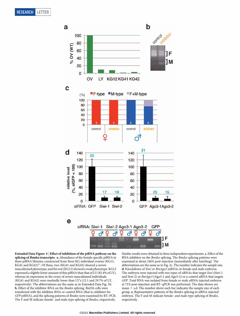
Extended Data Figure 4 | Effect of inhibition of the piRNA pathway on thesplicing of Bmdsx transcripts. a, Abundance of the female-specific piRNA inthree piRNA libraries constructed from three KG individual ovaries (KG12,KG41 and KG42)13. Of these, two (KG41 and KG42) showed a severemasculinized phenotype, and the rest (KG12) showed a weak phenotype. KG12expressed a slightly lower amount of this piRNA than that of LY (82.4% of LY),whereas its expression in the ovary of severe masculinized individuals(KG41 and KG42) were markedly lower than LY’s (12.1 and 29.7% of LY,respectively). The abbreviations are the same as in Extended Data Fig. 3d.b, Effect of the inhibitor RNA on the Bmdsx splicing. BmN4 cells weretransfected with the inhibitor RNA or control RNA (that is, inhibitor forGFP piRNA), and the splicing patterns of Bmdsx were examined by RT–PCR.The F and M indicate female- and male-type splicing of Bmdsx, respectively.
Similar results were obtained in three independent experiments. c, Effect of theRNA inhibitor on the Bmdsx splicing. The Bmdsx splicing patterns wereexamined at about 240 h post-injection (immediately after hatching). Theabbreviations are the same as in Fig. 2c. The number indicates the sample size.d, Knockdown of Siwi or BmAgo3 mRNAs in female and male embryos.The embryos were injected with two types of siRNAs that target Siwi (Siwi-1and Siwi-2) or BmAgo3 (Ago3-1 and Ago3-2) or a control siRNA that targetsGFP. Total RNA was isolated from female or male siRNA-injected embryosat 72 h post-injection and RT–qPCR was performed. The data shown aremean 1 s.d. The number above each bar indicates the sample size of eachgroup. e, Representative patterns of the Bmdsx splicing in siRNA-injectedembryos. The F and M indicate female- and male-type splicing of Bmdsx,respectively.
RESEARCH LETTER
Macmillan Publishers Limited. All rights reserved©2014
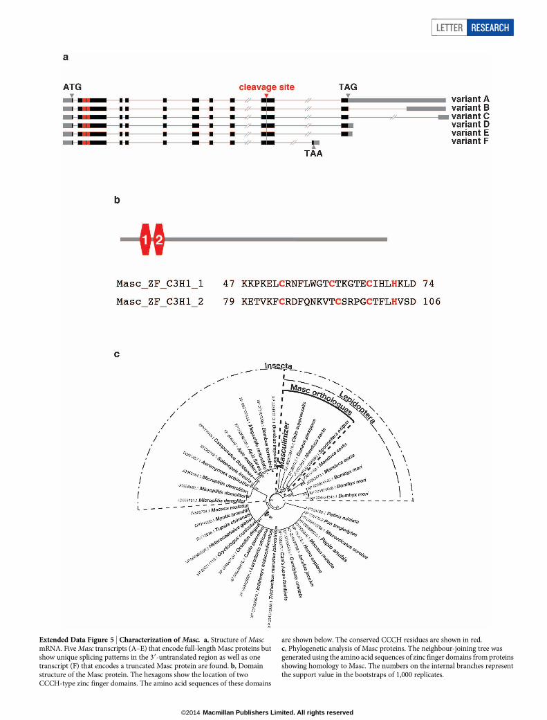
Extended Data Figure 5 | Characterization of Masc. a, Structure of MascmRNA. Five Masc transcripts (A–E) that encode full-length Masc proteins butshow unique splicing patterns in the 39-untranslated region as well as onetranscript (F) that encodes a truncated Masc protein are found. b, Domainstructure of the Masc protein. The hexagons show the location of twoCCCH-type zinc finger domains. The amino acid sequences of these domains
are shown below. The conserved CCCH residues are shown in red.c, Phylogenetic analysis of Masc proteins. The neighbour-joining tree wasgenerated using the amino acid sequences of zinc finger domains from proteinsshowing homology to Masc. The numbers on the internal branches representthe support value in the bootstraps of 1,000 replicates.
LETTER RESEARCH
Macmillan Publishers Limited. All rights reserved©2014
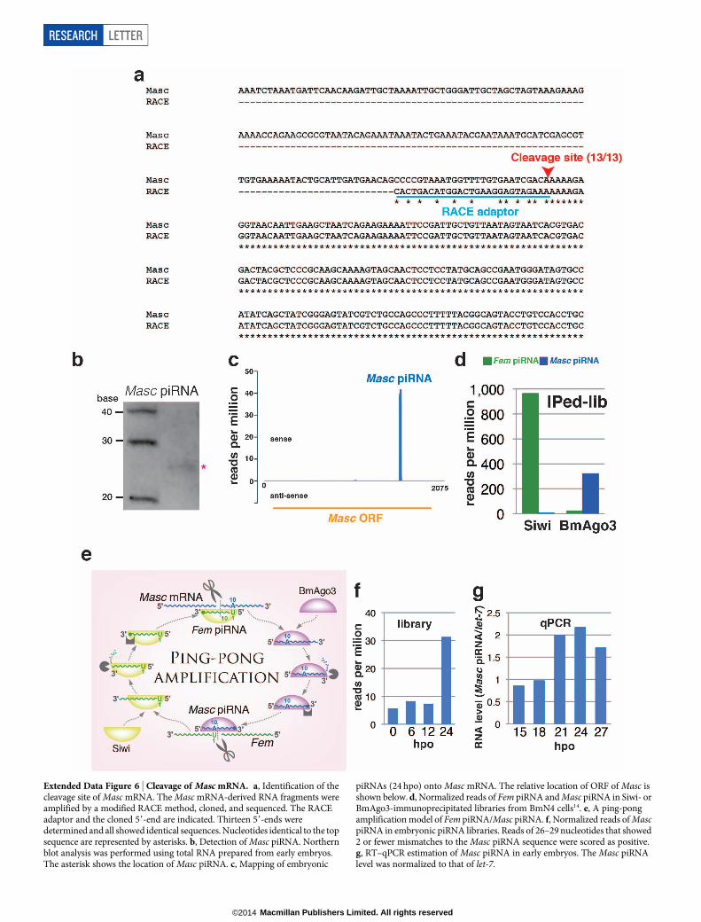
Extended Data Figure 6 | Cleavage of Masc mRNA. a, Identification of thecleavage site of Masc mRNA. The Masc mRNA-derived RNA fragments wereamplified by a modified RACE method, cloned, and sequenced. The RACEadaptor and the cloned 59-end are indicated. Thirteen 59-ends weredetermined and all showed identical sequences. Nucleotides identical to the topsequence are represented by asterisks. b, Detection of Masc piRNA. Northernblot analysis was performed using total RNA prepared from early embryos.The asterisk shows the location of Masc piRNA. c, Mapping of embryonic
piRNAs (24 hpo) onto Masc mRNA. The relative location of ORF of Masc isshown below. d, Normalized reads of Fem piRNA and Masc piRNA in Siwi- orBmAgo3-immunoprecipitated libraries from BmN4 cells14. e, A ping-pongamplification model of Fem piRNA/Masc piRNA. f, Normalized reads of MascpiRNA in embryonic piRNA libraries. Reads of 26–29 nucleotides that showed2 or fewer mismatches to the Masc piRNA sequence were scored as positive.g, RT–qPCR estimation of Masc piRNA in early embryos. The Masc piRNAlevel was normalized to that of let-7.
RESEARCH LETTER
Macmillan Publishers Limited. All rights reserved©2014
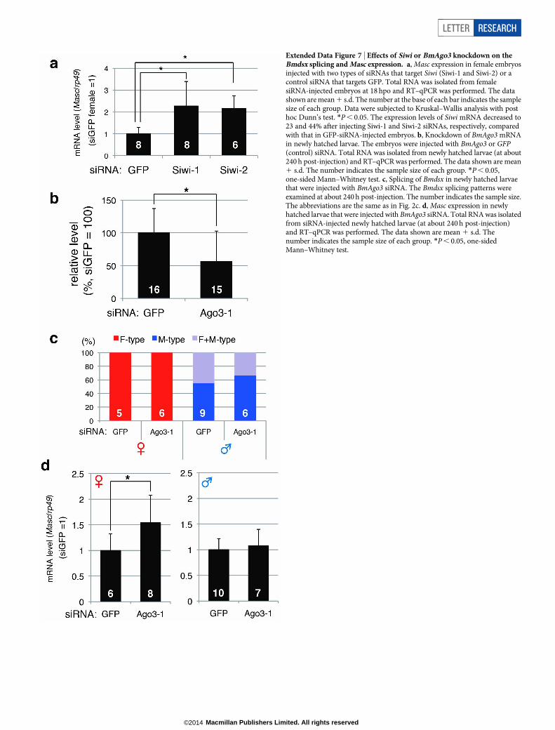
Extended Data Figure 7 | Effects of Siwi or BmAgo3 knockdown on theBmdsx splicing and Masc expression. a, Masc expression in female embryosinjected with two types of siRNAs that target Siwi (Siwi-1 and Siwi-2) or acontrol siRNA that targets GFP. Total RNA was isolated from femalesiRNA-injected embryos at 18 hpo and RT–qPCR was performed. The datashown are mean 1 s.d. The number at the base of each bar indicates the samplesize of each group. Data were subjected to Kruskal–Wallis analysis with posthoc Dunn’s test. *P , 0.05. The expression levels of Siwi mRNA decreased to23 and 44% after injecting Siwi-1 and Siwi-2 siRNAs, respectively, comparedwith that in GFP-siRNA-injected embryos. b, Knockdown of BmAgo3 mRNAin newly hatched larvae. The embryos were injected with BmAgo3 or GFP(control) siRNA. Total RNA was isolated from newly hatched larvae (at about240 h post-injection) and RT–qPCR was performed. The data shown are mean1 s.d. The number indicates the sample size of each group. *P , 0.05,one-sided Mann–Whitney test. c, Splicing of Bmdsx in newly hatched larvaethat were injected with BmAgo3 siRNA. The Bmdsx splicing patterns wereexamined at about 240 h post-injection. The number indicates the sample size.The abbreviations are the same as in Fig. 2c. d, Masc expression in newlyhatched larvae that were injected with BmAgo3 siRNA. Total RNA was isolatedfrom siRNA-injected newly hatched larvae (at about 240 h post-injection)and RT–qPCR was performed. The data shown are mean 1 s.d. Thenumber indicates the sample size of each group. *P , 0.05, one-sidedMann–Whitney test.
LETTER RESEARCH
Macmillan Publishers Limited. All rights reserved©2014
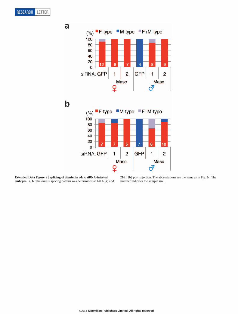
Extended Data Figure 8 | Splicing of Bmdsx in Masc siRNA-injectedembryos. a, b, The Bmdsx splicing pattern was determined at 144 h (a) and
216 h (b) post-injection. The abbreviations are the same as in Fig. 2c. Thenumber indicates the sample size.
RESEARCH LETTER
Macmillan Publishers Limited. All rights reserved©2014
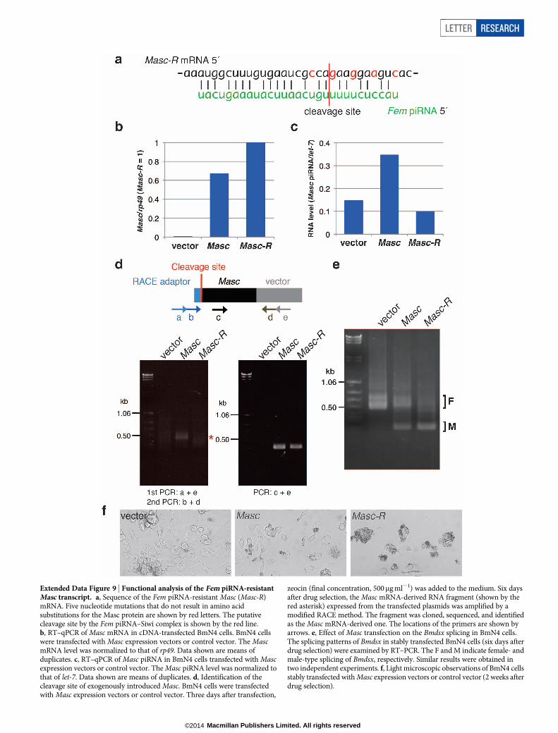
Extended Data Figure 9 | Functional analysis of the Fem piRNA-resistantMasc transcript. a, Sequence of the Fem piRNA-resistant Masc (Masc-R)mRNA. Five nucleotide mutations that do not result in amino acidsubstitutions for the Masc protein are shown by red letters. The putativecleavage site by the Fem piRNA–Siwi complex is shown by the red line.b, RT–qPCR of Masc mRNA in cDNA-transfected BmN4 cells. BmN4 cellswere transfected with Masc expression vectors or control vector. The MascmRNA level was normalized to that of rp49. Data shown are means ofduplicates. c, RT–qPCR of Masc piRNA in BmN4 cells transfected with Mascexpression vectors or control vector. The Masc piRNA level was normalized tothat of let-7. Data shown are means of duplicates. d, Identification of thecleavage site of exogenously introduced Masc. BmN4 cells were transfectedwith Masc expression vectors or control vector. Three days after transfection,
zeocin (final concentration, 500mg ml21) was added to the medium. Six daysafter drug selection, the Masc mRNA-derived RNA fragment (shown by thered asterisk) expressed from the transfected plasmids was amplified by amodified RACE method. The fragment was cloned, sequenced, and identifiedas the Masc mRNA-derived one. The locations of the primers are shown byarrows. e, Effect of Masc transfection on the Bmdsx splicing in BmN4 cells.The splicing patterns of Bmdsx in stably transfected BmN4 cells (six days afterdrug selection) were examined by RT–PCR. The F and M indicate female- andmale-type splicing of Bmdsx, respectively. Similar results were obtained intwo independent experiments. f, Light microscopic observations of BmN4 cellsstably transfected with Masc expression vectors or control vector (2 weeks afterdrug selection).
LETTER RESEARCH
Macmillan Publishers Limited. All rights reserved©2014
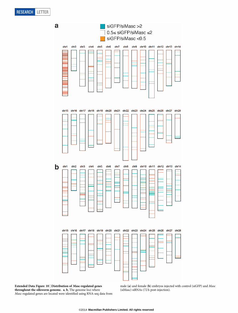
Extended Data Figure 10 | Distribution of Masc-regulated genesthroughout the silkworm genome. a, b, The genome loci whereMasc-regulated genes are located were identified using RNA-seq data from
male (a) and female (b) embryos injected with control (siGFP) and Masc(siMasc) siRNAs (72 h post-injection).
RESEARCH LETTER
Macmillan Publishers Limited. All rights reserved©2014