Embed Size (px)
Citation preview
Acta Biomaterialia 9 (2013) 8972–8982
Contents lists available at SciVerse ScienceDirect
Acta Biomaterialia
journal homepage: www.elsevier .com/locate /ac tabiomat
Silk hydrogels from non-mulberry and mulberry silkworm cocoonsprocessed with ionic liquids
1742-7061/$ - see front matter � 2013 Acta Materialia Inc. Published by Elsevier Ltd. All rights reserved.http://dx.doi.org/10.1016/j.actbio.2013.06.044
⇑ Corresponding authors at: 3Bs Research Group- Biomaterials, Biodegradablesand Biomimetics, University of Minho, Headquarters of the European Institute ofExcellence on Tissue Engineering and Regenerative Medicine, AvePark, ZonaIndustrial da Gandra, Caldas das Taipas, 4806-909 Guimarães, Portugal (S.S. Silva),Department of Biotechnology, Indian Institute of Technology Kharagpur, India (S. C.Kundu).
E-mail addresses: [email protected] (S.S. Silva), [email protected] (S.C. Kundu).
Simone S. Silva a,b,⇑, Elena G. Popa a,b, Manuela E. Gomes a,b, Mariana B. Oliveira a,b, Sunita Nayak c,Bano Subia c, João F. Mano a,b, Subhas C. Kundu c,⇑, Rui L. Reis a,b
a 3B’s Research Group – Biomaterials, Biodegradables and Biomimetics, University of Minho, Headquarters of the European Institute of Excellence on Tissue Engineeringand Regenerative Medicine, AvePark, Zona Industrial da Gandra, Caldas das Taipas, 4806-909 Guimarães, Portugalb ICVS/3B’s – PT Government Associate Laboratory, Braga/Guimarães, Portugalc Department of Biotechnology, Indian Institute of Technology, Kharagpur 721302, India
a r t i c l e i n f o a b s t r a c t
Article history:Received 20 February 2013Received in revised form 21 June 2013Accepted 26 June 2013Available online 9 July 2013
Keywords:Bombyx moriFibroinAntheraea mylittaIonic liquidsHydrogels
Matrices based on silk fibroin from the non-mulberry silkworm Antheraea mylitta and the mulberry silk-worm Bombyx mori have demonstrated good applicability in regenerative medicine. However, thecocoons of A. mylitta are underutilized in part due to their lack of solubility in traditional organic solvents.Therefore, the present work investigates the solubilization and processing of degummed fibers obtainedfrom the cocoons of both silkworm species into hydrogels using ionic liquids (ILs). The developed hydro-gels exhibited a rubbery consistency, viscoelastic behavior and rapid degradation in the presence of pro-tease XIV. Scanning electron and confocal microscopy images suggest that human adipose stem cells(hASCs) are able to adhere to and migrate at different levels within the hydrogel structures. Moreover,the MTS assay demonstrated the maintenance of cell metabolic activity for up to 28 days, while DNAquantification showed that hASCs were able to proliferate on the seeded hydrogels. The findings indicatethat complete IL removal from the fabricated hydrogels results in a positive hASCs cellular response. Thusthe present approach provides a unique opportunity to broaden the processability and application of silkfibroin obtained from A. mylitta cocoons for regenerative medicine, namely cartilage regeneration.
� 2013 Acta Materialia Inc. Published by Elsevier Ltd. All rights reserved.
1. Introduction
Recent investigations have shown that matrices based on silkfibroin from the non-mulberry silkworm Antheraea mylitta andthe mulberry silkworm Bombyx mori have diverse morphologies,controllable degradation and good biocompatibility [1–4]. Becauseof their versatility and properties silks are used as a biomaterial forbiomedical, biotechnological and tissue engineering applications[1,5]. Silk protein fibroin obtained from the non-mulberry Indiantropical tasar silkworm A. mylitta is utilized as a promising bioma-terial for tissue engineering and biomedical applications due to itsbiocompatibility, biodegradability and mechanical stability [6,7].The non-mulberry Indian tropical tasar silkworm A. mylitta silkprotein fibroin has a molecular weight of 395 kDa, with two sub-units each of approximately 197 kDa [6,8]. The additional feature
of A. mylitta silk fibroin is the presence of arginine–glycine–aspar-tic acid (RGD) motifs, integrin binding sites for cell attachment,proliferation and differentiation [8]. The presence of RGD motifson A. mylitta-based matrices provide a substrate for fibroblasts,chondrocytes, the osteogenic differentiation of human mesenchy-mal stem cells (hMSCs), rat bone marrow stromal cells (BSCs),and cardiomyocytes [6,7,9]. Thus silk fibroin from the non-mul-berry fed A. mylitta and mulberry fed B. mori differ not only in theiramino acid composition but also in the presence/absence of inte-grin binding RGD sequences [10,11].
On the other hand, previous studies conducted with silk fibroinshowed that hydrogel formation and the sol–gel transition weredependent on protein concentration, temperature, and pH, amongother parameters [12–16]. Different methods of silk-based hydro-gel formation, for example using carbon dioxide and chemicalcrosslinking, have been investigated [3,17,18]. Silk hydrogels havebeen previously characterized and investigated for pancreatic isletsco-encapsulated with extracellular matrix (ECM) proteins and mes-enchymal stromal cells [19]. Silk hydrogel-based delivery of bonemorphogenetic protein for the treatment of large bone defects hasbeen also investigated [20]. Despite promising findings on the useof A. mylitta fibroin protein isolated from the silk gland theircocoons are still underutilized, in part due to their insolubility in
S.S. Silva et al. / Acta Biomaterialia 9 (2013) 8972–8982 8973
water and the usual organic solvents. Earlier reports emphasizedthe use of certain ionic liquids (ILs) [21] to solubilize B. mori proteinfibroin, alone [22,23] or combined with chitosan [24]. To ourknowledge, to date only one report on the use of ILs as a solventfor Antheraea assamensis silk fibroin has been described [25]. How-ever, no work has been reported involving ILs in the dissolution andprocessing of A. mylitta silk fibroin. In spite of the widespread appli-cation of ILs as templates and solvents for different biomacromole-cules such as silk fibroin, chitin, and cellulose [24,26,27], issuesconcerning cellular response have not yet been fully explored.The present work reports the viability of silk protein fibroins ob-tained from the cocoons of non-mulberry fed A. mylitta and mul-berry fed B. mori through dissolution and their processability ashydrogels using an IL, 1-butyl-3-methyl imidazolium acetate (BMI-Ac). The fabricated silk hydrogels were characterized biochemicallyand biophysically. The use of mesenchymal stem cells (MSCs) is animportant tool in tissue engineering and regenerative medicine,mainly due to their plasticity and easy availability [28]. MSCs canbe obtained from many tissues, for example bone marrow and adi-pose tissue, and are recognized as a potential cell substitute forchondrocytes in cartilage regeneration [29]. MSCs have been usedto evaluate the cellular response of fabricated silk matrices, includ-ing hydrogels. An in vitro assessment was performed using humanadipose-derived stem cells (hASCs) to validate the use of bothfabricated silk hydrogels for cartilage regeneration. The mainadvantage of the present approach is that it provides a uniqueopportunity to broaden the application of Indian tasar silkwormA. mylitta fibroin obtained from cocoons for cartilage regeneration.
2. Materials and methods
2.1. Materials
Silk fibroin from cocoons of B. mori was kindly supplied by theAPPACDM (Castelo Branco, Portugal). The silk cocoons of A. mylittawere obtained from IIT Kharagpur (West Bengal, India). The ILBMIAc (Sigma Aldrich) was chosen as solvent and used withoutfurther purification. All other chemicals were reagent grade andwere used as received.
2.2. Methods
2.2.1. Isolation of silk protein fibroin from A. mylittaSilk protein fibroin was isolated from cocoons of A. mylitta fol-
lowing a standard extraction protocol [30]. Briefly, cocoons werecut into small pieces, boiled in 0.02 M Na2CO3 for 45–60 min andwashed with deionized water several times to remove sericin fromthe silk fibers. The degummed fibers were then dried completely atroom temperature.
2.2.2. Isolation of silk protein fibroin from B. moriThe degumming process was achieved by boiling the silk from
B. mori filaments for 1 h in water containing 1.1 g l�1 Na2CO3, fol-lowed by 30 min in water with 0.4 g l�1 Na2CO3 [17,31]. Finally,the resulting fibroin filaments were extensively rinsed in boilingdistilled water and air dried at room temperature.
2.2.3. Preparation of the silk fibroin-based hydrogelsTo prepare the hydrogels degummed fibers of cocoons of
A. mylitta were dissolved in BMIAc at 95 �C at a concentration of10 wt.%. The degummed fibers from B. mori were also dissolvedusing the same procedure. Both systems were kept under stirringfor 8 h. After dissolution the fiber/IL solutions were transferred topolystyrene molds, followed by gelation at 4 �C overnight. Thegelation process was completed by immersion of the molds in
ethanol to obtain silk hydrogels, identified as AM and BM, fromA. mylitta/IL and B. mori/IL solutions, respectively. The BMIAc re-moval procedure was carried out by immersing the materials inethanol for 24 h, followed by Soxhlet extraction with ethanol for5 days. During solvent removal aliquots of ethanol were collectedfor conductivity analysis. The conductivities of the aliquots of eth-anol were measured using a conductivimeter (inoLab Multilevel 3)with a Sonda WTW TetraCon 325 detector. After IL removal thesamples were treated with methanol/water (80/20 vol.%) for30 min to for induce formation of beta-sheets.
2.3. Characterization
2.3.1. Biochemical and biophysical characterizationThe amino acid composition of the samples, degummed fibroin
from A. mylitta filaments, degummed fibroin from B. mori filaments,AM hydrogel and BM hydrogel, were performed after hydrolysisand derivatization, followed by analysis in a Waters 600 HPLCgradient system equipped with a Waters 2487 UV detector.
2.3.2. Fourier transform infrared (FTIR) spectroscopyThe infrared spectra of the silk structures were obtained with a
Shimadzu-IR Prestige 21 spectrometer in the spectral region 4000–650 cm�1, with a resolution of 2 cm�1 for 32 scans. FTIR spectrawere obtained from the freeze-dried hydrogels to minimize theeffect of the presence of water. After that the samples were pow-dered, mixed with KBr and processed into pellets. Fourier self-deconvolution (FSD) of the infrared spectra covering the amide Iregion (1595–1705 cm�1) was performed using Origin� Software.To measure the relative areas of the amide I components FSD spec-tra were curve fitted to Gaussian line shape profiles. The deconvo-luted amide I spectra were area normalized and the relative areasof the single bands were used to determine the fraction of the sec-ondary structural elements. The band assignments and the detailedprocedure to determine b-sheet crystallinity were described previ-ously by Hu et al. [32].
2.3.3. Scanning electron microscopy (SEM)Freeze-dried hydrogel samples were observed using a Leica
Cambridge S360 scanning electron microscope. The materials werefixed using mutual conductive adhesive tape on aluminum stubsand covered with gold palladium using a sputter coater.
2.3.4. Differential scanning calorimetry (DSC)DSC measurements were performed using a TA instruments
DSC Q100 equipped with a cooling accessory to quantitativelycharacterize the bound and bulk water in the hydrogels. Approxi-mately 10 mg of hydrogel was transferred to a DSC aluminumpan. Subsequently the pan was first cooled to �40 �C and thenheated to 40 �C at a rate of 5 �C min�1 [16]. The linear baseline tointegrate peaks was determined, and then the melting temperature(Tm) and enthalpy of fusion (DH) were calculated from the DSCthermograms using TA instruments software.
2.3.5. Dynamical mechanical analysisViscoelastic measurements of the AM and BM hydrogels were
performed using a TRITEC2000B DMA from Triton Technology(UK) in compression mode. The hydrogels were always analyzedunder hydrated conditions through immersion of the samples ina Teflon reservoir containing phosphate-buffered saline (PBS), usedduring all analysis periods. After equilibration at 37 �C the DMAspectra were obtained over a frequency scan of 0.1–10 Hz. Theexperiments were performed under a constant strain amplitude(30 lm). The average value of four tests is reported for eachsample.
8974 S.S. Silva et al. / Acta Biomaterialia 9 (2013) 8972–8982
2.3.6. In vitro degradationIn vitro degradation of the silk hydrogels (AM and BM) was
evaluated for 28 days using protease XIV from Streptomyces griseuswith an activity of 4.5 U ml�1. Silk hydrogels measuring 6.5 mm indiameter, 4.5 mm in height were immersed in 5 ml of PBS contain-ing 2 U ml�1 protease enzyme. The samples were also incubated inPBS without enzyme under similar conditions for comparison. Allsystems were maintained at 37 �C. The enzyme solution was re-placed with freshly prepared solution every 7 days to maintainthe activity of the enzyme. Samples were collected at differenttime points, washed gently with distilled water, dried at 40 �Cand weighed. Percentage weight loss at respective time pointswas calculated.
2.3.7. Human adipose stem cell isolation and expansionhASCs were obtained from subcutaneous fat tissue, under
protocols established with the Plastic Surgery Department of theHospital da Prelada (Porto, Portugal) and isolated by enzymaticdigestion as previously described [33]. Briefly, the adipose tissuesamples were digested with 0.075% collagenase type II (Col II)(C6885, Sigma) in PBS (21600-044, Invitrogen) for 45 min at37 �C under gentle stirring. The digested tissue was filtered witha 100 lm filter mesh, and centrifuged at 1000g for 10 min at5 �C. Afterwards the cell suspension was washed for 10 min withlysis buffer (155 mM NH4Cl, 10 mM KHCO3, 0.1 mM ED-TA = 14.61 mg, pH 7.3), to remove erythrocytes, and additionallycentrifuged at 800g for 10 min at 5 �C. The adherent hASCs wereexpanded in basal medium with minimum essential medium(a-MEM) (12000-063, Invitrogen) with 10 vol.% fetal bovine serum(FBS) (10270, Invitrogen), 1 vol.% antibiotic–antimycotic and2.2 mg ml�1 NaHCO3 (S5761, Sigma) with medium changes every3–4 days. Cells were subcultured at a cell density of 3.5 � 103 -cells cm�2 until a sufficient cell number was achieved for use inthe experimental assays.
2.3.7.1. Cell seeding. Prior to the cell culture studies the hydrogelswere sterilized in an autoclave. The hASCs were seeded at a densityof 3 � 105 cells cm3 onto silk hydrogel samples with a diameter of7 mm and a height of 5 mm. All seeded hydrogels were subse-quently cultured for 28 days in either basal or chondrogenic med-ium in a 37 �C humidified atmosphere including 5% CO2. Thechondrogenic induction medium was composed of Dulbecco’smodified Eagle’s medium–low glucose (DMEM) (D5523, Sigma)supplemented with 3.8 mg ml�1 NaHCO3, 1 vol.% antibiotic–anti-mycotic solution, 5 vol.% FBS (SH3007103, Fisher Scientific), 1�ITS + 1 (insulin–transferrin–selenium liquid medium supplement,I2521, Sigma), 17 mM L-ascorbic acid (A4544, Sigma), 0.1 M so-dium pyruvate (P4562, Sigma), 35 mM L-proline (P5607, Sigma),1 mM dexamethasone (D2915, Sigma) and 10 ng ml�1 humantransforming growth factor-b1 (TGF-b1) (14-8348, eBioscience).Controls consisted of cell-seeded silk hydrogels cultured in basalmedium (without chondrogenic growth factor) and silk hydrogelswithout cells, maintained in culture for the same time periods un-der the same cell maintenance conditions. The culture mediumwas replaced twice per week. At the end of each culture time pointthe samples were retrieved, rinsed with PBS and evaluated interms of cellular behavior, by confocal analysis, MTS test, DNAquantification and histological evaluation.
2.3.7.2. Morphological analysis. The cell distribution and attach-ment of hASC cells on both the AM and BM constructs was ana-lyzed using confocal microscopy. The matrices were fixed with10% formalin for 30 min and then the samples were incubated with1% bovine serum albumin (BSA) for 30 min. The constructs werepermeabilized using 0.1% Triton X-100 for 5 min, then incubatedwith rhodamine–phalloidin for 20 min at room temperature,
followed by washing with PBS and staining with 5 lg ml�1 Hoechst33342 for 30 min. Fluorescence images from stained constructswere obtained using a confocal laser scanning microscope (Olym-pus FV1000). The morphology of the constructs was also observedusing a Leica Cambridge S360 scanning electron microscope. Theconstructs were fixed using mutual conductive adhesive tape onaluminum stubs and covered with gold palladium using a sputtercoater.
2.3.7.3. Viability and proliferation of human adipose-derived stemcells. The metabolic activity of hASCs seeded on the silk hydro-gels was evaluated using the CellTiter 96� AQueous One SolutionCell Proliferation Assay (MTS, Promega). Briefly, cell–hydrogelconstructs were placed in a mixture containing serum-free cellculture medium and MTS reagent at a 5:1 ratio and incubatedfor 3 h, at the end of which the optical density was read (OD490 nm). Cell quantification was performed using the fluorimet-ric PicoGreen double-stranded DNA assay according to the man-ufacturer’s instructions (Quant-iT™ PicoGreen� dsDNA Kit,P7589, Invitrogen). Briefly, cell–hydrogel constructs, collectedafter each time period, were rinsed with PBS, placed in micro-tubes containing 1 ml of ultra-pure water and stored at �80 �Cuntil analyzed. The fluorescence of the samples was measuredwith a microplate reader (Synergie HT, Izasa) at 480 and520 nm, and the DNA concentration was calculated using a stan-dard curve (0–2 lg ml�1) relating the amount of dsDNA to thefluorescence intensity.
2.3.7.4. Histological evaluation. Samples collected at the end of theculturing period were fixed with 3.7 vol.% formalin solution andstored at 4 �C until analysis. The samples were processed througha series of dehydration steps, embedded in paraffin (MicromEC350-2, Thermo Scientific) and sectioned at 3 lm (Microm HM355, Thermo Scientific). For hematoxylin and eosin (H&E) stain-ing the sections were colored with Papanicolaou Harris hematox-ylin (05-12011/L, Bio-optica) for 3 min, washed in running tapwater and stained with Eosin-Y (05-M10003, Bio-optica) for30 s. Toluidine blue staining was used to evaluate cartilageECM component deposition, namely glycosaminoglycans (GAGs).Toluidine blue staining solution was prepared by dissolving 1%toluidine blue in distilled water containing 0.5 g sodium borate(Riedelde-Haën). After filtering the solution the sections weredipped for 2–3 s. Finally, the slides were dehydrated through analcohol series from 30 to 100 vol.% alcohol. The stained cells werevisualized by reflected/transmitted light microscopy (Zeiss,Germany) and images were taken using a digital camera (AxionMRc5, Zeiss).
2.3.7.5. Immunohistochemical analysis. The tissue slides were depa-raffinized and an antigen retrieval step was performed (the slideswere dipped in 10 mM citrate buffer for 20 min at 95 �C). Nextthe sections were washed in PBS and endogenous peroxidase activ-ity was quenched with 0.3% hydrogen peroxide (31642, Sigma) for5–15 min. Sections were washed with PBS and blocked with 2.5%horse serum from a R.T.U. Vectastain� Universal Elite ABC Kit(PK-7200, Vector Laboratories) for 1 h to avoid non-specific stain-ing. The samples were then incubated with rabbit anti-collagen Iantibody (ab292, Abcam) and rabbit anti-collagen II antibody(ab34712, Abcam) for 1 h at room temperature in a humidifiedatmosphere. Next the slides were incubated with secondaryantibody for 1 h at room temperature and developed with a DABsubstrate kit for peroxidase (SK-4100, Vector Laboratories). Slideswere counterstained with hematoxylin, mounted and visualizedunder a light microscope.
S.S. Silva et al. / Acta Biomaterialia 9 (2013) 8972–8982 8975
2.4. Statistical analysis
All quantitative experiments were run in triplicate andthe results are expressed as means ± standard deviation forn = 3. Statistical analysis of the data was conducted by
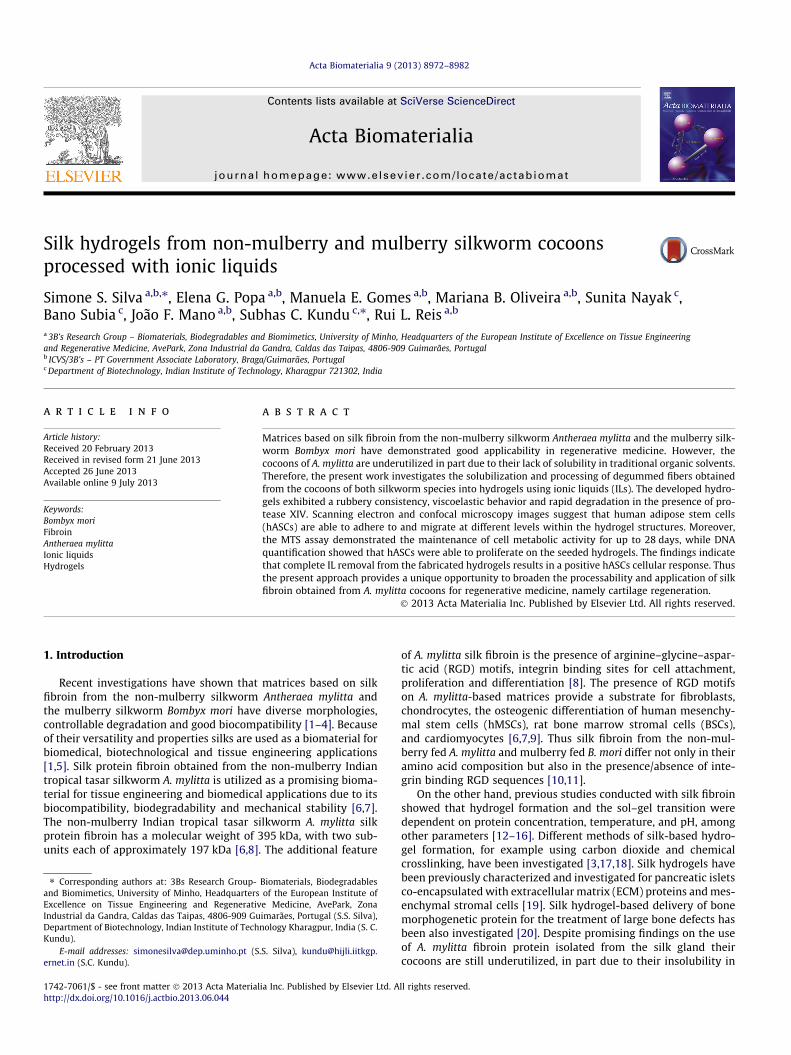
Fig. 1. Schematic representation of fabrication of silk hydrogels from non-mulberry and mdegummed fibers; (c) silk/BMIAc solution; (d) gelation of the silk/BMIAc solution; (e) fafibers; (c) silk/BMIAc solution; (d) gelation of the silk/BMIAc solution; (e) fabricated hyd
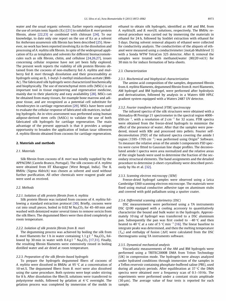
Fig. 2. (A) Conductivity measurements on aliquots of ethanol obtained after immersion odegummed fibers (A. mylitta and B. mori) and fabricated hydrogels (BM and AM).
two-way ANOVA with Bonferroni’s post test using Graph-PadPrism v. 5.0 for Windows (GraphPad Software, San Die-go, http://www.graphpad.com). Differences between thegroups at p < 0.05 were considered to be statisticallysignificant.
ulberry silk cocoons using BMIAc as solvent. (A): (a) Antheraea mylitta cocoons; (b)bricated hydrogels of A. mylitta (AM). (B): (a) Bombyx mori cocoons; (b) degummedrogels of B. mori (BM).
f the structures in ethanol and soxhlet extraction. (B) Amino acid composition of the
8976 S.S. Silva et al. / Acta Biomaterialia 9 (2013) 8972–8982
3. Results and discussion
Efficient dissolution and processing of silk fibroin from A. mylit-ta and B. mori cocoons was achieved using BMIAc at a concentra-tion of 10 wt.% (see Fig. 1). Both fibroin/BMIAc solutions werecolorless and viscous even at high silk concentrations (10 wt.%).They also showed good stability over a long period (1–2 weeks inan inert atmosphere). Despite the broad applicability of silk fibroinobtained from A. mylitta (Fig. 1Aa)[2,34,35], the degummed fibersshow a lack of solubility in common solvents such as lithium bro-mide, probably due to hydrogen bonding and the hydrophobic nat-ure of the fibroin [36]. Some studies [22,24] have suggested thatBMIAc is a good solvent for biomacromolecules like B. mori silkfibroin. However, to date the use of BMIAc in the dissolution offibroin from cocoons of A. mylitta has not been described in theliterature. Considering that A. mylitta cocoons are an underutilizedmaterial with a substantial amount of fibroin the present approachallows the dissolution and processing of A. mylitta degummedfibers with different shapes on the medium/large scale, increasingtheir applicability.
Upon cooling both viscous silk/BMIAc solutions resulted in theformation of gels (ionogels), which were molded into a cylindricalshape and subsequently immersed in an ethanol bath. The forma-tion of silk hydrogels from aqueous solutions can be affected by thetemperature, pH, and protein concentration [37]. In our approachsilk gelation was influenced by reducing the temperature duringmolding and immersion in an ethanol bath. A similar temperatureeffect was observed for the gelation of chitosan–silk/BMIAc solu-tions [24]. The mechanism of gelation of silk/IL solutions can beassociated with the formation of ionogels during cooling of thesolutions. In an ionogel the IL is immobilized in a way that involvesthe formation of a three-dimensional (3-D) network that is respon-sible for the solid-like behavior of the material [38,39]. Also, theeffect of temperature on the gelation response of the polymer/IL
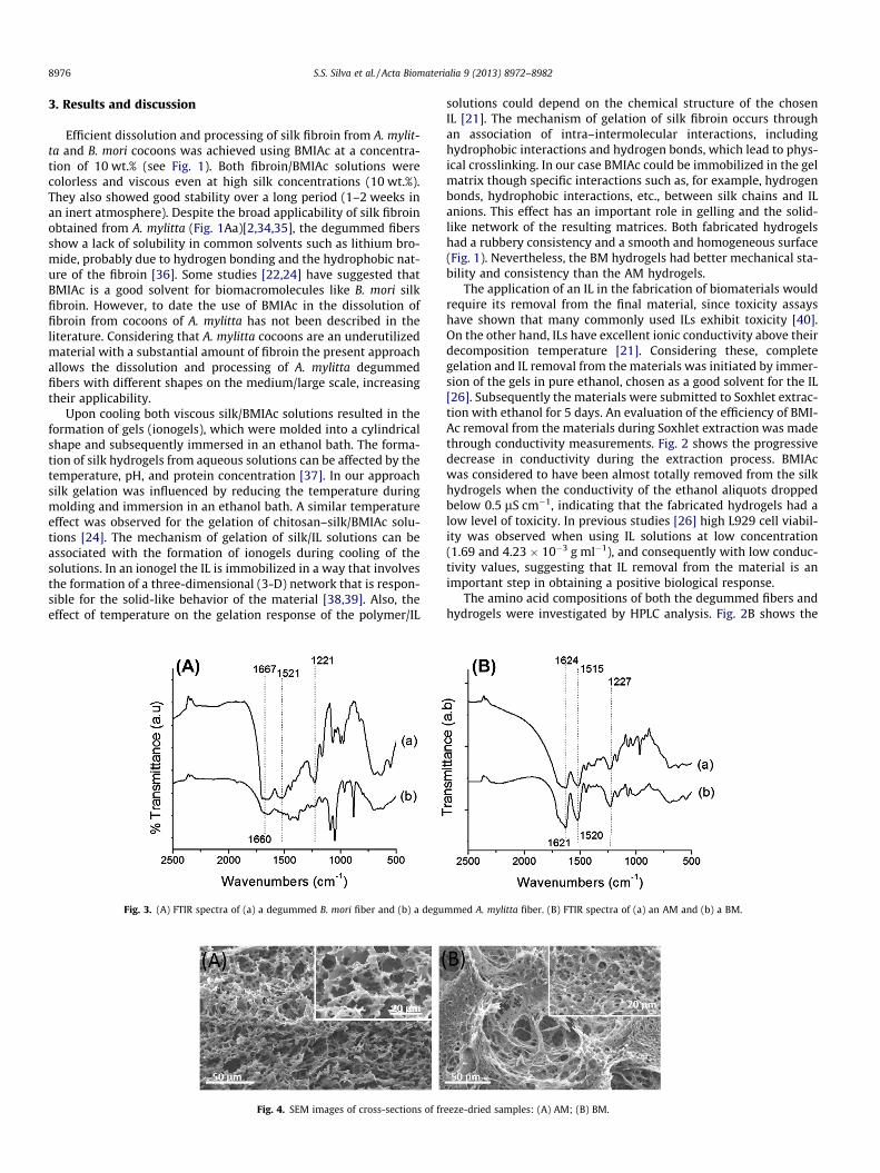
Fig. 3. (A) FTIR spectra of (a) a degummed B. mori fiber and (b) a degu
Fig. 4. SEM images of cross-sections of fr
solutions could depend on the chemical structure of the chosenIL [21]. The mechanism of gelation of silk fibroin occurs throughan association of intra–intermolecular interactions, includinghydrophobic interactions and hydrogen bonds, which lead to phys-ical crosslinking. In our case BMIAc could be immobilized in the gelmatrix though specific interactions such as, for example, hydrogenbonds, hydrophobic interactions, etc., between silk chains and ILanions. This effect has an important role in gelling and the solid-like network of the resulting matrices. Both fabricated hydrogelshad a rubbery consistency and a smooth and homogeneous surface(Fig. 1). Nevertheless, the BM hydrogels had better mechanical sta-bility and consistency than the AM hydrogels.
The application of an IL in the fabrication of biomaterials wouldrequire its removal from the final material, since toxicity assayshave shown that many commonly used ILs exhibit toxicity [40].On the other hand, ILs have excellent ionic conductivity above theirdecomposition temperature [21]. Considering these, completegelation and IL removal from the materials was initiated by immer-sion of the gels in pure ethanol, chosen as a good solvent for the IL[26]. Subsequently the materials were submitted to Soxhlet extrac-tion with ethanol for 5 days. An evaluation of the efficiency of BMI-Ac removal from the materials during Soxhlet extraction was madethrough conductivity measurements. Fig. 2 shows the progressivedecrease in conductivity during the extraction process. BMIAcwas considered to have been almost totally removed from the silkhydrogels when the conductivity of the ethanol aliquots droppedbelow 0.5 lS cm�1, indicating that the fabricated hydrogels had alow level of toxicity. In previous studies [26] high L929 cell viabil-ity was observed when using IL solutions at low concentration(1.69 and 4.23 � 10�3 g ml�1), and consequently with low conduc-tivity values, suggesting that IL removal from the material is animportant step in obtaining a positive biological response.
The amino acid compositions of both the degummed fibers andhydrogels were investigated by HPLC analysis. Fig. 2B shows the
mmed A. mylitta fiber. (B) FTIR spectra of (a) an AM and (b) a BM.
eeze-dried samples: (A) AM; (B) BM.
S.S. Silva et al. / Acta Biomaterialia 9 (2013) 8972–8982 8977
amino acids present in the degummed fibers and hydrogels (AMand BM). According to the literature [12] the primary structure ofB. mori silk fibroin consists of the amino acid sequence glycine–ser-ine–glycine–alanine–glycine–alanine, which is in agreement withthe obtained amino acid composition (Fig. 2B). The obtained aminoacid sequence of the degummed A. mylitta fiber is also in agree-ment with the findings reported by Datta et al. [8]. A. mylittafibroin is predominantly constituted of alanine, glycine and serineamino acids [8]. It also contains a number of RGD sequences thatare known to be effective in integrin-mediated cell attachment(AAN28165.1) [8]. By comparison, degummed A. mylitta fibers havehigher arginine and alanine content than degummed B. mori fibers.
Some studies have indicated that the degumming conditions forsilk fibroin protein could affect not only the mechanical propertiesbut also cell function [41,42]. Following these studies we calcu-lated the ratio of the molar concentrations of serine and glycineresidues present in the degummed silk fibers in order to establisha metric for comparing the amino acid composition between sam-ples. The values found were 0.22% and 0.33% for degummed B. moriand A. mylitta fibroin, respectively. Comparison between a citedmolar ratio value (0.19%) reported for a 100% silk fibroin sample[42] and those calculated for B. mori and A. mylitta fibroin con-firmed that both fibers were degummed.
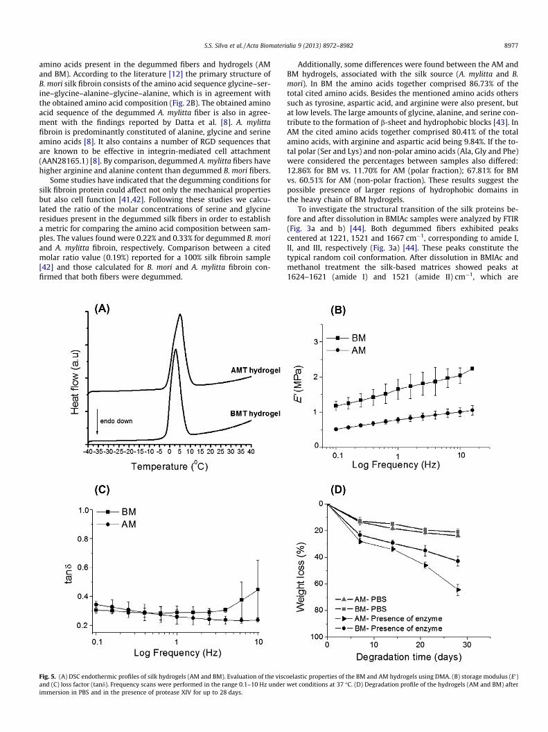
Fig. 5. (A) DSC endothermic profiles of silk hydrogels (AM and BM). Evaluation of the viscand (C) loss factor (tand). Frequency scans were performed in the range 0.1–10 Hz underimmersion in PBS and in the presence of protease XIV for up to 28 days.
Additionally, some differences were found between the AM andBM hydrogels, associated with the silk source (A. mylitta and B.mori). In BM the amino acids together comprised 86.73% of thetotal cited amino acids. Besides the mentioned amino acids otherssuch as tyrosine, aspartic acid, and arginine were also present, butat low levels. The large amounts of glycine, alanine, and serine con-tribute to the formation of b-sheet and hydrophobic blocks [43]. InAM the cited amino acids together comprised 80.41% of the totalamino acids, with arginine and aspartic acid being 9.84%. If the to-tal polar (Ser and Lys) and non-polar amino acids (Ala, Gly and Phe)were considered the percentages between samples also differed:12.86% for BM vs. 11.70% for AM (polar fraction); 67.81% for BMvs. 60.51% for AM (non-polar fraction). These results suggest thepossible presence of larger regions of hydrophobic domains inthe heavy chain of BM hydrogels.
To investigate the structural transition of the silk proteins be-fore and after dissolution in BMIAc samples were analyzed by FTIR(Fig. 3a and b) [44]. Both degummed fibers exhibited peakscentered at 1221, 1521 and 1667 cm�1, corresponding to amide I,II, and III, respectively (Fig. 3a) [44]. These peaks constitute thetypical random coil conformation. After dissolution in BMIAc andmethanol treatment the silk-based matrices showed peaks at1624–1621 (amide I) and 1521 (amide II) cm�1, which are
oelastic properties of the BM and AM hydrogels using DMA. (B) storage modulus (E0)wet conditions at 37 �C. (D) Degradation profile of the hydrogels (AM and BM) after
8978 S.S. Silva et al. / Acta Biomaterialia 9 (2013) 8972–8982
associated with intermolecular b-sheet bands (Fig. 3b) [24,35]. Ab-sence of the characteristic peaks of BMIAc [26] suggest that thissolvent could be almost totally removed by Soxhlet extraction.Thus the structural features of both hydrogels revealed that theuse of BMIAc did not affect the characteristic b-sheet formation in-duced by methanol treatment.
By analyzing curve fitting of the FTIR spectra (data not shown)of the amide I region between 1595 and 1705 cm�1 for both hydro-gels, taking into consideration that the regions 1600–1640 and1690–1705 cm�1 are associated with intermolecular b-sheet bands[32,45], the amount of secondary structure in the AM and BMhydrogels was determined. The results show that the BM sampleshave a higher b-sheet content (28.6%) than the AM samples(25.14%). Relatively little is known about the mechanisms of disso-lution of biopolymers such as silk fibroin in ILs. Some studies havesuggested that ILs disrupts the hydrogen bonding present in b-sheets [46], which in turn can affect the b-sheet content in thehydrogels. In fact, differences in b-sheet content between the AMand BM samples were found, supporting this statement.
Both the AM and BM hydrogels were freeze-dried to obtainscaffolds, whose morphologies were investigated by SEM (seeFig. 4A and B). By analyzing the morphology of the cross-sectionsof the freeze-dried hydrogels it was noted that they had a uniformporosity and pore size distribution in the range 10–50 lm. Never-theless, the BM hydrogels appeared as a compact structure (Fig. 4B)compared with the AM hydrogels (Fig. 4A). Similar pore sizes wereobserved in the case of B. mori scaffolds produced using silk fibroin
Fig. 6. (A) SEM images of hASCs seeded on both AM and BM hydrogels after 28 days. The(B) Confocal laser micrographs of hASCs cultured on AM and BM hydrogels after 28 dayHoechst 33342 for nuclei. Scale bar: left, 20 lm; upper right, 20 lm.
isolated from cocoons [47]. Furthermore, these freeze-dried struc-tures were too fragile to handle (data not shown).
The state of the water in the resulting silk hydrogels was char-acterized (Fig. 5A) by DSC analysis. In the DSC profiles of both silkhydrogels a sharp peak around �1 to 1.5 �C was observed. Accord-ing to the literature [48] bound (non-freezing) and bulk (freezing)water can be found in hydrogels. The relatively lower melting pointof water in the DSC curves of the hydrogels could be associatedwith bound and free water. Considering the enthalpy of fusion(DH) of the water (bound and free) in the silk hydrogels, the calcu-lated percentage water contents were 73.1 ± 5.1% and 80.4 ± 2.2%for AM and BM, respectively.
Fig. 5B and C presents the viscoelastic behavior of the hydrogels,with the storage (elastic) and loss (viscous) components of the com-plex modulus represented. The DMA results point to an increase inE0 for both hydrogels (Fig. 5B), although a significant difference wasobserved between BM and AM. In silk fibroin the presence ofb-sheets has a marked influence on their mechanical properties[5]. Thus the differences observed in the mechanical properties ofAM and BM can be related to the different contents of b-sheet, asdetermined by deconvolution of their FTIR spectra. Thus the highestb-sheet content, calculated for BM hydrogels, allowed the forma-tion of a more rigid network, which affected the mechanical prop-erties. The influence of the hydrogel composition on the lossfactor (tand) was also analyzed. The tand results indicate that thedamping capability is not different for the two materials and doesnot depend on the frequency (Fig. 5C). The elastic modulus of
upper left insets show SEM images of both hydrogels without cells. Scale bar 20 lm.s. The cells were stained with rhodamine–phalloidin for actin filaments (red) and
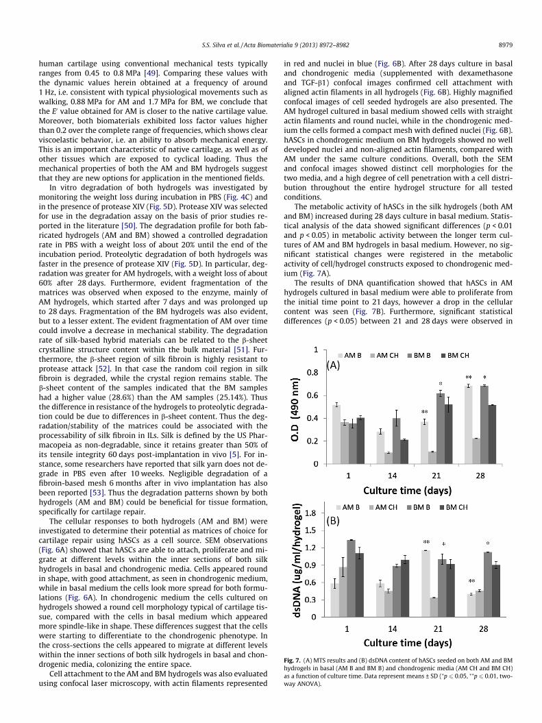
Fig. 7. (A) MTS results and (B) dsDNA content of hASCs seeded on both AM and BMhydrogels in basal (AM B and BM B) and chondrogenic media (AM CH and BM CH)as a function of culture time. Data represent means ± SD (⁄p 6 0.05, ⁄⁄p 6 0.01, two-way ANOVA).
S.S. Silva et al. / Acta Biomaterialia 9 (2013) 8972–8982 8979
human cartilage using conventional mechanical tests typicallyranges from 0.45 to 0.8 MPa [49]. Comparing these values withthe dynamic values herein obtained at a frequency of around1 Hz, i.e. consistent with typical physiological movements such aswalking, 0.88 MPa for AM and 1.7 MPa for BM, we conclude thatthe E0 value obtained for AM is closer to the native cartilage value.Moreover, both biomaterials exhibited loss factor values higherthan 0.2 over the complete range of frequencies, which shows clearviscoelastic behavior, i.e. an ability to absorb mechanical energy.This is an important characteristic of native cartilage, as well as ofother tissues which are exposed to cyclical loading. Thus themechanical properties of both the AM and BM hydrogels suggestthat they are new options for application in the mentioned fields.
In vitro degradation of both hydrogels was investigated bymonitoring the weight loss during incubation in PBS (Fig. 4C) andin the presence of protease XIV (Fig. 5D). Protease XIV was selectedfor use in the degradation assay on the basis of prior studies re-ported in the literature [50]. The degradation profile for both fab-ricated hydrogels (AM and BM) showed a controlled degradationrate in PBS with a weight loss of about 20% until the end of theincubation period. Proteolytic degradation of both hydrogels wasfaster in the presence of protease XIV (Fig. 5D). In particular, deg-radation was greater for AM hydrogels, with a weight loss of about60% after 28 days. Furthermore, evident fragmentation of thematrices was observed when exposed to the enzyme, mainly ofAM hydrogels, which started after 7 days and was prolonged upto 28 days. Fragmentation of the BM hydrogels was also evident,but to a lesser extent. The evident fragmentation of AM over timecould involve a decrease in mechanical stability. The degradationrate of silk-based hybrid materials can be related to the b-sheetcrystalline structure content within the bulk material [51]. Fur-thermore, the b-sheet region of silk fibroin is highly resistant toprotease attack [52]. In that case the random coil region in silkfibroin is degraded, while the crystal region remains stable. Theb-sheet content of the samples indicated that the BM sampleshad a higher value (28.6%) than the AM samples (25.14%). Thusthe difference in resistance of the hydrogels to proteolytic degrada-tion could be due to differences in b-sheet content. Thus the deg-radation/stability of the matrices could be associated with theprocessability of silk fibroin in ILs. Silk is defined by the US Phar-macopeia as non-degradable, since it retains greater than 50% ofits tensile integrity 60 days post-implantation in vivo [5]. For in-stance, some researchers have reported that silk yarn does not de-grade in PBS even after 10 weeks. Negligible degradation of afibroin-based mesh 6 months after in vivo implantation has alsobeen reported [53]. Thus the degradation patterns shown by bothhydrogels (AM and BM) could be beneficial for tissue formation,specifically for cartilage repair.
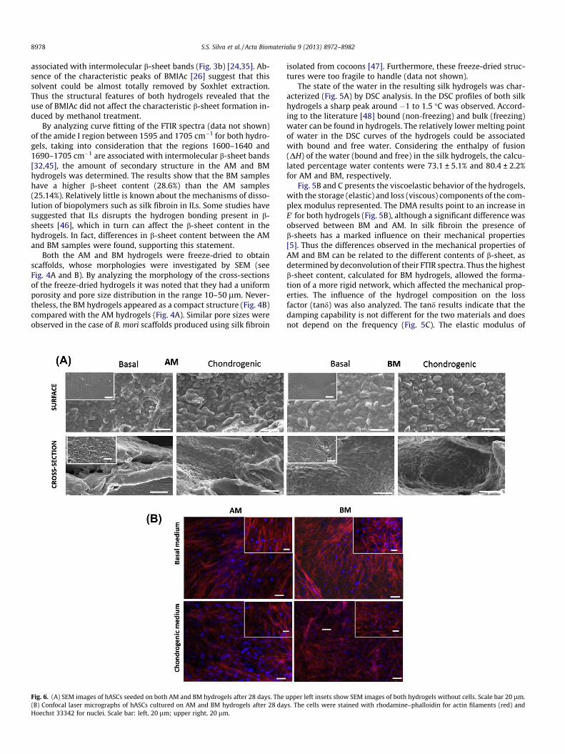
The cellular responses to both hydrogels (AM and BM) wereinvestigated to determine their potential as matrices of choice forcartilage repair using hASCs as a cell source. SEM observations(Fig. 6A) showed that hASCs are able to attach, proliferate and mi-grate at different levels within the inner sections of both silkhydrogels in basal and chondrogenic media. Cells appeared roundin shape, with good attachment, as seen in chondrogenic medium,while in basal medium the cells look more spread for both formu-lations (Fig. 6A). In chondrogenic medium the cells cultured onhydrogels showed a round cell morphology typical of cartilage tis-sue, compared with the cells in basal medium which appearedmore spindle-like in shape. These differences suggest that the cellswere starting to differentiate to the chondrogenic phenotype. Inthe cross-sections the cells appeared to migrate at different levelswithin the inner sections of both silk hydrogels in basal and chon-drogenic media, colonizing the entire space.
Cell attachment to the AM and BM hydrogels was also evaluatedusing confocal laser microscopy, with actin filaments represented
in red and nuclei in blue (Fig. 6B). After 28 days culture in basaland chondrogenic media (supplemented with dexamethasoneand TGF-b1) confocal images confirmed cell attachment withaligned actin filaments in all hydrogels (Fig. 6B). Highly magnifiedconfocal images of cell seeded hydrogels are also presented. TheAM hydrogel cultured in basal medium showed cells with straightactin filaments and round nuclei, while in the chondrogenic med-ium the cells formed a compact mesh with defined nuclei (Fig. 6B).hASCs in chondrogenic medium on BM hydrogels showed no welldeveloped nuclei and non-aligned actin filaments, compared withAM under the same culture conditions. Overall, both the SEMand confocal images showed distinct cell morphologies for thetwo media, and a high degree of cell penetration with a cell distri-bution throughout the entire hydrogel structure for all testedconditions.
The metabolic activity of hASCs in the silk hydrogels (both AMand BM) increased during 28 days culture in basal medium. Statis-tical analysis of the data showed significant differences (p < 0.01and p < 0.05) in metabolic activity between the longer term cul-tures of AM and BM hydrogels in basal medium. However, no sig-nificant statistical changes were registered in the metabolicactivity of cell/hydrogel constructs exposed to chondrogenic med-ium (Fig. 7A).
The results of DNA quantification showed that hASCs in AMhydrogels cultured in basal medium were able to proliferate fromthe initial time point to 21 days, however a drop in the cellularcontent was seen (Fig. 7B). Furthermore, significant statisticaldifferences (p < 0.05) between 21 and 28 days were observed in
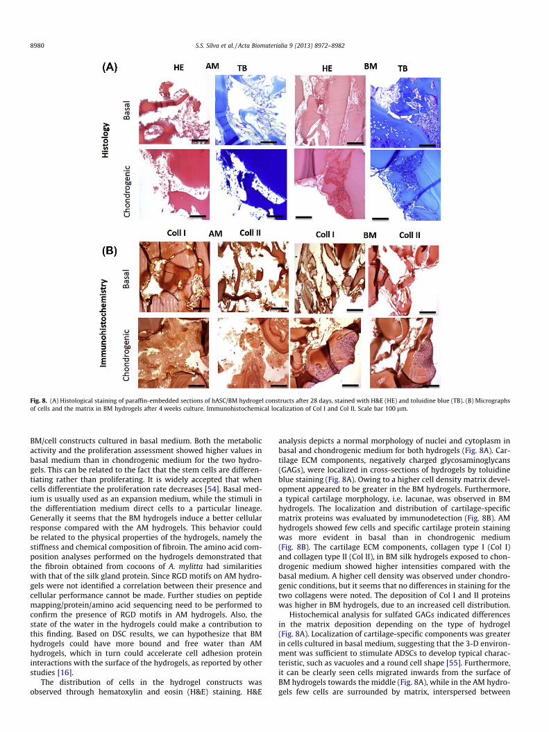
Fig. 8. (A) Histological staining of paraffin-embedded sections of hASC/BM hydrogel constructs after 28 days, stained with H&E (HE) and toluidine blue (TB). (B) Micrographsof cells and the matrix in BM hydrogels after 4 weeks culture. Immunohistochemical localization of Col I and Col II. Scale bar 100 lm.
8980 S.S. Silva et al. / Acta Biomaterialia 9 (2013) 8972–8982
BM/cell constructs cultured in basal medium. Both the metabolicactivity and the proliferation assessment showed higher values inbasal medium than in chondrogenic medium for the two hydro-gels. This can be related to the fact that the stem cells are differen-tiating rather than proliferating. It is widely accepted that whencells differentiate the proliferation rate decreases [54]. Basal med-ium is usually used as an expansion medium, while the stimuli inthe differentiation medium direct cells to a particular lineage.Generally it seems that the BM hydrogels induce a better cellularresponse compared with the AM hydrogels. This behavior couldbe related to the physical properties of the hydrogels, namely thestiffness and chemical composition of fibroin. The amino acid com-position analyses performed on the hydrogels demonstrated thatthe fibroin obtained from cocoons of A. mylitta had similaritieswith that of the silk gland protein. Since RGD motifs on AM hydro-gels were not identified a correlation between their presence andcellular performance cannot be made. Further studies on peptidemapping/protein/amino acid sequencing need to be performed toconfirm the presence of RGD motifs in AM hydrogels. Also, thestate of the water in the hydrogels could make a contribution tothis finding. Based on DSC results, we can hypothesize that BMhydrogels could have more bound and free water than AMhydrogels, which in turn could accelerate cell adhesion proteininteractions with the surface of the hydrogels, as reported by otherstudies [16].
The distribution of cells in the hydrogel constructs wasobserved through hematoxylin and eosin (H&E) staining. H&E
analysis depicts a normal morphology of nuclei and cytoplasm inbasal and chondrogenic medium for both hydrogels (Fig. 8A). Car-tilage ECM components, negatively charged glycosaminoglycans(GAGs), were localized in cross-sections of hydrogels by toluidineblue staining (Fig. 8A). Owing to a higher cell density matrix devel-opment appeared to be greater in the BM hydrogels. Furthermore,a typical cartilage morphology, i.e. lacunae, was observed in BMhydrogels. The localization and distribution of cartilage-specificmatrix proteins was evaluated by immunodetection (Fig. 8B). AMhydrogels showed few cells and specific cartilage protein stainingwas more evident in basal than in chondrogenic medium(Fig. 8B). The cartilage ECM components, collagen type I (Col I)and collagen type II (Col II), in BM silk hydrogels exposed to chon-drogenic medium showed higher intensities compared with thebasal medium. A higher cell density was observed under chondro-genic conditions, but it seems that no differences in staining for thetwo collagens were noted. The deposition of Col I and II proteinswas higher in BM hydrogels, due to an increased cell distribution.
Histochemical analysis for sulfated GAGs indicated differencesin the matrix deposition depending on the type of hydrogel(Fig. 8A). Localization of cartilage-specific components was greaterin cells cultured in basal medium, suggesting that the 3-D environ-ment was sufficient to stimulate ADSCs to develop typical charac-teristic, such as vacuoles and a round cell shape [55]. Furthermore,it can be clearly seen cells migrated inwards from the surface ofBM hydrogels towards the middle (Fig. 8A), while in the AM hydro-gels few cells are surrounded by matrix, interspersed between
S.S. Silva et al. / Acta Biomaterialia 9 (2013) 8972–8982 8981
short fragments of the hydrogels (Fig. 8A). Degradation of thematrices could happen during culture. For instance, it was ob-served that the AM hydrogels appeared more fragile with an openstructure compared with the initial state. These features suggestthat the hydrogels lose their stability due to degradation in culturemedium, confirmed by the weight loss profile, with values be-tween those in the presence or absence of enzyme (Fig. 4D). Thedifferences in the degradation behavior, the state of the waterpresent, the b-sheet content and the mechanical properties of thehydrogels could work together affecting the observed biologicaloutcomes.
4. Conclusions
This work has proposed an alternative route to explore silkfibers (fibroins) obtained from A. mylitta and B. mori cocoonsthrough their fabrication as hydrogels, using BMIAc. Both fabri-cated hydrogels had their own distinct properties, such as acontrolled degradation rate, tuned mechanical properties, visco-elastic behavior, and bound water, which were dependent on thefibroin source. The biological evaluation suggested that hADSCs re-spond differently to the architecture of silk-based hydrogels. Thedevelopment of a cartilage-specific matrix appears to depend onthe structure, composition, stiffness, and state of the water presentin the hydrogels. Thus the outcomes demonstrated herein suggestthat the use of ILs for the dissolution/processing of degummedfibers derived from A. mylitta cocoons into hydrogels could beinteresting in cartilage regeneration repair strategies. Furthermore,depending on the target tissue, the processing could be adapted,with the ILs being employed not only as solvent but also as reac-tion and hybridization medium. These opportunities will overcomethe restricted use of fibroin from the silk glands of live silkworms.
Acknowledgments
The authors acknowledge financial support from the Portu-guese Foundation for Science and Technology (Grants SFRH/BPD/45307/2008 and SFRH/BD/64070/2009), the Fundo Social Europeu,and the Programa Diferencial de Potencial Humano. This work waspartially supported by the FEDER through POCTEP 0330_IBERO-MARE_1_P and also by the Department of Biotechnology, Govern-ment of India. S.C.K. is grateful to R.L.R. and S.S.S. for theirexcellent hospitality during his stay at the 3B’s laboratory,Guimarães, Portugal. R.L.R. also offers his sincere thanks to S.C.K.for providing hospitality during his short visits to his laboratoryin the Indian Institute of Technology, Kharagpur.
Appendix A. Figures with essential color discrimination
Certain figures in this article, particularly Figures 1, 6 and 8, aredifficult to interpret in black and white. The full colour images canbe found in the on-line version, at http://dx.doi.org/10.1016/j.actbio.2013.06.044.
References
[1] Omenetto FG, Kaplan DL. New opportunities for an ancient material. Science2010;329(5991):528–31.
[2] Acharya C, Ghosh SK, Kundu SC. Silk fibroin film from non-mulberry tropicaltasar silkworms: a novel substrate for in vitro fibroblast culture. Acta Biomater2009;5(1):429–37.
[3] Rockwood DN, Preda RC, Yucel T, Wang X, Lovett ML, Kaplan DL. Materialsfabrication from Bombyx mori silk fibroin. Nat Protoc 2011;6(10):1612–31.
[4] Bhardwaj N, Kundu SC. Chondrogenic differentiation of rat MSCs on porousscaffolds of silk fibroin/chitosan blends. Biomaterials 2012;33(10):2848–57.
[5] Altman GH, Diaz F, Jakuba C, Calabro T, Horan RL, Chen J, et al. Silk-basedbiomaterials. Biomaterials 2003;24(3):401–16.
[6] Mandal BB, Kundu SC. A novel method for dissolution and stabilization of non-mulberry silk gland protein fibroin using anionic surfactant sodium dodecylsulfate. Biotechnol Bioeng 2008;99(6):1482–9.
[7] Mandal BB, Das S, Choudhury K, Kundu SC. Implication of silk film RGDavailability and surface roughness on cytoskeletal organization and proliferationof primary rat bone marrow cells. Tissue Eng A 2010;16(7):2391–403.
[8] Datta A, Ghosh AK, Kundu SC. Purification and characterization of fibroin fromthe tropical Saturniid silkworm, Antheraea mylitta. Insect Biochem Mol Biol2001;31(10):1013–8.
[9] Patra C, Talukdar S, Novoyatleva T, Velagala SR, Mühlfeld C, Kundu B, et al. Silkprotein fibroin from Antheraea mylitta for cardiac tissue engineering.Biomaterials 2012;33(9):2673–80.
[10] Datta A, Ghosh AK, Kundu SC. Differential expression of the fibroin gene indevelopmental stages of silkworm, Antheraea mylitta (Saturniidae). CompBiochem Physiol B Biochem Mol Biol 2001;129(1):197–204.
[11] Minoura N, Aiba SI, Higuchi M, Gotoh Y, Tsukada M, Imai Y. Attachment andgrowth of fibroblast cells on silk fibroin. Biochem Biophys Res Commun1995;208(2):511–6.
[12] Kim UJ, Park JY, Li CM, Jin HJ, Valluzzi R, Kaplan DL. Structure and properties ofsilk hydrogels. Biomacromolecules 2004;5(3):786–92.
[13] Nagarkar S, Nicolai T, Chassenieux C, Lele A. Structure and gelation mechanismof silk hydrogels. Phys Chem Chem Phys 2010;12(15):3834–44.
[14] Yucel T, Cebe P, Kaplan DL. Vortex-induced injectable silk fibroin hydrogels.Biophys J 2009;97(7):2044–50.
[15] Wang X, Kluge JA, Leisk GG, Kaplan DL. Sonication-induced gelation of silkfibroin for cell encapsulation. Biomaterials 2008;29(8):1054–64.
[16] Numata K, Katashima T, Sakai T. State of water, molecular structure, andcytotoxicity of silk hydrogels. Biomacromolecules 2011;12(6):2137–44.
[17] Silva SS, Motta A, Rodrigues MrT, Pinheiro AFM, Gomes ME, Mano JoF, et al.Novel genipin-cross-linked chitosan/silk fibroin sponges for cartilageengineering strategies. Biomacromolecules 2008;9(10):2764–74.
[18] Floren ML, Spilimbergo S, Motta A, Migliaresi C. Carbon dioxide induced silkprotein gelation for biomedical applications. Biomacromolecules2012;13(7):2060–72.
[19] Davis NE, Beenken-Rothkopf LN, Mirsoian A, Kojic N, Kaplan DL, Barron AE,et al. Enhanced function of pancreatic islets co-encapsulated with ECMproteins and mesenchymal stromal cells in a silk hydrogel. Biomaterials2012;33(28):6691–7.
[20] Diab T, Pritchard EM, Uhrig BA, Boerckel JD, Kaplan DL, Guldberg RE. A silkhydrogel-based delivery system of bone morphogenetic protein for thetreatment of large bone defects. J Mech Behav Biomed 2012;11:123–31.
[21] Anthony J, Brennecke J, Holbrey J, Maginn E, Mantz R, Trulove P, et al.Physicochemical properties of ionic liquids. In: Wasserscheid P, Welton T,editors. Ionic liquids in synthesis. Weinheim, Germany: Wiley-VCH; 2002.
[22] Gupta MK, Khokhar SK, Phillips DM, Sowards LA, Drummy LF, Kadakia MP,et al. Patterned silk films cast from ionic liquid solubilized fibroin as scaffoldsfor cell growth. Langmuir 2007;23(3):1315–9.
[23] Phillips DM, Drummy LF, Conrady DG, Fox DM, Naik RR, Stone MO, et al.Dissolution and regeneration of Bombyx mori silk fibroin using ionic liquids. JAm Chem Soc 2004;126(44):14350–1.
[24] Silva S, Santos T, Cerqueira M, Marques A, Reys L, Silva T, et al. The use of ionicliquids in the processing of chitosan/silk-based hydrogels for biomedicalapplications. Green Chem 2012;14:1463–70.
[25] Goujon N, Rajkhowa R, Wang X, Byrne N. Effect of solvent on ionic liquiddissolved regenerated Antheraea assamensis silk fibroin. J Appl Polym Sci2013;128(6):4411–6.
[26] Silva SS, Duarte ARC, Carvalho AP, Mano JF, Reis RL. Green processing of porouschitin structures for biomedical applications combining ionic liquids andsupercritical fluid technology. Acta Biomater 2011;7(3):1166–72.
[27] Swatloski RP, Spear SK, Holbrey JD, Rogers RD. Dissolution of cellulose withionic liquids. J Am Chem Soc 2002;124(18):4974–5.
[28] Caplan AI. Adult mesenchymal stem cells for tissue engineering versusregenerative medicine. J Cell Physiol 2007;213(2):341–7.
[29] Csaki C, Schneider PRA, Shakibaei M. Mesenchymal stem cells as a potentialpool for cartilage tissue engineering. Ann Anat 2008;190(5):395–412.
[30] Jin HJ, Park J, Karageorgiou V, Kim UJ, Valluzzi R, Cebe P, et al. Water-stable silkfilms with reduced b-sheet content. Adv Funct Mater 2005;15(8):1241–7.
[31] Silva SS, Maniglio D, Motta A, Mano JF, Reis RL, Migliaresi C. Genipin-modifiedsilk-fibroin nanometric nets. Macromol Biosci 2008;8(8):766–74.
[32] Hu X, Kaplan D, Cebe P. Determining beta-sheet crystallinity in fibrousproteins by thermal analysis and infrared spectroscopy. Macromolecules2006;39(18):6161–70.
[33] Rada T, Reis RL, Gomes ME. Adipose tissue-derived stem cells and theirapplication in bone and cartilage tissue engineering. Tissue Eng B2009;15(2):113–25.
[34] Acharya C, Ghosh S, Kundu S. Silk fibroin protein from mulberry and non-mulberry silkworms: cytotoxicity, biocompatibility and kinetics of L929murine fibroblast adhesion. J Mater Sci Mater Med 2008;19(8):2827–36.
[35] Mandal BB, Kundu SC. Non-bioengineered silk gland fibroin protein:characterization and evaluation of matrices for potential tissue engineeringapplications. Biotechnol Bioeng 2008;100(6):1237–50.
[36] Kundu SC, Kundu B, Talukdar S, Bano S, Nayak S, Kundu J, et al. Invited review.Nonmulberry silk biopolymers. Biopolymers 2012;97(6):455–67.
[37] Matsumoto A, Chen J, Collette AL, Kim U-J, Altman GH, Cebe P, et al.Mechanisms of silk fibroin sol–gel transitions. J Phys Chem B2006;110(43):21630–8.
8982 S.S. Silva et al. / Acta Biomaterialia 9 (2013) 8972–8982
[38] Le Bideau J, Viau L, Vioux A. Ionogels, ionic liquid based hybrid materials.Chem Soc Rev 2011;40(2):907–25.
[39] Néouze M-A, Bideau JL, Gaveau P, Bellayer S, Vioux A. Ionogels, new materialsarising from the confinement of ionic liquids within silica-derived networks.Chem Mat 2006;18(17):3931–6.
[40] Zhao DB, Liao YC, Zhang ZD. Toxicity of ionic liquids. Clean Soil Air Water2007;35(1):42–8.
[41] Jiang P, Liu H, Wang C, Wu L, Huang J, Guo C. Tensile behavior and morphologyof differently degummed silkworm (Bombyx mori) cocoon silk fibres. MaterLett 2006;60(7):919–25.
[42] Wray LS, Hu X, Gallego J, Georgakoudi I, Omenetto FG, Schmidt D, et al. Effectof processing on silk-based biomaterials: reproducibility and biocompatibility.J Biomed Mater Res B Appl Biomater 2011;99B(1):89–101.
[43] Jin H-J, Kaplan DL. Mechanism of silk processing in insects and spiders. Nature2003;424(6952):1057–61.
[44] Um IC, Kweon H, Park YH, Hudson S. Structural characteristics and propertiesof the regenerated silk fibroin prepared from formic acid. Int J Biol Macromol2001;29(2):91–7.
[45] Motta A, Maniglio D, Migliaresi C, Kim H-J, Wan X, Hu X, et al. Silk fibroinprocessing and thrombogenic responses. J Biomat Sci Polym E2009;20(13):1875–97.
[46] Sah M, Pramanik K. Regenerated silk fibroin from Bombyx mori silk cocoon fortissue engineering applications. Int J Environ Sci Technol 2010;1(5):404–8.
[47] Nazarov R, Jin H-J, Kaplan DL. Porous 3-D scaffolds from regenerated silkfibroin. Biomacromolecules 2004;5(3):718–26.
[48] Hoffman AS. Hydrogels for biomedical applications. Adv Drug Deliv Rev2002;54(1):3–12.
[49] Mansour JM. Kinesiology: the mechanics and pathomecanics of humanmovements. In: Oatis C, editor. Biomechanics of cartilage. Philadelphia,PA: Lippincott Williams and Wilkins; 2003. p. 66–79.
[50] Li M, Ogiso M, Minoura N. Enzymatic degradation behavior of porous silkfibroin sheets. Biomaterials 2003;24(2):357–65.
[51] Zhou J, Cao C, Ma X, Hu L, Chen L, Wang C. In vitro and in vivo degradationbehavior of aqueous-derived electrospun silk fibroin scaffolds. Polym DegradStabil 2010;95(9):1679–85.
[52] Yongpei H, Qin Z, Renchuan Y, Lingshuang W, Mingzhong L. The relationshipbetween secondary structure and biodegradation behavior of silk fibroinscaffolds. Adv Mater Sci Eng 2012;2012. p. 185905–10.
[53] Dal Pra I, Freddi G, Minic J, Chiarini A, Armato U. De novo engineering ofreticular connective tissue in vivo by silk fibroin nonwoven materials.Biomaterials 2005;26(14):1987–99.
[54] Geoffrey MC. Cell proliferation in development and differentiation. In: Thecell: a molecular approach. Sunderland, MA: Sinauer Associates; 2000.
[55] LeBaron RG, Athanasiou KA. Ex vivo synthesis of articular cartilage.Biomaterials 2000;21(24):2575–87.