Embed Size (px)
Citation preview
Biochem. J. (2010) 431, 169–178 (Printed in Great Britain) doi:10.1042/BJ20100409 169
REVIEW ARTICLEA nuclear glutathione cycle within the cell cyclePedro DIAZ VIVANCOS*, Tonja WOLFF†1, Jelena MARKOVIC†, Federico V. PALLARDO† and Christine H. FOYER‡2
*CEBAS-CSIC, Department of Plant Breeding, P.O. Box 164, 30100-Murcia, Campus de Espinardo, Spain, †Department of Physiology, University of Valencia-CIBERER, Av. BlascoIbanez 15, 46010 Valencia, Spain, and ‡Centre for Plant Sciences, Faculty of Biology, University of Leeds, Leeds LS2 9JT, U.K.
The complex antioxidant network of plant and animal cells hasthe thiol tripeptide GSH at its centre to buffer ROS (reactiveoxygen species) and facilitate cellular redox signalling whichcontrols growth, development and defence. GSH is found innearly every compartment of the cell, including the nucleus.Transport between the different intracellular compartments ispivotal to the regulation of cell proliferation. GSH co-localizeswith nuclear DNA at the early stages of proliferation in plantand animal cells. Moreover, GSH recruitment and sequestrationin the nucleus during the G1- and S-phases of the cell cycle hasa profound impact on cellular redox homoeostasis and on geneexpression. For example, the abundance of transcripts encodingstress and defence proteins is decreased when GSH is sequesteredin the nucleus. The functions of GSHn (nuclear GSH) are
considered in the present review in the context of whole-cell redoxhomoeostasis and signalling, as well as potential mechanisms forGSH transport into the nucleus. We also discuss the possible roleof GSHn as a regulator of nuclear proteins such as histones andPARP [poly(ADP-ribose) polymerase] that control geneticand epigenetic events. In this way, a high level of GSH in thenucleus may not only have an immediate effect on gene expressionpatterns, but also contribute to how cells retain a memory of thecellular redox environment that is transferred through generations.
Key words: antioxidant, cell cycle, epigenome, gene expression,glutathione, nucleus, oxidative signalling, poly(ADP-ribose)polymerase (PARP), redox state.
INTRODUCTION
The paradigm concerning ROS (reactive oxygen species) thattraditionally viewed these metabolites as potentially lethaland thus presenting an ever-present danger due to theirphysicochemical toxicity, has existed in redox biology for manyyears. More recent data challenge this view, indicating thatthese compounds are key determinants of redox status whoseincreased availability in the cell is favoured by programmedROS production or withdrawal of antioxidant capacity [1,2].Enhanced ROS accumulation influences many pathways of redoxsignalling and control [3]. ROS signalling is made possible byhomoeostatic regulation that is dependent on cellular antioxidantstatus. Antioxidants continuously process ROS and they arecrucial components of the cellular redox signalling network. Ithas been known for many years that the cellular redox state isa crucial regulator of the cell cycle [4–6]. Low levels of cellularoxidation triggered by superoxide and hydrogen peroxide activatecell signalling pathways leading to proliferation, and are requiredfor the correct mitogenic signalling [5,7,8]. An oxidation eventthat occurs early in the G1-phase of the cell cycle is a criticalregulatory step in the progression to S-phases [9]. Moreover, inthe G1-phase cellular GSH levels are low and an increase in totalGSH is subsequently necessary for the cells to progress fromthe G1- to the S-phase [10]. Thus the current model of cell-cycleregulation incorporates an intrinsic redox cycle, in which transientoxidations, perhaps related to bursts of ROS, serve to regulate keyproteins by processes such as thiol–disulfide exchange reactionsat critical cysteine residues and in this way regulate cell-cycle
progression or cause an arrest in the proliferation cycle [11].However, conditions that cause excessive or prolonged cellularoxidation arrest the cell cycle and trigger cell death. Uncontrolledoxidation which can damage DNA, leading to altered basesand damaged sugar residues, resulting in single- and double-stranded DNA breaks [12,13], is considered to be importantin this response. Strand breaks trigger a DDR (DNA damageresponse) by inducing the expression of molecular markersassociated with DNA damage repair, such as PARP [poly(ADP-ribose) polymerase], RAD51 and BRCA (breast cancer early-onset) family members [14–17]. An inherent part of the DDRis the activation of checkpoints with conserved key regulatorssuch as the ATM (ataxia telangiectasia mutated) and Rad3-relatedprotein kinases that arrest cell-cycle progression and stimulaterepair processes in order to preserve genome integrity [18–20]. Itshould be noted, however, that while the initiation of a DDR byoxidative DNA base damage is well characterized in mammalianand yeast cells [21,22], there is little evidence that oxidative stressleads to DNA damage or DDR induction in plants [12,23,24].Oxidative DNA modifications display a negative linear correlationwith nuclear GSH [25].
Until recently, much uncertainty has remained concerning theenzymes and antioxidants that afford protection to the nucleusand prevent DNA damage. However, the characterization of therecruitment of one of the cell’s major antioxidants, the thioltripeptide GSH (Figure 1), into the nucleus in the G1- and S-phasesof the cell cycle [26] provides a realistic and powerful mechanismfor strategic deployment of antioxidant defence mechanismsduring the cell cycle in animals and plants.
Abbreviations used: AP-1, activator protein-1; BAG, Bcl-2-associated athanogene; BSO, buthionine sulfoximine; CMFDA, 5-chloromethyl fluoresceindiacetate; DDR, DNA damage response; DEM, diethylmaleate; DHA, dehydroascorbate; γ-EC, γ-glutamyl cysteine; GCL, glutamate-cysteine ligase; GFP,green fluorescent protein; GR, glutathione reductase; GRX, glutaredoxin; GSH-S, glutathione synthetase; GST, glutathione transferase; NAC, N-acetyl-L-cysteine; NF-κB, nuclear factor κB; PARP, poly(ADP-ribose) polymerase; rml1, rootmeristemless1; ROS, reactive oxygen species; TRX, thioredoxin.
1 Present address: Max Planck Institute for Development Biology, Tubingen, Germany.2 To whom correspondence should be addressed (email [email protected]).
c© The Authors Journal compilation c© 2010 Biochemical Society
www.biochemj.org
Bio
chem
ical
Jo
urn
al
170 P. Diaz Vivancos and others
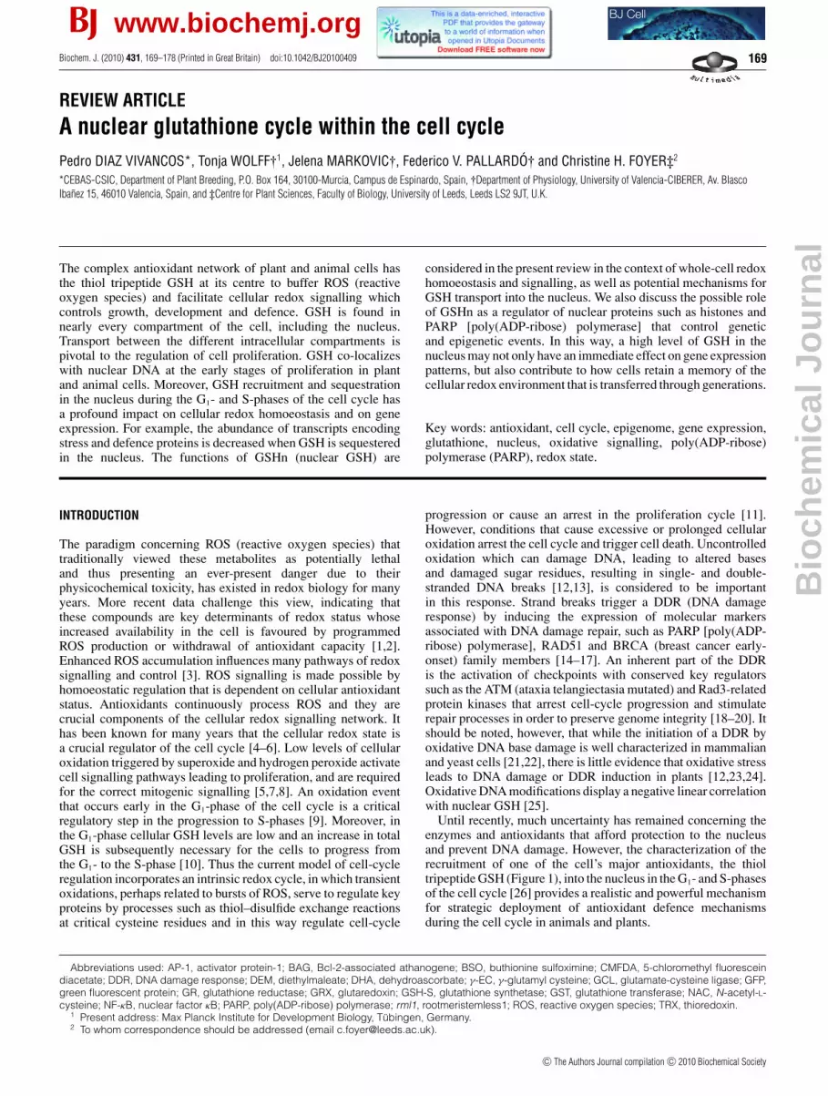
Figure 1 A simple scheme showing the pathway of glutathione synthesisand cycling between reduced (GSH) and oxidized (GSSG) forms
(1) γ -Glutamyl cysteine synthetase (glutamate–cysteine ligase); (2) glutathione synthetase;(3) glutathione reductase.
GLUTATHIONE SYNTHESIS AND INTRACELLULARCOMPARTMENTATION
Glutathione is synthesized in a two-step ATP-dependent pathway.The first reaction, which is catalysed by the enzyme γ -glutamylcysteine synthetase (encoded by the GSH1 gene in plants) alsocalled GCL (glutamate-cysteine ligase), produces γ -EC (γ -glutamyl cysteine). In the second step, catalysed by GSH-S(glutathione synthetase; encoded by the GSH2 gene in plants),glycine is added to form glutathione (Figure 1). Much of ourunderstanding of the functions of GSH in plants has comefrom studies on mutants altered in these enzymes or fromtransgenic plants where the activities of GCL or GSH-S have beenmanipulated [27,28]. For example, in the model plant Arabidopsisthaliana knockout mutations of GSH1 that completely blockglutathione synthesis are lethal [29]. This demonstrates that glu-tathione is essential for the plant survival, as is the case inmammalian and yeast cells.
Less severe mutations in the GSH1 gene, which result in de-creased glutathione contents, have been extremely useful inelucidating the functions of glutathione in plants. For example,the rml1 (rootmeristemless1) mutant, which has less than 5 %of wild-type glutathione contents, fails to develop a root apicalmeristem because all the cells arrest at the G1-phase of thecell cycle [28]. Intriguingly, this mutant shows a much lesssevere shoot than root phenotype because of redundancy betweenglutathione and TRXs (thioredoxins) in the control of shoot apicalmeristem function. The redundancy in TRX/glutathione functionswas demonstrated by combining the rml1 mutation with mutationsin the two genes encoding cytosolic/mitochondrial NADPH-TRXreductase (NTRA, NTRB) [30]. Other mutations in GSH1, resultingin decreases in tissue glutathione contents of between 25 and50% that of the wild-type plants, do not lead to an altered growthphenotype. However, lower glutathione levels result in a decreasedability to withstand biotic and abiotic stresses. For example,the cad2 mutant was identified by an enhanced sensitivity tocadmium, the rax1 mutant by altered expression of a cytosolicascorbate peroxidase gene and the pad2 mutant by decreasedcamalexin contents and enhanced sensitivity to pathogens [31].
In Arabidopsis leaves, the first step of GSH synthesis takesplace exclusively in the chloroplasts [32]. In other plant speciesthe situation is less clear and γ -EC production may also takeplace in the cytosol, as well as the chloroplasts [33]. While thesecond enzyme, GSH-S, is encoded by a single gene (GSH2),alternative splicing results in localization of the gene product inthe chloroplast and cytosol [33,34]. While GSH1 knockouts areembryonic lethal, GSH2-knockout mutants are seedling-lethal,probably reflecting partial replacement of GSH functions byγ -EC, which accumulates to very high levels in these plants[35]. The wild-type phenotype can be restored in gsh2 mutantsby complementing the mutant with targeted expression of theenzyme to the cytosol alone [35]. All of the other compartments
of the cell are dependent on the import of glutathione from thecytosol.
GSH synthesis is subject to multiple levels of control, but themost important of these are considered to be cysteine availabilityand GCL activity [2]. It has long been recognized that cysteineavailability can impose an important limitation on GSH synthesis.In general, however, up-regulation of the cysteine synthesispathway occurs concomitantly with that of GSH synthesis [27].The expression of the GSH1 or GSH2 genes is not modulated bymany known environmental or endogenous triggers, and markedincreases in the abundance of GSH1 or GSH2 transcripts haveonly been reported in response to jasmonic acid and in certainstress situations such as exposure to heavy metals or certainpathogens. Crucially, increases in hydrogen peroxide and otheroxidants, which are known to cause accumulation of glutathione,do not alter the expression of the GSH1 or GSH2 genes. However,GCL activity has long been known to be regulated by GSH[31,32,36] (Figure 1). The GCL homodimer is linked in plantsby two disulfide bonds, one of which is involved in redoxregulation [36]. Although the exact mechanistic details remain tobe resolved, activation through disulfide formation is consideredto be important in contributing to up-regulation of GSH synthesisin response to oxidative and other stresses [36]. While it remainsunclear precisely how redox regulation interacts mechanisticallywith the classic model of GSH feedback inhibition of GCLactivity, these two regulatory processes would appear to havedistinct functions from a physiological perspective. For example,feedback inhibition acts as a homoeostatic control mechanism torestrict the extent of cellular GSH accumulation and acceleratesynthesis in response to depletion and, conversely, covalent thiol–disulfide regulation allows enhanced synthesis, specifically inresponse to increased cellular oxidation.
There is strong interplay between GSH concentrations andGSH/GSSG ratio, where an accumulation of GSSG often causessubsequent increases in total glutathione [2]. Oxidative activationof GSH synthesis can be rationalized in terms of the homoeostaticmaintenance of glutathione redox potential, as this is relatedto [GSH]2/[GSSG] rather than to [GSH]/[GSSG]. At a constantGSH/GSSG ratio, increases in glutathione pool size entail subtle,but potentially important, decreases in redox potential.
GLUTATHIONE AND CELLULAR REDOX STATE
The redox potential of the cytosol, which is established by themajor redox buffers such as ascorbate, glutathione and NADP(H),is considered to be in the order of −300 mV. If we assume thatall three of these redox buffers are in equilibrium in the cytosol,then at NADP+/NADPH = 1 (representing a redox potential of−320 mV), there should be very little oxidized glutathione. Thusthe overall redox state of the glutathione [GSH/(GSH+2GSSG)]pool in a organ such as the leaf is considered to be between0.9 and 0.95 assuming that GR (glutathione reductase) activityallows the glutathione and NADP(H) couples to be in redoxequilibrium (i.e. at the same redox potential). However, a ratherdifferent picture emerges from the application of the in vivoprobes based on GFPs (green flourescent proteins) containingoxidizable thiol groups that are able to monitor glutathionestatus. Such measurements suggest that cytosolic GSH/GSSGratios are underestimated in whole-tissue measurements, and thatGSH/GSSG ratios can be considerably higher than 0.95 in certainintracellular compartments that therefore have a redox potentialof less than −320 mV [37]. This would confer a high sensitivityon the signalling function of glutathione redox potential mediated
c© The Authors Journal compilation c© 2010 Biochemical Society

A nuclear glutathione cycle 171
through GRX (glutaredoxin)-dependent changes in protein thiol–disulfide status.
Increases in GSSG relative to GSH are a useful indicator ofoxidative stress or ‘disulfide stress’. However, it should be notedthat an important property of cellular glutathione homoeostasisis that increased oxidation is generally accompanied or rapidlyfollowed by increases in the total pool size.
The concentration and/or redox state of the glutathionepool in each cellular compartment are important for cellularredox homoeostasis and redox signalling [38,39]. While itremains unclear how GSH/GSSG transporters function in inter-compartmental redox regulation, the transport of GSH betweenthe different cellular compartments is fundamental for themaintenance of cellular GSH levels and redox-based signallingpathways. Recently, a screen for Arabidopsis mutants that areinsensitive to the GCL-inhibitor BSO (buthionine sulfoximine),revealed the identity of the chloroplast γ -EC and GSHtransporter, called chloroquinone-like transporter [40]. However,whereas GSH transport mechanisms have been identified atthe plastid membrane [40] and at the plasma membrane andtonoplast membranes [41–44], no information is available on themechanism that regulate GSH transport into the nucleus. Untilrecent observations suggested that much of the cellular GSH poolcould be restricted to the nucleus [26], transport of GSH into thenucleus had not been thought to be important because nuclearpores were not considered to restrict diffusion of low-molecular-mass solutes such as GSH.
THE FUNCTIONS OF GLUTATHIONE
Glutathione is the major non-protein cellular thiol and is presentin many cellular compartments at millimolar concentrations. Thishigh abundance imposes a low cellular thiol–disulfide redoxpotential on the cell and allows GSH to function as a thiol buffer,maintaining cytoplasmic thiols in the reduced state. Like otherthiols, glutathione can undergo numerous redox reactions andalmost all GSH functions are linked to oxidation of the cysteinegroup. GSH is oxidized by ROS at high rates, but it is able tofunction as an efficient antioxidant scavenger and ‘sacrificial’nucleophile because it is present in the cell at relatively highconcentrations. Oxidized forms notably include disulfides, eitherwith another glutathione molecule to form GSSG or with adifferent thiol to form ‘mixed disulfides’, as well as moreoxidized forms in which the thiol group is converted into sulfenic,sulfinic or sulfonic acids [1]. However, glutathione functionsare not restricted to these compounds because of the enormouspotential array of glutathione conjugates that can be formed withelectrophilic species.
The cellular glutathione pool is an effective buffer or barrieragainst excessive oxidation protecting redox-sensitive mole-cules in the cell. The glutathione pool is maintained predominantlyin a reduced state because of the action of GR. GR is found inmany cellular compartments and has a very high affinity for thesubstrates GSSG and NADPH. In addition to chemical oxidation,GSH oxidation can also occur as a result of a number ofenzyme-catalysed reactions that use GSH to reduce hydrogenperoxide or other peroxides to water or the correspondingalcohol. It should be noted, however, that in plants enzymesthat have been annotated as glutathione peroxidases use TRXsmore efficiently than glutathione. However, certain plant GSTs(glutathione transferases) can catalyse glutathione peroxidationand GSH is also involved in peroxide metabolism through GRX-dependent peroxiredoxins. In addition, GSH is required for theregeneration of reduced ascorbate through the action of DHARs[DHA (dehydroascorbate) reductases]. GSH-dependent reduction
of DHA allows NADPH oxidation to be coupled to ROS removalvia ascorbate and glutathione pools.
The nucleophilic nature and the chemical reactivity of its γ -cysteine thiol group make GSH particularly suitable for a broadrange of functions in metabolism and signalling [1,2]. Glutathioneparticipates in post-transcriptional protein modification throughthiol–disulfide exchange and through S-glutathionylation, i.e.formation of a stable mixed disulfide between GSH and a proteinthiol, and in this way GSH protects proteins of irreversiblemodifications induced by oxidation or nitrosylation by reactivenitrogen species. Moreover, GSNO (S-nitrosoglutathione) has anumber of physiological functions, particularly as a signallingmolecule or as a reservoir of NO. In addition to its antioxidantactivities, glutathione is important in the detoxification ofxenobiotics through the action of GSTs that catalyse theconjugation to glutathione and phytochelatins, which contributeto heavy metal tolerance. In plants GSH also participates inthe regulation of sulfur metabolism with roles in the uptake,assimilation, transport and storage of reduced sulfur. As indicatedabove, cellular glutathione status has long been implicated inrelaying defence signals in plants [1,2,38]. For example, GSH isan important mediator of the salicylate-dependent suppression ofjasmonate signalling in plants that occurs upon pathogen or insectattack [45]. In animal cells, the glutathionylation of proteins suchas SERCA (sarcoplasmic/endoplasmic reticulum Ca2+-ATPase)pumps and myofibrils in muscle, and transcription factors suchas NF-κB (nuclear factor κB). In the latter case, the GSH-generating agent NAC (N-acetyl-L-cysteine) has been shown toenhance hypoxic apoptosis in mouse fibroblasts by blockingthe NF-κB survival pathway. Since GRXs, which specificallycatalyse reduction of protein-SSG mixed disulfides, reversed theinhibition of p65-NF-κB DNA binding in extracts from hypoxiccells plus NAC and restored NF-κB activity, GRX-dependentS-glutathionylation of p65-NF-κB is considered to be the mostprobable mediator of NF-κB inactivation and enhanced hypoxicapoptosis [46].
GLUTATHIONE-DEPENDENT REGULATION OF CELL PROLIFERATION
A considerable amount of evidence in the literature obtainedfrom studies on both animal and plant cells support the viewthat glutathione is a key regulator of cell proliferation. Earlystudies showed that GSH was an important factor in the controlof tumour growth [47], whereas low-molecular-mass thiols wereimplicated in the regulation of cell proliferation [48]. In a similarmanner to mammalian cells, plants cells in the G1-phase of thecell cycle were found to have a very low level of GSH [10].Moreover, an increase in the amount of total GSH was shownto be necessary for the cells to progress from the G1- to theS-phase of the cell cycle [10]. In contrast with the positive effectof GSH, addition of GSSG caused the cell cycle to arrest at G1 [49].As discussed above, depletion of GSH blocks the transition fromG1- to S-phase in the cell cycle of the root apical meristem, butredundancy between GSH and TRX functions are observed in thedevelopment of the shoot meristem [30]. GSH is also importantin the formation of nodules, which are specialized organelleswhere plants cells are in symbiotic association with rhizobialbacteria [50]. Although in Arabidopsis cells the GSH/GSSG ratiois high and constant throughout the cell cycle [51], proliferatinghuman colon epithelial cells were characterized by particularlyhigh GSH/GSSG ratios [52].
Recent studies have shown that the intracellular distributionof GSH is also a crucial factor in proliferation, particularlythe distribution of GSH between the nucleus and cytosol[26]. A number of methods are available for the detection
c© The Authors Journal compilation c© 2010 Biochemical Society

172 P. Diaz Vivancos and others
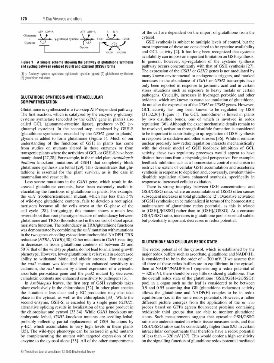
Figure 2 Confocal microscopy images showing GSH localization in thecytoplasm (a–c) and in the nucleus (d–f) of Arabidopsis cells
Double staining using Hoechst 33342 to localize nuclei (blue stain; a, c, d and f) and CMFDA(green stain; b, c, e and f) to track GSH, was performed on living cells at points where GSHwas localized throughout the cytoplasm (a–c) or in the nucleus (d–f). Confocal images (a) and(d) show blue Hoechst fluorescence, images (b) and (e) show distributions of green CMFDAfluorescence, and (c) and (f) show the two superimposed stains.
of GSH in cells. Pioneering work using monochlorobimane–GSH conjugates demonstrated the compartmentalization of GSHin the nucleus of hepatocytes [53]. Thereafter, microinjectionstudies revealed that GSH conjugates preferentially localize tonuclei [54]. Immunocytochemistry using specific antibodies andcomputer-supported transmission electron microscopy has beenused in plants to quantify the relative amounts of glutathionein the different compartments of the plant cell [55,56]. Real-time imaging is generally restricted to cytosol and mitochondrialcompartments, but it also provides a useful tool with whichto study the participation of glutathione in cellular redoxhomoeostasis [57]. These in vivo probes, which are based on GFPscontaining oxidizable thiol groups, were designed to ‘sense’ theglutathione redox potential by equilibration of their thiol–disulfidestatus with that of glutathione. To date, these redox-sensitive GFPshave largely been used to monitor glutathione status in the cytosol[57]. Confocal microscopy using CellTracker Green [CMFDA(5-chloromethyl fluorescein diacetate)] to detect GSH has provedto be a useful tool in the characterization of GSH distributionbetween the nucleus and cytoplasm in animal and plant (Figure 2)cells [26,51].
RECRUITMENT INTO THE NUCLEUS DURING THE CELL CYCLE
As discussed above there is a general consensus of opinionthat the cell cycle includes a redox cycle involving changesin the abundance of ROS and the intracellular partitioning ofGSH. Moreover, the hierarchy of redox-dependent regulatorycheckpoint changes during cell proliferation in a manner thatnot only is crucial for correct cell-cycle progression, butalso links cell-cycle progression to cell fate [58]. An oxidationevent is required at G1 [9] in order to stimulate mitogenicpathways that control the activities of CDKs (cyclin-dependentkinases) and initiate the phosphorylation cascade including thephosphorylation of the pRB (retinoblastoma protein) in orderto allow entry into S-phase and activate DNA replication andcell division [58,59]. Thereafter, a more general reduction ofthe cellular environment is required in order to enable the cellsto progress to the G2/M-phases [60]. GSH recruitment into thenucleus and concomitant regulation of glutathione homoeostasis
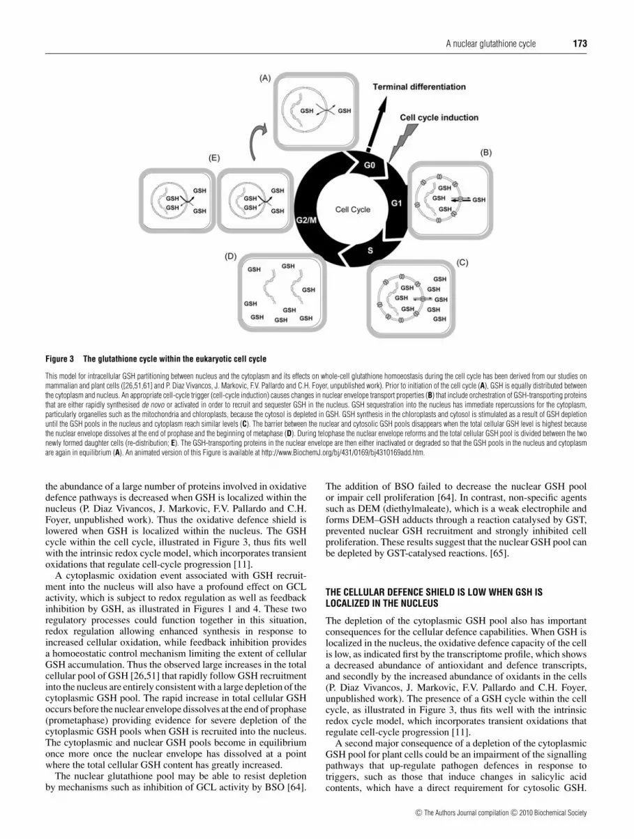
in plant [51] and mammalian [26] cells will not only haveimplications for the regulation of the abundance of ROS in thecytosol, but also have a profound impact on the redox stateof the nucleus and the cytosol with many repercussions forprocesses that are subject to redox regulation in each cellularcompartment. Combining information obtained from our studieson mammalian [26,61] and plant [51] cells, we propose amodel for the glutathione cycle that exists within the cell cycleillustrated in Figure 3 (see also the accompanying animationat http://www.BiochemJ.org/bj/431/0169/bj4310169add.htm). Inthis model, intracellular GSH partitioning and homoeostasisundergoes the following pattern during the cell cycle: (i) GSHis recruited and sequestered in the nucleus early (G1) in cellproliferation, (ii) as a result of GSH sequestration in the nucleus,the cytoplasm is starved of GSH, (iii) the sharp fall in cytosolicGSH availability and the accompanying change in cytosolic redoxstate triggers GSH synthesis in the cytoplasm, (iv) the totalGSH pool of the cells increases rapidly, (v) the nuclear envelopedissolves at the end of prophase/beginning of metaphase allowingequilibration between the cytosol and nuclear GSH pools duringthe G2- and M-phases, and (vi) the nuclear envelope re-formsduring telophase (before cytokinesis), the cells divide and thecellular GSH pool is re-distributed between the daughter cells.
THE IMPACT OF NUCLEAR GSH SEQUESTRATION ON THE REDOXPROCESSES IN THE CYTOPLASM
A depletion of cytoplasmic GSH upon GSH recruitment intothe nucleus is entirely consistent with previous observations ofan oxidation event occurring early in the G1-phase of the cellcycle [9,58]. The recruitment of GSH into the nucleus early incell proliferation will have a profound effect on the cytosolicGSH concentration without necessarily affecting the GSH/GSSGratios. The glutathione redox potential depends not only on theGSH/GSSG ratio of the cytosol, but also on the overall GSHconcentration. GSH depletion will greatly change the cytosolicglutathione redox potential of plant cells, which will rise fromvalues that are generally accepted to be well below −300 mV. Thiswill greatly alter signalling functions associated with glutathioneredox potential-mediated events such as GRX-dependent changesin protein thiol–disulfide status, where a change of 50 mVsignificantly alters the relationship between oxidized and reducedforms. Two considerations are important in analysing the effectsof changes in the cytosolic and nuclear GSH pools on thiol-based ROS sensors [1]. These are the thermodynamics (redoxpotential of oxidizable thiols) of the system and kinetics (ability tocompete with the antioxidative system). In oxyR, for example, thehydrogen-peroxide-reactive thiols have a midpoint redox potentialof approx. −0.18 V and a rate constant for the reaction withhydrogen peroxide that is comparable with peroxidases. Theseproperties mean that under optimal conditions where the redoxpotential of the glutathione pool is approx. −0.24 V, oxyR willgenerally be in its reduced inactive form. However, any increasein hydrogen peroxide availability or change in glutathione redoxpotential (or both) can readily cause oxidative activation ofthe sensor [1]. Redox-dependent shifts in oxyR–DNA contactssuggest that such a mechanism might operate in cells [62]. Sensoroxidation may also be facilitated by programmed withdrawal ofGSH, or it could be catalysed by specific peroxidases, as shownfor the yAP-1 system in yeast [63].
A depletion of cytoplasmic GSH upon GSH recruitment intothe nucleus is entirely consistent with previous observations of anoxidation event occurring early in the G1-phase of the cell cycle[9,58]. Comparisons of the plant cell transcriptome when GSH islocalized in the nucleus relative to cytoplasmic GSH show that
c© The Authors Journal compilation c© 2010 Biochemical Society
A nuclear glutathione cycle 173
Figure 3 The glutathione cycle within the eukaryotic cell cycle
This model for intracellular GSH partitioning between nucleus and the cytoplasm and its effects on whole-cell glutathione homoeostasis during the cell cycle has been derived from our studies onmammalian and plant cells ([26,51,61] and P. Diaz Vivancos, J. Markovic, F.V. Pallardo and C.H. Foyer, unpublished work). Prior to initiation of the cell cycle (A), GSH is equally distributed betweenthe cytoplasm and nucleus. An appropriate cell-cycle trigger (cell-cycle induction) causes changes in nuclear envelope transport properties (B) that include orchestration of GSH-transporting proteinsthat are either rapidly synthesised de novo or activated in order to recruit and sequester GSH in the nucleus. GSH sequestration into the nucleus has immediate repercussions for the cytoplasm,particularly organelles such as the mitochondria and chloroplasts, because the cytosol is depleted in GSH. GSH synthesis in the chloroplasts and cytosol is stimulated as a result of GSH depletionuntil the GSH pools in the nucleus and cytoplasm reach similar levels (C). The barrier between the nuclear and cytosolic GSH pools disappears when the total cellular GSH level is highest becausethe nuclear envelope dissolves at the end of prophase and the beginning of metaphase (D). During telophase the nuclear envelope reforms and the total cellular GSH pool is divided between the twonewly formed daughter cells (re-distribution; E). The GSH-transporting proteins in the nuclear envelope are then either inactivated or degraded so that the GSH pools in the nucleus and cytoplasmare again in equilibrium (A). An animated version of this Figure is available at http://www.BiochemJ.org/bj/431/0169/bj4310169add.htm.
the abundance of a large number of proteins involved in oxidativedefence pathways is decreased when GSH is localized within thenucleus (P. Diaz Vivancos, J. Markovic, F.V. Pallardo and C.H.Foyer, unpublished work). Thus the oxidative defence shield islowered when GSH is localized within the nucleus. The GSHcycle within the cell cycle, illustrated in Figure 3, thus fits wellwith the intrinsic redox cycle model, which incorporates transientoxidations that regulate cell-cycle progression [11].
A cytoplasmic oxidation event associated with GSH recruit-ment into the nucleus will also have a profound effect on GCLactivity, which is subject to redox regulation as well as feedbackinhibition by GSH, as illustrated in Figures 1 and 4. These tworegulatory processes could function together in this situation,redox regulation allowing enhanced synthesis in response toincreased cellular oxidation, while feedback inhibition providesa homoeostatic control mechanism limiting the extent of cellularGSH accumulation. Thus the observed large increases in the totalcellular pool of GSH [26,51] that rapidly follow GSH recruitmentinto the nucleus are entirely consistent with a large depletion of thecytoplasmic GSH pool. The rapid increase in total cellular GSHoccurs before the nuclear envelope dissolves at the end of prophase(prometaphase) providing evidence for severe depletion of thecytoplasmic GSH pools when GSH is recruited into the nucleus.The cytoplasmic and nuclear GSH pools become in equilibriumonce more once the nuclear envelope has dissolved at a pointwhere the total cellular GSH content has greatly increased.
The nuclear glutathione pool may be able to resist depletionby mechanisms such as inhibition of GCL activity by BSO [64].
The addition of BSO failed to decrease the nuclear GSH poolor impair cell proliferation [64]. In contrast, non-specific agentssuch as DEM (diethylmaleate), which is a weak electrophile andforms DEM–GSH adducts through a reaction catalysed by GST,prevented nuclear GSH recruitment and strongly inhibited cellproliferation. These results suggest that the nuclear GSH pool canbe depleted by GST-catalysed reactions. [65].
THE CELLULAR DEFENCE SHIELD IS LOW WHEN GSH ISLOCALIZED IN THE NUCLEUS
The depletion of the cytoplasmic GSH pool also has importantconsequences for the cellular defence capabilities. When GSH islocalized in the nucleus, the oxidative defence capacity of the cellis low, as indicated first by the transcriptome profile, which showsa decreased abundance of antioxidant and defence transcripts,and secondly by the increased abundance of oxidants in the cells(P. Diaz Vivancos, J. Markovic, F.V. Pallardo and C.H. Foyer,unpublished work). The presence of a GSH cycle within the cellcycle, as illustrated in Figure 3, thus fits well with the intrinsicredox cycle model, which incorporates transient oxidations thatregulate cell-cycle progression [11].
A second major consequence of a depletion of the cytoplasmicGSH pool for plant cells could be an impairment of the signallingpathways that up-regulate pathogen defences in response totriggers, such as those that induce changes in salicylic acidcontents, which have a direct requirement for cytosolic GSH.
c© The Authors Journal compilation c© 2010 Biochemical Society
174 P. Diaz Vivancos and others
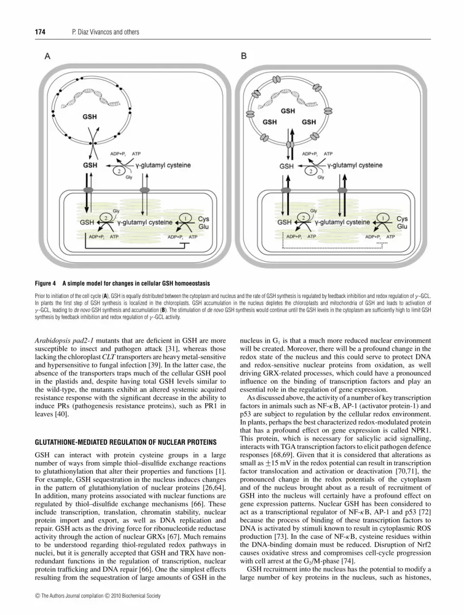
Figure 4 A simple model for changes in cellular GSH homoeostasis
Prior to initiation of the cell cycle (A), GSH is equally distributed between the cytoplasm and nucleus and the rate of GSH synthesis is regulated by feedback inhibition and redox regulation of γ -GCL.In plants the first step of GSH synthesis is localized in the chloroplasts. GSH accumulation in the nucleus depletes the chloroplasts and mitochondria of GSH and leads to activation ofγ -GCL, leading to de novo GSH synthesis and accumulation (B). The stimulation of de novo GSH synthesis would continue until the GSH levels in the cytoplasm are sufficiently high to limit GSHsynthesis by feedback inhibition and redox regulation of γ -GCL activity.
Arabidopsis pad2-1 mutants that are deficient in GSH are moresusceptible to insect and pathogen attack [31], whereas thoselacking the chloroplast CLT transporters are heavy metal-sensitiveand hypersensitive to fungal infection [39]. In the latter case, theabsence of the transporters traps much of the cellular GSH poolin the plastids and, despite having total GSH levels similar tothe wild-type, the mutants exhibit an altered systemic acquiredresistance response with the significant decrease in the ability toinduce PRs (pathogenesis resistance proteins), such as PR1 inleaves [40].
GLUTATHIONE-MEDIATED REGULATION OF NUCLEAR PROTEINS
GSH can interact with protein cysteine groups in a largenumber of ways from simple thiol–disulfide exchange reactionsto glutathionylation that alter their properties and functions [1].For example, GSH sequestration in the nucleus induces changesin the pattern of glutathionylation of nuclear proteins [26,64].In addition, many proteins associated with nuclear functions areregulated by thiol–disulfide exchange mechanisms [66]. Theseinclude transcription, translation, chromatin stability, nuclearprotein import and export, as well as DNA replication andrepair. GSH acts as the driving force for ribonucleotide reductaseactivity through the action of nuclear GRXs [67]. Much remainsto be understood regarding thiol-regulated redox pathways innuclei, but it is generally accepted that GSH and TRX have non-redundant functions in the regulation of transcription, nuclearprotein trafficking and DNA repair [66]. One the simplest effectsresulting from the sequestration of large amounts of GSH in the
nucleus in G1 is that a much more reduced nuclear environmentwill be created. Moreover, there will be a profound change in theredox state of the nucleus and this could serve to protect DNAand redox-sensitive nuclear proteins from oxidation, as welldriving GRX-related processes, which could have a pronouncedinfluence on the binding of transcription factors and play anessential role in the regulation of gene expression.
As discussed above, the activity of a number of key transcriptionfactors in animals such as NF-κB, AP-1 (activator protein-1) andp53 are subject to regulation by the cellular redox environment.In plants, perhaps the best characterized redox-modulated proteinthat has a profound effect on gene expression is called NPR1.This protein, which is necessary for salicylic acid signalling,interacts with TGA transcription factors to elicit pathogen defenceresponses [68,69]. Given that it is considered that alterations assmall as +−15 mV in the redox potential can result in transcriptionfactor translocation and activation or deactivation [70,71], thepronounced change in the redox potentials of the cytoplasmand of the nucleus brought about as a result of recruitment ofGSH into the nucleus will certainly have a profound effect ongene expression patterns. Nuclear GSH has been considered toact as a transcriptional regulator of NF-κB, AP-1 and p53 [72]because the process of binding of these transcription factors toDNA is activated by stimuli known to result in cytoplasmic ROSproduction [73]. In the case of NF-κB, cysteine residues withinthe DNA-binding domain must be reduced. Disruption of Nrf2causes oxidative stress and compromises cell-cycle progressionwith cell arrest at the G2/M-phase [74].
GSH recruitment into the nucleus has the potential to modify alarge number of key proteins in the nucleus, such as histones,
c© The Authors Journal compilation c© 2010 Biochemical Society

A nuclear glutathione cycle 175
telomerase and PARPs. The presence of high levels of GSHin the nucleus has already been shown to cause alterations intelomerase activities in co-ordination with changes in critical cell-cycle proteins, particularly Id2 and E2F4 [61]. The accumulationof GSH in the nucleus has the potential to control the struc-ture of chromatin and the dynamics of chromatin condensation[75]. Various residues in the histone tails are subject to post-translational modification by processes such as methylation(lysine and arginine), acetylation (lysine), phosphoryaltion (serineand threonine) and ubiquitination (lysine), thereby changingthe histone–DNA interaction which creates or blocks protein-binding sites. Different combinations of histone modificationsalter the recruitment of proteins to chromatin in a manner thateither represses or activates gene expression [76]. The effect ofa particular histone modification on gene expression dependson its spatial distribution across a gene region termed ‘thehistone-modification landscape’ and on the presence of othernearby modifications [77]. Thus post-translational modificationscan be predictive of the transcriptional ability of a givenregion of chromatin. Moreover, the combinational nature ofhistone N-terminal modifications provides a ‘histone code’ thatconsiderably extends the information of the genetic code [78].There are many ways in which a high nuclear GSH pool couldinfluence ‘the histone-modification landscape’, from effects onDNA or histone methylation to other histone modificationsvia modulation of enzymes such as the histone acetyl-transferases.
Although little information is available to date on how nuclearGSH influences ‘the histone-modification landscape’ there issome evidence that PARP activity is influenced by a high level ofGSH [51]. Poly(ADP-ribosyl)ation is a unique post-translationalprotein modification catalysed by PARPs, which tag long, linear ormultiply branched poly(ADP-ribose) polymers on target proteins,using NAD+ as a substrate. Most of the known physiologicalacceptors are nuclear proteins, which are almost exclusivelyinvolved in the metabolism of nucleic acids, the maintenance ofchromatin architecture or the modulation of cell-cycle activities[79,80]. PARP is also closely linked with cell-cycle regulationin mammals and with the control of stress tolerance in plants.Transgenic plants with down-regulated PARP activity displayenhanced stress tolerance [81,82]. Of the different antioxidantspresent in plant cells, only GSH showed a good correlationwith changes in PARP activity during cell proliferation [51]. Thechanges in PARP activity and associated decreases in the cellularpyridine nucleotide pools that showed correlations with increasesin total glutathione preceded changes in the expression of thePARP1 and PARP2 genes [51]. Thus it is possible that GSHn(nuclear GSH) has a direct effect on PARP activity, as well as theabundance of PARP-mRNAs as illustrated in Figure 5.
MECHANISMS OF GSH MOVEMENT TO THE NUCLEUS
Molecular trafficking across the nuclear envelope is controlled bythe nuclear pore complex [83]. While ions and small hydrophilicmolecules, such as glutathione, are considered to move rapidlyby diffusion, the nuclear envelope can maintain ion gradientsand support ATP-dependent membrane potentials. Moreover, thepermeability of the nuclear envelope membrane and associatedtransport were known to be greatly increased in proliferatingcells because of alterations in the characteristics of the pores.One pore-forming protein that has been linked to changes innuclear GSH content is Bcl-2, which is localized at the nuclearenvelope [83]. It has long been known that Bcl-2 is an intracellularregulator of apoptosis and there are numerous reports concerning
Figure 5 A simple model for the effects of GSH on nuclear proteins suchas PARP and on gene expression
One possible sequence of events would involve GSH transport into the nucleus as a result ofchanges to nuclear pore proteins such as Bcl-2 (A). Redox modification of histones, for exampleby glutathionylation of (B). Changes in the expression of genes such as PARP (C). Transportof PARP mRNAs (blue lines) to the cytoplasm; translation of PARP peptides (blue chain) andtransport of peptides to the nucleus (D). Direct effects of GSH on nuclear proteins such ashistones and PARP (E), i.e. activity, protein folding or activation etc.
its functions in programmed cell death. However, the potentialrole of Bcl-2 in cellular proliferation emerged only recently. GSHbinds to Bcl-2 in mitochondria [84] and overexpression of Bcl-2results in GSH recruitment into the nucleus [85]. Overexpressionof Bcl-2 in HeLa cells not only increased the total cellularglutathione level, but it also caused a re-distribution of cellularGSH, with accumulation in the nucleus leading to the hypothesisthat the nuclear compartmentalization of GSH was facilitated byBcl-2 [85]. Similarly, high nuclear Bcl-2 expression correlateswith higher nuclear GSH levels in rat CC531 colorectal cancercells [86]. We have found that Bcl-2 levels are higher in thenucleus of proliferating than in quiescent 3T3 fibroblasts, and thiscoincides with the high level of glutathione in the nucleus, as wellas with the intense nuclear transport regulated by the nuclear pores(P. Diaz Vivancos, J. Markovic, F.V. Pallardo and C.H. Foyer,unpublished work). It is now generally accepted that Bcl-2 is apore-forming protein and that it is a candidate GSH transporter[84]. The incorporation or activation of GSH transport proteinssuch as Bcl-2 allows the specific translocation of selectedsubstances, particularly GSH, into the nucleus. In accordancewith this view, we suggest that Bcl-2 becomes associated withthe nuclear envelope membrane prior to cell division and thatthis binding facilitates the translocation of glutathione to the cellnucleus, as illustrated in Figure 5. Bcl-2 family proteins could thusact as ‘gate-keepers’ or as docking proteins, capable of pullingother proteins and peptides out from the cytosol.
While plant cells do not contain Bcl-2 proteins, a number ofBAG (Bcl-2-associated athanogene) proteins have been described[87,88]. Of the seven BAG genes in the Arabidopsis genome,only the nuclear-localized AtBAG6 gene showed a differential
c© The Authors Journal compilation c© 2010 Biochemical Society

176 P. Diaz Vivancos and others
expression pattern when GSH was localized in the nucleus relativeto the cytoplasm (P. Diaz Vivancos, J. Markovic, F.V. Pallardo andC.H. Foyer, unpublished work).
It is also possible that GSTs are involved in nuclearcompartmentalization of GSH. Although the nuclear localizationof GSTs remains controversial, it is possible that at least twoGSTs are present in the nucleus of mammalian cells [89,90]. Forexample, approx. 10% of the cytosolic GST pool that consistedmainly of the Alpha GST class, particularly GSTA1-1, wasassociated with the outer nuclear membrane and approx. 10 %was compartmentalized in the nucleus hepatocytes [90]. To date,relatively few examples of nuclear GSTs have been identified inplants [91]. However, GSTU12 was localized in the nucleus in A.thaliana [92] and a member of the Phi class of GSTs, which actas transferases and peroxidases, was localized in the nucleus ofvine leaves [93].
CONCLUSIONS AND FUTURE DIRECTIONS
Since its discovery over a century ago, GSH has been shown tofulfil many crucial functions in animal and plant cells. The findingthat this essential redox metabolite is recruited into the nucleusduring the cell cycle in both animal and plant cells [26,51] isperhaps one of the most important findings related to central GSHfunctions to date. Many aspects of the process of GSH recruitmentinto the nucleus remain to be discovered particularly regardingthe mechanisms that facilitate GSH transport and sequestrationin the nucleus, the effects of high nuclear GSH on the activitiesand functions of nuclear proteins, and how GSH depletion altersredox processes in the cytoplasm.
The field of redox biology has recently witnessed a dramaticreappraisal of the significance of ROS and cellular oxidation. Formany years considered as only damaging agents to be suppressedor policed by the antioxidant system, ROS are now known to beimportant signalling molecules. The concept persists that ROSexert their principal effects through chemical toxicity that causesdamage, but enhanced or even irreversible oxidation can also beconsidered in terms of cell signalling and marking molecules forturnover. While the signalling compared with damage oppositionis largely irrelevant to the description of the basic biochemicalmechanisms by which ROS oxidize cellular components, thechoice between ‘damage’ and ‘signalling’ remains crucial inthe evaluation of the physiological significance of redox-regulatedmechanisms. Our view is that extensive evidence now supportsthe view that oxidative signalling has an important role in theregulation of cell proliferation, with glutathione acting as anessential regulator of the nuclear redox potentials and havingmultiple signalling functions. In this regard, it is importantto quantify the changes in the nuclear and cytoplasmic redoxpotential causes by nuclear GSH accumulation, particularly inrelation to protein targets. The Nernst equation could be used tocalculate the magnitude of changes in redox potential or redox-sensitive GFPs could be targeted to the nucleus as well as thecytosol to analyse effects of GSH movement on the redox potentialin each cellular compartment.
One of the most important future research directions in ourview will be to determine how the large GSH-mediated increasesin the redox state of the nucleus influence genetic and epigeneticprocesses. The redox regulation of nuclear proteins by GSH-dependent pathways will greatly improve our understanding ofnuclear processes and also provide new insights into the controlof cell fate, the inverse relationships between growth and defencein relation to stress, and also how to treat aging and diseasesassociated with uncontrolled oxidation.
ACKNOWLEDGEMENTS
We thank Dr Eva Serna and Ms Sonia Priego (UCIM-Central Research Unit, Faculty ofMedicine, University of Valencia, Spain) for their technical assistance in our laboratory.
FUNDING
Work in our laboratories was funded by the Biotechnology and Biological SciencesResearch Council [grant number BB/C51508X/1A.P] and the European Union [grantnumber PITN-GA-2008-215174:Chloroplast Signals]. P.D.V. thanks the Fundacion Seneca(Spain) for a postdoctoral research fellowship. J.M. and F.V.P. thank the Ministry of Scienceand Innovation-Spain [project number SAF2008-01338].
REFERENCES
1 Foyer, C. H. and Noctor, G. (2005) Redox homeostasis and antioxidant signalling: ametabolic interface between stress perception and physiological responses. Plant Cell 17,1866–1875
2 Foyer, C. H. and Noctor, G. (2009) Redox regulation in photosynthetic organisms:signaling, acclimation and practical implications. Antioxid. Redox Signaling 11, 862–905
3 Jones, D. P. (2006) Redefining oxidative stress. Antioxid. Redox Signaling 8, 1865–18794 Atzori, L., Dypbukt, J. M., Sundqvist, K., Cotgreave, I., Edman, C. C., Moldeus, P. and
Grafstrom, R. C. (1990) Growth-associated modifications of low-molecular-weight thiolsand protein sulfhydryls in human bronchial fibroblasts. J. Cell. Physiol. 143, 165–171
5 Davies, K. J. (1999) The broad spectrum of responses to oxidants in proliferating cells: anew paradigm for oxidative stress. IUBMB Life 48, 41–47
6 Hirt, H. (2000) Connecting oxidative stress, auxin, and cell cycle regulation through aplant mitogen-activated protein kinase pathway. Proc. Natl. Acad. Sci. U.S.A. 97,2405–2407
7 Oberley, L. W., Oberley, T. D. and Buettner, G. R. (1981) Cell division in normal andtransformed cells: the possible role of superoxide and hydrogen peroxide. Med.Hypotheses 7, 21–42
8 Pani, G., Colavitti, R., Bedogni, B., Anzevino, R., Borrello, S. and Galeotti, T. (2000) Aredox signaling mechanism for density-dependent inhibition of cell growth. J. Biol.Chem. 275, 38891–38899
9 Menon, S. G., Sarsour, E. H., Spitz, D. R., Higashikubo, R., Sturm, M., Zhang, H. andGoswami, P. C. (2003) Redox regulation of the G1 to S phase transition in the mouseembryo fibroblast cell cycle. Cancer Res. 63, 2109–2117
10 Kerk, N. M. and Feldman, L. J. (1995) A biochemical model for the initiation andmaintenance of the quiescent centre: implications for organization of root meristem.Development 121, 2825–2833
11 Menon, S. G. and Goswami, P. C. (2007) A redox cycle within the cell cycle: ring in theold with the new. Oncogene 26, 1101–1109
12 Amor, Y., Babiychuk, E., Inze, D. and Levine, A. (1998) The involvement ofpoly(ADP-ribose) polymerase in the oxidative stress responses in plants. FEBS Lett. 440,1–7
13 Roldan-Arjona, T. and Ariza, R. R. (2009) Repair and tolerance of oxidative DNA damagein plants. Mut. Res. 681, 169–179
14 Doutriaux, M. P., Couteau, F., Bergounioux, C. and White, C. (1998) Isolation andcharacterisation of the RAD51 and DMC1 homologs from Arabidopsis thaliana. Mol. Gen.Genet. 257, 283–291
15 Doucet-Chabeaud, G., Godon, C., Brutesco, C., de Murcia, G. and Kazmaier, M. (2001)Ionising radiation induces the expression of PARP-1 and PARP-2 genes in Arabidopsis.Mol. Gen. Genomics 265, 954–963
16 Heitzeberg, F., Chen, I. P., Hartung, F., Orel, N., Angelis, K. J. and Puchta, H. (2004) TheRad17 homologue of Arabidopsis is involved in the regulation of DNA damage repair andhomologous recombination. Plant J. 38, 954–968
17 Siaud, N., Dray, E., Gy, I., Gerard, E., Takvorian, N. and Doutriaux, M-P. (2004) Brca2 isinvolved in meiosis in Arabidopsis thaliana as suggested by its interaction with Dmc1.EMBO J. 23, 1392–1401
18 De Schutter, K., Joubes, J., Cools, T., Verkest, A., Corellou, F., Babiychuk, E., Van DerSchueren, E., Beeckman, T., Kushnir, S., Inze, D. and De Veylder, L. (2007) ArabidopsisWEE1 kinase controls cell cycle arrest in response to activation of the DNA integritycheckpoint. Plant Cell 19, 211–225
19 Harper, J. W. and Elledge, S. J. (2007) The DNA damage response: ten years after. Mol.Cell 28, 739–745
20 Cools, T. and De Veylder, T. (2009) DNA stress checkpoint control and plant development.Curr. Opin. Plant Biol. 12, 23–28
21 Hammond, E. M., Kaufmann, M. R. and Giaccia, A. J. (2007) Oxygen sensing and theDNA-damage response. Curr. Opin. Cell Biol. 19, 680–684
c© The Authors Journal compilation c© 2010 Biochemical Society

A nuclear glutathione cycle 177
22 Liu, C-Y., Lee, C-F. and Wei, Y-H. (2009) Role of reactive oxygen species-elicitedapoptosis in the pathophysiology of mitochondrial and neurodegenerative diseasesassociated with mitochondrial DNA mutations. J. Formos. Med. Assoc. 108, 599–611
23 Stavreva, D. A. and Gichner, T. (2002) DNA damage induced by hydrogen peroxide incultured tobacco cells is dependent on the cell growth stage. Mutat. Res. 514, 147–152
24 Filkowski, J., Kovalchuk, O. and Kovalchuk, I. (2004) Genome stability of vtc1, tt4 and tt5Arabidopsis thaliana mutants impaired in protection against oxidative stress. Plant J. 38,60–69
25 Green, R. M., Graham, M., O’Donovan, M. R., Chipman, J. K. and Hodges, N. J. (2006)Subcellular compartmentalization of glutathione: correlations with parameters of oxidativestress related to genotoxicity. Mutagenesis 21, 383–390
26 Markovic, J., Borras, C., Ortega, A., Sastre, J., Vina, J. and Pallardo, F. V. (2007)Glutathione is recruited into the nucleus in early phases of cell proliferation. J. Biol.Chem. 282, 20416–20424
27 Noctor, G. and Foyer, C. H. (1998) Ascorbate and glutathione: keeping active oxygenunder control. Annu. Rev. Plant Physiol. Plant Mol. Biol. 49, 249–279
28 Vernoux, T., Wilson, R. C., Seeley, K. A., Reichheld, J. P., Muroy, S., Brown, S., Maughan,S. C., Cobbett, C. S., Van Montagu, M., Inze, D. et al. (2000) The ROOTMERISTEMLESS1/CADMIUM SENSITIVE2 gene defines a glutathione-dependentpathway involved in initiation and maintenance of cell division during postembryonic rootdevelopment. Plant Cell 12, 97–110
29 Cairns, N. G., Pasternak, M., Wachter, A., Cobbett, C. S. and Meyer, A. J. (2006)Maturation of Arabidopsis seeds is dependent on glutathione biosynthesis within theembryo. Plant Physiol. 141, 446–455
30 Reichheld, J. P., Khafif, M., Riondet, C., Droux, M., Bonnard, G. and Meyer, Y. (2007)Inactivation of thioredoxin reductases reveals a complex interplay between thioredoxinand glutathione pathways in Arabidopsis development. Plant Cell 19, 1851–1865
31 Schlaeppi, K., Bodenhausen, N., Buchala, A., Mauch, F. and Reymond, P. (2008) Theglutathione-deficient mutant pad2-1 accumulates lower amounts of glucosinolates and ismore susceptible to the insect herbivore Spodoptera littoralis. Plant J. 55, 774–786
32 Meister, A. (1995) Glutathione biosynthesis and its inhibition. Methods Enzymol. 252,26–30
33 Wachter, A., Wolf, S., Steininger, H., Bogs, J. and Rausch, T. (2005) Differential targetingof GSH1 and GSH2 is achieved by multiple transcription initiation: implications for thecompartmentation of glutathione biosynthesis in the Brassicaceae. Plant J. 41, 15–30
34 Noctor, G., Gomez, L., Vanacker, H. and Foyer, C. H. (2002) Interactions betweenbiosynthesis, compartmentation and transport in the control of glutathione homeostasisand signalling. J. Exp. Bot. 53, 1283–1304
35 Pasternak, M., Lim, B., Wirtz, M., Hell, R., Cobbett, C. S. and Meyer, A. J. (2008)Restricting glutathione biosynthesis to the cytosol is sufficient for normal plantdevelopment. Plant J. 53, 999–1012
36 Hicks, L. M., Cahoon, R. E., Bonner, E. R., Rivard, R. S., Sheffield, J. and Jez, J. M. (2007)Thiol-based regulation of redox-active glutamate-cysteine ligase from Arabidopsisthaliana. Plant Cell 19, 2653–2661
37 Meyer, A. J., Brach, T., Marty, L., Kreye, S., Rouhier, N., Jacquot, J. P. and Hell, R. (2007)Redox-sensitive GFP in Arabidopsis thaliana is a quantitative biosensor for the redoxpotential of the cellular glutathione redox buffer. Plant J. 52, 973–986
38 May, M. J., Vernoux, T., Leaver, C., Van Montagu, M. and Inze, D. (1998) Glutathionehomeostasis in plants: implications for environmental sensing and plant development.J. Exp. Bot. 49, 649–667
39 Noctor, G., Arisi, A. C. M., Jouanin, L., Kunert, K. J., Rennenberg, H. and Foyer, C. H.(1998) Glutathione: biosynthesis, metabolism and relationship to stress toleranceexplored in transformed plants. J. Exp. Bot. 49, 623–647
40 Maughan, S. C., Pasternak, M., Cairns, N., Kiddle, G., Brach, T., Jarvis, R., Haas, F.,Nieuwland, J., Lim, B., Muller, C. et al. (2010) Plant homologs of the Plasmodiumfalciparum chloroquine-resistance transporter, PfCRT, are required for glutathionehomeostasis and stress responses. Proc. Natl. Acad. Sci. U.S.A. 107, 2331–2336
41 Lu, Y. P., Li, Z. S. and Rea, P. A. (1997) AtMRP1 gene of Arabidopsis encodes aglutathione S-conjugate pump: isolation and functional definition of a plant ATP-bindingcassette transporter gene. Proc. Natl. Acad. Sci. U.S.A. 94, 8243–8248
42 Foyer, C. H., Theodoulou, F. L. and Delrot, S. (2001) The functions of inter- andintracellular glutathione transport systems in plants. Trends Plant Sci. 6, 486–492
43 Gaedeke, N., Klein, M., Kolukisaoglu, U., Forestier, C., Muller, A., Ansorge, M., Becker, D.,Mamnun, Y., Kuchler, K., Schulz, B. et al. (2001) The Arabidopsis thaliana ABC transporterAtMRP5 controls root development and stomata movement. EMBO J. 20, 1875–1887
44 Liu, G., Sanchez-Fernandez, R., Li, Z. S. and Rea, P. A. (2001) Enhanced multispecificityof Arabidopsis vacuolar multidrug resistance-associated protein-type ATP-bindingcassette transporter, AtMRP2. J. Biol. Chem. 276, 8648–8656
45 Koornneef, A., Leon-Reyes, A., Ritsema, T., Verhage, A., Den Otter, F. C., Van Loon, L. C.and Pieterse, C. M. J. (2008) Kinetics of salicylate-mediated suppression of jasmonatesignaling reveal a role for redox modulation. Plant Physiol. 147, 1358–1368
46 Qanungo, S., Starke, D. W., Pai, H. V., Mieyal, J. J. and Nieminen, A. L. (2007)Glutathione supplementation potentiates hypoxic apoptosis by S-glutathionylation ofp65-NFκB. J. Biol. Chem. 282, 18427–18436
47 Kosower, N. S. and Kosower, E. M. (1978) The glutathione status of cells. Int. Rev. Cytol.54, 109–160
48 Harris, J. W. and Patt, H. M. (1969) Non-protein sulfhydryl content and cell-cycledynamics of Ehrlich ascites tumor. Exp. Cell Res. 56, 134–141
49 Potters, G., Horemans, N., Bellone, S., Caubergs, R. J., Trost, P., Guisez, Y. and Asard, H.(2004) Dehydroascorbate influences the plant cell cycle through aglutathione-independent reduction mechanism. Plant Physiol. 134, 1479–1487
50 Frendo, P., Harrison, J., Norman, C., Hernandez-Jimenez, M. J., Van de Sype, G.,Gilabert, A. and Puppo, A. (2005) Glutathione and homoglutathione play a critical role inthe nodulation process of Medicago truncatula. Mol. Plant Microb. Int. 18, 254–259
51 Pellny, T. K., Locato, V., Vivancos, P. D., Markovic, J., De Gara, L., Pallardo, F. V. andFoyer, C. H. (2009) Pyridine nucleotide cycling and control of intracellular redox state inrelation to poly(ADP-ribose) polymerase activity and nuclear localisation of glutathioneduring exponential growth of Arabidopsis cells in culture. Mol. Plant 2, 442–456
52 Nkabyo, Y. S., Ziegler, T. R., Gu, L. H., Watson, W. H. and Jones, D. P. (2002) Glutathioneand thioredoxin redox during differentiation in human colon epithelial (Caco-2) cells. Am.J. Physiol. Gastrointest. Liver Physiol. 283, G1352–G1359
53 Bellomo, G., Vairetti, M., Stivala, L., Mirabelli, F., Richelmi, P. and Orrenius, S. (1992)Demonstration of nuclear compartmentalization of glutathione in hepatocytes. Proc. Natl.Acad. Sci. U.S.A. 89, 4412–4416
54 Briviba, K., Fraser, G., Sies, H. and Ketterer, B. (1993) Distribution of themonochlorobimane-glutathione conjugate between nucleus and cytosol in isolatedhepatocytes. Biochem. J. 294, 631–633
55 Zechmann, B., Zellnig, G. and Muller, M. (2006) Immunocytochemical localization ofglutathione precursors in plant cells. J. Electron Microsc. 55, 173–181
56 Zechmann, B., Mauch, F., Sticher, L. and Muller, M. (2008) Sub-cellularimmunocytochemical analysis detects the highest concentrations of glutathione inmitochondria and not in plastids. J. Exp. Bot. 59, 4017–4027
57 Gutscher, M., Pauleau, A. L., Marty, L., Brach, T., Wabnitz, G. H., Samstag, Y., Meyer, A. J.and Dick, T. P. (2008) Real-time imaging of the intracellular glutathione redox potential.Nat. Methods 5, 553–559
58 Burhans, W. C. and Heintz, N. H. (2010) The cell cycle is a redox cycle: linkingphase-specific targets to cell fate. Free Radical Biol. Med. 47, 1282–1393
59 Carpenter, G. and Cohen, S. (1990) Epidermal growth factor. J. Biol. Chem. 265,7709–7712
60 Conour, J. E., Graham, W. V. and Gaskins, H. R. (2004) A combined in vitro/bioinformaticinvestigation of redox regulatory mechanisms governing cell cycle progression. Physiol.Genomics 18, 196–205
61 Pallardo, F. V., Markovic, J., Garcıa, J. L. and Vina, J. (2009) Role of nuclear glutathioneas a key regulator of cell proliferation. Mol. Aspects Med. 30, 77–85
62 Toledano, M. B., Kullik, I., Trinh, F., Baird, P. T., Schneider, T. D. and Storz, G. (1994)Redox-dependent shift of OxyR-DNA contacts along an extended DNA-binding site: amechanism for differential promoter selection. Cell 78, 897–909
63 Delauney, A., Pflieger, D., Barrault, M. B., Vinh, J. and Toledano, M. B. (2002) A thiolperoxidase is an H2O2 receptor and redox-transducer in gene activation. Cell 111, 1–11
64 Markovic, J., Mora, N. J., Broseta, A. M., Gimeno, A., de-la-Concepcion, N., Vina, J. andPallardo, F. V. (2009) Nuclear GSH depletion impairs cell proliferation in 3T3 fibroblasts.PLoS ONE 29, e6413
65 Britten, R. A., Green, J. A., Broughton, C., Browning, P. G., White, R. and Warenius, H. M.(1991) The relationship between nuclear glutathione levels and resistance to melphalan inhuman ovarian tumour cells. Biochem. Pharmacol. 41, 647–649
66 Go, Y.-M. and Jones, D. P. (2010) Redox control mechanisms in the nucleus:mechanisms and functions. Antioxid. Redox Signaling 13, 489–509
67 Reichard, P. and Thelander, L. (1979) Reduction of ribonucleotides. Ann. Rev. Biochem.48, 133–158
68 Mou, Z., Fan, W. and Dong, X. (2003) Inducers of plant systemic acquired resistanceregulate NPR1 function through redox changes. Cell 27, 935–944
69 Despres, C., Chubak, C., Rochon, A., Clark, R., Bethune, T., Desveaux, D. and Fobert,P. R. (2003) The Arabidopsis NPR1 disease resistance protein is a novel cofactor thatconfers redox regulation of DNA binding activity to the basic domain/leucine zippertranscription factor TGA1. Plant Cell 15, 2181–2191
70 Hutter, D. E., Till, B. G. and Greene, J. J. (1997) Redox state changes indensity-dependent regulation of proliferation. Exp. Cell Res. 232, 435–438
71 Sun, Y. and Oberley, L. W. (1996) Redox regulation of transcriptional activators. FreeRadical Biol. Med. 21, 335–348
72 Jang, J. H. and Surh, Y. J. (2003) Potentiation of cellular antioxidant capacity by Bcl-2:implications for its antiapoptotic function. Biochem. Pharmacol. 66, 1371–1379
73 Hansen, J. M., Go, Y. M. and Jones, D. P. (2006) Nuclear and mitochondrialcompartmentation of oxidative stress and redox signaling. Annu. Rev. Pharmacol. Toxicol.46, 215–234
c© The Authors Journal compilation c© 2010 Biochemical Society

178 P. Diaz Vivancos and others
74 Reddy, N. M., Kleeberger, S. R., Bream, J. H., Fallon, P. G., Kensler, T. W., Yamamoto, M.and Reddy, S. P. (2008) Genetic disruption of the Nrf2 compromises cell-cycleprogression by impairing GSH-induced redox signaling. Oncogene 27, 5821–5832
75 Bellomo, G., Palladini, G. and Vairetti, M. (1997) Intranuclear distribution, function andfate of glutathione and glutathione-S-conjugate in living rat hepatocytes studied byfluorescence microscopy. Microsc. Res. Tech. 36, 243–252
76 Berger, S. L. (2007) The complex language of chromatin regulation during transcription.Nature 447, 407–412
77 Bernstein, B. E., Meissner, A. and Lander, E. S. (2007) The mammalian epigenome.Cell 128, 669–681
78 Jenuwin, T. and Allis, C. D. (2001) Translating the histone code. Science 293, 1074–108079 Kraus, W. L. (2008) Transcriptional control by PARP-1: chromatin modulation,
enhancer-binding, co-regulation, and insulation. Curr. Opin. Cell Biol. 20, 294–30280 Rouleau, M., Aubin, R. A. and Poirier, G. G. (2004) Poly(ADP-ribosyl)ated chromatin
domains: access granted. J. Cell Sci. 117, 815–82581 De Block, M., Verduyn, C., De Brouwer, D. and Cornelissen, M. (2005) Poly(ADP-ribose)
polymerase in plants affects energy homeostasis, cell death and stress tolerance. Plant J.41, 95–106
82 Vanderauwera, S., De Block, M., Van de Steene, N., van de Cotte, B., Metzlaff, M. and VanBreusegem, F. (2007) Silencing of poly(ADP-ribose) polymerase in plants alters abioticstress signal transduction. Proc. Natl. Acad. Sci. U.S.A. 104, 15150–15155
83 Krajewski, S., Tanaka, S., Takayama, S., Schibler, M. J., Fenton, W. and Reed, J. C. (1993)Investigation of the subcellular-distribution of the Bcl-2 oncoprotein-residence in thenuclear-envelope, endoplasmic-reticulum, and outer mitochondrial-membranes. CancerRes. 53, 4701–4714
84 Zimmermann, A. K., Loucks, F. A., Schroeder, E. K., Bouchard, R. J., Tyler, K. L. andLinseman, D. A. (2007) Glutathione binding to the Bcl-2 homology-3 domain groove: amolecular basis for Bcl-2 antioxidant function at mitochondria. J. Biol. Chem. 282,29296–29304
85 Voehringer, D. W., McConkey, D. J., McDonnell, T. J., Brisbay, S. and Meyn, R. E. (1998)Bcl-2 expression causes redistribution of glutathione to the nucleus. Proc. Natl. Acad.Sci. U.S.A. 95, 2956–2960
86 Hoetelmans, R. W., Vahrmeijer, A. L., van Vlierberghe, R. L., Keijzer, R., van de Velde,C. J., Mulder, G. J. and Van Dierendonck, J. H. (2003) The role of various Bcl-2 domainsin the anti-proliferative effect and modulation of cellular glutathione levels: a prominentrole for the BH4 domain. Cell Prolif. 36, 35–44
87 Doukhanina, E. V., Chen, S., van der Zalm, E., Godzik, A., Reed, J. and Dickman, M. B.(2006) Identification and functional characterization of the BAG protein family inArabidopsis thaliana. J. Biol. Chem. 281, 18793–18801
88 Kabbage, M. and Dickman, M. B. (2008) The BAG proteins: a ubiquitous family ofchaperone regulators. Cell. Mol. Life Sci. 65, 1390–1402
89 Abei, M., Harada, S., Tanaka, N., McNeil, M. and Osuga, T. (1989)Immunohistochemical localization of human liver glutathione S-transferase (GST)isozymes with special reference to polymorphic GST1. Biochim. Biophys. Acta 995,279–284
90 Stella, L., Pallottini, V., Moreno, S., Leoni, S., De Maria, F., Turella, P., Federici, G.,Fabrini, R., Dawood, K. F. and Bello, M. L. (2007) Electrostatic association of glutathionetransferase to the nuclear membrane. Evidence of an enzyme defence barrier at the nuclearenvelope. J. Biol. Chem. 282, 6372–6379
91 Wagner, U., Edwards, R., Dixon, D. P. and Mauch, F. (2002) Probing the diversity ofthe Arabidopsis glutathione S-transferase gene family. Plant Mol. Biol. 49,515–532
92 Dixon, D. P., Hawkins, T. and Hussey P.J., Edwards R. (2008) Enzyme activities andsub-cellular localization of members of the Arabidopsis glutathione transferase superfamily. J. Exp. Bot. 60, 1207–1218
93 Valtaud, C., Foyer, C. H., Dixon, D. P., Fleurat-Lessard, P. and Bourbouloux, A. (2009)Systemic effects on leaf glutathione metabolism and defence protein expression causedby esca infection in grapevines. Funct. Plant Biol. 36, 260–279
Received 19 March 2010/28 July 2010; accepted 2 August 2010Published on the Internet 28 September 2010, doi:10.1042/BJ20100409
c© The Authors Journal compilation c© 2010 Biochemical Society


