Embed Size (px)
Citation preview
299
INTRODUCTION
The frequency of spinal trauma, which indeveloped countries affects 70% of cases underthe age of 40, has gradually increased over thelast 50 years. Typical causes of spinal trauma in-clude road traffic accidents (50%), sports ac-tivities (25%), occupational accidents (20%)and accidental falls (5%) (19).
Neurological injuries occur in 10-14% ofcases of spinal trauma; 85% of these cases pres-ent at the time of the trauma, between 5-10%originate shortly afterwards, and between 5-10% become evident at a later time (9). Diag-nostic and therapeutic advancements have in-creased patient survival rates; however, littlecan be done to reverse frank spinal cord injury.Improvements in survival rates have led to aparallel increase in the number of younger in-valids. It is in part for this reason that preven-tion, rapid diagnosis and proper patient man-agement are of paramount importance in mini-mizing neurological patient deficit (16).
Timely radiological diagnosis is fundamentalin the modern management of patients withspinal trauma. Mastery of this includes a thor-ough knowledge of the anatomy and biome-chanics of the spine, an understanding of themechanism of the trauma, experience in the
correct execution of radiological examinations,knowledge of the indications and limitations ofthe individual imaging techniques under con-sideration and a correct appreciation of the im-portance of the correlation between the imag-ing findings and the clinical signs and symp-toms.
Practically speaking, the spinal column hastwo functions: to facilitate stability and move-ment, and to protect the spinal cord and spinalnerves. Consequently, we have two separate yetclosely connected considerations when evaluat-ing the patient with spinal trauma: the spinalcolumn itself and the underlying neural andmeningeal structures. At the level of the presentstate of the art, medical imaging techniquesmake it possible to glean information concern-ing all components of the spine, including itscontents and the perivertebral tissues. Conven-tional radiology, CT and MRI are the tech-niques currently available, although the mostadvanced techniques do not necessarily repre-sent the best ones in every case (2, 12, 15).
Fundamental diagnostic information is pro-vided by conventional x-rays which, when com-plemented by conventional tomography, are thebest choice for evaluating acute trauma of thespinal column itself. Radiographic analysis alsomakes it possible to perform the examination in
5.2
CT IN SPINAL TRAUMA EMERGENCIES
S. Perugini, S. Ghirlanda, R. Rossi, M.G. Bonetti, U. Salvolini
a number of projections without moving the pa-tient. In addition, the entire spine as well as oth-er body parts can be examined at the same time.According to the literature, 43% of patientswith burst spinal fractures may have otherspinal injuries. Associated both distal and prox-imal traumatic spinal lesions are present in 10-17% of patients. For example, in patients withcervical injuries, 12% also have thoracic frac-tures and 3% reveal lumbar fractures.
With regard to conventional radiography,particular attention should be paid to evaluat-ing the following parameters in the patient withspinal trauma:
Vertebral alignment abnormalities:– reversal of normal spinal curvature– abrupt increase in the spinal curvature– alignment of the articular processes, lami-
nae, spinous processes, transverse processesand vertebral bodies
– widening of the interspinous/interlaminarspaces
Abnormalities of the perivertebral soft tissues:– enlargement of the retropharyngeal and
retrotracheal spaces– enlargement of the thoracic and lumbar
paraspinal spaces
Disk/Articular abnormalities:– abnormalities of the intervertebral disk
space– abnormalities of the posterior spinal articu-
lar facet processes (zygapophyses).
It is also necessary to evaluate spinal stabili-ty in patients with spinal trauma. The term in-stability in this context indicates a posttraumat-ic state of intersegmental hypermobility thatmay require surgical restabilization. In order toproperly discuss and study the spinal columnfor possible instability, the spine has been di-vided into three columns (6, 7): anterior col-umn, middle column and posterior column.Practically speaking, the involvement of a sin-gle column does not necessarily entail instabili-ty, however, the involvement of two adjacentcolumns usually does.
Recent studies (5) have identified five radio-logical signs of spinal instability associated withspinal injuries. These five signs can be summa-rized as follows:
1) anterior or posterior vertebral subluxa-tion greater than 2 mm
2) enlargement in the interlaminar space ofmore than 2 mm
3) enlargement in the joint space betweenthe articular facet processes; malalignment ofthe same a loss of contact between contiguousfacets
4) fracture involving the posterior cortex ofthe vertebral bodies
5) enlargement in the lateral dimension ofthe central spinal canal of more than 2 mm asmeasured by the interpedicular distance be-tween adjacent vertebrae.
It is also essential to know the mechanism ofthe trauma as well as the biomechanics of thespine, which make it possible to identify the ar-eas most at risk of injury. The highest risk areasare those sites at which a greater degree of mo-bility exists, such as the cervical and lumbarspinal segments, or where a junction exists be-tween a relatively mobile segment and one thatis less so or not at all (e.g., the cervico-thoracicjunction, the thoraco-lumbar junction, the lum-bo-sacral junction).
On the basis of the dynamics of trauma, it ispossible to recognize injuries caused by: simpleflexion, flexion-distraction, flexion disloca-tion, flexion and compression, simple exten-sion, extension-distraction, extension-disloca-tion, “shearing” forces, and rotation. Althoughthere is overlap between the various types,each of these traumatic mechanisms may causea particular type of fracture.
To summarize, when investigating traumaticlesions of the spine one must: understand themechanism of the trauma; ensure that the pa-tient is only moved once it has been determinedthat it is possible to do so with safety by expertpersonnel and preferably under medical guid-ance; remember that patients with spine traumanot infrequently have polytrauma and that neu-rological injuries can conceal involvement of in-ternal organs (e.g., the spleen); perform a pre-
300 V. SPINAL EMERGENCIES
liminary medical imaging investigation that willfocus clinical attention to relevant areas andsurvey the areas at highest risk; interrupt the di-agnostic evaluation if a lesion requiring emer-gency treatment is detected; demand that a spe-cialist interpret the medical images and that aradiologist be present when these examinationsare carried out.
TECHNIQUES
Although one must consider a technique’slimits and advantages (18), only thorough in-vestigations provide reliable diagnostic infor-mation. Naturally the imaging modality chosenand the parameters selected will vary accordingto the type of injury and the imaging equipmentavailable.
The CT investigation technique initially in-volves a lateral scanogram in the cervical, tho-racic and lumbar areas in order to centre theaxial sections of the spine and an anteroposte-rior scanogram in the thoracic spine in order tonumber the vertebrae. Axial sections are thenangled to the direction of the intervertebraldisk. A second set of stacked axial images arethen obtained using relatively thin sectionwidth and without space between the sections(this will enable multiplanar reconstructions tobe performed). For the cervical spine it is ad-visable to use a slice thickness of 2 mm, where-as at the thoracic and lumbar levels it is possi-ble to use slice thicknesses of up to 5 mm.Thicker slices will not reveal fractures or othertypes of injury due to the partial volume effect.The use of thin (2 mm) slices makes it possibleto obtain more reliable electronic reconstruc-tions on the coronal, lateral and oblique planes.The acquisition field of view (FOV) must bewide enough to cover the entire volume beingexamined (e.g., cervical spine: 25 cm; cervi-cothoracic: 35 or 50 cm; lumbar: 35 or 50 cm).The data postprocessing FOV must be of 15 or18 cm in order to be able to visualize the spinalcolumn as well as the perivertebral structures.Larger postprocessing FOV’s are used forstudying the surrounding tissues (e.g., lung, liv-er, kidney and spleen), especially when the clin-
ical situation or trauma dynamics suggest in-juries of these organs.
The introduction of spiral and multislice CTinto clinical practice has considerably expand-ed the diagnostic possibilities of CT. A majoradvantage of this technique is the ability of ac-quiring volume data rapidly, enabling subse-quent processing in multiple planes.
As a result, motion artefacts are reduced andone can obtain reconstructed images of excel-lent quality. These advantages make spiral CTparticularly well suited to studying acute trau-ma patients (17).
Conventional radiography, as compared toCT, has a sensitivity of 60%, a specificity of100% and positive and negative predictive val-ues of 100% and 85%, respectively (13). SpiralCT on the other hand has a sensitivity of 90%,a 100% specificity and positive and negativepredictive values of 100% and 95%, respec-tively (1, 11).
The spinal segment most at risk of injury intrauma patients is C1-2. A spiral CT of thisspinal segment is preferably performed at thesame time as the cranial CT (4, 20). Anotherhigh risk level is the cervico-thoracic junction,one that is often inadequately visualized withconventional x-rays (22). The CT screening ofthis level is also considered valid from an eco-nomic point of view (21). Overall, high resolu-tion spiral CT examinations offer a detailedanalysis of the spine, generating informationthat sometimes is of decisive importance intreatment planning (8, 10, 11).
An optimized spiral CT technique of thecervical spine includes: for routine imaging, aslice thickness of 3 mm (pitch 1) is used withsection reconstructions acquired at 1.5 mmintervals; for higher resolution imaging, aslice thickness of 1 mm with 1.5 mm intervals(pitch 1.5) is recommended with section re-constructions acquired at 0.75 mm intervals.At the thoracic and/or lumbar levels the spi-ral technique suggested is: for routine imag-ing, a slice thickness of 5 mm with 5 mm in-tervals (pitch 1), with reconstructions ac-quired at 2.5 mm intervals; for more detailedexaminations, the same protocol is used as forthe cervical spine.
5.2 CT IN SPINAL TRAUMA EMERGENCIES 301

However, CT principally focuses upon thebony structures. Obviously, the spinal cord isgenerally poorly visualized. When studying theCT images, the data must always be viewedwith windows suited to both bony structures aswell as the soft tissues. The investigation mustbe complemented by electronic reconstructionsin the sagittal, coronal and oblique planes. Ifpossible, three-dimensional reconstructions arealso performed.
INDICATIONS
Correct diagnostic medical imaging of thepatient with spinal trauma demands that thespine be examined in the sagittal and coronalplanes. Axial imaging alone is inadequate. Itshould also be pointed out that the clinical sta-tus of spine trauma patients varies greatly as dothe clinical and diagnostic priorities. Four dif-ferent clinical situations can be encountered:severe polytrauma patient in a coma, patientwith spinal cord damage not in a coma, patientwith radicular signs/symptoms, and patientwithout neurological deficits but with clinicalsuspicion of spine damage.
1) Severe polytrauma patient in a comaIn this case, and until proven otherwise, the
patient must be considered to have spine injury.The cranial trauma and the state of coma re-quire CT scanogram(s) extending from the topof the skull through the cervical spine in the lateral projection. With certain types of CTequipment, it is possible to obtain oblique projection scanograms. The current state ofscanography only permits the determination ofgross vertebral alignment alterations in thefrontal, lateral and oblique projections; in thelatter projection it is sometimes possible to seedislocations of the posterior spinal facet joints.The scanogram can also be used to evaluate thethoracolumbar spine, analysing the same in-juries as in the cervical spine.
Therefore, while at the current time thescanogram does not replace conventional radi-ography, it nevertheless can constitute the ini-tial examination in the spine trauma patient.
2) Patient with spinal cord injury not in a comaIn this case the CT should be performed in
patients when it is essential for emergency sur-gical planning and in patients where MRI is notpossible. Importantly, the CT examinationmakes it possible to discover if traumatic bonyencroachment into the central spinal canal ispresent (7).
3) Patient without neurological deficitCT should be performed in spinal areas
where conventional x-rays are inconclusive orin order to better define the extent of an injurydetected using x-rays.
4) Patient with radicular symptomsThe CT examination should focus on the
spinal level indicated by the clinical examina-tion in order to search for a traumatic abnor-mality giving rise to radicular compression(e.g., traumatic disk herniation, bony fracturefragment, vertebral subluxation).
SEMEIOTICS
CT provides an excellent analysis of thebony spinal structures. However, while frac-tures along the coronal and sagittal planes arewell visualized, fractures in the axial plane arepoorly identified. Dislocations in the axialplane can also be difficult to determine fromthe axial images. Nevertheless, multiplanar re-constructions when of high resolution qualitycan visualize these abnormalities in the axialplane, and lessens this limitation of CT. Finally,posttraumatic swelling of the paravertebral softtissues is well seen on CT examinations.
Radiography and CT
For descriptive purposes, vertebral columntrauma can be divided into: trauma of the prox-imal cervical spine (from the occipital condylesto C2) (case nos. 1, 2, 3, respectively, Figs. 5.1,5.2, 5.3); trauma of the mid-distal cervical spine(from C3 to C7) (case nos. 3, 4, 5, respectively,Figs. 5.3, 5.4, 5.5); trauma of the thoracolum-
302 V. SPINAL EMERGENCIES

bar spine (case nos. 6 and 7, respectively, Figs.5.6, 5.7).
Proximal (craniad) cervical trauma
Fractures of the occipital condyle are rareand are typically caused by vertical compres-sion; they are examined very well with thinslice CT; atlanto-occipital dislocation is usually
fatal and therefore rarely observed on radiog-raphy. In non-fatal cases, lateral projection x-rays show marked thickening of the periverte-bral soft tissues associated with atlanto-occip-ital dislocation.
C1 fractures can involve the anterior or pos-terior arch, and because C1 constitutes a bonyring, they are almost always seen in combina-tion. The mechanisms of trauma are essentiallyhyperextension (i.e., whiplash) or vertical com-
5.2 CT IN SPINAL TRAUMA EMERGENCIES 303
Fig. 5.1 - Acute cervical spine trauma. a), b) Axial CT of the cervical region includes the distance extending from the entire thicknessof the skull base to at least the bottom of C2, scanned with 2mm thickness or less slices. c), d) Contiguous axial sections show a com-minuted fracture of the anterior arch of the C1 with associated involvement of a lateral mass. The posterior arch of C1 is intact.
a c
b d
pression. Each of these causes a different typeof fracture: hyperextension trauma results in afracture of the posterior arch of C1, compres-sion trauma causes a fracture of both the ante-rior and posterior arches of C1. Posterior arch
fractures are clearly visible on both x-rays andCT images.
Fractures of the anterior and posterior archof C1, or Jefferson’s fractures, also result in thedislocation of the articular facets of C1 in rela-
304 V. SPINAL EMERGENCIES
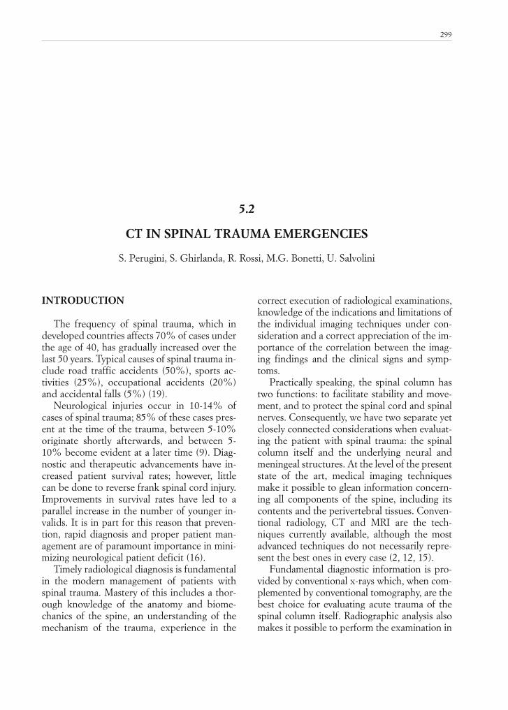
Fig. 5.1 - (Cont.) e) A reconstructed sagittal section demon-strates the relationship between the anterior arch of the C1 andthe odontoid process. f1), f2) Reconstructed coronal sectionsshow the normal relationship between the occipital condyles(f2) and the lateral masses of C2 (f1); there is no lateral disloca-tion of the lateral articular masses of C2.
Fig. 5.2 - Acute cervical hyperextension trauma. a) Lateral x-ray of the cervical spine shows a hangman fracture associatedwith anterior subluxation of C2 and bilateral fractures of theposterior arch of C1. b) Lateral scanogram and c) d) axial CTconfirms the x-ray findings of the C1 and C2 fractures.
e
a
b
f1
f2
5.2 CT IN SPINAL TRAUMA EMERGENCIES 305
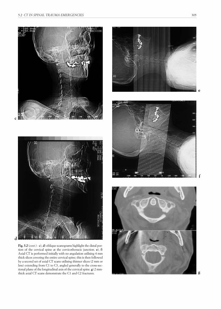
Fig. 5.2 (cont.) - c), d) oblique scanograms highlight the distal por-tion of the cervical spine at the cervicothoracic junction. e), f)Axial CT is performed initially with no angulation utilising 4 mmthick slices covering the entire cervical spine; this is then followedby a second set of axial CT scans utilising thinner slices (2 mm orless) extending from C1 to C3, angled generally to the cross-sec-tional plane of the longitudinal axis of the cervical spine. g) 2 mm-thick axial CT scans demonstrate the C1 and C2 fractures.
c
d
e
f
g
tion to those of C2. This alteration is clearly vis-ible on the open mouth anteroposterior projec-tion radiograph that is principally used forstudying the odontoid process. This projectionalso makes it possible to measure the degree oflateral dislocation of the articular facets which,if greater than 7 mm, suggests a rupture of thetransverse atlantal ligament. Axial CT makes itpossible to visualize the fractures, however theexamination must be performed using thincontiguous slices complemented by reconstruc-
tions in the sagittal and coronal planes in orderto show the subluxation of the articular facetprocesses.
306 V. SPINAL EMERGENCIES
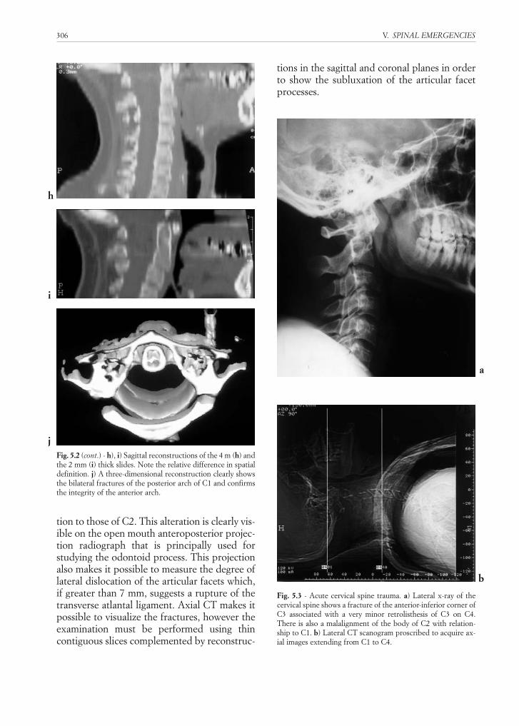
Fig. 5.2 (cont.) - h), i) Sagittal reconstructions of the 4 m (h) andthe 2 mm (i) thick slides. Note the relative difference in spatialdefinition. j) A three-dimensional reconstruction clearly showsthe bilateral fractures of the posterior arch of C1 and confirmsthe integrity of the anterior arch.
Fig. 5.3 - Acute cervical spine trauma. a) Lateral x-ray of thecervical spine shows a fracture of the anterior-inferior corner ofC3 associated with a very minor retrolisthesis of C3 on C4.There is also a malalignment of the body of C2 with relation-ship to C1. b) Lateral CT scanogram proscribed to acquire ax-ial images extending from C1 to C4.
h
a
b
i
j
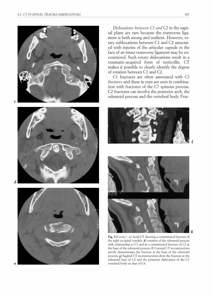
Dislocations between C1 and C2 in the sagit-tal plane are rare because the transverse liga-ment is both strong and resilient. However, ro-tary subluxations between C1 and C2 associat-ed with injuries of the articular capsule in theface of an intact transverse ligament may be en-countered. Such rotary dislocations result in atraumatic-acquired form of torticollis. CTmakes it possible to clearly identify the degreeof rotation between C1 and C2.
C1 fractures are often associated with C2fractures and these in turn are seen in combina-tion with fractures of the C7 spinous process.C2 fractures can involve the posterior arch, theodontoid process and the vertebral body. Frac-
5.2 CT IN SPINAL TRAUMA EMERGENCIES 307
Fig. 5.3 (cont.) - c) Axial CT showing a comminuted fracture ofthe right occipital condyle, d) rotation of the odontoid processwith relationship to C1 and e) a comminuted fracture of C2 atthe base of the odontoid process. f) Coronal CT reconstructionpoorly demonstrates the fracture at the base of the odontoidprocess. g) Sagittal CT reconstructions show the fracture at theodontoid base of C2 and the posterior dislocation of the C3vertebral body on that of C4.
c
d
f
g
e
308 V. SPINAL EMERGENCIES
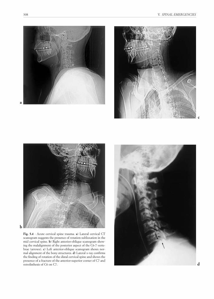
Fig. 5.4 - Acute cervical spine trauma. a) Lateral cervical CTscanogram suggests the presence of rotation-subluxation in themid cervical spine. b) Right anterior-oblique scanogram show-ing the malalignment of the posterior aspect of the C6-7 verte-brae (arrows). c) Left anterior-oblique scanogram shows nor-mal alignment of the bony structures. d) Lateral x-ray confirmsthe finding of rotation of the distal cervical spine and shows thepresence of a fracture of the anterior-superior corner of C7 andretrolisthesis of C6 on C7.
a
c
d
b

5.2 CT IN SPINAL TRAUMA EMERGENCIES 309
Fig. 5.4 (cont.) - e) Anterior-posterior x-ray shows an abruptdeviation of the spinous processes of C6 and higher to the rightin relationship to the spinous processes of C7 and below. f), g)High resolution axial CT of the cervico-occipital shows a de-viation to the right of the C2 spinous process in the absence ofa fracture. h) A digital summation of the C1 and C2 images (f+ g = h) show a degree of atlanto-axial rotation, which ne-vertheless may within physiological limits. i) Fracture of the ri-ght C6 lamina and the right posterior articular spinal facet pro-cesses of C6-7.
e
g
h
i
f

tures of the body, caused by various types oftraumatic forces, are classified as three differenttypes, depending upon the site of the injury. C2fractures are associated with neurological in-juries in 20% of cases (2, 3).
Hyperextension C2 fractures, or hangman’sfractures, affect the pedicles bilaterally eithersymmetrically or asymmetrically and can be as-sociated with fractures of the inferior-anteriorcorner of the vertebral body itself; there is oftena related subluxation of C2 on C3. Radiographyin such cases will clearly demonstrate these
fracture-dislocations. CT must be performedusing thin slices, extending from the foramenmagnum through C4 and must be comple-mented by reconstructions in the sagittal andcoronal planes. CT is necessary when the pa-tient feels pain in the upper neck, when cervi-cal spine x-rays are negative or when radiogra-phy shows thickening of the perivertebral softtissues.
Limitations of CT include the often poor vi-sualization of linear fractures that developalong the axial plane of section. In this case it is
310 V. SPINAL EMERGENCIES
Fig. 5.4 (cont.) - j), k), l) Coronal reconstructions showing afracture of C7 (j), fractures of the lateral masses (k) and lateraldisplacement of the spinous process of C6 (l). m), n), o), p) Sa-gittal reconstructions: the reconstruction on the left side (m)demonstrates comminuted fractures of the posterior articularspinal facet processes; the reconstruction on the right side (p)shows normal relationships of the posterior articular spinal fa-cet processes; n-o) adjacent parasagittal reconstructions showthe fracture of the body of C7 and the greater intersegmentalrotational subluxation to the right of midline (n).
jm
n
o
p
k
l

5.2 CT IN SPINAL TRAUMA EMERGENCIES 311
Fig. 5.5 - Acute cervical spine trauma. a) Lateral radiographconducted shows anterior subluxation of C5 on C6, an increasein the interlaminar distance and dislocation of the posterior ar-ticular facets. b) Anteroposterior radiograph shows deviationof the spinous process of C5 to the right indicating a probablerotational dislocation. c, d) Right-oblique projection radi-ographs confirms the anterior dislocation of the articular facetsassociated with probable fracture of the pedicle of C5. d) Left-oblique projection radiograph shows a similar finding, but witha smaller displacement than that of the contralateral side. Notethe malalignment of the contralateral vertebral pedicles.
a
c
d
b

312 V. SPINAL EMERGENCIES
Fig. 5.5 (cont.) - e) Lateral CT scanogram . f) Axial CT showsasymmetric dislocation of the body of C5 with relationship toC6. g) Axial CT shows a fracture of the right pedicle of C5. h)Image summation technique including the bodies of C5 and C6showing the C5-6 anterior subluxation. i) Oblique-sagittal re-construction method from preceding axial image set.
e
h
i
f
g
essential to supplement the examination withhigh resolution multiplanar reconstructionsand with conventional tomography.
Mid-distal (caudal) cervical spine trauma
Cervical spine trauma associated with minorspinal fractures does not in general result in
neural tissue injury, with the exception of casesin which there is underlying congenital-ac-quired stenosis of the central spinal canal.
In adults the mid-distal cervical region is thelevel of the spine that is most frequently affect-ed in cases of trauma. This is especially com-mon at the C5-C7 level (81% according toBerquist) (2, 3). Distal cervical spine traumaticinjuries are categorized according to the mech-anism of the trauma: hyperflexion, hyperexten-sion and axial compression.
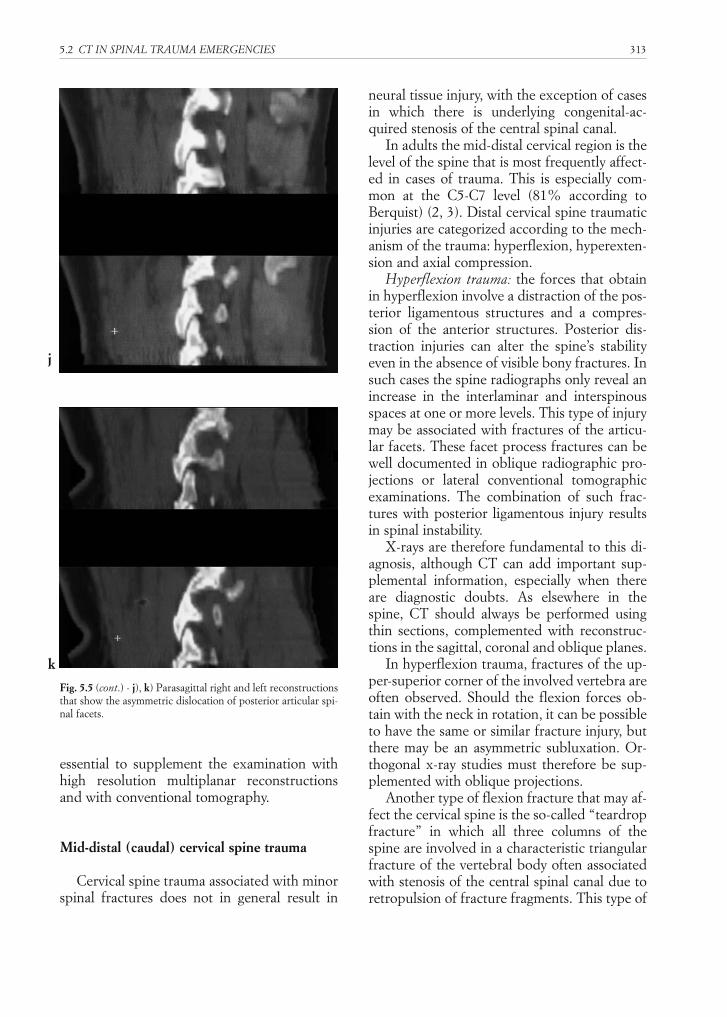
Hyperflexion trauma: the forces that obtainin hyperflexion involve a distraction of the pos-terior ligamentous structures and a compres-sion of the anterior structures. Posterior dis-traction injuries can alter the spine’s stabilityeven in the absence of visible bony fractures. Insuch cases the spine radiographs only reveal anincrease in the interlaminar and interspinousspaces at one or more levels. This type of injurymay be associated with fractures of the articu-lar facets. These facet process fractures can bewell documented in oblique radiographic pro-jections or lateral conventional tomographicexaminations. The combination of such frac-tures with posterior ligamentous injury resultsin spinal instability.
X-rays are therefore fundamental to this di-agnosis, although CT can add important sup-plemental information, especially when thereare diagnostic doubts. As elsewhere in thespine, CT should always be performed usingthin sections, complemented with reconstruc-tions in the sagittal, coronal and oblique planes.
In hyperflexion trauma, fractures of the up-per-superior corner of the involved vertebra areoften observed. Should the flexion forces ob-tain with the neck in rotation, it can be possibleto have the same or similar fracture injury, butthere may be an asymmetric subluxation. Or-thogonal x-ray studies must therefore be sup-plemented with oblique projections.
Another type of flexion fracture that may af-fect the cervical spine is the so-called “teardropfracture” in which all three columns of thespine are involved in a characteristic triangularfracture of the vertebral body often associatedwith stenosis of the central spinal canal due toretropulsion of fracture fragments. This type of
5.2 CT IN SPINAL TRAUMA EMERGENCIES 313
Fig. 5.5 (cont.) - j), k) Parasagittal right and left reconstructionsthat show the asymmetric dislocation of posterior articular spi-nal facets.
j
k

314 V. SPINAL EMERGENCIES
Fig. 5.6 - Acute thoracic spine compression trauma. a) Lateralradiograph of the thoracolumbar junction shows compressionof T12 with poor definition of the T11-12 disc space. b) An-teroposterior radiograph showing an increase in the interpedic-ular distance of T12 as compared to the vertebrae above andbelow; this indicates the presence of a burst fracture of T12. c)Lateral CT scanogram confirms the fracture of T12 and showsthe anterior wedging of T8. .
a b
c
5.2 CT IN SPINAL TRAUMA EMERGENCIES 315
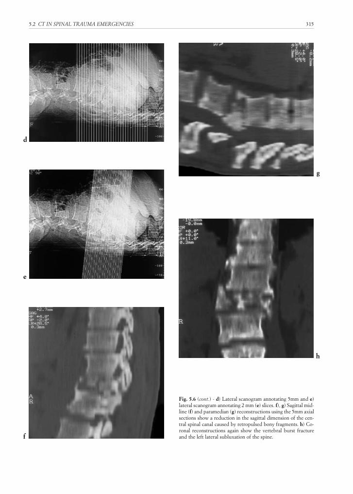
Fig. 5.6 (cont.) - d) Lateral scanogram annotating 5mm and e)lateral scanogram annotating 2 mm (e) slices. f), g) Sagittal mid-line (f) and paramedian (g) reconstructions using the 5mm axialsections show a reduction in the sagittal dimension of the cen-tral spinal canal caused by retropulsed bony fragments. h) Co-ronal reconstructions again show the vertebral burst fractureand the left lateral subluxation of the spine.
d
g
h
e
f
fracture should be differentiated from burstfractures that may be stable. CT enables the dif-ferentiation between these two types of fractureby showing the characteristic involvement ofthe three pillars in teardrop fractures.
Spinal cord injury in hyperflexion fracturesis common when there is a vertebral dislocationassociated with a subluxation of the posteriorarticular facets bilaterally, and in cases of tear-drop fractures. Cervical spinal nerve injuriesare more commonly observed in unilateral frac-ture-dislocations.
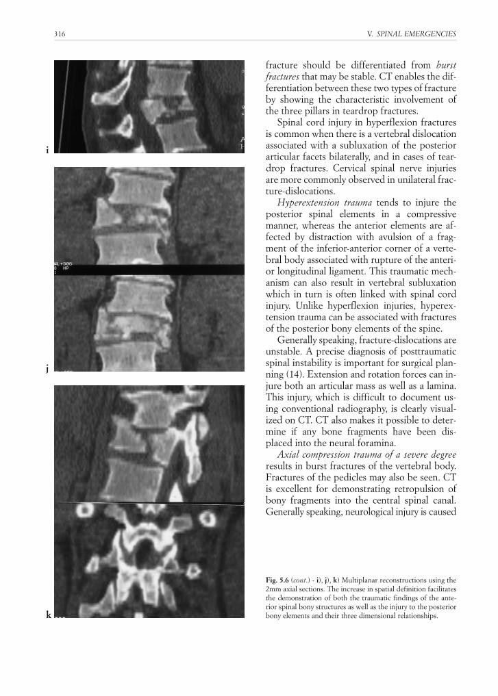
Hyperextension trauma tends to injure theposterior spinal elements in a compressivemanner, whereas the anterior elements are af-fected by distraction with avulsion of a frag-ment of the inferior-anterior corner of a verte-bral body associated with rupture of the anteri-or longitudinal ligament. This traumatic mech-anism can also result in vertebral subluxationwhich in turn is often linked with spinal cordinjury. Unlike hyperflexion injuries, hyperex-tension trauma can be associated with fracturesof the posterior bony elements of the spine.
Generally speaking, fracture-dislocations areunstable. A precise diagnosis of posttraumaticspinal instability is important for surgical plan-ning (14). Extension and rotation forces can in-jure both an articular mass as well as a lamina.This injury, which is difficult to document us-ing conventional radiography, is clearly visual-ized on CT. CT also makes it possible to deter-mine if any bone fragments have been dis-placed into the neural foramina.
Axial compression trauma of a severe degreeresults in burst fractures of the vertebral body.Fractures of the pedicles may also be seen. CTis excellent for demonstrating retropulsion ofbony fragments into the central spinal canal.Generally speaking, neurological injury is caused
316 V. SPINAL EMERGENCIES
Fig. 5.6 (cont.) - i), j), k) Multiplanar reconstructions using the2mm axial sections. The increase in spatial definition facilitatesthe demonstration of both the traumatic findings of the ante-rior spinal bony structures as well as the injury to the posteriorbony elements and their three dimensional relationships.
i
j
k
5.2 CT IN SPINAL TRAUMA EMERGENCIES 317
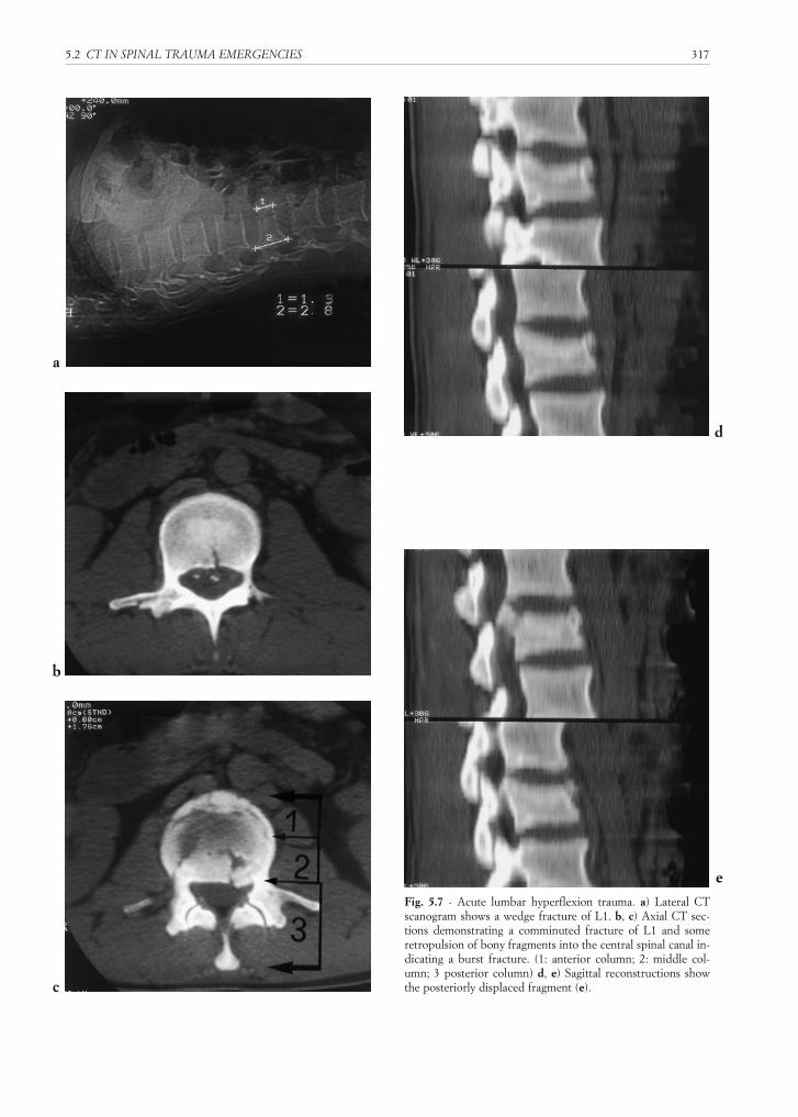
Fig. 5.7 - Acute lumbar hyperflexion trauma. a) Lateral CTscanogram shows a wedge fracture of L1. b, c) Axial CT sec-tions demonstrating a comminuted fracture of L1 and someretropulsion of bony fragments into the central spinal canal in-dicating a burst fracture. (1: anterior column; 2: middle col-umn; 3 posterior column) d, e) Sagittal reconstructions showthe posteriorly displaced fragment (e).
a
d
e
b
c

by this posterior displacement of bone with di-rect trauma to the spinal nerves/roots andspinal cord.
CT may also prove essential in differentiat-ing between certain types of fractures, for ex-ample in distinguishing between teardrop andburst fractures. CT must be performed usingthin slices, examining the vertebrae above andbelow the focus of the trauma. It is of vital im-portance to supplement the examination withimage reconstructions in the sagittal and coro-nal planes in order to better demonstrate thethree dimensional relationships of the spinaltrauma.
Trauma of the thoracic and lumbar segmentsof the spinal column
Most thoracolumbar spinal trauma (3, 5) oc-curs at and near the thoraco-lumbar junction,between T12 and L2. In this area there is ananatomical change in the orientation of the pos-terior articular spinal facet joints and a transi-tion from a relatively hypomobile spinal seg-ment to one with greater mobility.
The mechanisms of trauma are the same asthose described for the cervical spine, althoughthe majority of thoracolumbar injuries occur inhyperflexion.
318 V. SPINAL EMERGENCIES
Fig. 5.7 (cont.) - f) Three-dimensional (3-D) CT sagittal plane sectional reconstruction in the sagittal plane shows a relative increase inthe density of the vertebral marrow in three adjacent vertebrae (darker vertebral body shading), indicating that there is marrow com-pression and concentration of bony trabeculae. g) 3-D CT reconstruction demonstrating a superficial overview of the vertebral trauma.h), i) 3-D CT reconstruction of L1 shows the posterior vertebral body fracture, the central spinal canal stenosis and the widening of theposterior spinal facet joint articular space on the left side (arrows).
f h
g i

Flexion trauma entails a compression of theanterior structures and a distraction of the pos-terior spinal-perispinal tissues. Anterior com-pression results in wedge-shaped compressionfractures of the vertebral body, burst fractures,fracture-dislocations and distraction-fractures,depending in part upon the forces applied. Dis-traction-fractures may affect the articular facetprocesses or laminae.
Anteroposterior and lateral x-rays make itpossible to identify the vertebral compressionas well as burst fractures. In some cases, therelevant alterations can be quite minor, suchas a slight curve of the anterior margin of thevertebral body or condensation of the bonytrabeculae. Multiple, adjacent compressionfractures are common. In compression frac-tures, a loss of structural integrity of the pos-terior vertebral cortical surface can be sus-pected when the anterior height of the verte-bral body is reduced by more than one-halfthat of the rear.
CT demonstrates the posterior retropulsionof bony fragments in cases of burst fractures,the dimensions of the central spinal canal andneural foramina, and documents fractures ofthe posterior bony elements.
Fractures caused by flexion forces can resultin intersegmental subluxation, especially at thethoraco-lumbar junction. When there is an in-volvement of the posterior and middle columnsat the lumbar level, the fracture-dislocation isunstable. However, such instability is onlyrarely observed in the thoracic spine due to thestrong paravertebral muscles, the connectedribs and the orientation of the posterior spinalfacet joints.
Vertical compression trauma resulting inburst fractures is most commonly observed atthe thoraco-lumbar junction. Using conven-tional x-rays it is possible to show the compres-sion and fragmentation of the vertebral body,including disruptions of the posterior vertebralcortical margin. However, CT shows the in-volvement of the posterior vertebral cortex bet-ter than conventional x-rays and makes it pos-sible to visualize any migration of fragments in-to the central spinal canal. CT also makes itpossible to highlight fractures of the posterior
bony spinal elements that can indicate spinalinstability.
Single burst fractures of vertebral bodies areassociated with fractures at other levels thatmay or may not be contiguous in up to 43% ofcases. An imaging survey of the entire spinalcolumn is therefore imperative once a burstfracture is identified.
Hyperextension trauma is not commonly en-countered in the thoracolumbar spine. Whenthis is the mechanism of the trauma, isolatedfractures of the spinous and transverse process-es may be encountered. In the lumbar spine thelatter can be associated with renal trauma.
In cases of torsion trauma CT reveals evensmall fractures of the articular facet processes,on the condition that thin contiguous sectionsare used.
CONCLUSIONS
In cases of spinal trauma, CT provides addi-tional information to that obtained by conven-tional x-rays, thus permitting a more sensitiveexamination of the bony structures of thespinal column. Nevertheless, CT investigationsare only carried out after x-rays have shown orsuggested an injury. Contiguous, thin, stackedCT sections must be acquired in order to ob-tain optimal sagittal, coronal and oblique planereconstructions. CT also reveals traumatic le-sions of the adjacent soft tissue structures, how-ever these abnormalities are typically gross;subtle injuries will be overlooked on CT.
REFERENCES
1. Berne JD, Velmahos GC, El-Tawil Q et al: Value of com-plete cervical helical computed tomographic scanning inidentifying cervical spine injury in the unevaluable blunttrauma patient with multiple injuries: a prospective study. JTrauma, 47(5):896-902, 1999.
2. Berquist TH: Spinal trauma. Da: Trauma Radiology. Editedby Mc Cort – Churchill Livingstone: 31-74, 1990.
3. Berquist TH, Cabanella ME: The spine. Da: Imaging of theorthopedic trauma. Edited by TH Berquist – Raven Press:93-206, 1992.
4. Cusmano F, Ferrozzi F, Uccelli M et al: Upper cervicalspine fracture: sources of misdiagnosis. Radiol Med,98(4):230-235, 1999.
5.2 CT IN SPINAL TRAUMA EMERGENCIES 319

5. Daffner RH, Deeb ZL, Goldeberg AL et al: The radiologi-cal assessment of post-traumatic vertebral stability. SkeletalRadiol, 19:103-108, 1990.
6. Denis F: The three column spine and its significance in theclassification of the acute thoraco-lumbar spinal injuries.Spine, 8:817-831, 1983.
7. Denis F: Spinal instability as defined by three-column spineconcept in acute spinal trauma. Clin Orthop, 189:65-73, 1984.
8. El-Khoury GY, Kathol MH, Daniel WW: Imaging of acuteinjuries of the cervical spine: value of plain radiography,CT, and MR imaging. AJR, 164(1):43-50, 1995.
9. Guareschi B: Le lesioni traumatiche vertebrali. Da A.P.C.:Rivisitiamo la radiologia tradizionale. L’urgenza radiologi-ca, pp. 39-50.
10. Katz MA, Beredjiklian PK, Vresilovic EJ et al: Computedtomographic scanning of cervical spine fractures: does itinfluence treatment? J Orthop Trauma, 13(5):338-343,1999.
11. LeBlang SD, Nunez DB Jr: Helical CT of cervical spine andsoft tissue injuries of the neck. Radiol Clin North Am,37(3):512-532, 1999.
12. Leite CC, Escobar BE, Bazan C 3rd et al: MRI of cervical fa-cet dislocation. Neuroradiology, 39(8):583-588, 1997.
13. Mace SE: Emergency evaluation of cervical spine injuries:CT versus plain radiographs. Ann Emerg Med, 14(10):973-975, 1985.
14. Monti C, Malaguti MC, Bettini N et al: La TC nei traumiacuti del rachide. Da: La diagnostica per immagini nelle
“urgenze”, a cura di Romagnoli e Del Vecchio, Idelson, Na-poli: 191-198, 1991.
15. Murphey MD, Batnitzky S, Bramble JM: Diagnostic ima-ging of spinal trauma. Radiol Clin North Am, 27:855-873,1989.
16. Narayan RK: Emergency room management of the head-injured patient. Da: Textbook of head injury. Edited by DPBeker and SK Gaudeman, W.B. Saunders: 23-66, 1989.
17. Nuñez DB Jr, Quencer RM: The role of helical CT in theassessment of cervical spine injuries. AJR, 171:951-957,1998.
18. Olsen WL, Chakeres DW, Berry I et al: Traumatismes durachis et de la moelle. Da: Imagerie du rachis et de la moel-le, Scanner, IRM, Ultrasons. Edited by C Manelfe, Vi-got:387-426, 1989.
19. Pistolesi GF, Bergamo Andreis IA: L’imaging diagnosticodel rachide. Edizione Libreria Cortina, Verona: 637-706,1987.
20. Schmieder K, Hentsch A, Engelhardt M et al: Results ofspiral CT in patients with fractures of the craniocervicaljunction suspected on conventional radiographs. Eur Ra-diol, 9(5):1008, 1999.
21. Tan E, Schweitzer ME, Vaccaro L et al: Is computed tomo-graphy of nonvisualized C7-T1 cost-effective?. J Spinal Di-sord, 12(6):472-476, 1999.
22. Tehranzadeh J, Bonk RT, Ansari A et al: Efficacy of limitedCT for nonvisualized lower cervical spine in patients withblunt trauma. Skeletal Radiol, 23(5):349-352, 1994.
320 V. SPINAL EMERGENCIES


