Embed Size (px)
Citation preview
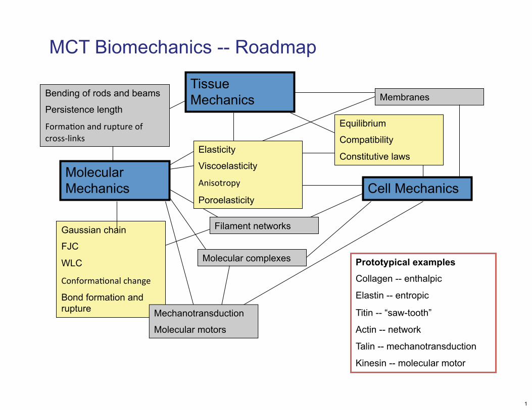
MCT Biomechanics -- Roadmap
Tissue Mechanics
Equilibrium
Compatibility
Constitutive laws
Membranes
Gaussian chain
FJC
WLC
Conformatonal change
Bond formation and rupture
Filament networks
Molecular complexes
Cell Mechanics Molecular Mechanics
Prototypical examples
Collagen -- enthalpic
Elastin -- entropic
Titin -- “saw-tooth”
Actin -- network
Talin -- mechanotransduction
Kinesin -- molecular motor
Elasticity
Viscoelasticity
Anisotropy
Poroelasticity
Bending of rods and beams
Persistence length
Formaton and rupture of cross-links
Mechanotransduction
Molecular motors
1
Biomechanics in the news: Mechanism of the microtubule motor dynein
The cover of Science removed due to copyright restrictions.
2
Image from The Inner Life of the Cell removed due to copyright restrictions.
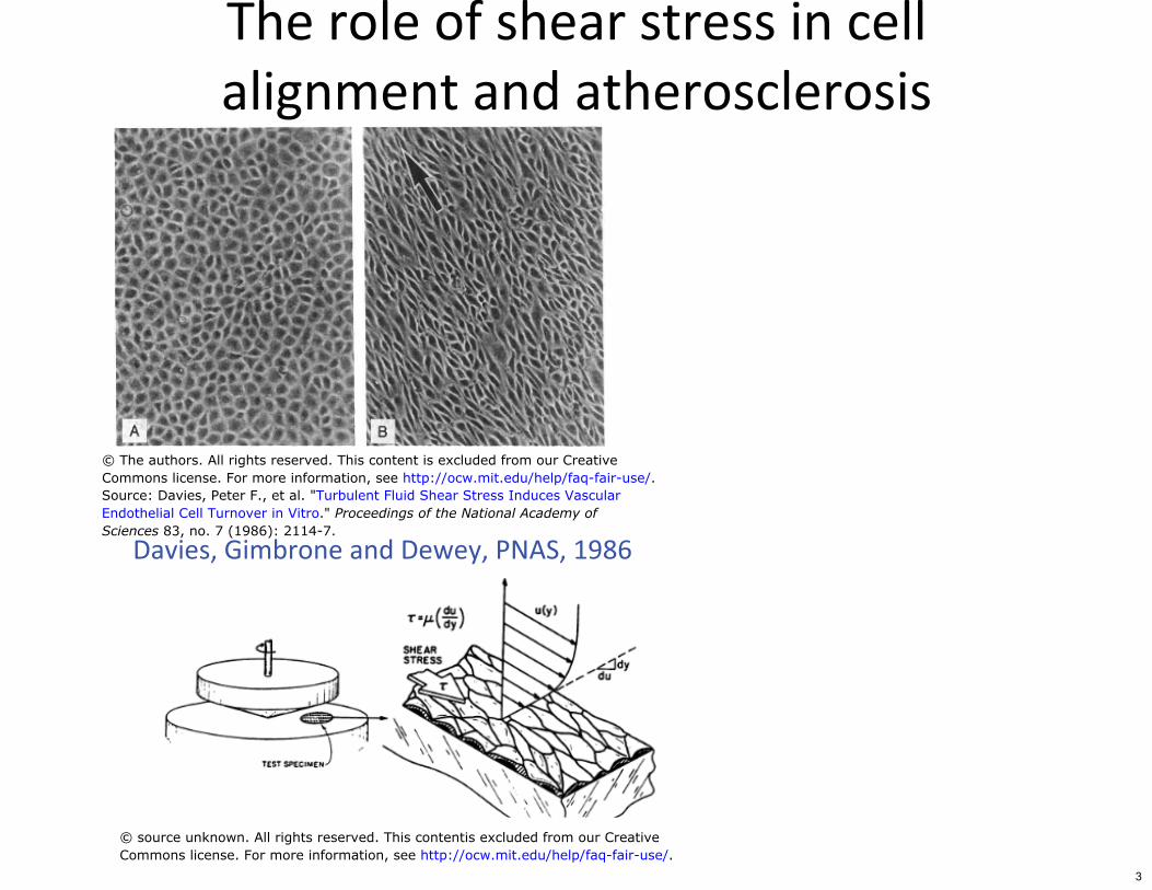
The role of shear stress in cell alignment and atherosclerosis
Source: Davies, Peter F., et al. "Turbulent Fluid Shear Stress Induces VascularEndothelial Cell Turnover in Vitro." Proceedings of the National Academy ofSciences 83, no. 7 (1986): 2114-7.
Davies, Gimbrone and Dewey, PNAS, 1986
© source unknown. All rights reserved. This contentis excluded from our CreativeCommons license. For more information, see http://ocw.mit.edu/help/faq-fair-use/.
© The authors. All rights reserved. This content is excluded from our CreativeCommons license. For more information, see http://ocw.mit.edu/help/faq-fair-use/.
3
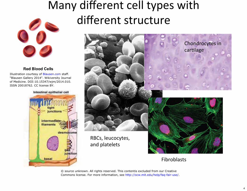
Many diferent cell types with diferent structure
Chondrocytes in cartlage
Fibroblasts
RBCs, leucocytes, and platelets
© source unknown. All rights reserved. This contentis excluded from our CreativeCommons license. For more information, see http://ocw.mit.edu/help/faq-fair-use/.
Illustration courtesy of Blausen.com staff."Blausen Gallery 2014". Wikiversity Journalof Medicine. DOI:10.15347/wjm/2014.010.ISSN 20018762. CC license BY.
4
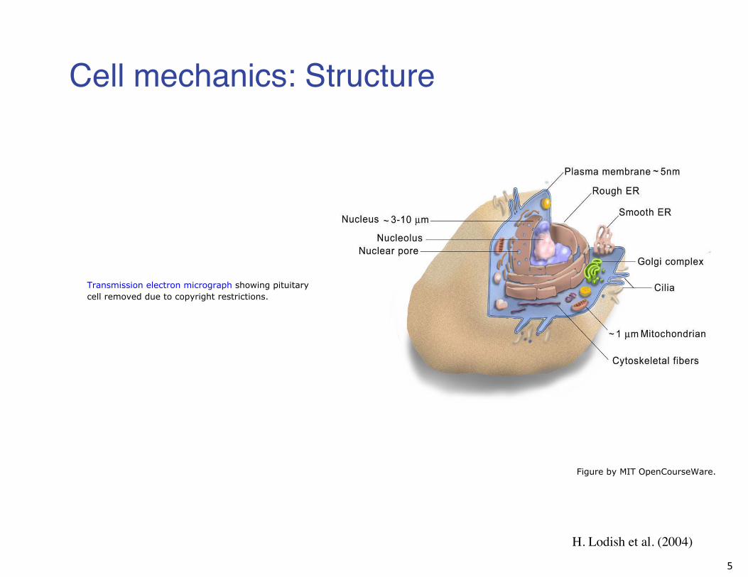
Cell mechanics: Structure
Transmission electron micrograph showing pituitarycell removed due to copyright restrictions.
H. Lodish et al. (2004)
5
Figure by MIT OpenCourseWare.
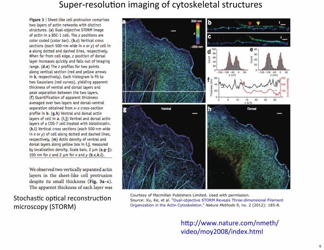
Super-resoluton imaging of cytoskeletal structures
Stochastc optcal reconstructon microscopy (STORM)
htp://www.nature.com/nmeth/ video/moy2008/index.html
Courtesy of Macmillan Publishers Limited. Used with permission.Source: Xu, Ke, et al. "Dual-objective STORM Reveals Three-dimensional FilamentOrganization in the Actin Cytoskeleton." Nature Methods 9, no. 2 (2012): 185-8.
6

Actn/spectrin structure in axons
Figure 4 removed due to copyright restrictions.Source: Xu, Ke, et al. "Actin, Spectrin, and Associated Proteins form a PeriodicCytoskeletal Structure in Axons." Science 339, no. 6118 (2013): 452-6.
Stochastc optcal reconstructon Xu, Science, 201�microscopy (STORM)
7
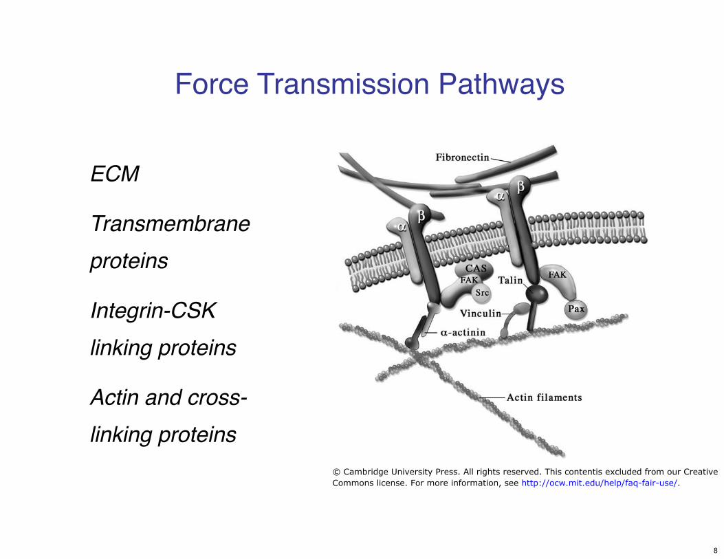
Force Transmission Pathways
ECM
Transmembrane
proteins
Integrin-CSK
linking proteins
Actin and cross-
linking proteins
© Cambridge University Press. All rights reserved. This contentis excluded from our CreativeCommons license. For more information, see http://ocw.mit.edu/help/faq-fair-use/.
8
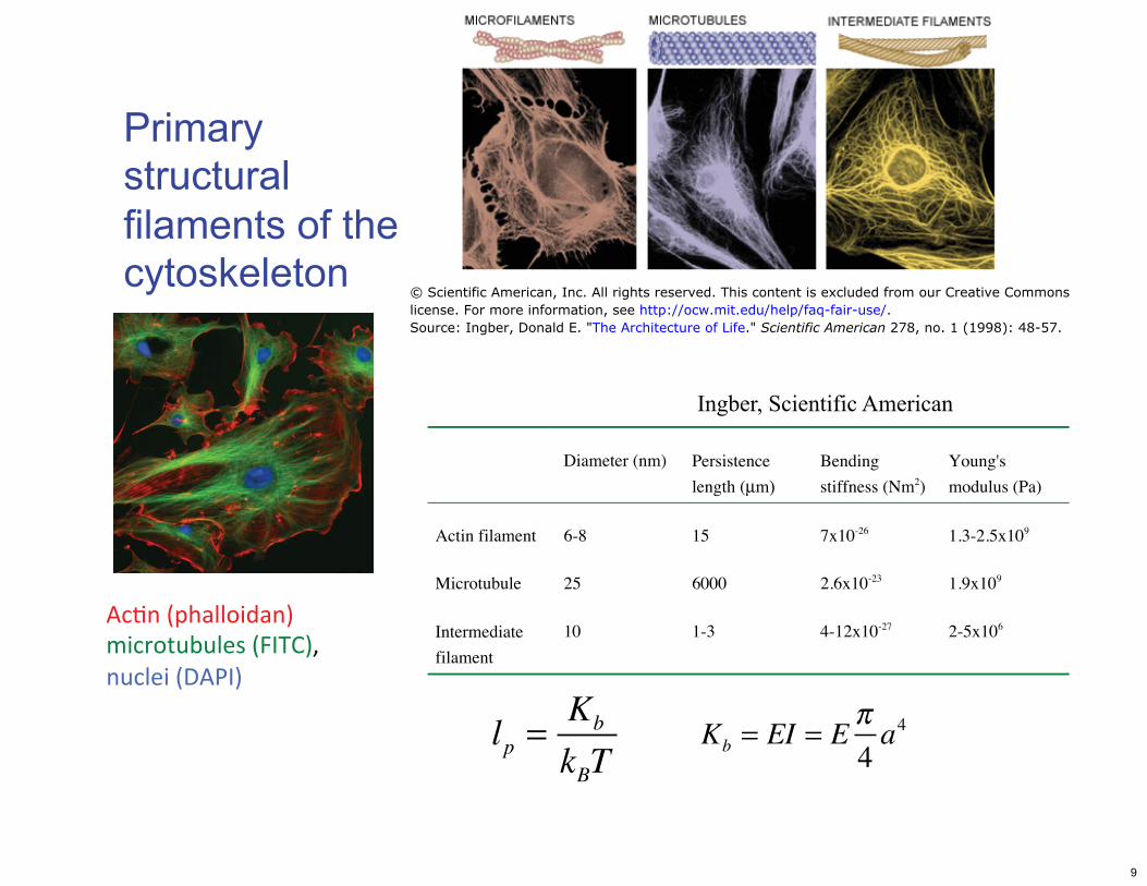
Primary structural filaments of the cytoskeleton
Ingber, Scientific American
Actn (phalloidan) microtubules (FITC), nuclei (DAPI)
Diameter (nm) Persistence Bending Young's
length (μm) stiffness (Nm2) modulus (Pa)
Actin filament 6-8
Microtubule 25
Intermediate
filament
10
lp = Kb
kBT
15 7x10-26
6000 2.6x10⁻²³
1-3 4-12x10-27
Kb = EI = E π 4 a4
1.3-2.5x10⁹
1.9x10⁹
2-5x10⁶
© Scientific American, Inc. All rights reserved. This content is excluded from our Creative Commonslicense. For more information, see http://ocw.mit.edu/help/faq-fair-use/.Source: Ingber, Donald E. "The Architecture of Life." Scientific American 278, no. 1 (1998): 48-57.
9
Mechanical factors in integrin
adhesion
Figures 1 & 2 removed due to copyright restrictions.Source: Roca-Cusachs, Pere, et al. "Finding the Weakest Link–exploring Integrin-mediatedMechanical Molecular Pathways." Journal of Cell Science 125, no. 13 (2012): 3025-038.
10

11
Image from The Inner Life of the Cell removed due to copyright restrictions.
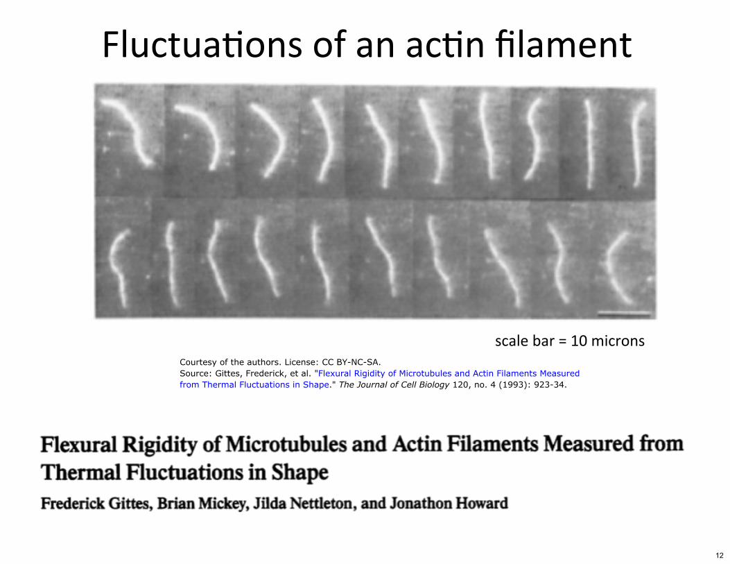
Fluctuatons of an actn flament
scale bar = 10 microns Courtesy of the authors. License: CC BY-NC-SA.Source: Gittes, Frederick, et al. "Flexural Rigidity of Microtubules and Actin Filaments Measuredfrom Thermal Fluctuations in Shape." The Journal of Cell Biology 120, no. 4 (1993): 923-34.
12

Actin microfilaments
Image of G-actin monomers polymerizing into F-actinfilaments removed due to copyright restrictions.
Courtesy of John Hartwig. Used with permission.
Structure of actin. Image courtesy of Dr. Willy Wriggers.Used with permission.
13
Cytoskeletal structure
Figure 1 removed due to copyright restrictions.Source: Hirokawa, Nobutaka. "Kinesin and Dynein Superfamily Proteins and theMechanism of Organelle Transport." Science 279, no. 5350 (1998): 519-26.
14
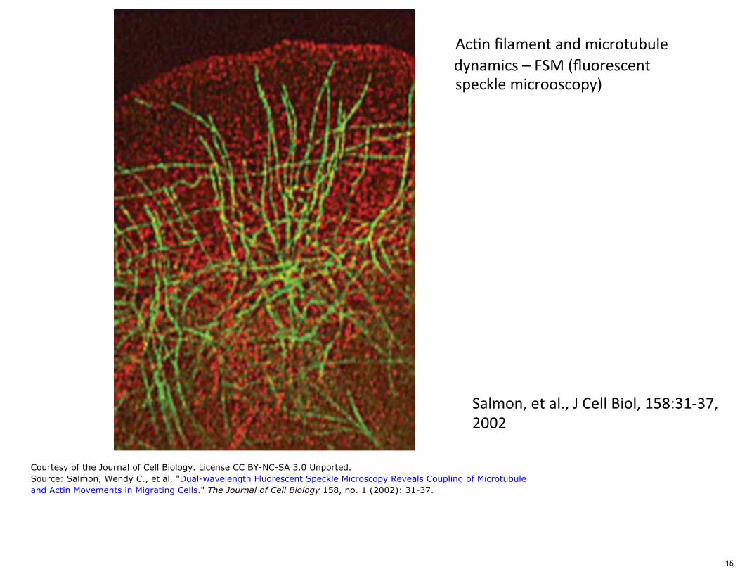
Actn flament and microtubule
speckle microoscopy)
Salmon, et al., � Cell Biol, 158:�1-��, 2002
Courtesy of the Journal of Cell Biology. License CC BY-NC-SA 3.0 Unported.Source: Salmon, Wendy C., et al. "Dual-wavelength Fluorescent Speckle Microscopy Reveals Coupling of Microtubuleand Actin Movements in Migrating Cells." The Journal of Cell Biology 158, no. 1 (2002): 31-37.
dynamics – FSM (fluorescent
15
Vascular
Transvascular
Convection
Interstitial
100 μm
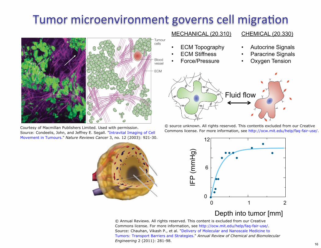
Tumor microenvironment governs cell migraton MECHANICAL (20.310) CHEMICAL (20.330)
Tumour cells
• ECM Topography • Autocrine Signals • ECM Stiffness • Paracrine Signals
Blood • Force/Pressure • Oxygen Tension vessel
ECM
Fluid flow
IFP
(mm
Hg)
12
6
0 0 1 2
Depth into tumor [mm]
© source unknown. All rights reserved. This contentis excluded from our CreativeCourtesy of Macmillan Publishers Limited. Used with permission.Commons license. For more information, see http://ocw.mit.edu/help/faq-fair-use/.Source: Condeelis, John, and Jeffrey E. Segall. "Intravital Imaging of Cell
Movement in Tumours." Nature Reviews Cancer 3, no. 12 (2003): 921-30.
© Annual Reviews. All rights reserved. This content is excluded from our CreativeCommons license. For more information, see http://ocw.mit.edu/help/faq-fair-use/.Source: Chauhan, Vikash P., et al. "Delivery of Molecular and Nanoscale Medicine toTumors: Transport Barriers and Strategies." Annual Review of Chemical and BiomolecularEngineering 2 (2011): 281-98.
16
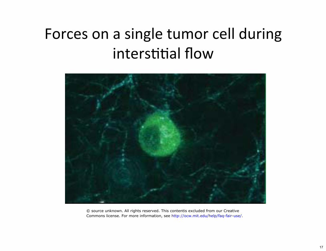
Forces on a single tumor cell during
© source unknown. All rights reserved. This contentis excluded from our CreativeCommons license. For more information, see http://ocw.mit.edu/help/faq-fair-use/.
inters((al flow
17
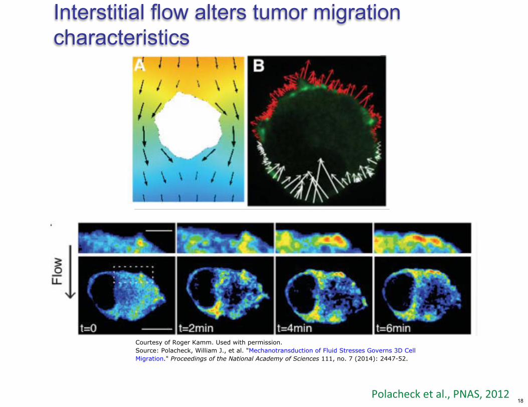
Interstitial flow alters tumor migration characteristics
%��� ��������2#�%&�'#�34(3�
Courtesy of Roger Kamm. Used with permission.Source: Polacheck, William J., et al. "Mechanotransduction of Fluid Stresses Governs 3D CellMigration." Proceedings of the National Academy of Sciences 111, no. 7 (2014): 2447-52.
18
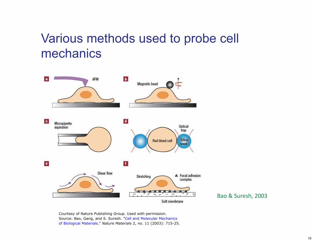
Various methods used to probe cell mechanics
����D�'���� #�344:�
Courtesy of Nature Publishing Group. Used with permission.Source: Bao, Gang, and S. Suresh. "Cell and Molecular Mechanicsof Biological Materials." Nature Materials 2, no. 11 (2003): 715-25.
19
������
64�-2/��&8�3��
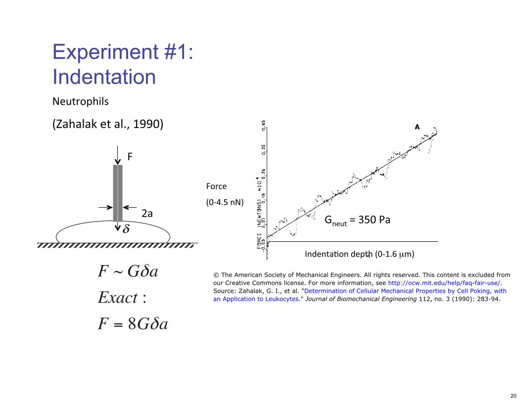
Experiment #1: Indentation�
;������������� �64�(2+�μ�8�
&������ ����
6E� ��������2#�())48�
��
$ �@�:/4�%��δ ����
F ~ Gδa © The American Society of Mechanical Engineers. All rights reserved. This content is excluded fromour Creative Commons license. For more information, see http://ocw.mit.edu/help/faq-fair-use/.
Exact :Source: Zahalak, G. I., et al. "Determination of Cellular Mechanical Properties by Cell Poking, withan Application to Leukocytes." Journal of Biomechanical Engineering 112, no. 3 (1990): 283-94.
F = 8Gδa
20
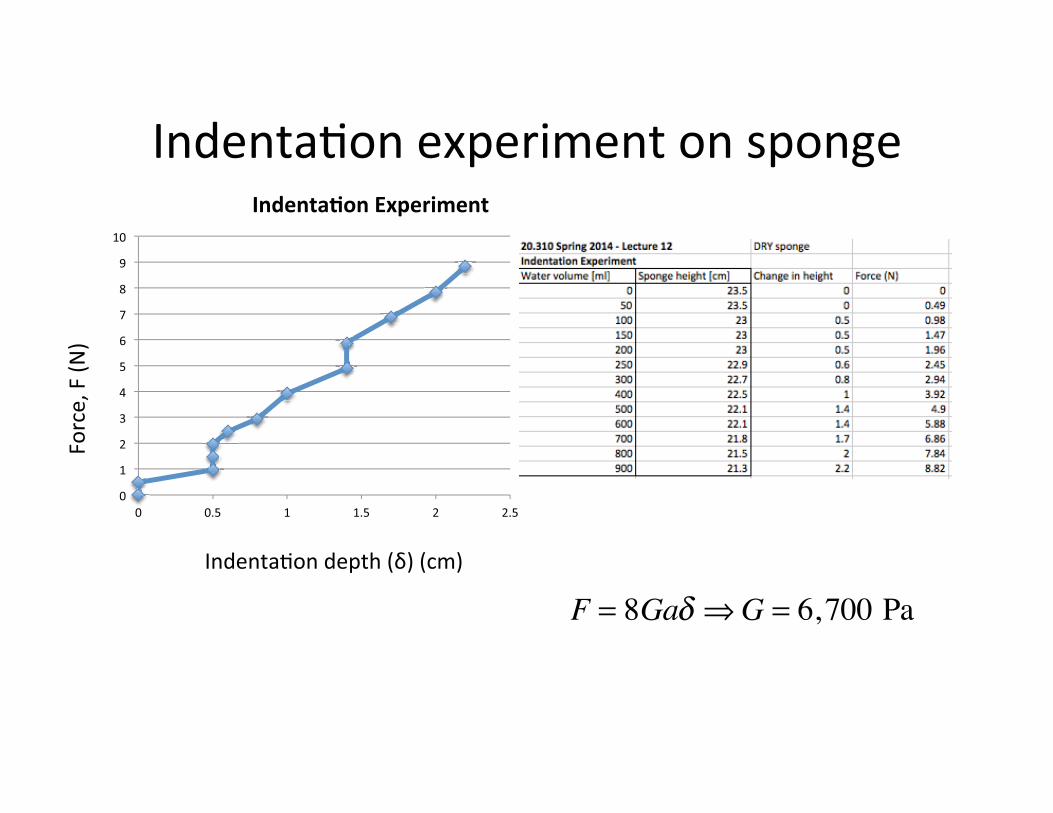
;����������5�������������������
;������������� �6F8�6��8�
�����#���6&
8�
4�
(�
3�
:�
-�
/�
+�
>�
*�
)�
(4�
4� 42/� (� (2/� 3� 32/�
����������� �������
������6&8�
F = 8Gaδ ⇒G = 6,700 Pa
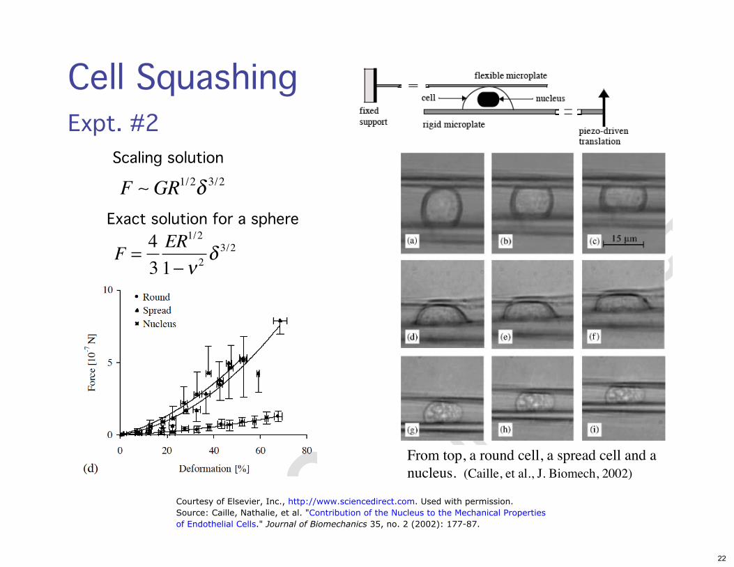
Cell SquashingExpt. #2
F ∼ GR1/2δ 3/2
4F =
3
ER1/2
1−ν 2δ 3/2
Exact solution for a sphere
Scaling solution
From top, a round cell, a spread cell and a nucleus. (Caille, et al., J. Biomech, 2002)
Courtesy of Elsevier, Inc., http://www.sciencedirect.com. Used with permission.Source: Caille, Nathalie, et al. "Contribution of the Nucleus to the Mechanical Propertiesof Endothelial Cells." Journal of Biomechanics 35, no. 2 (2002): 177-87.
22
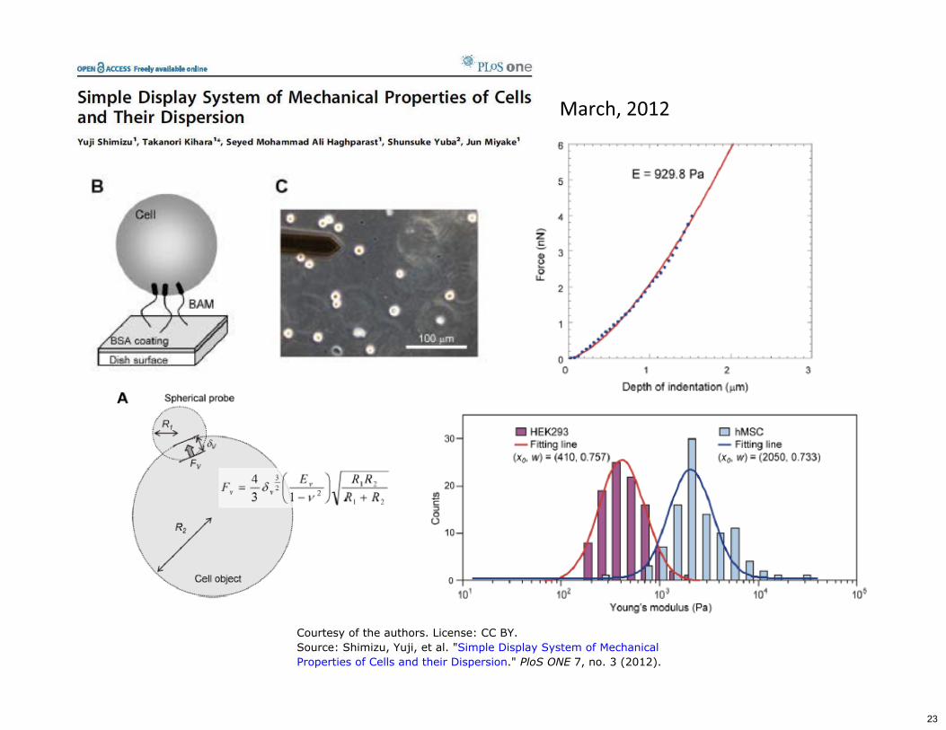
���� #�34(3�
Courtesy of the authors. License: CC BY.Source: Shimizu, Yuji, et al. "Simple Display System of MechanicalProperties of Cells and their Dispersion." PloS ONE 7, no. 3 (2012).
23
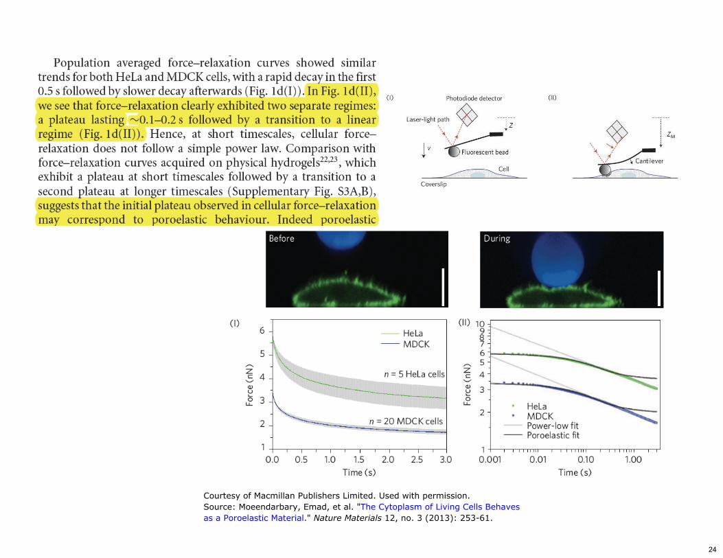
Courtesy of Macmillan Publishers Limited. Used with permission.Source: Moeendarbary, Emad, et al. "The Cytoplasm of Living Cells Behavesas a Poroelastic Material." Nature Materials 12, no. 3 (2013): 253-61.
24
G����������HH��������:!������5�
Figures removed due to copyright restrictions.
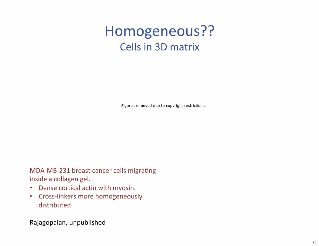
�!�����3:(���������������������������������������������2�• !������������������ �������2���• ������������������ ������������
������������� �.�J�������#�������� ��� �
25
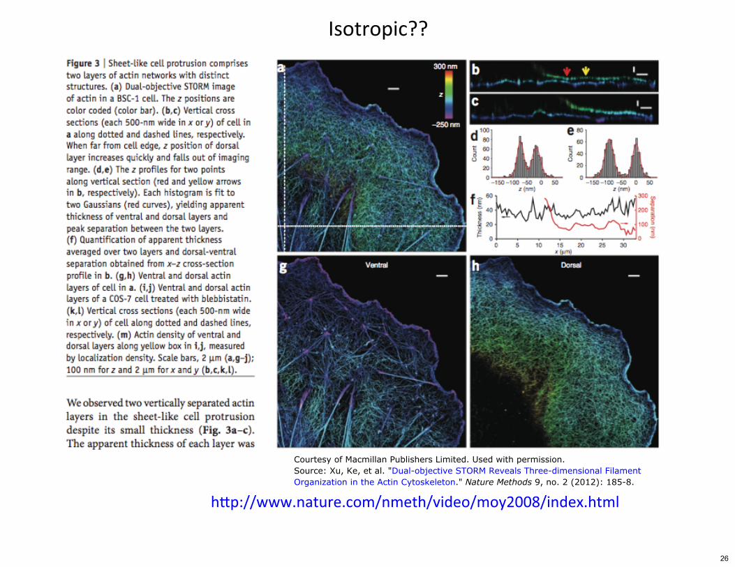
;��������HH�
Courtesy of Macmillan Publishers Limited. Used with permission.Source: Xu, Ke, et al. "Dual-objective STORM Reveals Three-dimensional FilamentOrganization in the Actin Cytoskeleton." Nature Methods 9, no. 2 (2012): 185-8.
0��11���2������2���1���� 1"����1���344*1����52 ����
26
���������0�����������
;����������
6E� ��������2#�())48�
;������������� �64�(2+�μ�8�
������
64�-2/��&8�
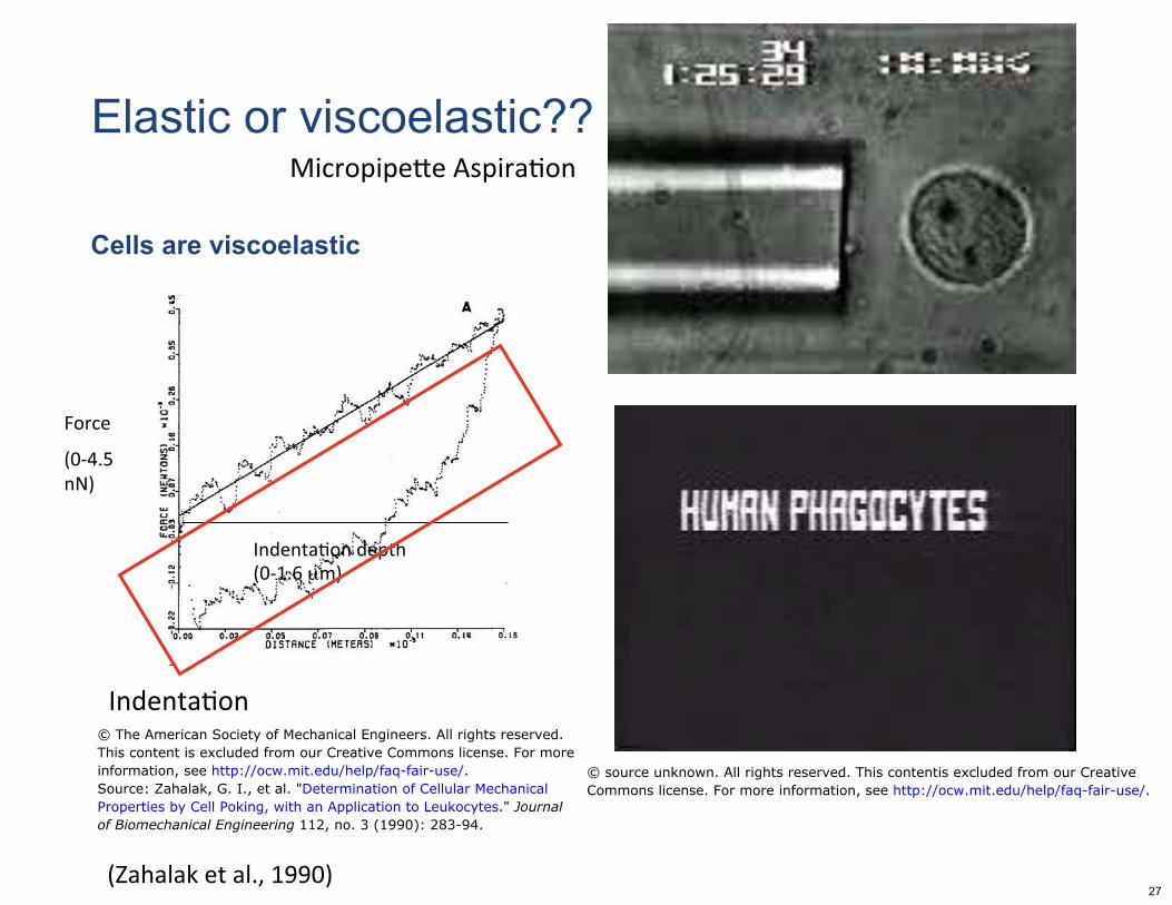
Cells are viscoelastic�
Elastic or viscoelastic??�
© The American Society of Mechanical Engineers. All rights reserved.This content is excluded from our Creative Commons license. For moreinformation, see http://ocw.mit.edu/help/faq-fair-use/. © source unknown. All rights reserved. This contentis excluded from our CreativeSource: Zahalak, G. I., et al. "Determination of Cellular Mechanical Commons license. For more information, see http://ocw.mit.edu/help/faq-fair-use/.Properties by Cell Poking, with an Application to Leukocytes." Journalof Biomechanical Engineering 112, no. 3 (1990): 283-94.
27
MIT OpenCourseWarehttp://ocw.mit.edu
20.310J / 3.053J / 6.024J / 2.797J Molecular, Cellular, and Tissue BiomechanicsSpring 2015
For information about citing these materials or our Terms of Use, visit: http://ocw.mit.edu/terms.


