Embed Size (px)
Citation preview
1
MEDICAL STUDIES IN ENGLISH
CLINICAL SKILLS: YEAR 1
STUDENT HANDOUT
2015
Contents:
1. Vital signs
2. Symptoms and signs of vital organ failure
3. Basic life support in adults
4. Basic life support in infants and children
5. Newborn resuscitation
6. First aid for the injured
7. First aid for environmental injuries and bites
8. Poisoning and burns
9. Immobilisation and transport devices for trauma patients
Department of Clinical Skills
University of Split School of Medicine
2
1.VITAL SIGNS
Irena Zakarija-Grković, MD, FRACGP, IBCLC, PhD
Introduction
Vital signs (from Latin signa vitae) are measurements of the body’s most basic functions. The
four main vital signs routinely monitored are:
1. Body temperature
2. Pulse
3. Respiration
4. Blood pressure
These measurements are taken to help assess the general physical health of a person, give
clues to possible diseases and show progress toward recovery.
Vital signs can be measured in a medical setting, at home, at the site of a medical emergency
or elsewhere. Measurement of vital signs is a routine part of any physical examination.
Body temperature
The temperature should always be recorded as part of the initial clinical examination of the
patient. The normal body temperature ranges from 36.6°C to 37.2°C. In very hot weather the
temperature may rise up to 0.5°C higher. Fever is defined as a morning oral temperature of
>37.2 °C (>98.9 °F) or an afternoon oral temperature of >37.7 °C (>99.9 °F). A temperature
between 37.3°C and 38.5°C is classified as a ‘low-grade fever’, whereas a body temperature
above 38.5°C is considered a ‘high- grade fever’.
Body temperature is measured using temperature devices inserted on or into the rectum,
mouth, axilla, skin, or ear. Some devices (laryngoscopes, bronchoscopes, rectal probes) may
have temperature-sensing probes that can record temperature continually. The most common
way to measure body temperature was (and still is in many countries) with a mercury
thermometer; because of glass breakage and the possibility of subsequent mercury
contamination, many developed countries use digital thermometers with disposable probe
covers to measure temperature from all of the body sites listed above. Disposable
temperature-sensitive strips that measure skin temperature are also used.
Oral temperatures are most commonly measured in adults, but rectal temperatures are the
most accurate because environmental factors that increase or decrease temperature
measurements have the least effect on the rectal area. The oral temperature is normally lower
than the rectal temperature by 0.5°C to 0.7°C. Axillary temperature may be 0.5°C lower than
the oral reading. There is a diurnal variation; body temperature is lowest in the morning and
reaches a peak between 6:00 and 10:00pm. The pattern of the fever may be helpful in
diagnosis.
Very high temperatures (hyperpyrexia, defined as above 41.5°C) are very serious and may
result in death. The causes include: heat stroke from exposure or excessive exertion, for
3
example in marathon runners; intracranial haemorrhage; malignant hyperthermia (a group of
genetically determined disorders in which hyperpyrexia occurs in response to various
anaesthetic agents or muscle relaxants); neuroleptic malignant syndrome; infections and
hypothalamic disease.
Hypothermia is defined as a temperature less than 35°C. Normal thermometers do not record
below 35°C and therefore special low- reading thermometers must be used where
hypothermia is suspected. Causes of hypothermia include: prolonged exposure to cold and
hypothyroidism.
Pulse
Pulse rate refers to the number of heart beats per minute. It can be measured centrally or
peripherally. When palpated, the pulse is felt best where the artery can be compressed
against bone: inside of neck (carotid artery), inside of elbow (brachial artery), inside or wrist
(radial artery), behind the knee (popliteal artery), behind the ankle (posterior tibial artery)
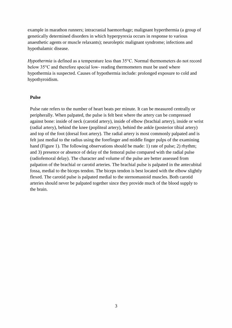
and top of the foot (dorsal foot artery). The radial artery is most commonly palpated and is
felt just medial to the radius using the forefinger and middle finger pulps of the examining
hand (Figure 1). The following observations should be made: 1) rate of pulse; 2) rhythm;
and 3) presence or absence of delay of the femoral pulse compared with the radial pulse
(radiofemoral delay). The character and volume of the pulse are better assessed from
palpation of the brachial or carotid arteries. The brachial pulse is palpated in the antecubital
fossa, medial to the biceps tendon. The biceps tendon is best located with the elbow slightly
flexed. The carotid pulse is palpated medial to the sternomastoid muscles. Both carotid
arteries should never be palpated together since they provide much of the blood supply to
the brain.
4
The normal resting heart rate in adults is between 60 and 100 beats per minute. Formal
counting over 30 seconds, multiplied by two, is an accurate way of determining the heart
rate. Bradycardia (Greek bradus slow, kardia heart) is defined as a heart rate less than 60
beats per minute and is due to a variety of causes including physiological, drugs,
hypothyroidism, hypothermia, arrhythmia. Tachycardia (Greek tachus swift, kardia heart) is
defined as a heart rate over 100 beats per minute and can be cause by a hyperdynamic
circulation, drugs, arrhythmias etc.
The rhythm of the pulse can be regular or irregular. An irregular rhythm can be completely
irregular with no pattern or it can be regularly irregular.
Radiofemoral delay refers to a noticeable delay in the arrival of the femoral pulse wave. It
can be detected by simultaneously palpating the radial and femoral pulse. It suggests the
diagnosis of coarctation of the aorta. This lesion can cause upper limb hypertension. It is
also useful to palpate both radial pulses together to detect radial-radial inequality in timing
or volume, usually due to a large arterial occlusion by an atherosclerotic plaque or
aneurysm.
Respiration
The respiration rate is the number of breaths a person takes per minute. The rate is usually
measured when a person is at rest and simply involves counting the number of breaths for
one minute by counting how many times the chest rises. Normal respiration rates for an
adult person at rest range from 12-16 breaths per minute. Respiration rates may increase
with fever, illness, exercise, etc. When checking respiration, it is important to also note
whether a person has any difficulty breathing. Dyspnoea (Greek dys, bad and pnoia,
breathing) or shortness of breath is often defined as an unexpected awareness of breathing.
Tachypnoea refers to a rapid respiratory rate. Look to see whether the accessory muscles of
respiration are being used. These muscles include the sternomastoids, the platysma and the
5
strap muscles of the neck. Characteristically the accessory muscles cause elevation of the
shoulders with inspiration and aid respiration by increasing chest expansion.
Blood pressure
Measurement of the arterial blood pressure is essential. Usually indirect measurements are
obtained with a sphygmomanometer (Greek sphygmos, pulsing and manos, thin). The
systolic blood pressure is the peak pressure that occurs in the artery following ventricular
systole and diastolic blood pressure is the level to which the arterial blood pressure falls
during ventricular diastole. It is expressed in millimetres of mercury (mmHg) or kilopascals
(kPa). Normal blood pressure is at or below 120/80mmHg. It may normally vary between
arms by up to 10mmHg. It should be taken in a lying and standing position. A fall in blood
pressure or more than 15mmHg (systolic) or 10mmHg (diastolic) on standing is abnormal
(postural hypotension).
The usual blood pressure cuff width is 12.5cm. This is suitable for a normal-sized adult
forearm. However, in obese patients, the normal -sized cuff will overestimate the blood
pressure and therefore a large cuff must be used. Smaller sizes are available for children.
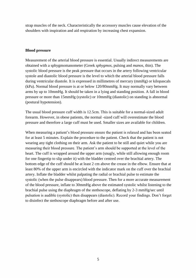
When measuring a patient’s blood pressure ensure the patient is relaxed and has been seated
for at least 5 minutes. Explain the procedure to the patient. Check that the patient is not
wearing any tight clothing on their arm. Ask the patient to be still and quiet while you are
measuring their blood pressure. The patient’s arm should be supported at the level of the
heart. The cuff is wrapped around the upper arm (snugly, while still allowing enough room
for one fingertip to slip under it) with the bladder centred over the brachial artery. The
bottom edge of the cuff should be at least 2 cm above the crease in the elbow. Ensure that at
least 80% of the upper arm is encircled with the indicator mark on the cuff over the brachial
artery. Inflate the bladder whilst palpating the radial or brachial pulse to estimate the
systolic (when the pulse disappears) blood pressure. Then for a more accurate measurement
of the blood pressure, inflate to 30mmHg above the estimated systolic whilst listening to the
brachial pulse using the diaphragm of the stethoscope, deflating by 2-3 mmHg/sec until
pulsation is audible (systolic) then disappears (diastolic). Record your findings. Don’t forget
to disinfect the stethoscope diaphragm before and after use.
6
Literature
• Clinical Examination: A Systematic Guide to Physical Diagnosis, 7th Edition, Talley
& O'Connor
• Šimunović V.J. (ed.): Basic & General Clinical Skills. Charlestone, SC, USA;
CreateSpace Independent Publishing Platform: 2013
7
2. CLINICAL SYMPTOMS AND SIGNS OF VITAL ORGAN FAILURE
Mladen Carev, M.D. Ph.D., Antonela Bunoza, M.D.
1. INTRODUCTORY REMARKS
1.1. CLINICAL SYMPTOMS AND SIGNS
The clinical symptom is a subjective indicator of a disease or a change in the patient’s
condition, as experienced by the patient himself. Unlike the clinical symptom, the clinical
sign is an objective indicator of a medical condition, and it can be detected by the clinical
examination of the patient. For example, the clinical symptoms may be difficult swallowing,
headache, sense of fear, while clinical signs may be ascites (accumulation of fluid in the
abdomen), splenomegaly (enlarged spleen), jaundice, and cough. Both the signs and
symptoms are usually non-specific, but their combination leads to correct clinical diagnosis.
The clinical syndrome is the term for a group of symptoms (and/or medical signs) that
normally appear together.
1.2. INTENSIVE CARE UNIT (ICU)
The intensive Care Unit (ICU) is a specially designed hospital unit where the life
support and maintenance of vital organs functions is provided to patients who are critically ill
and usually require a constant and invasive monitoring. The physicians working in intensive
care units are usually referred to as intensivists. This group of specialists has certification in a
variety of specialties including mostly anesthesia, followed by internal medicine, surgery and
emergency medicine. Furthermore, there is also a highly educated nursing staff for working
with these most difficult patients.
Patients are admitted to the ICU in several ways: directly from an emergency
department after diagnosis and surgical treatment, or from other hospital departments if they
have significant deterioration of their medical condition. It is also possible for a patient to be
admitted immediately after surgery, especially if the surgery is a demanding one and/or the
patient has multiple risk factors for postoperative complications. The most common
conditions that are treated in the ICU are trauma, sepsis and multiple organ failure. Patients
who are admitted to the ICU usually require support for hemodynamic instability
(hypertension/hypotension, life-threatening arrhythmias), as well as mechanical ventilation
because of the inability to maintain the airway or breathe sufficiently. Besides, many of them
8
have acute renal failure, gastrointestinal dysfunction and liver failure. Unfortunately,
frequently it comes to multiple organ failure at the same time
2. VITAL ORGANS FAILURE IN THE ICU
By definition organ failure is an altered organ function in the critically ill patients
requiring urgent medical intervention, in order to maintain homeostasis. Organ failure may be
acute, occurring through short period, and chronic, occurring gradually. In addition to failure
of any single vital organ, there is also a possibility of the occurrence of failure of two or more
vital organ systems, which is usually termed multiple organ dysfunction syndrome (MODS)
(Table 1).
The most common variables indicating organ dysfunction are:
Arterial hypoxemia (PaO2/FiO2<300)*
Acute oliguria (urinary output < 0.5 mL/kg/h during a minimum of 2 hours despite
adequate fluid resuscitation) and/or creatinine increase >44.2 μmol/L
Coagulation disorders (INR>1.5 or aPTT>60 seconds)**
Ileus (absence of peristalsis)
Thrombocytopenia (platelets<100,000/mm3)
Hyperbilirubinemia (bilirubin>70 μmol/L)
Increased lactate levels (>2 mmol/L)
*PaO2 – partial pressure of oxygen in arterial blood (mmHg), FiO2 – fraction of inspired
oxygen
**INR (International Normalized Ratio) – coagulation test measuring extrinsic pathway of
coagulation. INR is the ratio between the coagulation time of a sample of blood and the
normal coagulation time, when coagulation takes place in certain standardized conditions;
normal range is 0.8-1.2
aPTT (activated partial thromboplastin time) – a performance indicator of the efficacy of both
the "intrinsic" and the common coagulation pathway; normal range is 30-40 seconds.
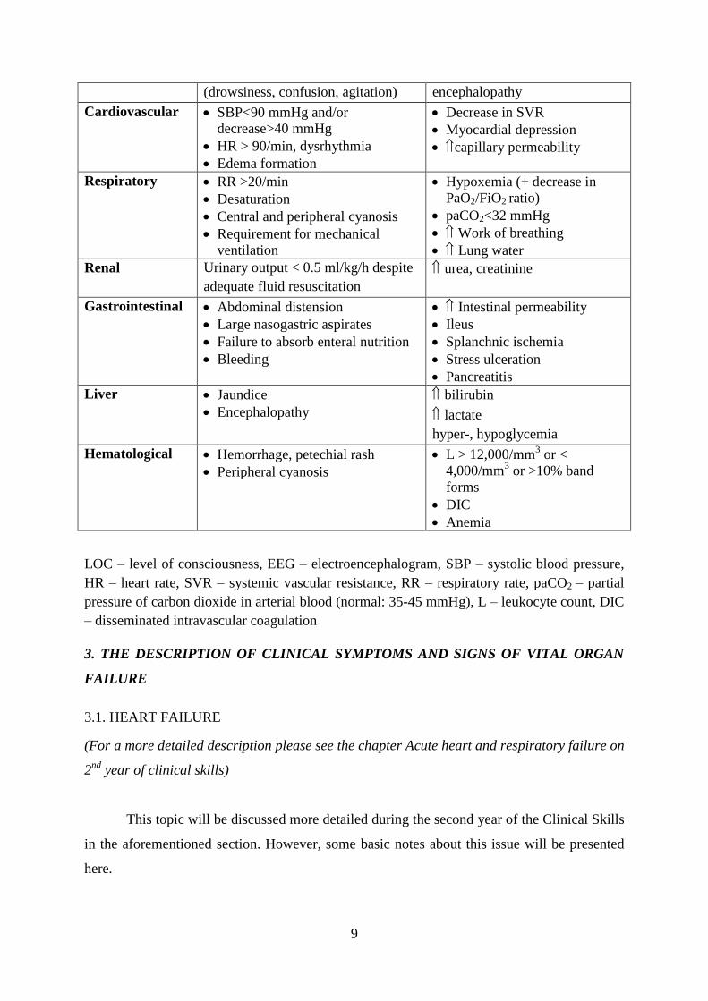
Table 1. The most commonly affected organs in MODS with description of clinical
manifestations
ORGAN
SYSTEM
ASSOCIATED CLINICAL
FEATURES
PHYSIOLOGICAL AND
BIOCHEMICAL CHANGES
Neurological Decreased LOC/encephalopathy Abnormal EEG with metabolic
9
(drowsiness, confusion, agitation) encephalopathy
Cardiovascular SBP<90 mmHg and/or
decrease>40 mmHg
HR > 90/min, dysrhythmia
Edema formation
Decrease in SVR
Myocardial depression
capillary permeability
Respiratory RR >20/min
Desaturation
Central and peripheral cyanosis
Requirement for mechanical
ventilation
Hypoxemia (+ decrease in
PaO2/FiO2 ratio)
paCO2<32 mmHg
Work of breathing
Lung water
Renal Urinary output < 0.5 ml/kg/h despite
adequate fluid resuscitation
urea, creatinine
Gastrointestinal Abdominal distension
Large nasogastric aspirates
Failure to absorb enteral nutrition
Bleeding
Intestinal permeability
Ileus
Splanchnic ischemia
Stress ulceration
Pancreatitis
Liver Jaundice
Encephalopathy
bilirubin
lactate
hyper-, hypoglycemia
Hematological Hemorrhage, petechial rash
Peripheral cyanosis
L > 12,000/mm3 or <
4,000/mm3 or >10% band
forms
DIC
Anemia
LOC – level of consciousness, EEG – electroencephalogram, SBP – systolic blood pressure,
HR – heart rate, SVR – systemic vascular resistance, RR – respiratory rate, paCO2 – partial
pressure of carbon dioxide in arterial blood (normal: 35-45 mmHg), L – leukocyte count, DIC
– disseminated intravascular coagulation
3. THE DESCRIPTION OF CLINICAL SYMPTOMS AND SIGNS OF VITAL ORGAN
FAILURE
3.1. HEART FAILURE
(For a more detailed description please see the chapter Acute heart and respiratory failure on
2nd
year of clinical skills)
This topic will be discussed more detailed during the second year of the Clinical Skills
in the aforementioned section. However, some basic notes about this issue will be presented
here.
10
Heart failure is a clinical syndrome that occurs due to changes in the function of the
heart as a pump, and is typically manifested by the characteristic clinical signs and symptoms.
These signs and symptoms arise because cardiac function does not meet the metabolic needs
of the organism. Because of the increase in retrograde pressure there is a congestion of
organs, and consequently the fluid accumulates retrograde ("backward") from one or both
ventricles. Therefore, it is sometimes called the congestive heart failure (congestion means the
accumulation of blood in a part of the body).
Acute heart failure is an emergency, life-threatening condition that is typically treated
in intensive care units. The most common causes are acute coronary syndrome, hypertensive
crisis, cardiac arrhythmias, valvular heart disease and myocarditis.
Acute heart failure can affect the right or left or both ventricles. Acute heart failure can
affect the right or left or both ventricles. Since the heart muscle can not eject enough blood,
there is a stasis of blood in various organs. In the right-sided heart failure the blood is
accumulated in the liver, digestive system and extremities. Due to the increased pressure in
that part of circulation there is a leakage of fluid from blood vessels into the tissues, which
results in the occurrence of enlarged liver, ascites, edema of lower legs and ankles, and the
distension of neck veins. Acute right-sided heart failure can also be a consequence of
pulmonary embolism when embolus closes pulmonary artery flow. In left-sided heart failure
there is a stasis of blood in pulmonary circulation with increased pulmonary venous pressure
and consequent fluid extravasation in alveoli. Fluid-filled alveoli prevent an adequate gas
exchange at the alveolar-capillary membrane and tissue oxygenation. Besides, acute
pulmonary edema and cardiogenic shock may develop. The main clinical signs are shallow
and rapid breathing, possible frothy and pink sputum, decrease in oxygen saturation,
cyanosis, tachycardia, and chest pain. With the development of cardiogenic shock there is
also hypotension.
The diagnosis is based on clinical presentation, medical history, chest X-rays, ECG,
echocardiography, and coronary angiography. It is necessary as soon as possible to determine
the cause of acute heart failure and administer appropriate treatment in an urgent manner.
The treatment depends on the clinical picture and the condition leading to acute heart
failure. The assessment of hemodynamic status of the patient is a very important issue. The
cardiogenic pulmonary edema is treated with oxygen, diuretics (drugs that stimulate urinary
output), vasodilators, and if necessary, mechanical ventilation. Treatment of cardiogenic
shock is based on inotropes (drugs that enhance the contractility of the heart muscle) and
vasoconstrictor drugs, as well as mechanical ventilation. Other possible emergency treatment
11
options are mechanical support, principally by using intra-aortic balloon pump (decreases
afterload and improves coronary blood flow), as well as urgent cardiac surgery. In the most
severe cases heart transplantation is needed.
3.2. RESPIRATORY FAILURE
(For a more detailed description please see the chapter Acute heart and respiratory failure on
2nd
year of clinical skills)
The main role of breathing is to maintain normal partial pressures of oxygen (PaO2)
and carbon dioxide (PaCO2), as well as pH in arterial blood. Therefore, respiratory failure, by
definition, is an inadequate gas exchange by the respiratory system, i.e. PaO2 and PaCO2 can
not be maintained in the normal range. Respiratory failure can also be defined as a clinical
syndrome in which the respiratory system fails in one or both of its essential functions of gas
exchange: oxygenation and/or elimination of CO2. It can be caused not only by a lung disease,
but also by a heart disease, respiratory muscles weakness, chest deformities, and the loss of
central control of breathing (events in the brain).
Regarding lung disease as the cause of respiratory failure, it should be noted that it can
occur due to many different direct and indirect insults that damage the lung tissue and lead to
accumulation of fluid in the lungs (pulmonary edema). The lungs are frequently injured
during severe sepsis. Respiratory failure usually occurs within 24-48 hours of the injury or
illness. It is manifested by rapid and shallow breathing. The skin may become blue, which is
termed cyanosis (blue skin and mucous membranes caused by increased concentration of > 50
g/L of reduced hemoglobin). The shortage of oxygen can cause very easily severe
complications in other tissues and organs. Without treatment the death occurs in most
patients. Diagnosis should be based on clinical presentation, medical history, laboratory tests
(blood gas analysis) and radiographs of the lungs. The treatment options are application of
oxygen through a mask, with endotracheal intubation and mechanical ventilation in severe
cases; positive pressure produced by mechanical ventilation helps keeping the alveoli open.
Pulmonary edema is treated with diuretics, and bacterial pneumonia with antibiotics.
12
3.3. LIVER FAILURE
Liver failure is a complex multiple system disease due to liver injury, and is caused by
inability of liver to perform its normal synthetic and metabolic function. There are two forms
of liver failure: acute and chronic.
3.3.1. ACUTE LIVER FAILURE
Main causes are acute viral hepatitis, the toxicity of paracetamol (acetaminophen),
mushrooms poisoning and some toxins. Prior to the availability of liver transplantation, the
mortality was as high as 80%. The classic features are acute onset, jaundice (usually first
visible in the sclera) and encephalopathy (syndrome characterized by impaired function of the
brain). It is also possible to encounter brain edema and kidney failure. The time period
between the development of jaundice and onset of encephalopathy is important for the
classification of acute liver failure; on this basis it can be hyperacute (interval 0-7 days), acute
(8-28 days) and subacute (28 days to 12 weeks). Also, the presence of encephalopathy is a
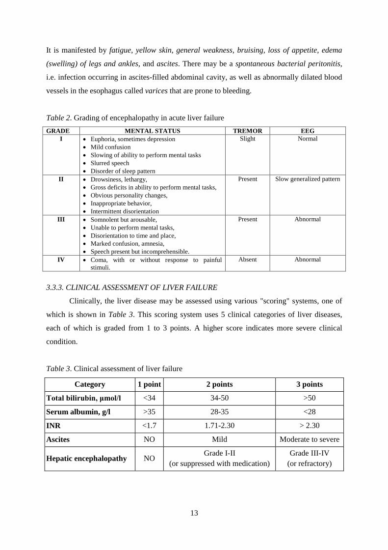
necessary prerequisite for the diagnosis of acute liver failure. The changes range from mild to
severe personality changes, confusion and deep coma. Typically 4 grades of encephalopathy
are described (Table 2). Patients with grades III and IV encephalopathy usually have brain
edema and increased intracranial pressure. According to some authors, there is also Grade 0
encephalopathy (subclinical encephalopathy or minimal hepatic encephalopathy) manifested
by normal mental status, but minimal changes in memory, concentration, intellectual function,
and co-
Anyway, this acute onset is usually characterized by weakness, fatigue and loss of
appetite. Less frequently, patients complain of abdominal pain and fever. Clinical signs
include jaundice, enlarged liver, and pain under the right rib cage. Ascites, i.e. fluid
accumulation in the abdominal cavity, is frequently seen. Urinary output may also be
gradually reduced (oliguria). Besides, the liver does not produce sufficient quantities of
clotting factors and the body has difficulty in controlling bleeding; a patient develops the
symptoms of poor blood coagulation - gastrointestinal bleeding, petechiae on the skin, and
bleeding from puncture sites.
3.3.2. CHRONIC LIVER FAILURE
Chronic liver failure is a consequence of diseases leading to a gradual deterioration in
functioning of hepatocytes and the development of cirrhosis. It evolves slowly over the years.
13
It is manifested by fatigue, yellow skin, general weakness, bruising, loss of appetite, edema
(swelling) of legs and ankles, and ascites. There may be a spontaneous bacterial peritonitis,
i.e. infection occurring in ascites-filled abdominal cavity, as well as abnormally dilated blood
vessels in the esophagus called varices that are prone to bleeding.
Table 2. Grading of encephalopathy in acute liver failure
GRADE MENTAL STATUS TREMOR EEG
I Euphoria, sometimes depression
Mild confusion
Slowing of ability to perform mental tasks
Slurred speech
Disorder of sleep pattern
Slight Normal
II Drowsiness, lethargy,
Gross deficits in ability to perform mental tasks,
Obvious personality changes,
Inappropriate behavior,
Intermittent disorientation
Present Slow generalized pattern
III Somnolent but arousable,
Unable to perform mental tasks,
Disorientation to time and place,
Marked confusion, amnesia,
Speech present but incomprehensible.
Present Abnormal
IV Coma, with or without response to painful
stimuli.
Absent Abnormal
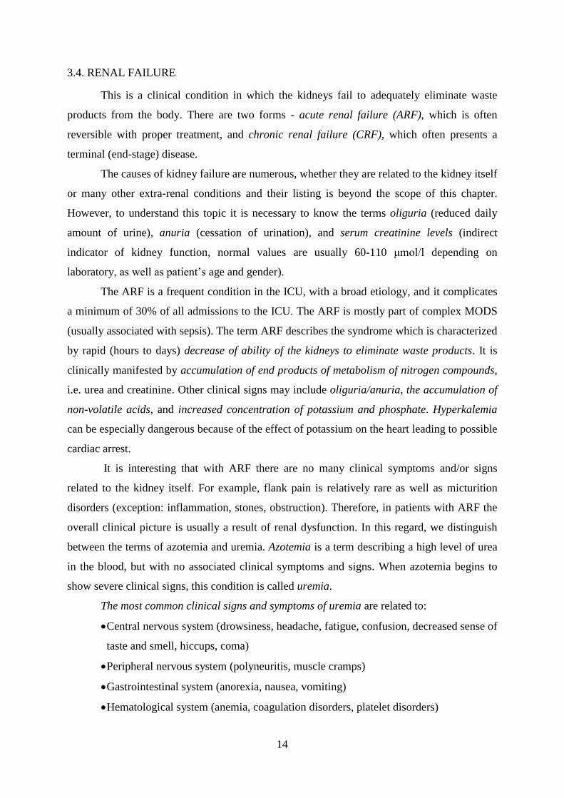
3.3.3. CLINICAL ASSESSMENT OF LIVER FAILURE
Clinically, the liver disease may be assessed using various "scoring" systems, one of
which is shown in Table 3. This scoring system uses 5 clinical categories of liver diseases,
each of which is graded from 1 to 3 points. A higher score indicates more severe clinical
condition.
Table 3. Clinical assessment of liver failure
Category 1 point 2 points 3 points
Total bilirubin, μmol/l <34 34-50 >50
Serum albumin, g/l >35 28-35 <28
INR <1.7 1.71-2.30 > 2.30
Ascites NO Mild Moderate to severe
Hepatic encephalopathy NO Grade I-II
(or suppressed with medication)
Grade III-IV
(or refractory)
14
3.4. RENAL FAILURE
This is a clinical condition in which the kidneys fail to adequately eliminate waste
products from the body. There are two forms - acute renal failure (ARF), which is often
reversible with proper treatment, and chronic renal failure (CRF), which often presents a
terminal (end-stage) disease.
The causes of kidney failure are numerous, whether they are related to the kidney itself
or many other extra-renal conditions and their listing is beyond the scope of this chapter.
However, to understand this topic it is necessary to know the terms oliguria (reduced daily
amount of urine), anuria (cessation of urination), and serum creatinine levels (indirect
indicator of kidney function, normal values are usually 60-110 μmol/l depending on
laboratory, as well as patient’s age and gender).
The ARF is a frequent condition in the ICU, with a broad etiology, and it complicates
a minimum of 30% of all admissions to the ICU. The ARF is mostly part of complex MODS
(usually associated with sepsis). The term ARF describes the syndrome which is characterized
by rapid (hours to days) decrease of ability of the kidneys to eliminate waste products. It is
clinically manifested by accumulation of end products of metabolism of nitrogen compounds,
i.e. urea and creatinine. Other clinical signs may include oliguria/anuria, the accumulation of
non-volatile acids, and increased concentration of potassium and phosphate. Hyperkalemia
can be especially dangerous because of the effect of potassium on the heart leading to possible
cardiac arrest.
It is interesting that with ARF there are no many clinical symptoms and/or signs
related to the kidney itself. For example, flank pain is relatively rare as well as micturition
disorders (exception: inflammation, stones, obstruction). Therefore, in patients with ARF the
overall clinical picture is usually a result of renal dysfunction. In this regard, we distinguish
between the terms of azotemia and uremia. Azotemia is a term describing a high level of urea
in the blood, but with no associated clinical symptoms and signs. When azotemia begins to
show severe clinical signs, this condition is called uremia.
The most common clinical signs and symptoms of uremia are related to:
Central nervous system (drowsiness, headache, fatigue, confusion, decreased sense of
taste and smell, hiccups, coma)
Peripheral nervous system (polyneuritis, muscle cramps)
Gastrointestinal system (anorexia, nausea, vomiting)
Hematological system (anemia, coagulation disorders, platelet disorders)
15
Cardiovascular system (pericarditis, coronary artery disease, hypertension,
pulmonary edema, peripheral edema)
The skin (itching, dryness)
The bones (osteomalacia)
Other (weight loss, fetor ex ore - a characteristic odor of ammonia)
In advanced cases of ARF, the renal replacement therapy (RRT) should be used. It
may be intermittent ("conventional" hemodialysis) or continuous. The latter is very popular in
the ICU, especially in hemodynamically unstable patients.
Chronic renal failure indicates the gradual loss of kidney function. As stated before,
the main role of the kidneys is to filter waste products and excess fluid from the body. When
chronic kidney disease is at an advanced stage, the dangerous levels of fluid, electrolytes and
waste are retained in the body. Many diseases can lead to chronic renal failure, like diabetes
mellitus, hypertension, polycystic kidney disease, glomerulonephritis, etc. Clinical signs and
symptoms are often nonspecific. Patients may complain of nausea, fatigue and loss of
appetite, may have high blood pressure, anemia, and reduced immunity. There is also a fluid
retention, leading to swelling of the legs and arms. If excess fluid accumulates in the area
around the heart or lungs, there is a chest pain and shortness of breath. These patients are
usually in the chronic hemodialysis program.
3.5. GASTROINTESTINAL DYSFUNCTION IN THE ICU
Gastrointestinal (GI) dysfunction in the ICU may be manifested as acute pancreatitis,
ileus, intestinal ischemia or infarction, GI bleeding and perforation of the abdominal organ.
Acute pancreatitis is manifested with severe abdominal pain usually radiating to the
back, nausea and vomiting, and hemodynamic and respiratory compromise in the most severe
cases. Tachypnea, dyspnea and hypoxia may indicate pleural effusions, relatively common in
this disease. The classic signs of hemorrhagic pancreatitis - ecchymoses in the flank (Grey
Turner’s sign) or umbilicus (Cullen’s sign) are not commonly present. Increased blood levels
of pancreatic enzymes (amylase, lipase) are helpful in diagnosis, as well as radiological
examination. Acute pancreatitis can be relatively mild and limited disease, but also an
16
extremely difficult condition (acute necrotizing pancreatitis), which may lead to MODS and
death.
Bowel obstruction (ileus) is relatively common among critically ill patients. It is
manifested usually with colicky abdominal pain, vomiting, dehydration, and absence of
normal peristalsis. Obstruction of the small intestine may be mechanical, when there is a
physical barrier to the aboral progression of intestinal contents, or paralytic, when some other
diseases inhibit the normal peristalsis (metabolic diseases, medications, neurogenic causes,
peritonitis).
Intestinal ischemia (mesenteric ischemia) is a condition in which any injury or
inflammation of the small intestine leads to inadequate blood supply. The cause is very
frequently long-term hypotension, as well as local vasoconstriction or blood clot. It is
commonly seen in elderly people suffering from cardiac arrhythmias (especially atrial
fibrillation). Apart from the general poor condition of the patient, the leading clinical
symptom is always a pain, in some cases followed by nausea, vomiting, diarrhea, and blood
in the rectum.
Acute GI bleeding is a common cause of admission to the ICU. It may originate from
the upper (more often) or lower GI tract. Peptic ulcer disease accounts for 75% of upper GI
bleeding (especially duodenal ulcers), while other causes are varices of the esophagus,
inflammation (esophagitis, duodenitis) and so-called Mallory Weiss syndrome (bleeding from
the mucosa at the connection point between the esophagus and stomach, usually as a result of
strong vomiting or coughing). Bleeding from the lower GI tract is typically caused by colonic
diverticulosis, polyps or tumors of the colon, angiodysplasia, and inflammatory bowel disease
(ulcerative colitis, Crohn's disease). Medical history of the bleeding patient may be
overwhelmed with the existence of ulcer disease or taking NSAIDs (non steroid anti-
inflammatory drugs – ibuprofen, diclofenac, aspirin, etc.). Regarding clinical features, the
bleeding is often without accompanying pain, especially in elderly patients. The signs of
hypovolemia may be present; pallor, sweating, tachycardia, oliguria, and changes in
consciousness. Hematemesis (vomiting blood) and melena (black, tarry stools with
characteristic odor) are common with bleeding from the upper GI tract. A history of vomiting
and retching preceding hematemesis suggests Mallory–Weiss syndrome. Hematochezia is the
17
passage of bright red blood from the rectum, in the form of pure blood or admixed with stool.
It usually represents a bleeding from the lower GI tract.
Acute abdomen - this term denotes a sudden, strong abdominal pain, of unknown
etiology, which lasts for less than 24 hours indicating an abdominal pathologic condition
that, if left undiscovered and untreated, would have a deleterious effect on the patient’s health
status.
The location of the pain can give valuable information about etiology; epigastric pain
may suggest ulcer perforation, the pain under the right rib cage indicates cholecystitis,
cholangitis, or subphrenic abscess, the pain in the right lower quadrant indicates appendicitis,
incarcerated hernia, or ectopic pregnancy. The periumbilical pain may indicate the beginning
of appendicitis, mesenteric ischemia, obstruction of the small intestine, while the pain in the
left lower quadrant indicates the possible diverticulitis, torsion of an ovarian cyst, or renal
colic. Of course, all possible causes are not listed here. Also important is the nature of the
pain; for example episodic or spasmodic pain usually occurs due to obstruction of hollow
organs or structures (bowel obstruction, acute cholecystitis), while permanent, severe pain
may indicate acute inflammation or perforation of organs (appendicitis). Here are some
clinical examples; sudden, strong and sharp pain (like "knife stab"), located in the epigastrium
is the leading symptom of perforation of gastroduodenal ulcers. Periumbilical pain that is
gradually moved over time and placed in the lower right quadrant (ileocecal region) refers to
acute appendicitis. The sudden and sharp pain, located below the right costal margin with
expansion towards the middle of the abdominal wall and the right scapula, suggests acute
cholecystitis and cholelithiasis. Strong and sharp pain radiating in the form of a belt toward
the back speaks mostly for acute pancreatitis, especially if occurring after eating fatty foods.
The intense pain with bowel cramps and bloating associated with the absence of stool and
flatulence is a sign of ileus. Acute colicky pain in the lumbar region radiating to the genital
region and toward the lower limbs occurs during attacks of kidney stones. Diffuse abdominal
pain associated with extreme tension of the abdominal muscles (i.e. muscular defense) and
vomiting, suggest diffuse peritonitis, an extremely dangerous condition.
18
4. CLINICAL ASSESSMENT OF ORGAN DYSFUNCTION IN ICU
4.1. INTRODUCTION
The clinical assessment of disease severity is an essential component of medical
practice. It influences the need and speed for supportive and specific therapy. In addition, it
can predict the outcome of the disease in certain patients.
This section will briefly explain some of the most commonly used clinical scoring
systems for assessing dysfunction of organs and organ systems in the ICU. Here it is very
important to emphasize that students are not supposed to learn all these scoring systems by
heart (except maybe the Glasgow Coma Score - GCS), since they are all available through
numerous practical “online” applications. For students the most important thing here would be
to conclude what clinical and laboratory variables are used in daily practice to define organ
dysfunction. Clinical assessment of liver and kidney failure was shown in previous chapters
4.2. OVERVIEW OF COMMONLY USED SCORING SYSTEMS OF ORGAN
DYSFUNCTION
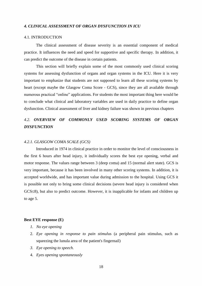
4.2.1. GLASGOW COMA SCALE (GCS)
Introduced in 1974 in clinical practice in order to monitor the level of consciousness in
the first 6 hours after head injury, it individually scores the best eye opening, verbal and
motor response. The values range between 3 (deep coma) and 15 (normal alert state). GCS is
very important, because it has been involved in many other scoring systems. In addition, it is
accepted worldwide, and has important value during admission to the hospital. Using GCS it
is possible not only to bring some clinical decisions (severe head injury is considered when
GCS8), but also to predict outcome. However, it is inapplicable for infants and children up
to age 5.
Best EYE response (E)
1. No eye opening
2. Eye opening in response to pain stimulus (a peripheral pain stimulus, such as
squeezing the lunula area of the patient's fingernail)
3. Eye opening to speech.
4. Eyes opening spontaneously
19
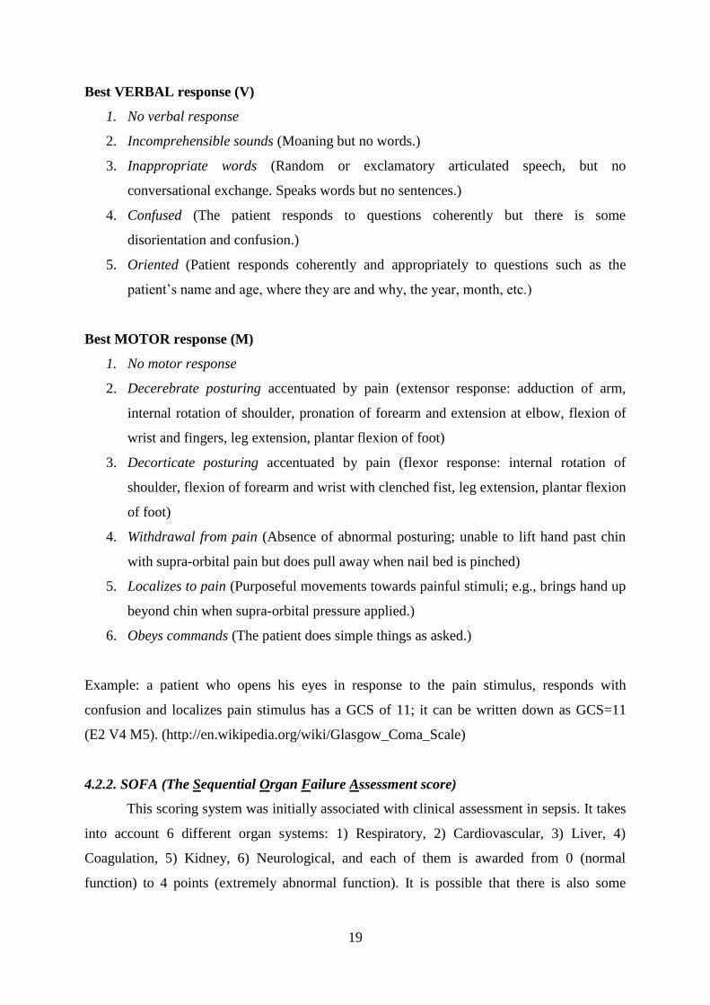
Best VERBAL response (V)
1. No verbal response
2. Incomprehensible sounds (Moaning but no words.)
3. Inappropriate words (Random or exclamatory articulated speech, but no
conversational exchange. Speaks words but no sentences.)
4. Confused (The patient responds to questions coherently but there is some
disorientation and confusion.)
5. Oriented (Patient responds coherently and appropriately to questions such as the
patient’s name and age, where they are and why, the year, month, etc.)
Best MOTOR response (M)
1. No motor response
2. Decerebrate posturing accentuated by pain (extensor response: adduction of arm,
internal rotation of shoulder, pronation of forearm and extension at elbow, flexion of
wrist and fingers, leg extension, plantar flexion of foot)
3. Decorticate posturing accentuated by pain (flexor response: internal rotation of
shoulder, flexion of forearm and wrist with clenched fist, leg extension, plantar flexion
of foot)
4. Withdrawal from pain (Absence of abnormal posturing; unable to lift hand past chin
with supra-orbital pain but does pull away when nail bed is pinched)
5. Localizes to pain (Purposeful movements towards painful stimuli; e.g., brings hand up
beyond chin when supra-orbital pressure applied.)
6. Obeys commands (The patient does simple things as asked.)
Example: a patient who opens his eyes in response to the pain stimulus, responds with
confusion and localizes pain stimulus has a GCS of 11; it can be written down as GCS=11
(E2 V4 M5). (http://en.wikipedia.org/wiki/Glasgow_Coma_Scale)
4.2.2. SOFA (The Sequential Organ Failure Assessment score)
This scoring system was initially associated with clinical assessment in sepsis. It takes
into account 6 different organ systems: 1) Respiratory, 2) Cardiovascular, 3) Liver, 4)
Coagulation, 5) Kidney, 6) Neurological, and each of them is awarded from 0 (normal
function) to 4 points (extremely abnormal function). It is possible that there is also some
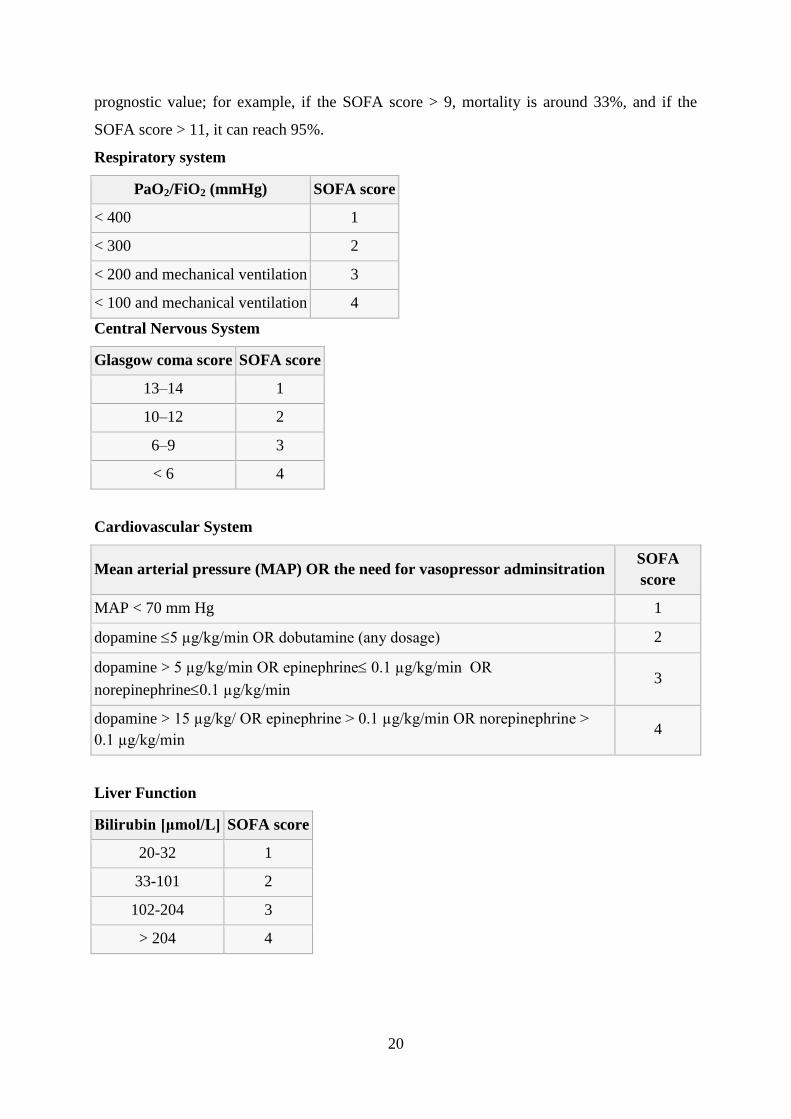
20
prognostic value; for example, if the SOFA score > 9, mortality is around 33%, and if the
SOFA score > 11, it can reach 95%.
Respiratory system
PaO2/FiO2 (mmHg) SOFA score
< 400 1
< 300 2
< 200 and mechanical ventilation 3
< 100 and mechanical ventilation 4
Central Nervous System
Glasgow coma score SOFA score
13–14 1
10–12 2
6–9 3
< 6 4
Cardiovascular System
Mean arterial pressure (MAP) OR the need for vasopressor adminsitration SOFA
score
MAP < 70 mm Hg 1
dopamine 5 µg/kg/min OR dobutamine (any dosage) 2
dopamine > 5 µg/kg/min OR epinephrine 0.1 µg/kg/min OR
norepinephrine0.1 µg/kg/min 3
dopamine > 15 µg/kg/ OR epinephrine > 0.1 µg/kg/min OR norepinephrine >
0.1 µg/kg/min 4
Liver Function
Bilirubin [μmol/L] SOFA score
20-32 1
33-101 2
102-204 3
> 204 4
21
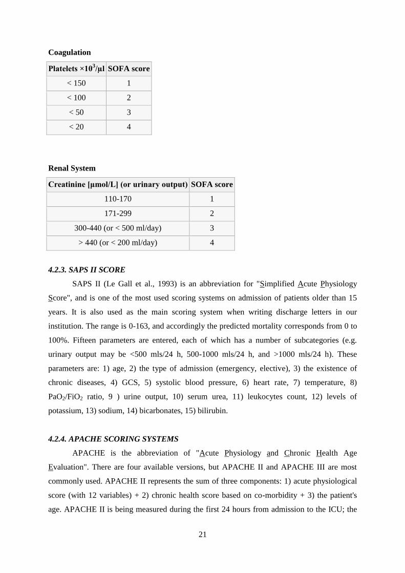
Coagulation
Platelets ×103/µl SOFA score
< 150 1
< 100 2
< 50 3
< 20 4
Renal System
Creatinine [μmol/L] (or urinary output) SOFA score
110-170 1
171-299 2
300-440 (or < 500 ml/day) 3
> 440 (or < 200 ml/day) 4
4.2.3. SAPS II SCORE
SAPS II (Le Gall et al., 1993) is an abbreviation for "Simplified Acute Physiology
Score", and is one of the most used scoring systems on admission of patients older than 15
years. It is also used as the main scoring system when writing discharge letters in our
institution. The range is 0-163, and accordingly the predicted mortality corresponds from 0 to
100%. Fifteen parameters are entered, each of which has a number of subcategories (e.g.
urinary output may be <500 mls/24 h, 500-1000 mls/24 h, and >1000 mls/24 h). These
parameters are: 1) age, 2) the type of admission (emergency, elective), 3) the existence of
chronic diseases, 4) GCS, 5) systolic blood pressure, 6) heart rate, 7) temperature, 8)
PaO2/FiO2 ratio, 9 ) urine output, 10) serum urea, 11) leukocytes count, 12) levels of
potassium, 13) sodium, 14) bicarbonates, 15) bilirubin.
4.2.4. APACHE SCORING SYSTEMS
APACHE is the abbreviation of "Acute Physiology and Chronic Health Age
Evaluation". There are four available versions, but APACHE II and APACHE III are most
commonly used. APACHE II represents the sum of three components: 1) acute physiological
score (with 12 variables) + 2) chronic health score based on co-morbidity + 3) the patient's
age. APACHE II is being measured during the first 24 hours from admission to the ICU; the
22
maximum possible value is 71. The value of the APACHE II >25 carries predictive mortality
of 50%, and the value of >35% carries predictive mortality of 80%. APACHE III scoring
system is slightly more complex, and less frequently used. It is based on up to 20
physiological variables along with some additional information. The values range from 0-299.
LITERATURE:
1. Marino PL. The ICU book. 3rd
edition. Philadelphia: Lippincott Williams & Wilkins;
2007.
2. Ivančević Ž. i sur. MSD priručnik dijagnostike i terapije. 2. hrvatsko izdanje. Split:
Placebo; 2010.
3. Butterworth J, Mackey DC, Wasnick J. Morgan and Mikhail's Clinical Anesthesiology.
5th edition. New York: McGraw-Hill Companies; 2013.
4. Ivančević Ž, Rumboldt Z, Bergovec M, Silobrčić V, Bruketa D. Harrison - Principi
interne medicine. 1. hrvatsko izdanje. Split: Placebo; 1997.
5. Miller RD, Eriksson LI, Fleisher LA, Wiener-Kronish JP, Young WL. Miller's
Anesthesia. 7th
edition. Philadelphia: Churchill Livingstone, Elsevier; 2009.
6. Papadakos PJ, Szalados JE. Critical care: the requisites in anesthesiology. Philadelphia:
Elsevier Mosby; 2005.
7. Bongard FS, Sue DY, Vintch JR. Current Diagnosis & Treatment Critical Care. 3rd
edition. New York: McGraw-Hill; 2008.
8. Bersten AD, Soni N. Oh's Intensive Care Manual. 6th
edition. Philadelphia: Butterworth-
Heinemann Elsevier; 2009.
9. Vincent J-L. Yearbook of Intensive Care and Emergency Medicine 2010. Heidelberg:
Springer; 2010.
10. Bone RC et al. Definitions for sepsis and organ failure and guidelines for the use of
innovative therapies in sepsis. The ACCP/SCCM Consensus Conference Committee.
American College of Chest Physicians/Society of Critical Care Medicine. Chest.
1992;101(6):1644-55.
11. American College of Chest Physicians/Society of Critical Care Medicine Consensus
Conference: definitions for sepsis and organ failure and guidelines for the use of
innovative therapies in sepsis. Crit Care Med 1992; 20:864-74.
12. Le Gall JR, Lemeshow S, Saulnier F. A New Simplified Acute Physiology Score (SAPS
II) Based on a European/North American Multicenter Study. JAMA. 1993;270:2957-2963
13. http://www.sfar.org/scores2/saps2.html
14. http://www.sfar.org/scores2/apache22.html
15. http://www.quesgen.com/ApacheIII.php
23
3. BASIC LIFE SUPPORT IN ADULTS
Mihajlo Lojpur, M.D., Ph.D.
1. VITAL SIGNS
Vital signs include taking the patient’s pulse, respiration, blood pressure, and
temperature.
1. Pulse
The ventricles (right and left) have two phases: diastole or the time when the ventricles 'rest'
so they can fill with blood, and systole, the time when the ventricles contract to send blood
either to the lungs (from the right side of the heart), or to the rest of the body (from the left
side of the heart).
The pulse represents the variation in blood pressure from diastole to systole. During diastole
blood pressure falls, but increases after systole as the heart pumps more blood into the
arteries. You feel this difference when taking your pulse.
When taking a patient’s pulse, you should be concerned with two factors: rate and character.
For pulse rate, you will have to determine the number of beats per minute. Pulse rate is
classified as normal, rapid, or slow. A normal pulse rate for adults is between 60 to
80 beats per minute. Any pulse rate above 100 beats per minute is rapid (tachycardia),
while a rate below 60 beats per minute is slow (bradycardia).
Normal pulse rates for different ages (per min.)
newborn
(0-30 days
old)
infants
(1 — 11
months)
children
(1 — 10 years)
children over 10 years &
adults,
including seniors
*well-trained
adult
athletes
70 - 190 80 - 120 70 - 130 60 - 100 40 - 60
NOTE: An athlete may have a normal at-rest pulse rate between 40 and 60
beats per minute. This is a slow pulse rate, but is not an indication of poor health.
Pulse character is the rhythm and force of the pulse.
o Pulse rhythm is evaluated as regular or irregular. When intervals
between beats are constant,
the pulse is regular, and when intervals are not
constant, the pulse is described as irregular.
o Pulse force refers to the pressure of the pulse wave as it expands the
artery. Pulse force is determined as full or thready. A
full pulse feels as if a strong wave has
24
passed under your fingertips. When the pulse feels weak and thin, the
pulse is described as thready.
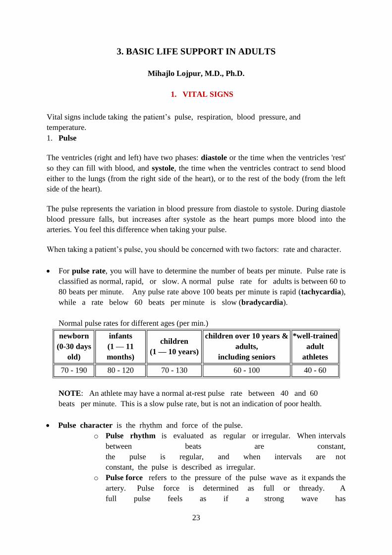
The pulse rate and character can be determined at a number of points throughout the
body.
The most common site to determine a patient’s pulse is the radial pulse but in an
emergency situation better sites to check for a pulse are carotid and femoral artery.
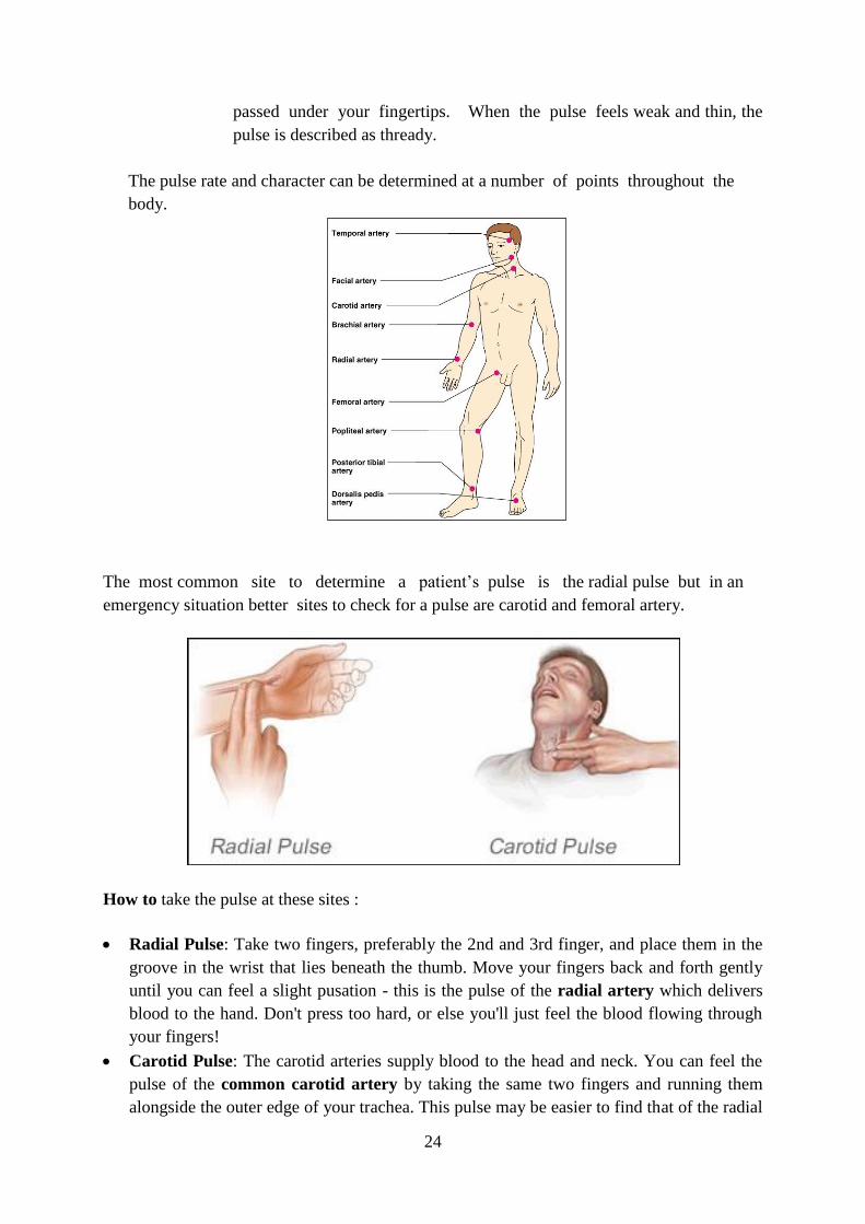
How to take the pulse at these sites :
Radial Pulse: Take two fingers, preferably the 2nd and 3rd finger, and place them in the
groove in the wrist that lies beneath the thumb. Move your fingers back and forth gently
until you can feel a slight pusation - this is the pulse of the radial artery which delivers
blood to the hand. Don't press too hard, or else you'll just feel the blood flowing through
your fingers!
Carotid Pulse: The carotid arteries supply blood to the head and neck. You can feel the
pulse of the common carotid artery by taking the same two fingers and running them
alongside the outer edge of your trachea. This pulse may be easier to find that of the radial
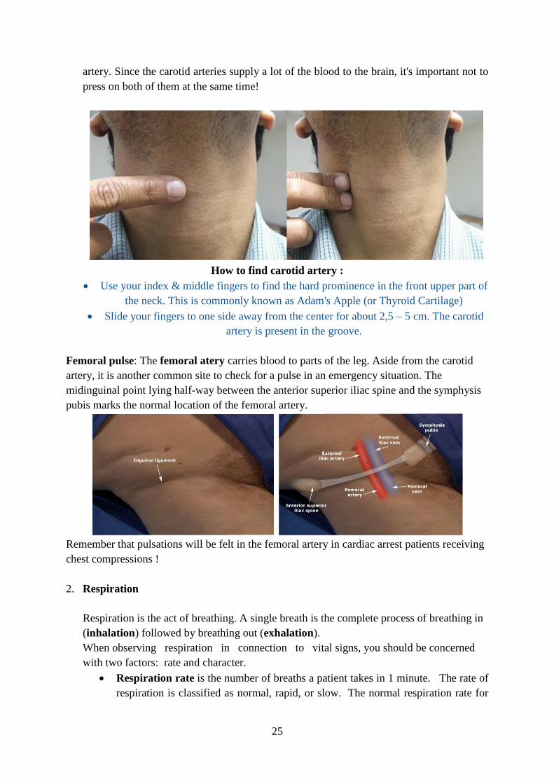
25
artery. Since the carotid arteries supply a lot of the blood to the brain, it's important not to
press on both of them at the same time!
How to find carotid artery :
Use your index & middle fingers to find the hard prominence in the front upper part of
the neck. This is commonly known as Adam's Apple (or Thyroid Cartilage)
Slide your fingers to one side away from the center for about 2,5 – 5 cm. The carotid
artery is present in the groove.
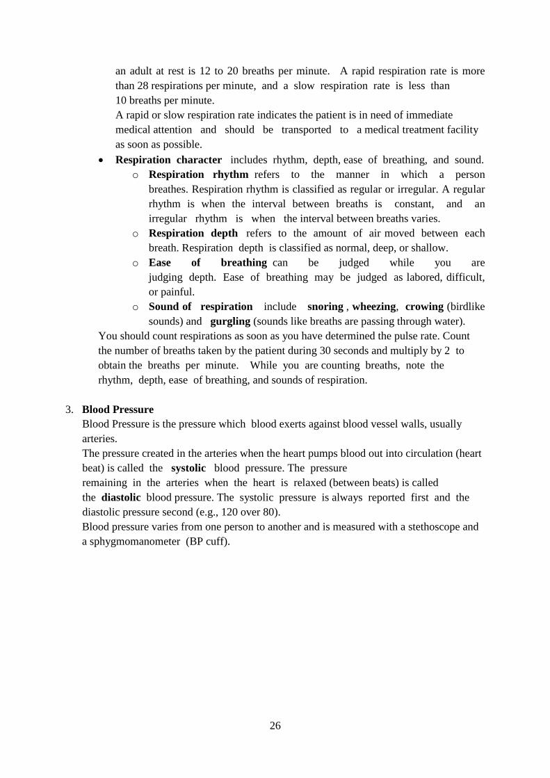
Femoral pulse: The femoral atery carries blood to parts of the leg. Aside from the carotid
artery, it is another common site to check for a pulse in an emergency situation. The
midinguinal point lying half-way between the anterior superior iliac spine and the symphysis
pubis marks the normal location of the femoral artery.
Remember that pulsations will be felt in the femoral artery in cardiac arrest patients receiving
chest compressions !
2. Respiration
Respiration is the act of breathing. A single breath is the complete process of breathing in
(inhalation) followed by breathing out (exhalation).
When observing respiration in connection to vital signs, you should be concerned
with two factors: rate and character.
Respiration rate is the number of breaths a patient takes in 1 minute. The rate of
respiration is classified as normal, rapid, or slow. The normal respiration rate for
26
an adult at rest is 12 to 20 breaths per minute. A rapid respiration rate is more
than 28 respirations per minute, and a slow respiration rate is less than
10 breaths per minute.
A rapid or slow respiration rate indicates the patient is in need of immediate
medical attention and should be transported to a medical treatment facility
as soon as possible.
Respiration character includes rhythm, depth, ease of breathing, and sound.
o Respiration rhythm refers to the manner in which a person
breathes. Respiration rhythm is classified as regular or irregular. A regular
rhythm is when the interval between breaths is constant, and an
irregular rhythm is when the interval between breaths varies.
o Respiration depth refers to the amount of air moved between each
breath. Respiration depth is classified as normal, deep, or shallow.
o Ease of breathing can be judged while you are
judging depth. Ease of breathing may be judged as labored, difficult,
or painful.
o Sound of respiration include snoring , wheezing, crowing (birdlike
sounds) and gurgling (sounds like breaths are passing through water).
You should count respirations as soon as you have determined the pulse rate. Count
the number of breaths taken by the patient during 30 seconds and multiply by 2 to
obtain the breaths per minute. While you are counting breaths, note the
rhythm, depth, ease of breathing, and sounds of respiration.
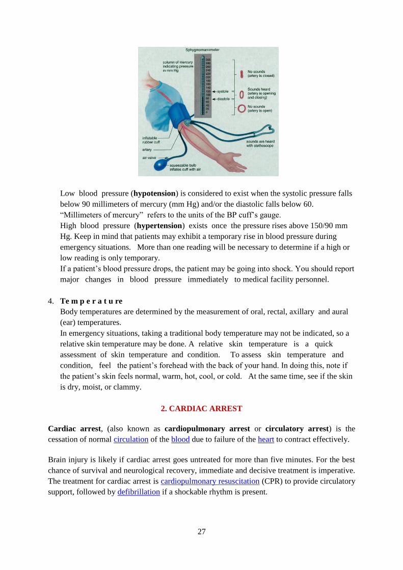
3. Blood Pressure
Blood Pressure is the pressure which blood exerts against blood vessel walls, usually
arteries.
The pressure created in the arteries when the heart pumps blood out into circulation (heart
beat) is called the systolic blood pressure. The pressure
remaining in the arteries when the heart is relaxed (between beats) is called
the diastolic blood pressure. The systolic pressure is always reported first and the
diastolic pressure second (e.g., 120 over 80).
Blood pressure varies from one person to another and is measured with a stethoscope and
a sphygmomanometer (BP cuff).
27
Low blood pressure (hypotension) is considered to exist when the systolic pressure falls
below 90 millimeters of mercury (mm Hg) and/or the diastolic falls below 60.
“Millimeters of mercury” refers to the units of the BP cuff’s gauge.
High blood pressure (hypertension) exists once the pressure rises above 150/90 mm
Hg. Keep in mind that patients may exhibit a temporary rise in blood pressure during
emergency situations. More than one reading will be necessary to determine if a high or
low reading is only temporary.
If a patient’s blood pressure drops, the patient may be going into shock. You should report
major changes in blood pressure immediately to medical facility personnel.
4. Te m p e r a t u re
Body temperatures are determined by the measurement of oral, rectal, axillary and aural
(ear) temperatures.
In emergency situations, taking a traditional body temperature may not be indicated, so a
relative skin temperature may be done. A relative skin temperature is a quick
assessment of skin temperature and condition. To assess skin temperature and
condition, feel the patient’s forehead with the back of your hand. In doing this, note if
the patient’s skin feels normal, warm, hot, cool, or cold. At the same time, see if the skin
is dry, moist, or clammy.
2. CARDIAC ARREST
Cardiac arrest, (also known as cardiopulmonary arrest or circulatory arrest) is the
cessation of normal circulation of the blood due to failure of the heart to contract effectively.
Brain injury is likely if cardiac arrest goes untreated for more than five minutes. For the best
chance of survival and neurological recovery, immediate and decisive treatment is imperative.
The treatment for cardiac arrest is cardiopulmonary resuscitation (CPR) to provide circulatory
support, followed by defibrillation if a shockable rhythm is present.
28
Causes
Coronary heart disease is the leading cause of sudden cardiac arrest. Many other cardiac and
non-cardiac conditions also increase ones risk
Approximately 60–70% of cardiac arrest is related to cardiac disease.
o Among adults, ischemic heart disease is the predominant cause of arrest. No
less than 30% of them at autopsy showing signs of recent myocardial
infarction.
o A number of other cardiac abnormalities can increase the risk of cardiac arrest
including: cardiomyopathy, cardiac rhythm disturbances, hypertensive heart
disease, congestive heart failure...
Cardiac arrest is unrelated to heart problems in 35% of cases.
o The most common non-cardiac causes: trauma, non-trauma related bleeding
(such as gastrointestinal bleeding, aortic rupture, and intracranial hemorrhage),
overdose, drowning and pulmonary embolism.
In infants and children, the most common cause of cardiac arrest is respiratory arrest.
Respiratory disorders most often resulting in cardiac arrest include airway obstruction, smoke
inhalation, drowning, infection and sudden infant death syndrome. In adults, the opposite
usually occurs - cardiac arrest leads to respiratory arrest.
Signs and symptoms
Cardiac arrest is an abrupt cessation of pump function in the heart, as evidenced by the
absence of a palpable pulse. Arrested blood circulation prevents delivery of oxygen to the
body. Due to inadequate cerebral perfusion, the patient will be unconscious and will have
stopped breathing.
Diagnosis
The main diagnostic criterion to diagnose a cardiac arrest is lack of circulation, however there
are a number of ways of determining this.
1. A cardiac arrest is usually diagnosed clinically by the absence of a pulse. In many cases
lack of carotid pulse is the gold standard for diagnosing cardiac arrest, but lack of a pulse
(particularly in the peripheral pulses) may be a result of other conditions (e.g. shock), or
simply an error on the part of the rescuer. Studies have shown that rescuers often make a
mistake when checking the carotid pulse in an emergency, whether they are healthcare
professionals or lay persons.
29
Owing to the inaccuracy in this method of diagnosis, some bodies such as the European
Resuscitation Council (ERC) have de-emphasised its importance. The Resuscitation
Council (UK), in line with the ERC's recommendations and those of the American Heart
Association, have suggested that the technique should be used only by healthcare
professionals with specific training and expertise, and even then that it should be viewed
in conjunction with other indicators such as agonal respiration.
2. Various other methods for detecting circulation have been proposed. Guidelines following
the 2000 International Liaison Committee on Resuscitation (ILCOR) recommendations
were for rescuers to look for "signs of circulation", but not specifically the pulse. These
signs included coughing, gasping, colour, twitching and movement.
However, in face of evidence that these guidelines were ineffective, the current
recommendation of ILCOR is that cardiac arrest should be diagnosed in all
casualties who are unconscious and not breathing normally.
3. ADULT BASIC LIFE SUPPORT
Basic life support includes the maintenance of an airway and the support of breathing and
the circulation without using equipment other than a simple airway device or protective
shield. A combination of expired air ventilation (rescue breathing) and chest compression is
known as cardiopulmonary resuscitation (CPR), which forms the basis of modern basic
life support.
The term "cardiac arrest" implies a sudden interruption of cardiac output, which may be
reversible with appropriate treatment. It is important that those who may be present at the
scene of a cardiac arrest should have learnt the appropriate resuscitation skills and be able to
put them into practice.
Simplification of the BLS sequence continues to be a feature of these guidelines, but, in
addition, there is now advice on who should be taught what skills, particularly chest-
compression-only or chest compression and ventilation.
All rescuers, trained or not, should provide chest compressions to victims of cardiac arrest :
• If a bystander is not trained in CPR, he or she should provide compression-only CPR
for the adult victim who suddenly collapses, with an emphasis to “push hard and fast”
on the center of the chest, or follow the directions of the EMS dispatcher. The rescuer
should continue compression-only CPR until an AED arrives and is ready for use or
EMS providers or other responders take over care of the victim.
• All trained lay rescuers should, at a minimum, provide chest compressions for victims
of cardiac arrest. In addition, if the trained lay rescuer is able to perform rescue
breaths, compressions and breaths should be provided in a ratio of 30 compressions to
30
2 breaths. The rescuer should continue CPR until an AED arrives and is ready for use
or EMS providers take over care of the victim.
Continued emphasis has been placed on high-quality CPR (with chest compressions of
adequate rate and depth, allowing complete chest recoil after each compression, minimizing
interruptions in compressions, and avoiding excessive ventilation) :
• Compression rate should be at least 100/min (rather than “approximately” 100/min).
• Compression depth for adults has been changed from the range of 4 to 5 cm to at
least 5 cm
Adult basic life support
A. Adult basic life support sequence
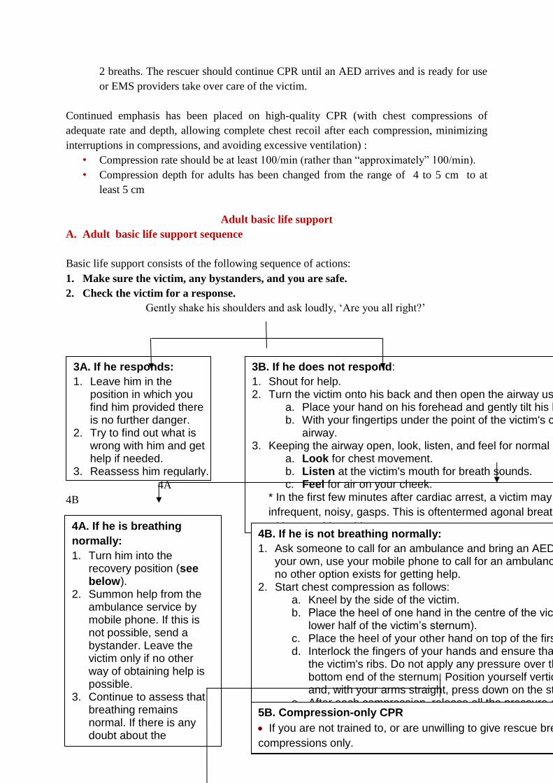
Basic life support consists of the following sequence of actions:
1. Make sure the victim, any bystanders, and you are safe.
2. Check the victim for a response.
Gently shake his shoulders and ask loudly, ‘Are you all right?’
4A
4B
3A. If he responds:
1. Leave him in the position in which you find him provided there is no further danger.
2. Try to find out what is wrong with him and get help if needed.
3. Reassess him regularly.
3B. If he does not respond:
1. Shout for help. 2. Turn the victim onto his back and then open the airway using head tilt and chin lift:
a. Place your hand on his forehead and gently tilt his head back. b. With your fingertips under the point of the victim's chin, lift the chin to open the
airway. 3. Keeping the airway open, look, listen, and feel for normal breathing :
a. Look for chest movement. b. Listen at the victim's mouth for breath sounds. c. Feel for air on your cheek.
* In the first few minutes after cardiac arrest, a victim may be barely breathing, or taking
infrequent, noisy, gasps. This is oftentermed agonal breathing and must not be confused
with normal breathing.
*Look, listen, and feel for no more than 10 s to determine if the victim is breathing
normally. If
you have any doubt whether breathing is normal, act as if it is not normal.
4A. If he is breathing
normally:
1. Turn him into the recovery position (see below).
2. Summon help from the ambulance service by mobile phone. If this is not possible, send a bystander. Leave the victim only if no other way of obtaining help is possible.
3. Continue to assess that breathing remains normal. If there is any doubt about the presence of normal breathing, start CPR
4B. If he is not breathing normally:
1. Ask someone to call for an ambulance and bring an AED if available. If you are on your own, use your mobile phone to call for an ambulance. Leave the victim only when no other option exists for getting help.
2. Start chest compression as follows: a. Kneel by the side of the victim. b. Place the heel of one hand in the centre of the victim’s chest (which is the
lower half of the victim’s sternum). c. Place the heel of your other hand on top of the first hand. d. Interlock the fingers of your hands and ensure that pressure is not applied over
the victim's ribs. Do not apply any pressure over the upper abdomen or the bottom end of the sternum. Position yourself vertically above the victim's chest and, with your arms straight, press down on the sternum 5 - 6 cm.
e. After each compression, release all the pressure on the chest without losing contact between your hands and the sternum.
f. Repeat at a rate of 100 - 120 per min
g. Compression and release should take an equal amount of time.
5B. Compression-only CPR
If you are not trained to, or are unwilling to give rescue breaths, give chest
compressions only.
If chest compressions only are given, these should be continuous at a rate of 100 - 120
min-1.
31
5A 6
5A 6
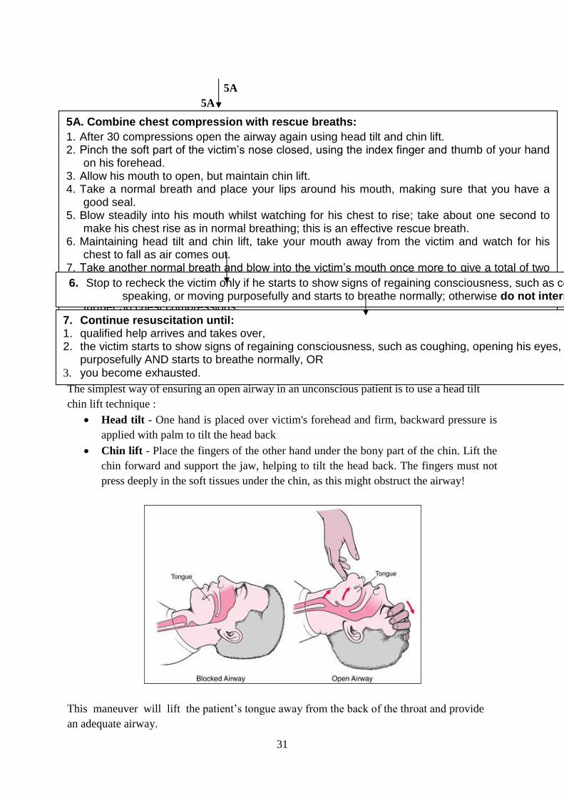
Head tilt and chin lift
The simplest way of ensuring an open airway in an unconscious patient is to use a head tilt
chin lift technique :
Head tilt - One hand is placed over victim's forehead and firm, backward pressure is
applied with palm to tilt the head back
Chin lift - Place the fingers of the other hand under the bony part of the chin. Lift the
chin forward and support the jaw, helping to tilt the head back. The fingers must not
press deeply in the soft tissues under the chin, as this might obstruct the airway!
This maneuver will lift the patient’s tongue away from the back of the throat and provide
an adequate airway.
5A. Combine chest compression with rescue breaths:
1. After 30 compressions open the airway again using head tilt and chin lift. 2. Pinch the soft part of the victim’s nose closed, using the index finger and thumb of your hand
on his forehead. 3. Allow his mouth to open, but maintain chin lift. 4. Take a normal breath and place your lips around his mouth, making sure that you have a
good seal. 5. Blow steadily into his mouth whilst watching for his chest to rise; take about one second to
make his chest rise as in normal breathing; this is an effective rescue breath. 6. Maintaining head tilt and chin lift, take your mouth away from the victim and watch for his
chest to fall as air comes out. 7. Take another normal breath and blow into the victim’s mouth once more to give a total of two
effective rescue breaths. The two breaths should not take more than 5 s. 8. Then return your hands without delay to the correct position on the sternum and give a
further 30 chest compressions. 9. Continue with chest compressions and rescue breaths in a ratio of 30:2.
6. Stop to recheck the victim only if he starts to show signs of regaining consciousness, such as coughing, opening his eyes, speaking, or moving purposefully and starts to breathe normally; otherwise do not interrupt resuscitation.
7. Continue resuscitation until: 1. qualified help arrives and takes over, 2. the victim starts to show signs of regaining consciousness, such as coughing, opening his eyes, speaking, or moving
purposefully AND starts to breathe normally, OR 3. you become exhausted.

32
NOTE : The jaw thrust technique is not recommended for lay rescuers because it is difficult to
learn and perform. Therefore, the lay rescuer should open the airway using a head-tilt, chin-
lift manoeuvre for both injured and non-injured victims.
Head tilt /chin lift
Jaw thrust
Look, listen and feel for normal breathing
You can check the breathing by placing your ears near the patients mouth and nose and listen
or feel for air coming out. Look also for the rise and fall of the chest, this will indicate that the
patient is breathing.
Look, listen, and feel for no more than 10 s to determine if the victim is breathing normally.
If you have any doubt whether breathing is normal, act as if it is not normal.
NOTE : Rescuers are often warned against mistaking agonal breathing, which is a series of
noisy gasps occurring in around 40% of cardiac arrest victims, for normal breathing.
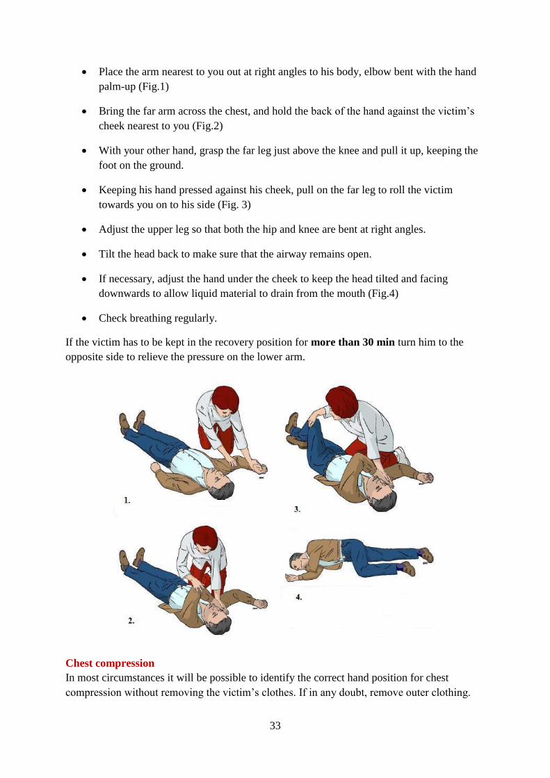
Recovery position
The position should be stable, near a true lateral position with the head dependent, and with
no pressure on the chest to impair breathing.
The RC(UK) recommends the following sequence of actions to place a victim in the recovery
position:
Remove the victim’s glasses, if present.
Kneel beside the victim and make sure that both his legs are straight.
33
Place the arm nearest to you out at right angles to his body, elbow bent with the hand
palm-up (Fig.1)
Bring the far arm across the chest, and hold the back of the hand against the victim’s
cheek nearest to you (Fig.2)
With your other hand, grasp the far leg just above the knee and pull it up, keeping the
foot on the ground.
Keeping his hand pressed against his cheek, pull on the far leg to roll the victim
towards you on to his side (Fig. 3)
Adjust the upper leg so that both the hip and knee are bent at right angles.
Tilt the head back to make sure that the airway remains open.
If necessary, adjust the hand under the cheek to keep the head tilted and facing
downwards to allow liquid material to drain from the mouth (Fig.4)
Check breathing regularly.
If the victim has to be kept in the recovery position for more than 30 min turn him to the
opposite side to relieve the pressure on the lower arm.
Chest compression
In most circumstances it will be possible to identify the correct hand position for chest
compression without removing the victim’s clothes. If in any doubt, remove outer clothing.
34
Each time compressions are resumed on an adult, the rescuer should place his hands on the
lower half of the sternum.
Performing chest compression:
a. Compress the chest at a rate of 100-120 min-1.
b. Each time compressions are resumed, place your hands without delay ‘in the centre of
the chest’ (see above).
c. Pay attention to achieving the full compression depth of 5-6 cm (for an adult).
d. Allow the chest to recoil completely after each compression.
e. Take approximately the same amount of time for compression and relaxation.
f. Minimise interruptions in chest compression.
g. Do not rely on a palpable carotid or femoral pulse as a gauge of effective arterial flow.
h. ‘Compression rate’ refers to the speed at which compressions are given, not the total
number delivered in each minute. The number delivered is determined not only by the
rate, but also by the number of interruptions to open the airway, deliver rescue breaths,
and allow AED analysis.
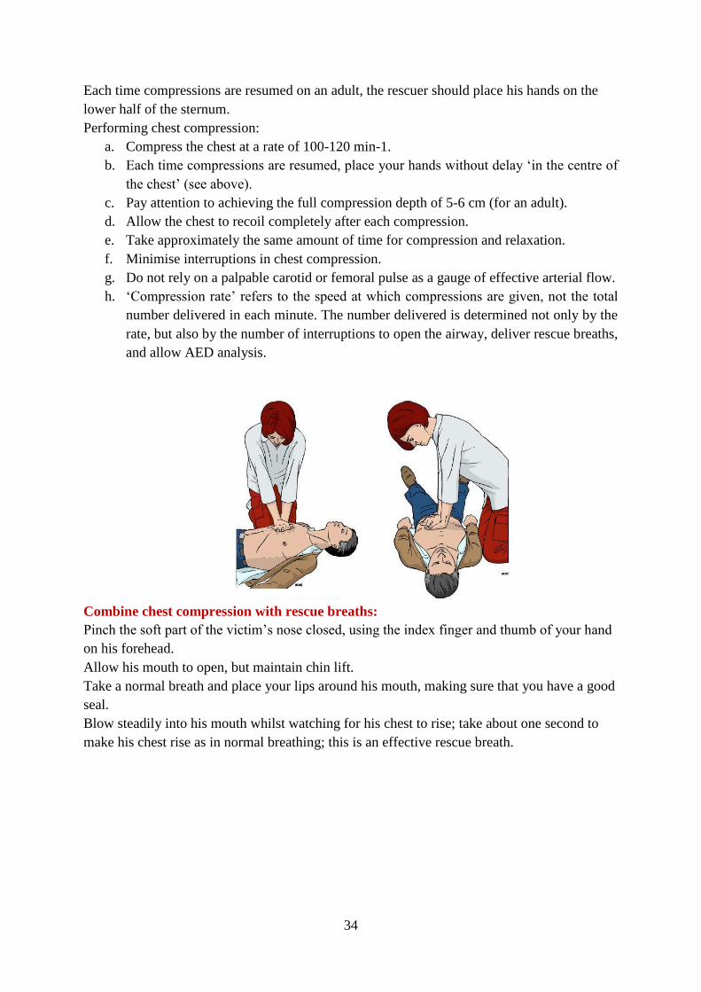
Combine chest compression with rescue breaths:
Pinch the soft part of the victim’s nose closed, using the index finger and thumb of your hand
on his forehead.
Allow his mouth to open, but maintain chin lift.
Take a normal breath and place your lips around his mouth, making sure that you have a good
seal.
Blow steadily into his mouth whilst watching for his chest to rise; take about one second to
make his chest rise as in normal breathing; this is an effective rescue breath.
35
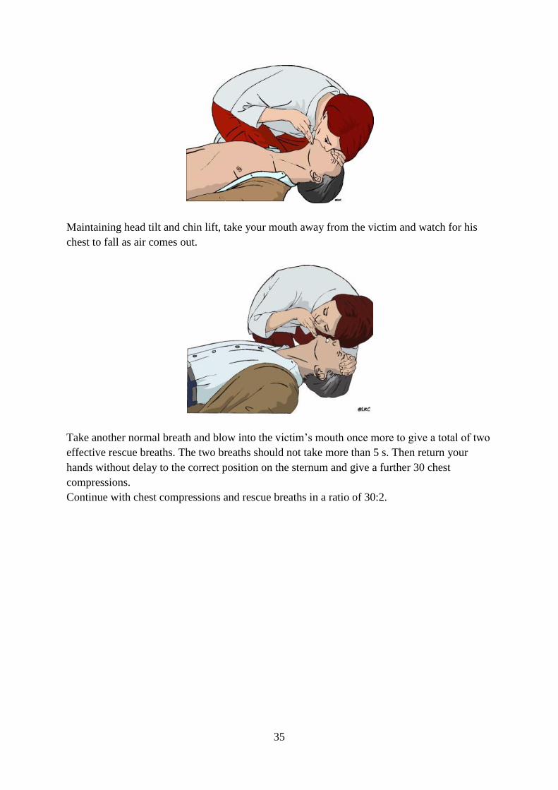
Maintaining head tilt and chin lift, take your mouth away from the victim and watch for his
chest to fall as air comes out.
Take another normal breath and blow into the victim’s mouth once more to give a total of two
effective rescue breaths. The two breaths should not take more than 5 s. Then return your
hands without delay to the correct position on the sternum and give a further 30 chest
compressions.
Continue with chest compressions and rescue breaths in a ratio of 30:2.
36
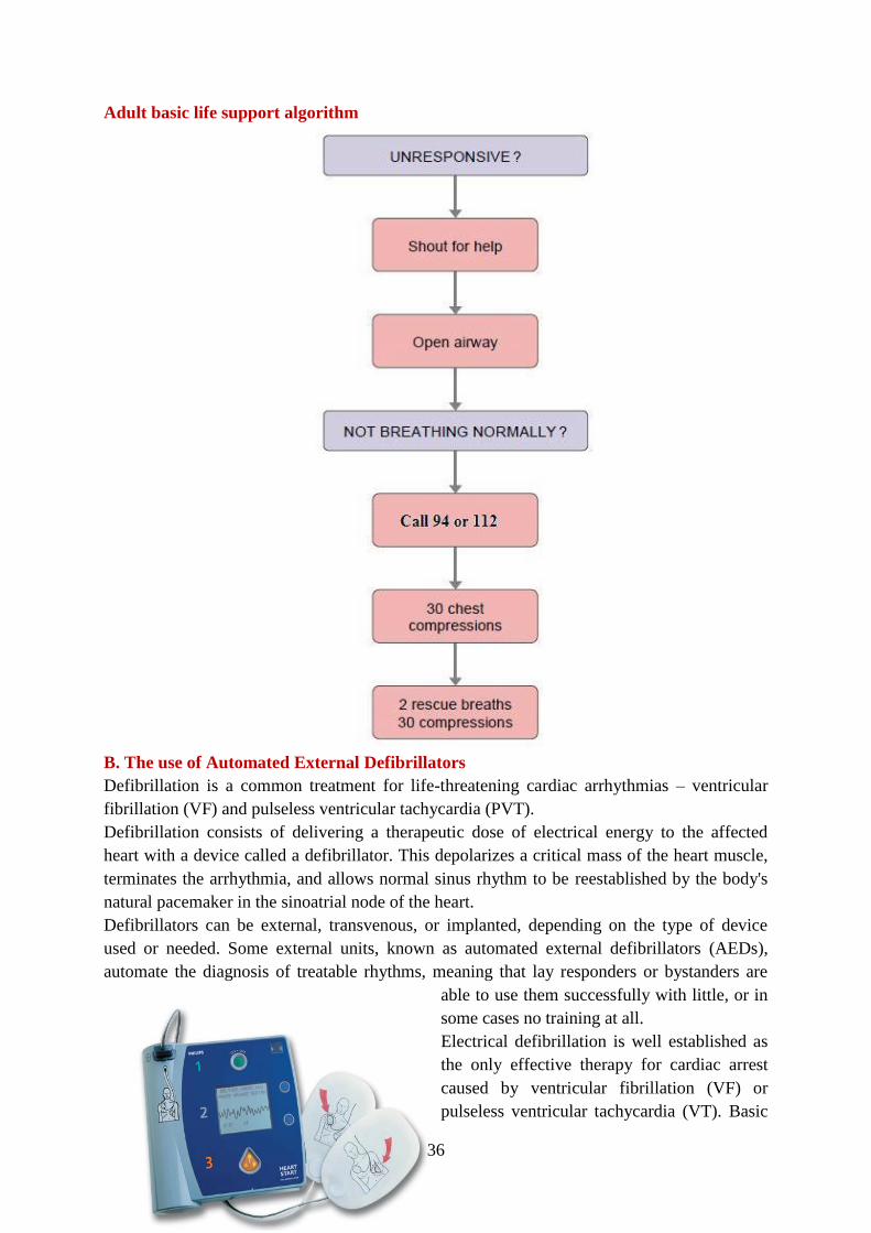
Adult basic life support algorithm
B. The use of Automated External Defibrillators
Defibrillation is a common treatment for life-threatening cardiac arrhythmias – ventricular
fibrillation (VF) and pulseless ventricular tachycardia (PVT).
Defibrillation consists of delivering a therapeutic dose of electrical energy to the affected
heart with a device called a defibrillator. This depolarizes a critical mass of the heart muscle,
terminates the arrhythmia, and allows normal sinus rhythm to be reestablished by the body's
natural pacemaker in the sinoatrial node of the heart.
Defibrillators can be external, transvenous, or implanted, depending on the type of device
used or needed. Some external units, known as automated external defibrillators (AEDs),
automate the diagnosis of treatable rhythms, meaning that lay responders or bystanders are
able to use them successfully with little, or in
some cases no training at all.
Electrical defibrillation is well established as
the only effective therapy for cardiac arrest
caused by ventricular fibrillation (VF) or
pulseless ventricular tachycardia (VT). Basic
37
life support will help to maintain a shockable rhythm but is not a definitive treatment.
The scientific evidence to support early defibrillation is overwhelming; the delay from
collapse to delivery of the first shock is the single most important determinant of survival :
If defibrillation is delivered promptly, survival rates as high as 75% have been
reported.
The chances of successful defibrillation decline at a rate of about 10% with each
minute of delay
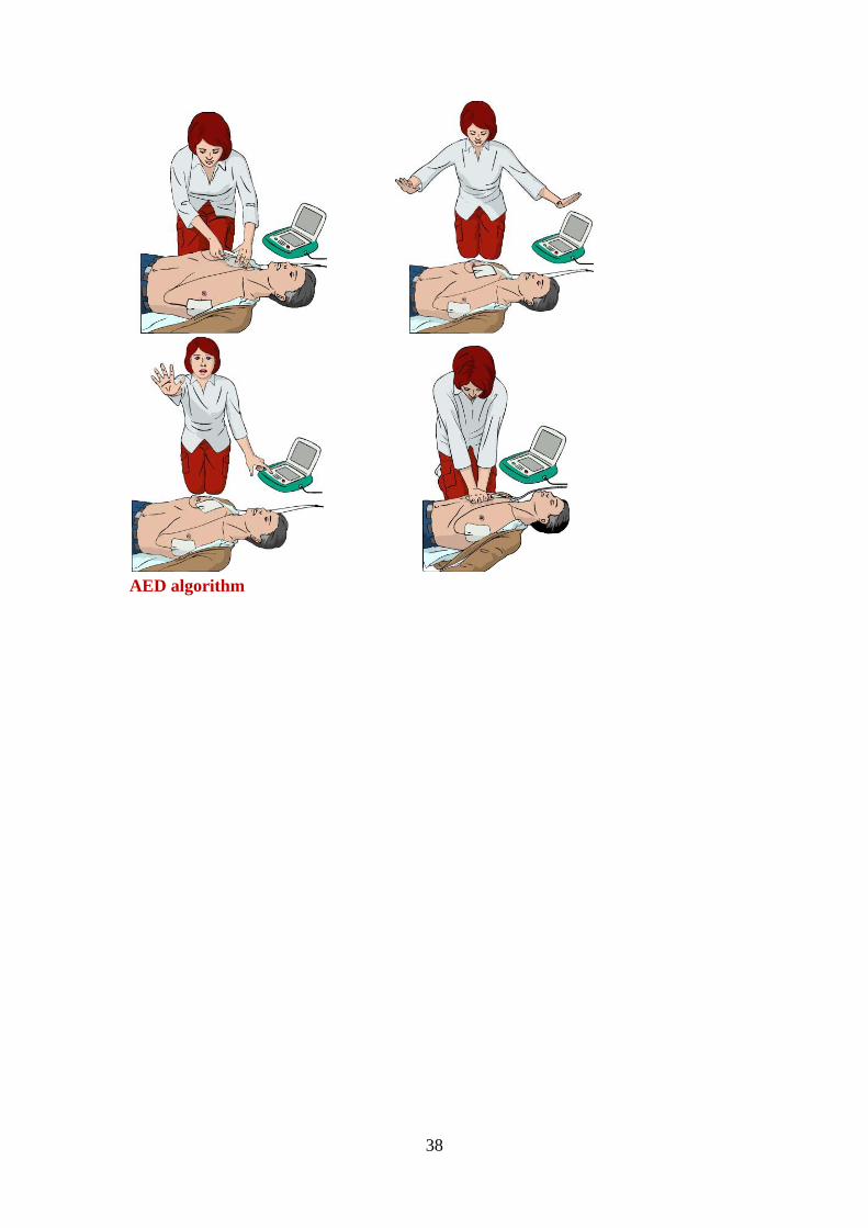
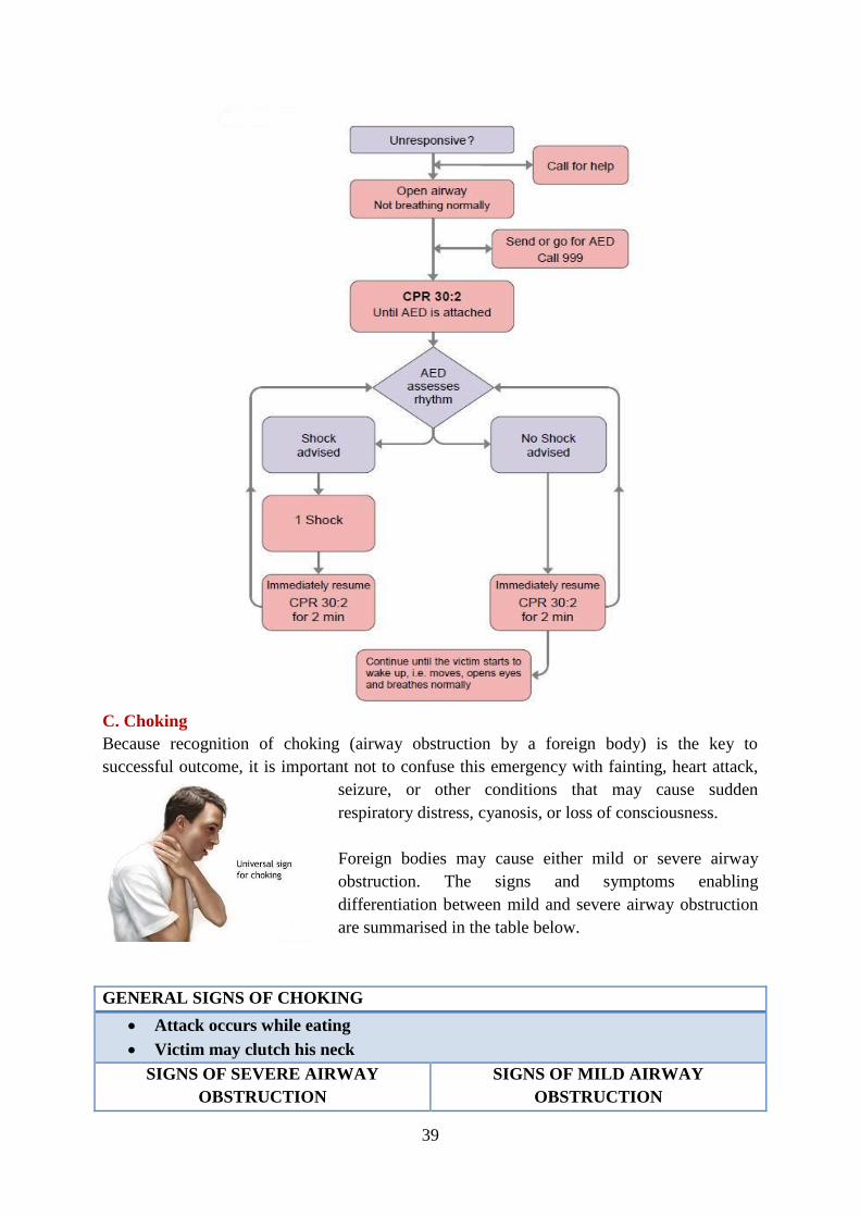
Sequence of actions when using an automated external defibrillator
The following sequence applies to the use of both semi-automatic and automatic AEDs in a
victim who is found to be unconscious and not breathing normally.
1. Follow the adult BLS sequence as described earlier. Do not delay starting CPR unless
the AED is available immediately.
2. As soon as the AED arrives:
If more than one rescuer is present, continue CPR while the AED is switched on. If
you are alone, stop CPR and switch on the AED.
Follow the voice / visual prompts.
Attach the electrode pads to the patient’s bare chest.
Ensure that nobody touches the victim while the AED is analysing the rhythm.
3A 3B
4. Continue to follow the AED prompts until:
qualified help arrives and takes over OR
the victim starts to show signs of regaining consciousness, such as coughing, opening
his eyes, speaking, or moving purposefully AND starts to breathe normally OR
you become exhausted.
3A. If a shock is indicated:
Ensure that nobody touches the victim.
Push the shock button as directed (fully-automatic AEDs
will deliver the shock automatically).
Continue as directed by the voice / visual prompts.
Minimise, as far as possible, interruptions in chest
compression.
3B. If no shock is indicated:
Resume CPR immediately using a ratio of 30
compressions to 2 rescue breaths.
Continue as directed by the voice / visual prompts.
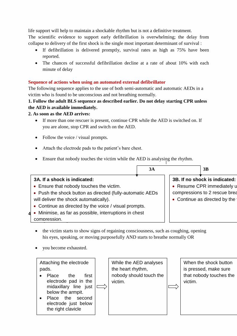
Attaching the electrode
pads.
Place the first electrode pad in the midaxillary line just below the armpit.
Place the second electrode just below the right clavicle
While the AED analyses
the heart rhythm,
nobody should touch the
victim.
When the shock button
is pressed, make sure
that nobody touches the
victim.
After the shock the AED
will prompt you to start
CPR.
Do not wait — start CPR
immediately and
alternate 30 chest
compressions with 2
rescue breaths
38
AED algorithm
39
C. Choking
Because recognition of choking (airway obstruction by a foreign body) is the key to
successful outcome, it is important not to confuse this emergency with fainting, heart attack,
seizure, or other conditions that may cause sudden
respiratory distress, cyanosis, or loss of consciousness.
Foreign bodies may cause either mild or severe airway
obstruction. The signs and symptoms enabling
differentiation between mild and severe airway obstruction
are summarised in the table below.
GENERAL SIGNS OF CHOKING
Attack occurs while eating
Victim may clutch his neck
SIGNS OF SEVERE AIRWAY
OBSTRUCTION
SIGNS OF MILD AIRWAY
OBSTRUCTION
40
Response to question „Are you choking?“
Victim unable to speak
Victim may respond by nodding
Other signs
Victim unable to breathe
Breathing sounds wheezy
Attempts at coughing are silent
Victim may be unconscious
Response to question „Are you choking?“
Victim speaks and answers yes
Other signs
Victim is able to speak, cough and
breathe
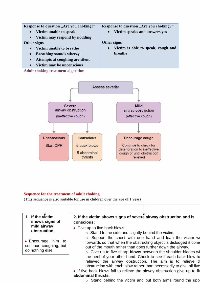
Adult choking treatment algorithm
Sequence for the treatment of adult choking
(This sequence is also suitable for use in children over the age of 1 year)
1. If the victim shows signs of mild airway obstruction:
Encourage him to continue coughing, but do nothing else.
2. If the victim shows signs of severe airway obstruction and is
conscious:
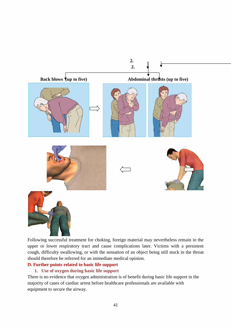
Give up to five back blows.
o Stand to the side and slightly behind the victim.
o Support the chest with one hand and lean the victim well
forwards so that when the obstructing object is dislodged it comes out of the mouth rather than goes further down the airway.
o Give up to five sharp blows between the shoulder blades with
the heel of your other hand. Check to see if each back blow has relieved the airway obstruction. The aim is to relieve the obstruction with each blow rather than necessarily to give all five.
If five back blows fail to relieve the airway obstruction give up to five abdominal thrusts.
o Stand behind the victim and put both arms round the upper
3. If the victim becomes unconscious:
Support the victim
carefully to the ground.
Call an ambulance
immediately.
Begin CPR – 30 : 2
o Each time the airway
is opened for rescue
breaths, look for
foreign material in
the throat. If visible,
remove it.
41
2. 3.
2. 3.
Back blows (up to five) Abdominal thrusts (up to five)
Following successful treatment for choking, foreign material may nevertheless remain in the
upper or lower respiratory tract and cause complications later. Victims with a persistent
cough, difficulty swallowing, or with the sensation of an object being still stuck in the throat
should therefore be referred for an immediate medical opinion.
D. Further points related to basic life support
1. Use of oxygen during basic life support
There is no evidence that oxygen administration is of benefit during basic life support in the
majority of cases of cardiac arrest before healthcare professionals are available with
equipment to secure the airway.
42
Its use may lead to interruption in chest compressions, and is not recommended, except in
cases of drowning (see below).
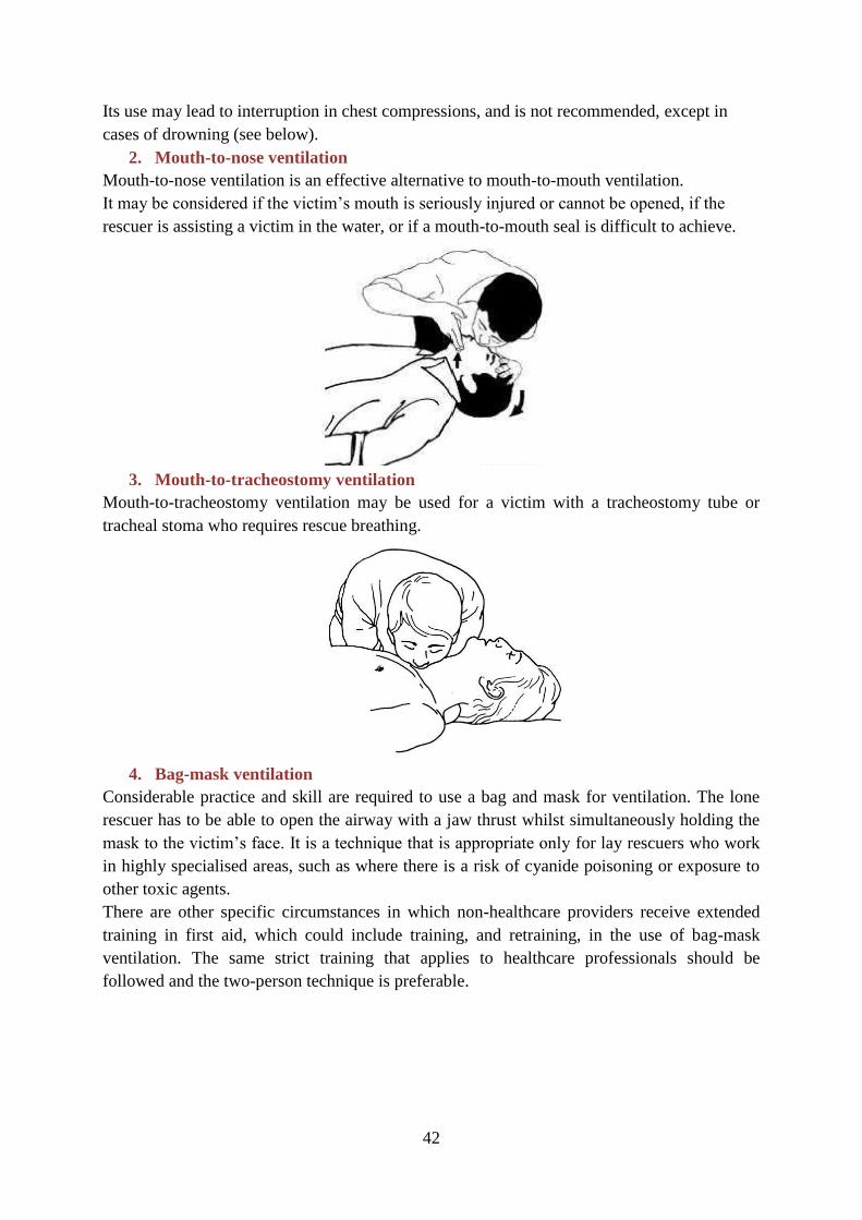
2. Mouth-to-nose ventilation
Mouth-to-nose ventilation is an effective alternative to mouth-to-mouth ventilation.
It may be considered if the victim’s mouth is seriously injured or cannot be opened, if the
rescuer is assisting a victim in the water, or if a mouth-to-mouth seal is difficult to achieve.
3. Mouth-to-tracheostomy ventilation
Mouth-to-tracheostomy ventilation may be used for a victim with a tracheostomy tube or
tracheal stoma who requires rescue breathing.
4. Bag-mask ventilation
Considerable practice and skill are required to use a bag and mask for ventilation. The lone
rescuer has to be able to open the airway with a jaw thrust whilst simultaneously holding the
mask to the victim’s face. It is a technique that is appropriate only for lay rescuers who work
in highly specialised areas, such as where there is a risk of cyanide poisoning or exposure to
other toxic agents.
There are other specific circumstances in which non-healthcare providers receive extended
training in first aid, which could include training, and retraining, in the use of bag-mask
ventilation. The same strict training that applies to healthcare professionals should be
followed and the two-person technique is preferable.
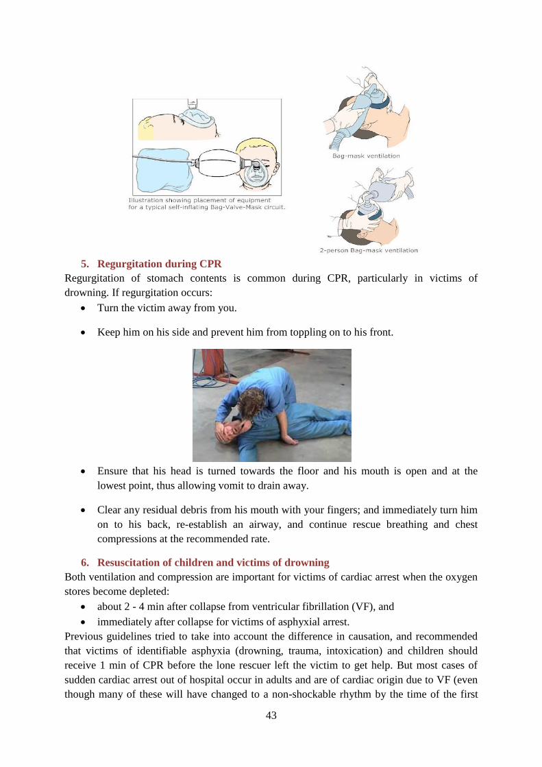
43
5. Regurgitation during CPR
Regurgitation of stomach contents is common during CPR, particularly in victims of
drowning. If regurgitation occurs:
Turn the victim away from you.
Keep him on his side and prevent him from toppling on to his front.
Ensure that his head is turned towards the floor and his mouth is open and at the
lowest point, thus allowing vomit to drain away.
Clear any residual debris from his mouth with your fingers; and immediately turn him
on to his back, re-establish an airway, and continue rescue breathing and chest
compressions at the recommended rate.
6. Resuscitation of children and victims of drowning
Both ventilation and compression are important for victims of cardiac arrest when the oxygen
stores become depleted:
about 2 - 4 min after collapse from ventricular fibrillation (VF), and
immediately after collapse for victims of asphyxial arrest.
Previous guidelines tried to take into account the difference in causation, and recommended
that victims of identifiable asphyxia (drowning, trauma, intoxication) and children should
receive 1 min of CPR before the lone rescuer left the victim to get help. But most cases of
sudden cardiac arrest out of hospital occur in adults and are of cardiac origin due to VF (even
though many of these will have changed to a non-shockable rhythm by the time of the first
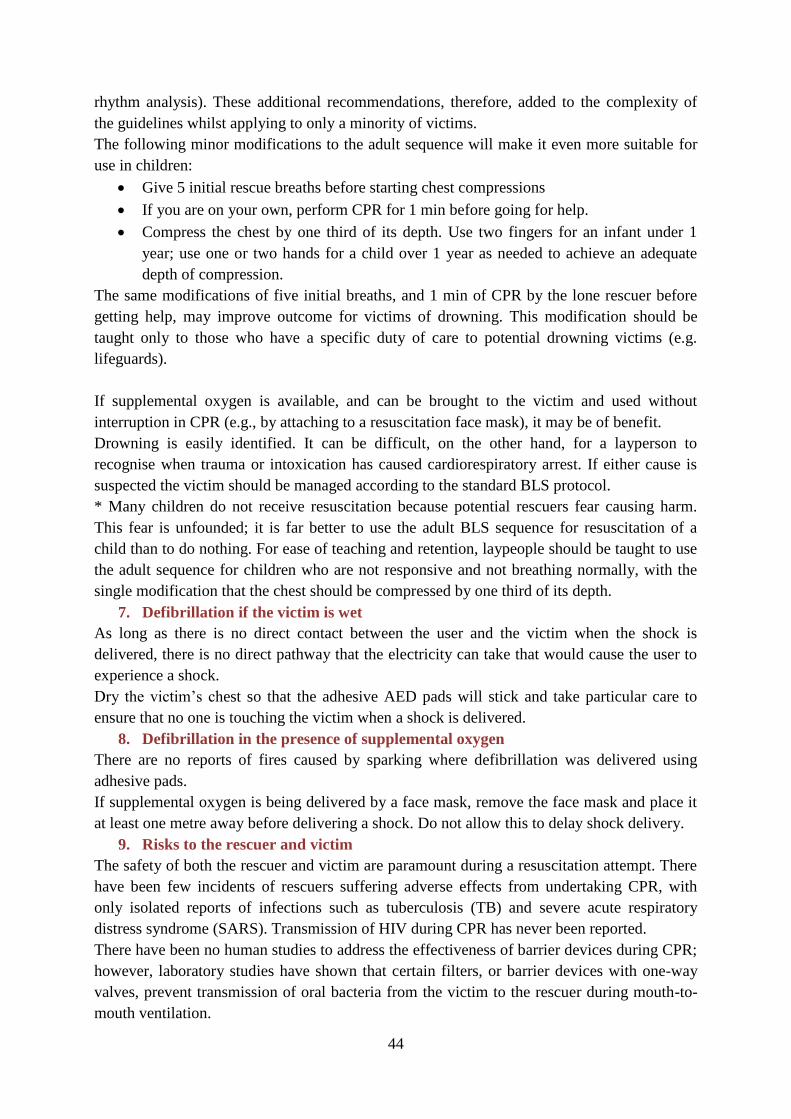
44
rhythm analysis). These additional recommendations, therefore, added to the complexity of
the guidelines whilst applying to only a minority of victims.
The following minor modifications to the adult sequence will make it even more suitable for
use in children:
Give 5 initial rescue breaths before starting chest compressions
If you are on your own, perform CPR for 1 min before going for help.
Compress the chest by one third of its depth. Use two fingers for an infant under 1
year; use one or two hands for a child over 1 year as needed to achieve an adequate
depth of compression.
The same modifications of five initial breaths, and 1 min of CPR by the lone rescuer before
getting help, may improve outcome for victims of drowning. This modification should be
taught only to those who have a specific duty of care to potential drowning victims (e.g.
lifeguards).
If supplemental oxygen is available, and can be brought to the victim and used without
interruption in CPR (e.g., by attaching to a resuscitation face mask), it may be of benefit.
Drowning is easily identified. It can be difficult, on the other hand, for a layperson to
recognise when trauma or intoxication has caused cardiorespiratory arrest. If either cause is
suspected the victim should be managed according to the standard BLS protocol.
* Many children do not receive resuscitation because potential rescuers fear causing harm.
This fear is unfounded; it is far better to use the adult BLS sequence for resuscitation of a
child than to do nothing. For ease of teaching and retention, laypeople should be taught to use
the adult sequence for children who are not responsive and not breathing normally, with the
single modification that the chest should be compressed by one third of its depth.
7. Defibrillation if the victim is wet
As long as there is no direct contact between the user and the victim when the shock is
delivered, there is no direct pathway that the electricity can take that would cause the user to
experience a shock.
Dry the victim’s chest so that the adhesive AED pads will stick and take particular care to
ensure that no one is touching the victim when a shock is delivered.
8. Defibrillation in the presence of supplemental oxygen
There are no reports of fires caused by sparking where defibrillation was delivered using
adhesive pads.
If supplemental oxygen is being delivered by a face mask, remove the face mask and place it
at least one metre away before delivering a shock. Do not allow this to delay shock delivery.
9. Risks to the rescuer and victim
The safety of both the rescuer and victim are paramount during a resuscitation attempt. There
have been few incidents of rescuers suffering adverse effects from undertaking CPR, with
only isolated reports of infections such as tuberculosis (TB) and severe acute respiratory
distress syndrome (SARS). Transmission of HIV during CPR has never been reported.
There have been no human studies to address the effectiveness of barrier devices during CPR;
however, laboratory studies have shown that certain filters, or barrier devices with one-way
valves, prevent transmission of oral bacteria from the victim to the rescuer during mouth-to-
mouth ventilation.
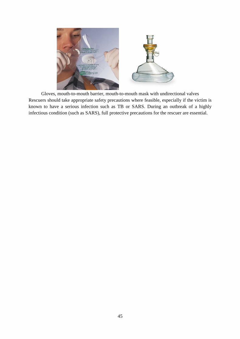
45
Gloves, mouth-to-mouth barrier, mouth-to-mouth mask with undirectional valves
Rescuers should take appropriate safety precautions where feasible, especially if the victim is
known to have a serious infection such as TB or SARS. During an outbreak of a highly
infectious condition (such as SARS), full protective precautions for the rescuer are essential.
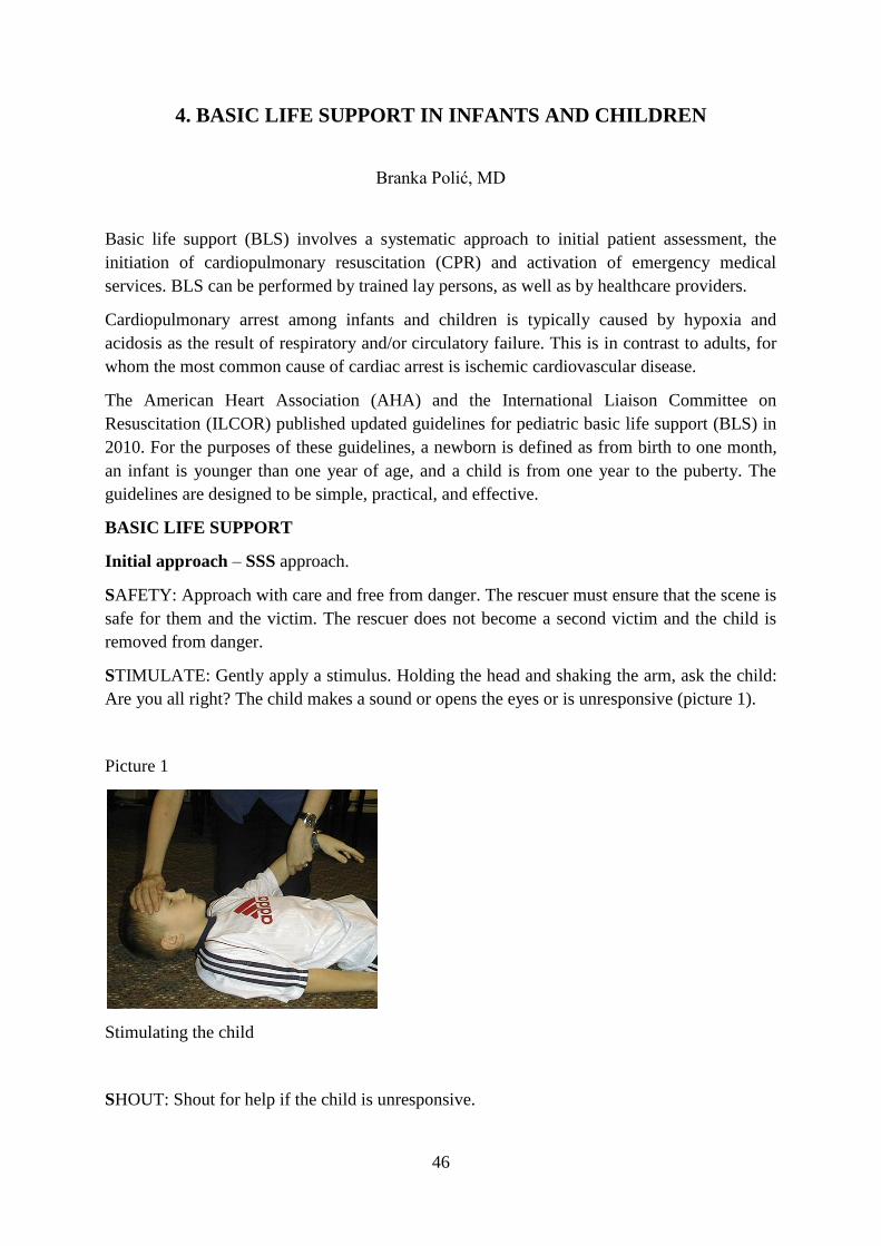
46
4. BASIC LIFE SUPPORT IN INFANTS AND CHILDREN
Branka Polić, MD
Basic life support (BLS) involves a systematic approach to initial patient assessment, the
initiation of cardiopulmonary resuscitation (CPR) and activation of emergency medical
services. BLS can be performed by trained lay persons, as well as by healthcare providers.
Cardiopulmonary arrest among infants and children is typically caused by hypoxia and
acidosis as the result of respiratory and/or circulatory failure. This is in contrast to adults, for
whom the most common cause of cardiac arrest is ischemic cardiovascular disease.
The American Heart Association (AHA) and the International Liaison Committee on
Resuscitation (ILCOR) published updated guidelines for pediatric basic life support (BLS) in
2010. For the purposes of these guidelines, a newborn is defined as from birth to one month,
an infant is younger than one year of age, and a child is from one year to the puberty. The
guidelines are designed to be simple, practical, and effective.
BASIC LIFE SUPPORT
Initial approach – SSS approach.
SAFETY: Approach with care and free from danger. The rescuer must ensure that the scene is
safe for them and the victim. The rescuer does not become a second victim and the child is
removed from danger.
STIMULATE: Gently apply a stimulus. Holding the head and shaking the arm, ask the child:
Are you all right? The child makes a sound or opens the eyes or is unresponsive (picture 1).
Picture 1
Stimulating the child
SHOUT: Shout for help if the child is unresponsive.
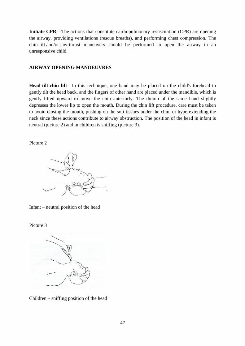
47
Initiate CPR—The actions that constitute cardiopulmonary resuscitation (CPR) are opening
the airway, providing ventilations (rescue breaths), and performing chest compression. The
chin-lift and/or jaw-thrust maneuvers should be performed to open the airway in an
unresponsive child.
AIRWAY OPENING MANOEUVRES
Head-tilt-chin lift—In this technique, one hand may be placed on the child's forehead to
gently tilt the head back, and the fingers of other hand are placed under the mandible, which is
gently lifted upward to move the chin anteriorly. The thumb of the same hand slightly
depresses the lower lip to open the mouth. During the chin lift procedure, care must be taken
to avoid closing the mouth, pushing on the soft tissues under the chin, or hyperextending the
neck since these actions contribute to airway obstruction. The position of the head in infant is
neutral (picture 2) and in children is sniffing (picture 3).
Picture 2
Infant – neutral position of the head
Picture 3
Children – sniffing position of the head
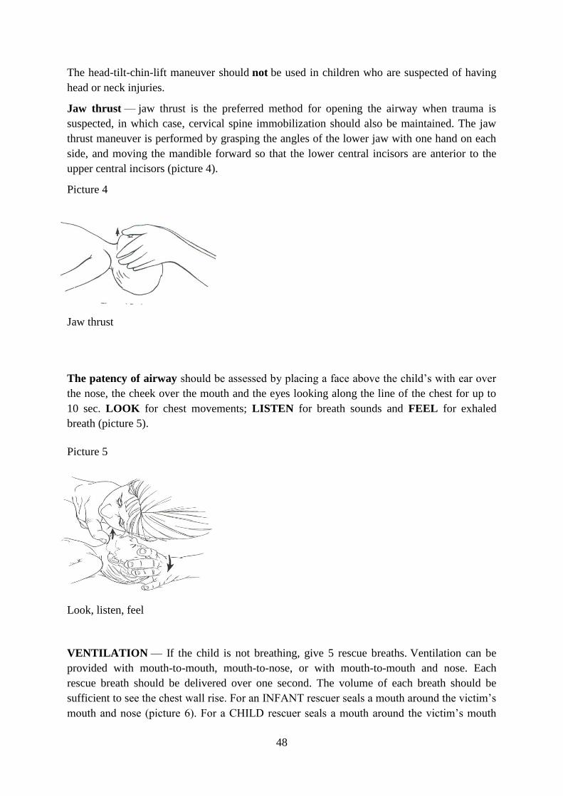
48
The head-tilt-chin-lift maneuver should not be used in children who are suspected of having
head or neck injuries.
Jaw thrust — jaw thrust is the preferred method for opening the airway when trauma is
suspected, in which case, cervical spine immobilization should also be maintained. The jaw
thrust maneuver is performed by grasping the angles of the lower jaw with one hand on each
side, and moving the mandible forward so that the lower central incisors are anterior to the
upper central incisors (picture 4).
Picture 4
Jaw thrust
The patency of airway should be assessed by placing a face above the child’s with ear over
the nose, the cheek over the mouth and the eyes looking along the line of the chest for up to
10 sec. LOOK for chest movements; LISTEN for breath sounds and FEEL for exhaled
breath (picture 5).
Picture 5
Look, listen, feel
VENTILATION — If the child is not breathing, give 5 rescue breaths. Ventilation can be
provided with mouth-to-mouth, mouth-to-nose, or with mouth-to-mouth and nose. Each
rescue breath should be delivered over one second. The volume of each breath should be
sufficient to see the chest wall rise. For an INFANT rescuer seals a mouth around the victim’s
mouth and nose (picture 6). For a CHILD rescuer seals a mouth around the victim’s mouth
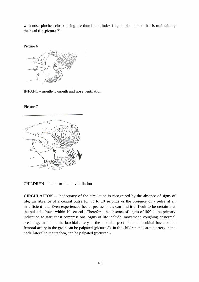
49
with nose pinched closed using the thumb and index fingers of the hand that is maintaining
the head tilt (picture 7).
Picture 6
INFANT - mouth-to-mouth and nose ventilation
Picture 7
.
.
CHILDREN - mouth-to-mouth ventilation
CIRCULATION -- Inadequacy of the circulation is recognized by the absence of signs of
life, the absence of a central pulse for up to 10 seconds or the presence of a pulse at an
insufficient rate. Even experienced health professionals can find it difficult to be certain that
the pulse is absent within 10 seconds. Therefore, the absence of ‘signs of life’ is the primary
indication to start chest compressions. Signs of life include: movement, coughing or normal
breathing. In infants the brachial artery in the medial aspect of the antecubital fossa or the
femoral artery in the groin can be palpated (picture 8). In the children the carotid artery in the
neck, lateral to the trachea, can be palpated (picture 9).
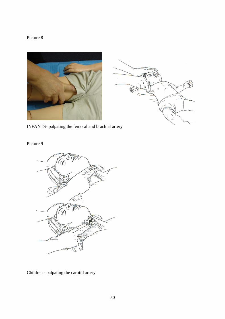
50
Picture 8
INFANTS- palpating the femoral and brachial artery
Picture 9
Children - palpating the carotid artery
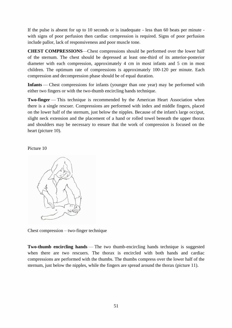
51
If the pulse is absent for up to 10 seconds or is inadequate - less than 60 beats per minute -
with signs of poor perfusion then cardiac compression is required. Signs of poor perfusion
include pallor, lack of responsiveness and poor muscle tone.
CHEST COMPRESSIONS—Chest compressions should be performed over the lower half
of the sternum. The chest should be depressed at least one-third of its anterior-posterior
diameter with each compression, approximately 4 cm in most infants and 5 cm in most
children. The optimum rate of compressions is approximately 100-120 per minute. Each
compression and decompression phase should be of equal duration.
Infants — Chest compressions for infants (younger than one year) may be performed with
either two fingers or with the two-thumb encircling hands technique.
Two-finger — This technique is recommended by the American Heart Association when
there is a single rescuer. Compressions are performed with index and middle fingers, placed
on the lower half of the sternum, just below the nipples. Because of the infant's large occiput,
slight neck extension and the placement of a hand or rolled towel beneath the upper thorax
and shoulders may be necessary to ensure that the work of compression is focused on the
heart (picture 10).
Picture 10
Chest compression – two-finger technique
Two-thumb encircling hands — The two thumb-encircling hands technique is suggested
when there are two rescuers. The thorax is encircled with both hands and cardiac
compressions are performed with the thumbs. The thumbs compress over the lower half of the
sternum, just below the nipples, while the fingers are spread around the thorax (picture 11).
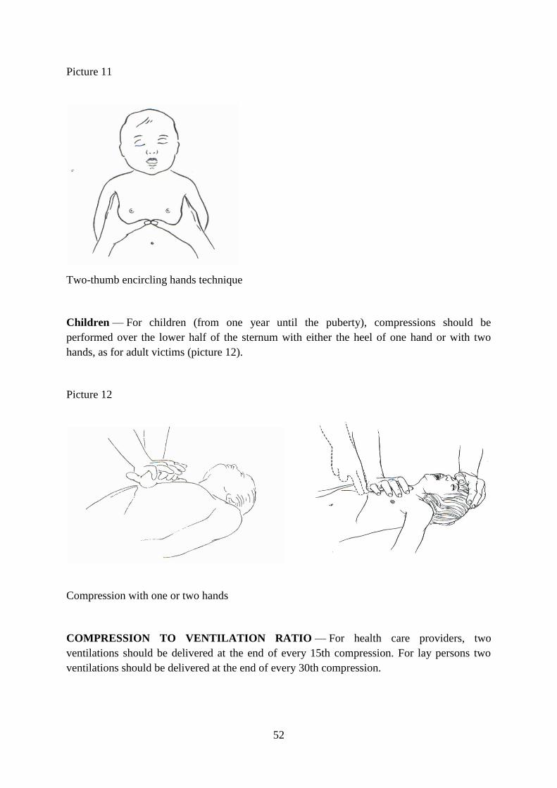
52
Picture 11
Two-thumb encircling hands technique
Children — For children (from one year until the puberty), compressions should be
performed over the lower half of the sternum with either the heel of one hand or with two
hands, as for adult victims (picture 12).
Picture 12
Compression with one or two hands
COMPRESSION TO VENTILATION RATIO — For health care providers, two
ventilations should be delivered at the end of every 15th compression. For lay persons two
ventilations should be delivered at the end of every 30th compression.
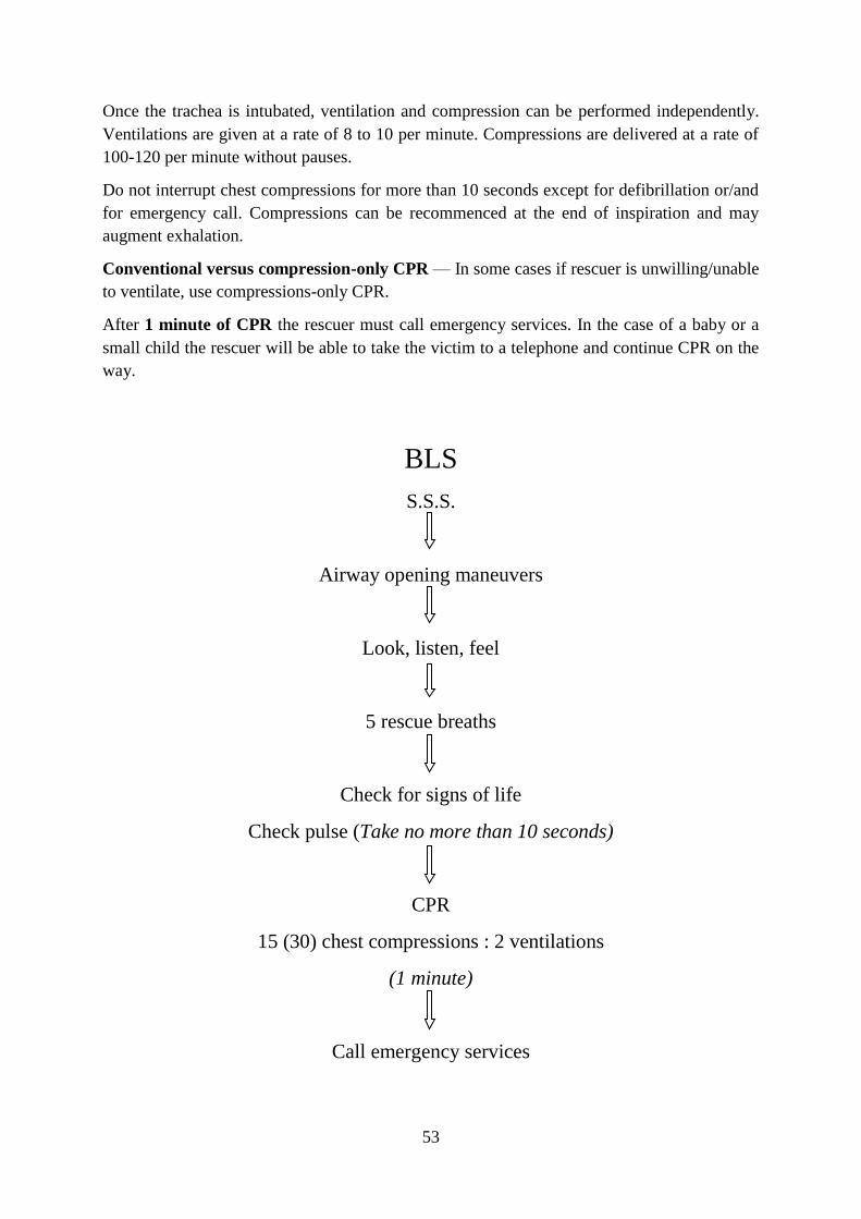
53
Once the trachea is intubated, ventilation and compression can be performed independently.
Ventilations are given at a rate of 8 to 10 per minute. Compressions are delivered at a rate of
100-120 per minute without pauses.
Do not interrupt chest compressions for more than 10 seconds except for defibrillation or/and
for emergency call. Compressions can be recommenced at the end of inspiration and may
augment exhalation.
Conventional versus compression-only CPR — In some cases if rescuer is unwilling/unable
to ventilate, use compressions-only CPR.
After 1 minute of CPR the rescuer must call emergency services. In the case of a baby or a
small child the rescuer will be able to take the victim to a telephone and continue CPR on the
way.
BLS
S.S.S.
Airway opening maneuvers
Look, listen, feel
5 rescue breaths
Check for signs of life
Check pulse (Take no more than 10 seconds)
CPR
15 (30) chest compressions : 2 ventilations
(1 minute)
Call emergency services
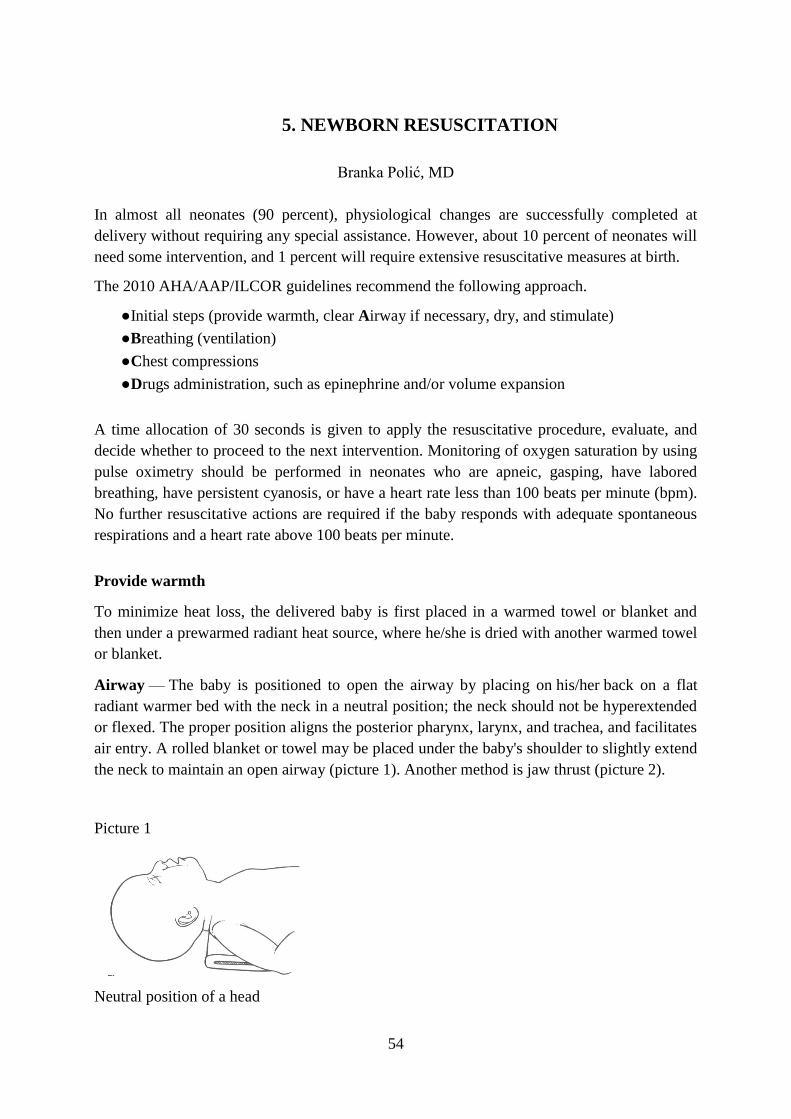
54
5. NEWBORN RESUSCITATION
Branka Polić, MD
In almost all neonates (90 percent), physiological changes are successfully completed at
delivery without requiring any special assistance. However, about 10 percent of neonates will
need some intervention, and 1 percent will require extensive resuscitative measures at birth.
The 2010 AHA/AAP/ILCOR guidelines recommend the following approach.
●Initial steps (provide warmth, clear Airway if necessary, dry, and stimulate)
●Breathing (ventilation)
●Chest compressions
●Drugs administration, such as epinephrine and/or volume expansion
A time allocation of 30 seconds is given to apply the resuscitative procedure, evaluate, and
decide whether to proceed to the next intervention. Monitoring of oxygen saturation by using
pulse oximetry should be performed in neonates who are apneic, gasping, have labored
breathing, have persistent cyanosis, or have a heart rate less than 100 beats per minute (bpm).
No further resuscitative actions are required if the baby responds with adequate spontaneous
respirations and a heart rate above 100 beats per minute.
Provide warmth
To minimize heat loss, the delivered baby is first placed in a warmed towel or blanket and
then under a prewarmed radiant heat source, where he/she is dried with another warmed towel
or blanket.
Airway — The baby is positioned to open the airway by placing on his/her back on a flat
radiant warmer bed with the neck in a neutral position; the neck should not be hyperextended
or flexed. The proper position aligns the posterior pharynx, larynx, and trachea, and facilitates
air entry. A rolled blanket or towel may be placed under the baby's shoulder to slightly extend
the neck to maintain an open airway (picture 1). Another method is jaw thrust (picture 2).
Picture 1
Neutral position of a head
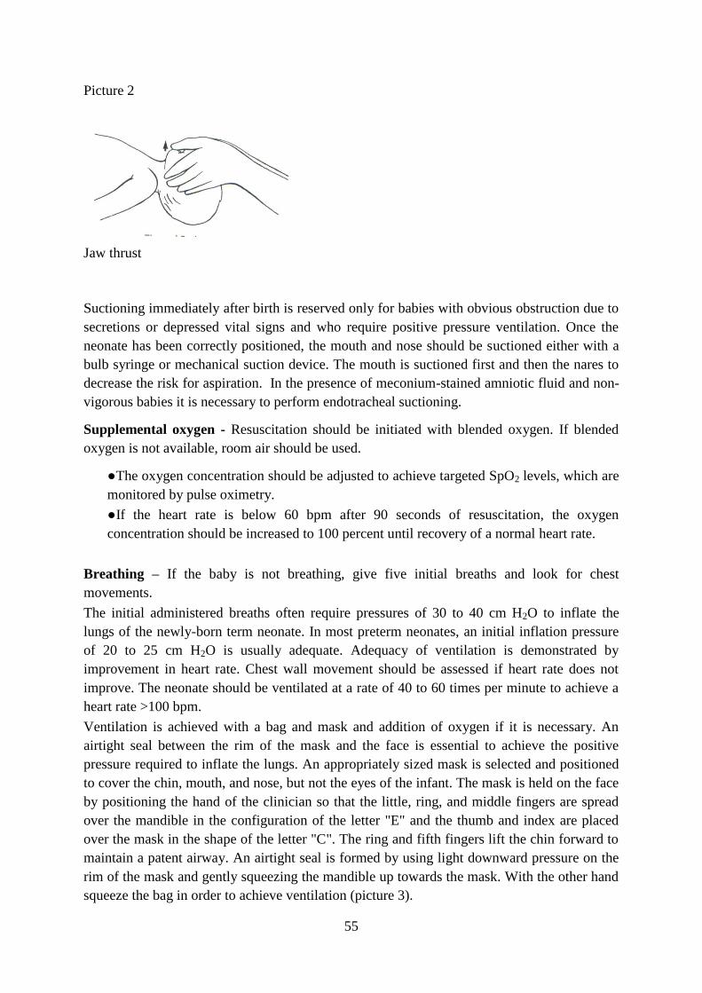
55
Picture 2
Jaw thrust
Suctioning immediately after birth is reserved only for babies with obvious obstruction due to
secretions or depressed vital signs and who require positive pressure ventilation. Once the
neonate has been correctly positioned, the mouth and nose should be suctioned either with a
bulb syringe or mechanical suction device. The mouth is suctioned first and then the nares to
decrease the risk for aspiration. In the presence of meconium-stained amniotic fluid and non-
vigorous babies it is necessary to perform endotracheal suctioning.
Supplemental oxygen - Resuscitation should be initiated with blended oxygen. If blended
oxygen is not available, room air should be used.
●The oxygen concentration should be adjusted to achieve targeted SpO2 levels, which are
monitored by pulse oximetry.
●If the heart rate is below 60 bpm after 90 seconds of resuscitation, the oxygen
concentration should be increased to 100 percent until recovery of a normal heart rate.
Breathing – If the baby is not breathing, give five initial breaths and look for chest
movements.
The initial administered breaths often require pressures of 30 to 40 cm H2O to inflate the
lungs of the newly-born term neonate. In most preterm neonates, an initial inflation pressure
of 20 to 25 cm H2O is usually adequate. Adequacy of ventilation is demonstrated by
improvement in heart rate. Chest wall movement should be assessed if heart rate does not
improve. The neonate should be ventilated at a rate of 40 to 60 times per minute to achieve a
heart rate >100 bpm.
Ventilation is achieved with a bag and mask and addition of oxygen if it is necessary. An
airtight seal between the rim of the mask and the face is essential to achieve the positive
pressure required to inflate the lungs. An appropriately sized mask is selected and positioned
to cover the chin, mouth, and nose, but not the eyes of the infant. The mask is held on the face
by positioning the hand of the clinician so that the little, ring, and middle fingers are spread
over the mandible in the configuration of the letter "E" and the thumb and index are placed
over the mask in the shape of the letter "C". The ring and fifth fingers lift the chin forward to
maintain a patent airway. An airtight seal is formed by using light downward pressure on the
rim of the mask and gently squeezing the mandible up towards the mask. With the other hand
squeeze the bag in order to achieve ventilation (picture 3).
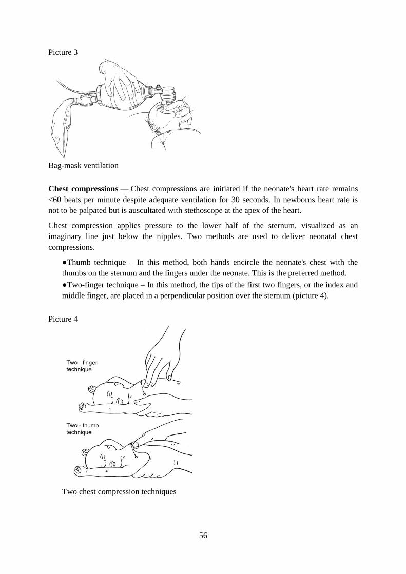
56
Picture 3
Bag-mask ventilation
Chest compressions — Chest compressions are initiated if the neonate's heart rate remains
<60 beats per minute despite adequate ventilation for 30 seconds. In newborns heart rate is
not to be palpated but is auscultated with stethoscope at the apex of the heart.
Chest compression applies pressure to the lower half of the sternum, visualized as an
imaginary line just below the nipples. Two methods are used to deliver neonatal chest
compressions.
●Thumb technique – In this method, both hands encircle the neonate's chest with the
thumbs on the sternum and the fingers under the neonate. This is the preferred method.
●Two-finger technique – In this method, the tips of the first two fingers, or the index and
middle finger, are placed in a perpendicular position over the sternum (picture 4).
Picture 4
Two chest compression techniques
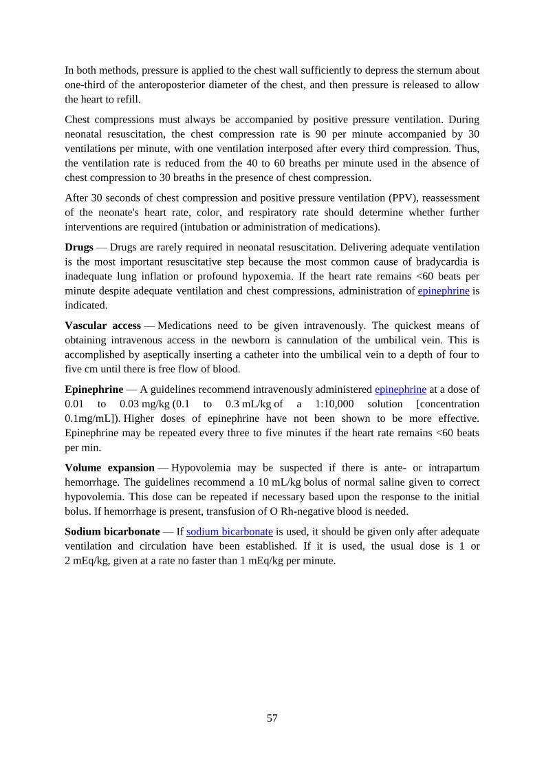
57
In both methods, pressure is applied to the chest wall sufficiently to depress the sternum about
one-third of the anteroposterior diameter of the chest, and then pressure is released to allow
the heart to refill.
Chest compressions must always be accompanied by positive pressure ventilation. During
neonatal resuscitation, the chest compression rate is 90 per minute accompanied by 30
ventilations per minute, with one ventilation interposed after every third compression. Thus,
the ventilation rate is reduced from the 40 to 60 breaths per minute used in the absence of
chest compression to 30 breaths in the presence of chest compression.
After 30 seconds of chest compression and positive pressure ventilation (PPV), reassessment
of the neonate's heart rate, color, and respiratory rate should determine whether further
interventions are required (intubation or administration of medications).
Drugs — Drugs are rarely required in neonatal resuscitation. Delivering adequate ventilation
is the most important resuscitative step because the most common cause of bradycardia is
inadequate lung inflation or profound hypoxemia. If the heart rate remains <60 beats per
minute despite adequate ventilation and chest compressions, administration of epinephrine is
indicated.
Vascular access — Medications need to be given intravenously. The quickest means of
obtaining intravenous access in the newborn is cannulation of the umbilical vein. This is
accomplished by aseptically inserting a catheter into the umbilical vein to a depth of four to
five cm until there is free flow of blood.
Epinephrine — A guidelines recommend intravenously administered epinephrine at a dose of
0.01 to 0.03 mg/kg (0.1 to 0.3 mL/kg of a 1:10,000 solution [concentration
0.1mg/mL]). Higher doses of epinephrine have not been shown to be more effective.
Epinephrine may be repeated every three to five minutes if the heart rate remains <60 beats
per min.
Volume expansion — Hypovolemia may be suspected if there is ante- or intrapartum
hemorrhage. The guidelines recommend a 10 mL/kg bolus of normal saline given to correct
hypovolemia. This dose can be repeated if necessary based upon the response to the initial
bolus. If hemorrhage is present, transfusion of O Rh-negative blood is needed.
Sodium bicarbonate — If sodium bicarbonate is used, it should be given only after adequate
ventilation and circulation have been established. If it is used, the usual dose is 1 or
2 mEq/kg, given at a rate no faster than 1 mEq/kg per minute.
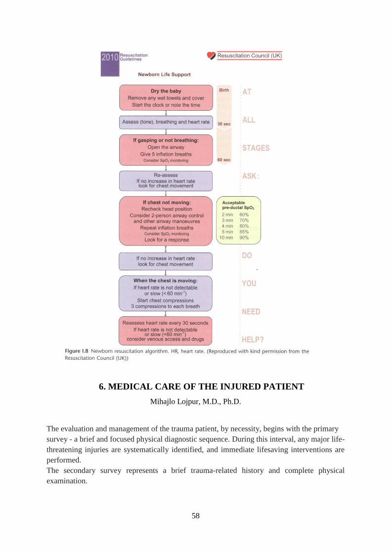
58
6. MEDICAL CARE OF THE INJURED PATIENT
Mihajlo Lojpur, M.D., Ph.D.
The evaluation and management of the trauma patient, by necessity, begins with the primary
survey - a brief and focused physical diagnostic sequence. During this interval, any major life-
threatening injuries are systematically identified, and immediate lifesaving interventions are
performed.
The secondary survey represents a brief trauma-related history and complete physical
examination.
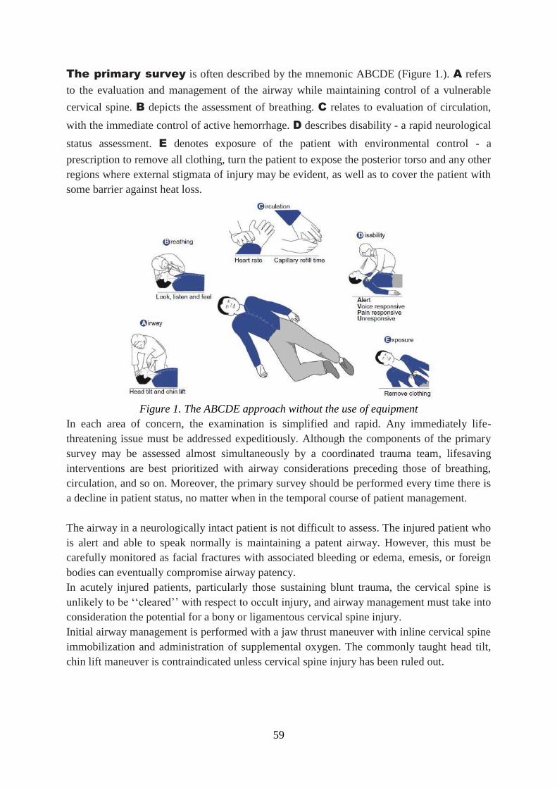
59
The primary survey is often described by the mnemonic ABCDE (Figure 1.). A refers
to the evaluation and management of the airway while maintaining control of a vulnerable
cervical spine. B depicts the assessment of breathing. C relates to evaluation of circulation,
with the immediate control of active hemorrhage. D describes disability - a rapid neurological
status assessment. E denotes exposure of the patient with environmental control - a
prescription to remove all clothing, turn the patient to expose the posterior torso and any other
regions where external stigmata of injury may be evident, as well as to cover the patient with
some barrier against heat loss.
Figure 1. The ABCDE approach without the use of equipment
In each area of concern, the examination is simplified and rapid. Any immediately life-
threatening issue must be addressed expeditiously. Although the components of the primary
survey may be assessed almost simultaneously by a coordinated trauma team, lifesaving
interventions are best prioritized with airway considerations preceding those of breathing,
circulation, and so on. Moreover, the primary survey should be performed every time there is
a decline in patient status, no matter when in the temporal course of patient management.
The airway in a neurologically intact patient is not difficult to assess. The injured patient who
is alert and able to speak normally is maintaining a patent airway. However, this must be
carefully monitored as facial fractures with associated bleeding or edema, emesis, or foreign
bodies can eventually compromise airway patency.
In acutely injured patients, particularly those sustaining blunt trauma, the cervical spine is
unlikely to be ‘‘cleared’’ with respect to occult injury, and airway management must take into
consideration the potential for a bony or ligamentous cervical spine injury.
Initial airway management is performed with a jaw thrust maneuver with inline cervical spine
immobilization and administration of supplemental oxygen. The commonly taught head tilt,
chin lift maneuver is contraindicated unless cervical spine injury has been ruled out.
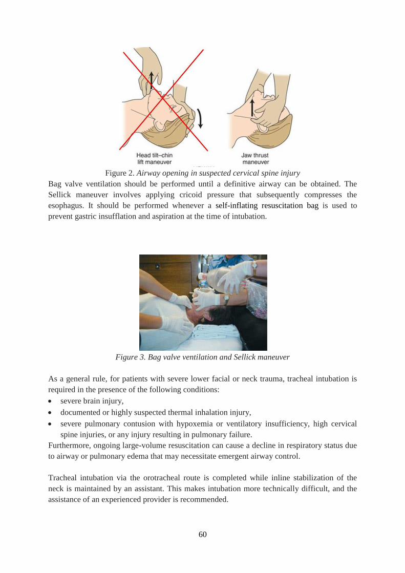
60
Figure 2. Airway opening in suspected cervical spine injury
Bag valve ventilation should be performed until a definitive airway can be obtained. The
Sellick maneuver involves applying cricoid pressure that subsequently compresses the
esophagus. It should be performed whenever a self-inflating resuscitation bag is used to
prevent gastric insufflation and aspiration at the time of intubation.
Figure 3. Bag valve ventilation and Sellick maneuver
As a general rule, for patients with severe lower facial or neck trauma, tracheal intubation is
required in the presence of the following conditions:
severe brain injury,
documented or highly suspected thermal inhalation injury,
severe pulmonary contusion with hypoxemia or ventilatory insufficiency, high cervical
spine injuries, or any injury resulting in pulmonary failure.
Furthermore, ongoing large-volume resuscitation can cause a decline in respiratory status due
to airway or pulmonary edema that may necessitate emergent airway control.
Tracheal intubation via the orotracheal route is completed while inline stabilization of the
neck is maintained by an assistant. This makes intubation more technically difficult, and the
assistance of an experienced provider is recommended.
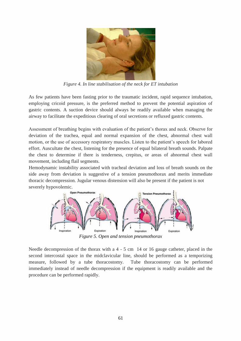
61
Figure 4. In line stabilisation of the neck for ET intubation
As few patients have been fasting prior to the traumatic incident, rapid sequence intubation,
employing cricoid pressure, is the preferred method to prevent the potential aspiration of
gastric contents. A suction device should always be readily available when managing the
airway to facilitate the expeditious clearing of oral secretions or refluxed gastric contents.
Assessment of breathing begins with evaluation of the patient’s thorax and neck. Observe for
deviation of the trachea, equal and normal expansion of the chest, abnormal chest wall
motion, or the use of accessory respiratory muscles. Listen to the patient’s speech for labored
effort. Auscultate the chest, listening for the presence of equal bilateral breath sounds. Palpate
the chest to determine if there is tenderness, crepitus, or areas of abnormal chest wall
movement, including flail segments.
Hemodynamic instability associated with tracheal deviation and loss of breath sounds on the
side away from deviation is suggestive of a tension pneumothorax and merits immediate
thoracic decompression. Jugular venous distension will also be present if the patient is not
severely hypovolemic.
Figure 5. Open and tension pneumothorax
Needle decompression of the thorax with a 4 - 5 cm 14 or 16 gauge catheter, placed in the
second intercostal space in the midclavicular line, should be performed as a temporizing
measure, followed by a tube thoracostomy. Tube thoracostomy can be performed
immediately instead of needle decompression if the equipment is readily available and the
procedure can be performed rapidly.
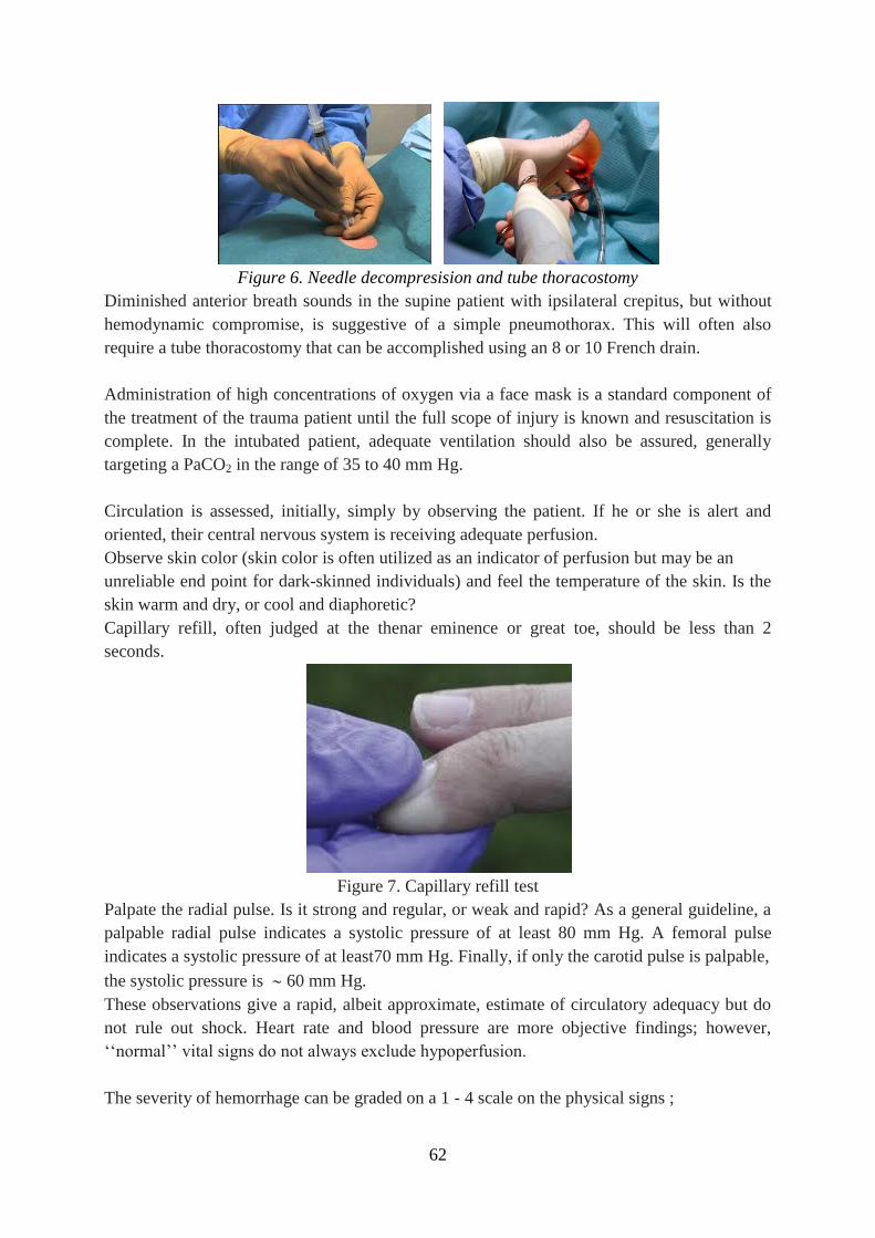
62
Figure 6. Needle decompresision and tube thoracostomy
Diminished anterior breath sounds in the supine patient with ipsilateral crepitus, but without
hemodynamic compromise, is suggestive of a simple pneumothorax. This will often also
require a tube thoracostomy that can be accomplished using an 8 or 10 French drain.
Administration of high concentrations of oxygen via a face mask is a standard component of
the treatment of the trauma patient until the full scope of injury is known and resuscitation is
complete. In the intubated patient, adequate ventilation should also be assured, generally
targeting a PaCO2 in the range of 35 to 40 mm Hg.
Circulation is assessed, initially, simply by observing the patient. If he or she is alert and
oriented, their central nervous system is receiving adequate perfusion.
Observe skin color (skin color is often utilized as an indicator of perfusion but may be an
unreliable end point for dark-skinned individuals) and feel the temperature of the skin. Is the
skin warm and dry, or cool and diaphoretic?
Capillary refill, often judged at the thenar eminence or great toe, should be less than 2
seconds.
Figure 7. Capillary refill test
Palpate the radial pulse. Is it strong and regular, or weak and rapid? As a general guideline, a
palpable radial pulse indicates a systolic pressure of at least 80 mm Hg. A femoral pulse
indicates a systolic pressure of at least70 mm Hg. Finally, if only the carotid pulse is palpable,
the systolic pressure is 60 mm Hg.
These observations give a rapid, albeit approximate, estimate of circulatory adequacy but do
not rule out shock. Heart rate and blood pressure are more objective findings; however,
‘‘normal’’ vital signs do not always exclude hypoperfusion.
The severity of hemorrhage can be graded on a 1 - 4 scale on the physical signs ;
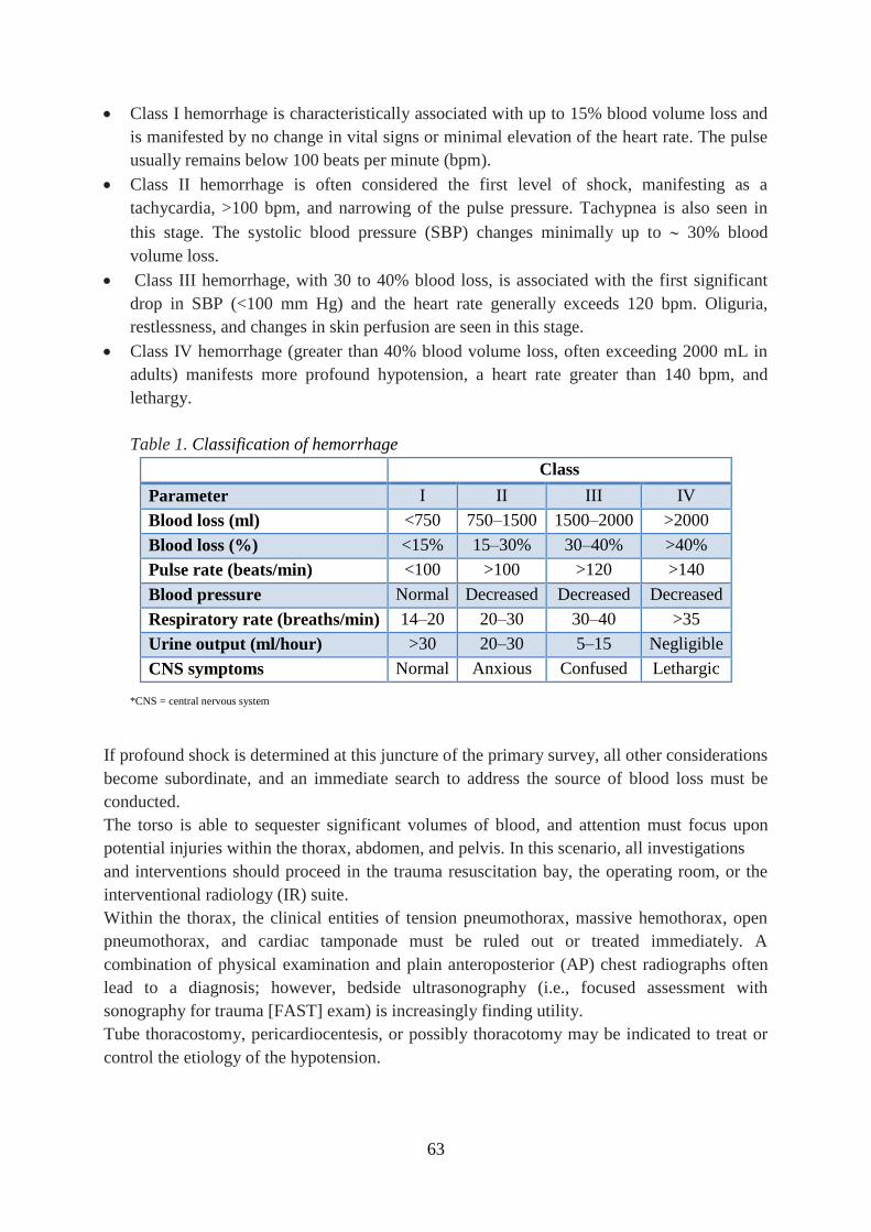
63
Class I hemorrhage is characteristically associated with up to 15% blood volume loss and
is manifested by no change in vital signs or minimal elevation of the heart rate. The pulse
usually remains below 100 beats per minute (bpm).
Class II hemorrhage is often considered the first level of shock, manifesting as a
tachycardia, >100 bpm, and narrowing of the pulse pressure. Tachypnea is also seen in
this stage. The systolic blood pressure (SBP) changes minimally up to 30% blood
volume loss.
Class III hemorrhage, with 30 to 40% blood loss, is associated with the first significant
drop in SBP (<100 mm Hg) and the heart rate generally exceeds 120 bpm. Oliguria,
restlessness, and changes in skin perfusion are seen in this stage.
Class IV hemorrhage (greater than 40% blood volume loss, often exceeding 2000 mL in
adults) manifests more profound hypotension, a heart rate greater than 140 bpm, and
lethargy.
Table 1. Classification of hemorrhage
Class
Parameter I II III IV
Blood loss (ml) <750 750–1500 1500–2000 >2000
Blood loss (%) <15% 15–30% 30–40% >40%
Pulse rate (beats/min) <100 >100 >120 >140
Blood pressure Normal Decreased Decreased Decreased
Respiratory rate (breaths/min) 14–20 20–30 30–40 >35
Urine output (ml/hour) >30 20–30 5–15 Negligible
CNS symptoms Normal Anxious Confused Lethargic
*CNS = central nervous system
If profound shock is determined at this juncture of the primary survey, all other considerations
become subordinate, and an immediate search to address the source of blood loss must be
conducted.
The torso is able to sequester significant volumes of blood, and attention must focus upon
potential injuries within the thorax, abdomen, and pelvis. In this scenario, all investigations
and interventions should proceed in the trauma resuscitation bay, the operating room, or the
interventional radiology (IR) suite.
Within the thorax, the clinical entities of tension pneumothorax, massive hemothorax, open
pneumothorax, and cardiac tamponade must be ruled out or treated immediately. A
combination of physical examination and plain anteroposterior (AP) chest radiographs often
lead to a diagnosis; however, bedside ultrasonography (i.e., focused assessment with
sonography for trauma [FAST] exam) is increasingly finding utility.
Tube thoracostomy, pericardiocentesis, or possibly thoracotomy may be indicated to treat or
control the etiology of the hypotension.
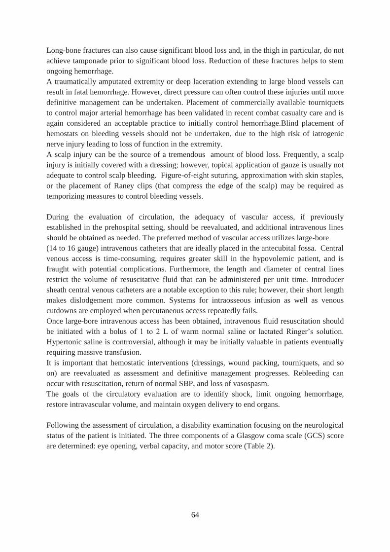
64
Long-bone fractures can also cause significant blood loss and, in the thigh in particular, do not
achieve tamponade prior to significant blood loss. Reduction of these fractures helps to stem
ongoing hemorrhage.
A traumatically amputated extremity or deep laceration extending to large blood vessels can
result in fatal hemorrhage. However, direct pressure can often control these injuries until more
definitive management can be undertaken. Placement of commercially available tourniquets
to control major arterial hemorrhage has been validated in recent combat casualty care and is
again considered an acceptable practice to initially control hemorrhage.Blind placement of
hemostats on bleeding vessels should not be undertaken, due to the high risk of iatrogenic
nerve injury leading to loss of function in the extremity.
A scalp injury can be the source of a tremendous amount of blood loss. Frequently, a scalp
injury is initially covered with a dressing; however, topical application of gauze is usually not
adequate to control scalp bleeding. Figure-of-eight suturing, approximation with skin staples,
or the placement of Raney clips (that compress the edge of the scalp) may be required as
temporizing measures to control bleeding vessels.
During the evaluation of circulation, the adequacy of vascular access, if previously
established in the prehospital setting, should be reevaluated, and additional intravenous lines
should be obtained as needed. The preferred method of vascular access utilizes large-bore
(14 to 16 gauge) intravenous catheters that are ideally placed in the antecubital fossa. Central
venous access is time-consuming, requires greater skill in the hypovolemic patient, and is
fraught with potential complications. Furthermore, the length and diameter of central lines
restrict the volume of resuscitative fluid that can be administered per unit time. Introducer
sheath central venous catheters are a notable exception to this rule; however, their short length
makes dislodgement more common. Systems for intraosseous infusion as well as venous
cutdowns are employed when percutaneous access repeatedly fails.
Once large-bore intravenous access has been obtained, intravenous fluid resuscitation should
be initiated with a bolus of 1 to 2 L of warm normal saline or lactated Ringer’s solution.
Hypertonic saline is controversial, although it may be initially valuable in patients eventually
requiring massive transfusion.
It is important that hemostatic interventions (dressings, wound packing, tourniquets, and so
on) are reevaluated as assessment and definitive management progresses. Rebleeding can
occur with resuscitation, return of normal SBP, and loss of vasospasm.
The goals of the circulatory evaluation are to identify shock, limit ongoing hemorrhage,
restore intravascular volume, and maintain oxygen delivery to end organs.
Following the assessment of circulation, a disability examination focusing on the neurological
status of the patient is initiated. The three components of a Glasgow coma scale (GCS) score
are determined: eye opening, verbal capacity, and motor score (Table 2).
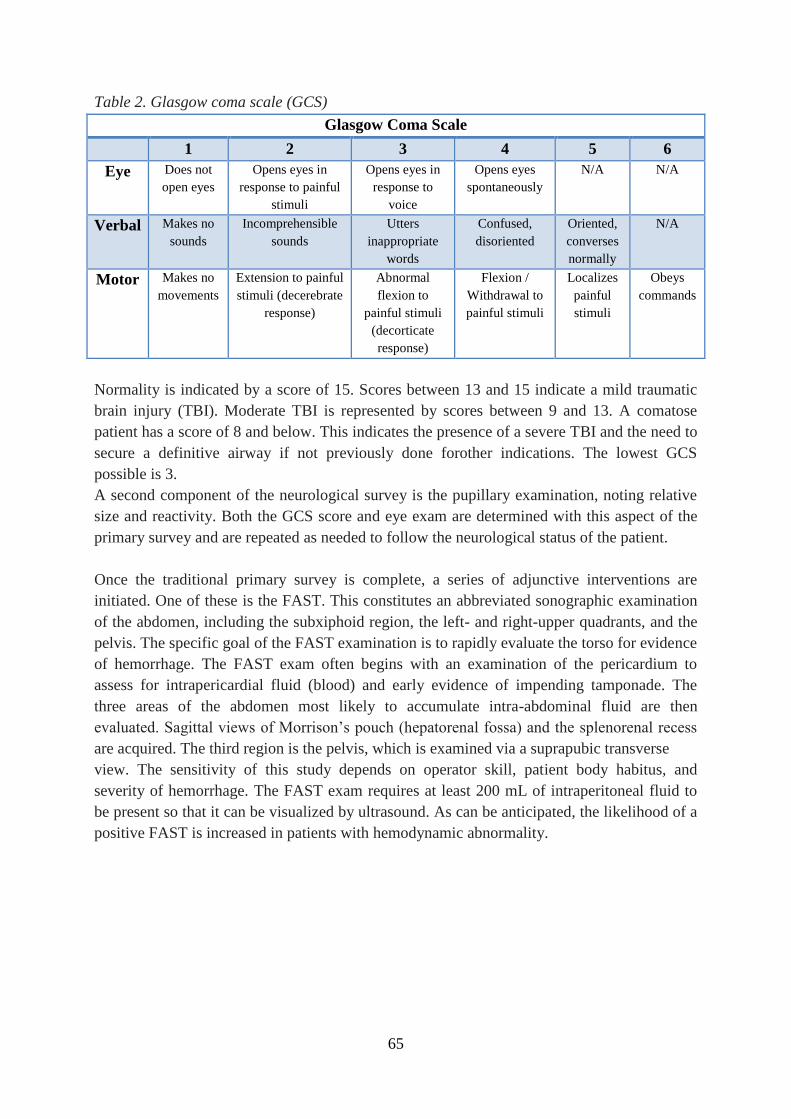
65
Table 2. Glasgow coma scale (GCS)
Glasgow Coma Scale
1 2 3 4 5 6
Eye Does not
open eyes
Opens eyes in
response to painful
stimuli
Opens eyes in
response to
voice
Opens eyes
spontaneously
N/A N/A
Verbal Makes no
sounds
Incomprehensible
sounds
Utters
inappropriate
words
Confused,
disoriented
Oriented,
converses
normally
N/A
Motor Makes no
movements
Extension to painful
stimuli (decerebrate
response)
Abnormal
flexion to
painful stimuli
(decorticate
response)
Flexion /
Withdrawal to
painful stimuli
Localizes
painful
stimuli
Obeys
commands
Normality is indicated by a score of 15. Scores between 13 and 15 indicate a mild traumatic
brain injury (TBI). Moderate TBI is represented by scores between 9 and 13. A comatose
patient has a score of 8 and below. This indicates the presence of a severe TBI and the need to
secure a definitive airway if not previously done forother indications. The lowest GCS
possible is 3.
A second component of the neurological survey is the pupillary examination, noting relative
size and reactivity. Both the GCS score and eye exam are determined with this aspect of the
primary survey and are repeated as needed to follow the neurological status of the patient.
Once the traditional primary survey is complete, a series of adjunctive interventions are
initiated. One of these is the FAST. This constitutes an abbreviated sonographic examination
of the abdomen, including the subxiphoid region, the left- and right-upper quadrants, and the
pelvis. The specific goal of the FAST examination is to rapidly evaluate the torso for evidence
of hemorrhage. The FAST exam often begins with an examination of the pericardium to
assess for intrapericardial fluid (blood) and early evidence of impending tamponade. The
three areas of the abdomen most likely to accumulate intra-abdominal fluid are then
evaluated. Sagittal views of Morrison’s pouch (hepatorenal fossa) and the splenorenal recess
are acquired. The third region is the pelvis, which is examined via a suprapubic transverse
view. The sensitivity of this study depends on operator skill, patient body habitus, and
severity of hemorrhage. The FAST exam requires at least 200 mL of intraperitoneal fluid to
be present so that it can be visualized by ultrasound. As can be anticipated, the likelihood of a
positive FAST is increased in patients with hemodynamic abnormality.
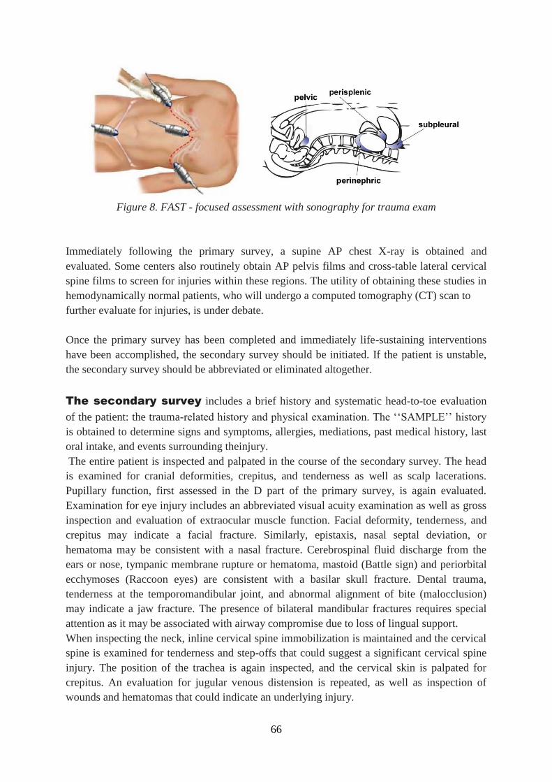
66
Figure 8. FAST - focused assessment with sonography for trauma exam
Immediately following the primary survey, a supine AP chest X-ray is obtained and
evaluated. Some centers also routinely obtain AP pelvis films and cross-table lateral cervical
spine films to screen for injuries within these regions. The utility of obtaining these studies in
hemodynamically normal patients, who will undergo a computed tomography (CT) scan to
further evaluate for injuries, is under debate.
Once the primary survey has been completed and immediately life-sustaining interventions
have been accomplished, the secondary survey should be initiated. If the patient is unstable,
the secondary survey should be abbreviated or eliminated altogether.
The secondary survey includes a brief history and systematic head-to-toe evaluation
of the patient: the trauma-related history and physical examination. The ‘‘SAMPLE’’ history
is obtained to determine signs and symptoms, allergies, mediations, past medical history, last
oral intake, and events surrounding theinjury.
The entire patient is inspected and palpated in the course of the secondary survey. The head
is examined for cranial deformities, crepitus, and tenderness as well as scalp lacerations.
Pupillary function, first assessed in the D part of the primary survey, is again evaluated.
Examination for eye injury includes an abbreviated visual acuity examination as well as gross
inspection and evaluation of extraocular muscle function. Facial deformity, tenderness, and
crepitus may indicate a facial fracture. Similarly, epistaxis, nasal septal deviation, or
hematoma may be consistent with a nasal fracture. Cerebrospinal fluid discharge from the
ears or nose, tympanic membrane rupture or hematoma, mastoid (Battle sign) and periorbital
ecchymoses (Raccoon eyes) are consistent with a basilar skull fracture. Dental trauma,
tenderness at the temporomandibular joint, and abnormal alignment of bite (malocclusion)
may indicate a jaw fracture. The presence of bilateral mandibular fractures requires special
attention as it may be associated with airway compromise due to loss of lingual support.
When inspecting the neck, inline cervical spine immobilization is maintained and the cervical
spine is examined for tenderness and step-offs that could suggest a significant cervical spine
injury. The position of the trachea is again inspected, and the cervical skin is palpated for
crepitus. An evaluation for jugular venous distension is repeated, as well as inspection of
wounds and hematomas that could indicate an underlying injury.
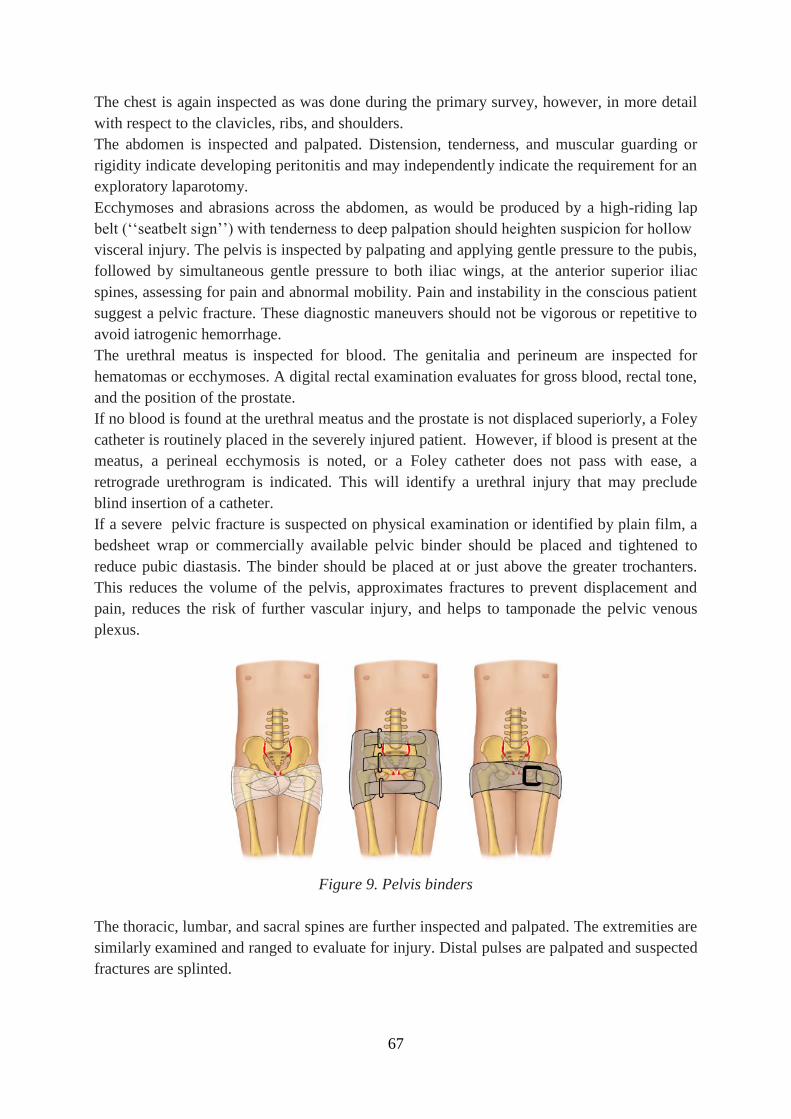
67
The chest is again inspected as was done during the primary survey, however, in more detail
with respect to the clavicles, ribs, and shoulders.
The abdomen is inspected and palpated. Distension, tenderness, and muscular guarding or
rigidity indicate developing peritonitis and may independently indicate the requirement for an
exploratory laparotomy.
Ecchymoses and abrasions across the abdomen, as would be produced by a high-riding lap
belt (‘‘seatbelt sign’’) with tenderness to deep palpation should heighten suspicion for hollow
visceral injury. The pelvis is inspected by palpating and applying gentle pressure to the pubis,
followed by simultaneous gentle pressure to both iliac wings, at the anterior superior iliac
spines, assessing for pain and abnormal mobility. Pain and instability in the conscious patient
suggest a pelvic fracture. These diagnostic maneuvers should not be vigorous or repetitive to
avoid iatrogenic hemorrhage.
The urethral meatus is inspected for blood. The genitalia and perineum are inspected for
hematomas or ecchymoses. A digital rectal examination evaluates for gross blood, rectal tone,
and the position of the prostate.
If no blood is found at the urethral meatus and the prostate is not displaced superiorly, a Foley
catheter is routinely placed in the severely injured patient. However, if blood is present at the
meatus, a perineal ecchymosis is noted, or a Foley catheter does not pass with ease, a
retrograde urethrogram is indicated. This will identify a urethral injury that may preclude
blind insertion of a catheter.
If a severe pelvic fracture is suspected on physical examination or identified by plain film, a
bedsheet wrap or commercially available pelvic binder should be placed and tightened to
reduce pubic diastasis. The binder should be placed at or just above the greater trochanters.
This reduces the volume of the pelvis, approximates fractures to prevent displacement and
pain, reduces the risk of further vascular injury, and helps to tamponade the pelvic venous
plexus.
Figure 9. Pelvis binders
The thoracic, lumbar, and sacral spines are further inspected and palpated. The extremities are
similarly examined and ranged to evaluate for injury. Distal pulses are palpated and suspected
fractures are splinted.
68
Once the initial evaluation has been completed, to the level of detail appropriate for the
patient’s hemodynamic stability, a decision point has been reached. In the stable patient,
further ancillary studies are routinely undertaken.
Laboratory analyses are routinely obtained including complete blood count, chemistry panel,
coagulation panel, arterial blood gas, liver function tests, urinalysis, and toxicology screening.
The advent of point-of-care testing renders the results of some of these laboratories available
in time to assist in the acute decision making. If the patient’s status allows, further detailed
physical examination of injuries, plain radiographs of suspected fractures, and a 12-lead
electrocardiogram, in patients with a cardiac history or risk factors, are completed.
A CT scan is an important adjunct to the physical examination and plain radiographs in the
assessment and triage of the multiply injured trauma patient. It should be emphasized that
movement to the CT suite is reserved for the hemodynamically stable patient, as access to the
patient and resources allowing intervention for sudden destabilization are temporarily
compromised.
An unenhanced CT scan of the head is obtained in patients with significant loss of
consciousness, with a GCS <15, or with significant cranial trauma on physical examination. A
facial CT scan is employed in a patient with evidence of facial trauma, based upon the
secondary survey. A CT scan of the cervical spine is obtained in a patient with cervical spine
tenderness to palpation, a depressed mental status, or significant distracting injury. Torso
scans, including the chest, abdomen, and pelvis, are obtained to rule out great vessel,
pulmonary, solid organ, hollow viscus, and bony injuries as dictated by mechanism and
physical signs of trauma. Many busy trauma centers have adopted the routine use of
combining multiple CT examinations (‘‘pan-scanning’’) in an effort to avoid missed injuries.
Most trauma patients who require the care of an interventional radiologist have undergone a
primary and secondary survey. Through physical examination and imaging studies, injuries
requiring the care of an interventional radiologist may be identified early in this initial
evaluation. These include potentially devastating injuries, such as splenic and hepatic
lacerations, great vessel disruptions, and pelvic fractures, all of which can lead to profound
hemorrhage. These are usually heralded by active intravenous contrast extravasation,
aneurysmal dilatation, or vascular dissection on screening CT studies. The interventional
radiologist may find him- or herself involved in the management of these patients within
minutes of injury identification, even while the trauma patient remains hemodynamically
abnormal. Moreover, because of the need for emergent intervention to control hemorrhage,
the interventional radiologist is often consulted in the care of critically injured trauma patients
while the initial workup is incomplete.
Changes in patient status mandate expedient reevaluation, regardless of physical location.
This rapid reassessment, initiated with a repetition of the primary survey, facilitates the
identification of missed injuries or the failure of current management strategies to control
problems that have already been identified.
The hemodynamically abnormal patient, who does not respond to initial resuscitation, will
usually be taken emergently to the operating room to control hemorrhage. These patients will
69
likely undergo exploration of the body cavity or cavities with the highest probability of
harboring the site of exsanguination.
This assessment is based on clinical suspicion, derived from the mechanism of injury, the
superficial injury pattern, the FAST examination, AP chest and pelvis pelvic radiographs, the
output of thoracostomy tubes, and, if performed, diagnostic peritoneal lavage. Though these
patients may have been taken to the operating room, their ongoing hemorrhage may still
require the assistance of the interventional radiologist because of the injury location and
issues of inaccessibility.
Liver lacerations and hemorrhage from pelvic fractures are infamously difficult to control
even with packing and damage control techniques. The patient who has had an emergent
abdominal exploration or thoracotomy may be brought from the operating room directly to the
IR suite for angiography and embolization based solely on the findings in the trauma room
and in the operating room. However, if operating room support is adequate in terms of
fluoroscopy, angiographic equipment, and technical training and, if patient status demands,
the interventional radiologist can also perform selective embolization in the operating room.
This may be needed in the patient whose total trauma burden remains unknown and frequent
reevaluation is requisite. Active resuscitation is usually proceeding for such patients. A hybrid
operating room with fixed fluoroscopic equipment would be the ideal setting for a trauma
laparotomy, which is complemented by an endovascular intervention. Alternatively, the use of
a portable fluoroscopy unit (C-arm) may be the only means to accomplish the endovascular
intervention.
The trauma patient who initially presents with hemodynamic instability and responds well to
initial volume resuscitation but subsequently manifests recurrent hypotension (‘‘transient
responder’’) is a patient who has persistent bleeding and may benefit significantly from
interventional radiologic techniques. This patient is often temporarily stable enough to obtain
CT scans that can demonstrate the injuries requiring intervention. The specific injuries are
better defined, allowing angiography to be optimally targeted.
Blood product utilizationis an area of trauma management where a widely accepted algorithm
has not yet emerged. Generally, early trauma resuscitation is initiated with readily available
crystalloid intravenous fluids: normal saline or lactated Ringer’s solution. If shock persists
after 2 to 3 L of crystalloid resuscitation in the emergency department, packed red blood cell
(PRBC) transfusions are initiated.
These are usually not type matched, in the face of time urgency, and are either type specific
or, most often, O- for females and males, respectively. Traditionally, 4 to 6 units PRBC are
transfused prior to the initiation of fresh frozen plasma, platelet, and cryoprecipitate units.
The optimal end point for intravascular resuscitation is also often obscure. Some have argued
that permissive hypotensive resuscitation, with goals of SBP as low as 70 mm Hg, may be
sufficient; however, this paradigm has only been applied to penetrating trauma. Generally, the
goal of an SBP of at least 90 mm Hg is the goal of intravenous fluid and blood product
resuscitation.
In the patient with persistent hemorrhage, with an ongoing transfusion requirement, the
appropriate mix of PRBC, plasma, platelets, and cryoprecipitate is unknown.
70
Targeting correction of laboratory values such as partial thromboplastin time, prothrombin
time, and international normalized ratio (INR), serum fibrinogen level, and platelet count and
function is difficult due to the time delay between obtaining the specimen, processing, and
reporting of the laboratory study. Point of care testing such as thromboelastography is not
universally available and standardized.
Current literature supports increasing the relative quantity of plasma transfused with respect
to each unit of PRBC. Combat casualty care experience, recently gleaned from the Iraq war,
demonstrates a higher survival rate in patients transfused with plasma : PRBC ratio of 1:1 in
distinction from those transfused with ratios of 1:2. However, a randomized prospective study
has yet to be completed in the civilian trauma population where blunt trauma is the norm.
Despite the absence of consensus regarding hemostatic resuscitation, other traditional clinical
targets remain less controversial. These include the directive to transfuse with blood products
until massive bleeding has been definitively controlled. Resuscitation goals of mean arterial
pressures approximating 65 mm Hg, heart rate <100 bpm, arterial oxygen saturation >94%,
central venous pressure of 8 to 12 mm Hg, improved base deficit or lactate to near normal,
mixed venous oxygenation saturation >70%, and urine output exceeding 0.5 mL/kg/h are
commonly accepted.
Goals and corrections for coagulopathy, either demonstrated visually at the time of surgery or
by laboratory values, have consensus. Examples include fresh frozen plasma for an INR >1.5,
cryoprecipitate to maintain fibrinogen >100, and platelets to maintain the platelet count
>50,000 mL.
Special consideration must be made to the patient’s temperature. Hypothermia is associated
with worsened patient outcome and is identified as part of the deadly triad along with
coagulopathy and acidosis.
Once the physical examination of the patient is complete, the patient should be covered with
warm sheets and blankets. Optimal care would include warming blankets, warmed
intravenous fluids and blood products, as well as heated ventilator circuits. This emphasis on
warming is of the utmost importance while the patient is in the operating room where
exposure and evaporation contribute to heat loss.
71
7. INJURIES CAUSED BY ENVIRONMENTAL FACTORS
Nenad Karanović, MD, PhD
Drowning
Introduction
Drowning is a severe public health problem because of its prevalence, morbidity and
mortality. The classic image of a victim helplessly pounding on the water trying to stay afloat
and desperately trying to breathe are rarely seen. More frequently the victim is seen floating
or quietly sinking below the surface. The victim of drowning usually cannot shout or call for
help because of laryngospasm, or lack of air required for phonation. In doing so, a typical
upright victim is noticed in the water with outstretched hands pounding and spraying with
occasional submersion and emergence of the head or upper body, so that to a casual observer
it might seem like a game. Victim may several times submerge and reappears on the surface.
Children can be seen on the surface for only 10 to 20 seconds before permanent submersion.
Unlike them, adults can stand on the surface gasping for life up to 60 seconds.
Annually worldwide deaths caused by drowning are high. According to the World Health
Organization, during the year 2000, around 449 000 people drowned, while another 1.3
million died from drowning. However, the exact number of victims is probably higher, but it
is difficult to determine it because many deaths caused by drowning are not reported
regularly. Such a large number of deaths put drowning as the second cause of accidental
deaths, especially in children, immediately after traffic accidents. Data for Croatia report on
average a hundred deaths caused by drowning per year, while data on morbidity due to
drowning are not known.
It is estimated that 40-45% of drowning occurs during swimming. Drowning is the cause of
60% of deaths in diving accidents, while nautical sports cause 90% of death cases.
The definition of drowning:
The new definition classifies drowning as the process leading to damage of respiratory
function due to submersion or immersion in a liquid medium.
Risk factors:
Drowning is most often observed in different activities related to water (swimming, diving,
etc.), but can be caused by trauma too. Furthermore, men are more likely to drown than
females. Children up to 5 years of age have the highest rate of drowning, as well as younger
people between 15 and 29 years of age. Alcohol consumption is a significant risk factor both
in adolescents and in adults. Various chronic diseases or acute events like myocardial
infarction, hypertensive crisis, diabetes mellitus (especially hypoglycemia), severe depressive
or anxiety disorders, epilepsy, and certain congenital syndromes and genetic factors (eg, type
1 "Long-QT syndrome") can contribute to the higher incidence of drowning.
72
The pathophysiology of drowning is complex. Primarily duration of hypoxia (lack of
oxygen) is responsible for mortality and morbidity later. Besides it, the consequences of
drowning can be affected by body's response to stress, injury to the lungs due to aspiration
(inhaling liquid or recovered mass), environmental factors such as hypothermia and individual
ability to adapt.
More recent findings have shown that the differences in drowning, depending on the tonicity
of water, are only theoretical and achievable in laboratory conditions, while not clinically
relevant. So ultimately there is no difference between immersion in fresh or salt water.
The consequences of drowning are manifested primarily by impact on the cardiovascular
system, respiratory and central nervous system. Primary CNS injury is the most important
factor related to the outcome and future quality of life of the surviving victims.
Treatment: Treatment success and later consequences largely depend on the speed of
providing the necessary care and treatment. The basis of treatment is to establish adequate
oxygenation (oxygen saturation) and perfusion (blood flow) of the tissues. Treatment is
accessed in two stages. The first stage is at the scene and during transport to the hospital,
while the second phase of treatment takes place in a hospital.
Outpatient treatment of drowning
Immediately on the scene: Adequate resuscitation at the scene of the accident is of utmost
importance for the survival and future quality of life.
The primary objectives are arranged in order of priority:
- The fast cessation of hypoxia - has the greatest impact on subsequent outcome
- Establishment of cardiovascular stability
- Prevention of further hypothermia
- Rapid and appropriate transport to a hospital
In the water or the sea: Be careful regarding the safety of rescuers, who should not be exposed
to unnecessary risk. During the rescue of victims who are conscious, it is safe to use auxiliary
floating objects that can be thrown to the victim and hold such an object between the victim
and saviour. It's very dangerous to try to directly approach the victim, because of the danger
of drowning the saviour, despite extraordinary swimming and training skills.
- Start rescue breathing "mouth to mouth" in apneic victims in the water. Do not attempt
to drain the water from the respiratory tract. Use floating objects or resources to
facilitate implementation of the aforementioned modes of breathing. The
implementation of measures of rescue breathing in the water is extremely difficult and
requires a well trained and physically fit rescuer.
- External cardiac massage cannot be performed in water. Therefore, it is necessary to
pull the victim out on a stable surface, shore or boat deck and after that immediately
begin cardiac compressions if indicated.
- The victim must be kept in a supine position, if possible, especially when being
pulled out of the water. For pulling the victim into the boat at least two people should
be present .
73
- If necessary, remove the vomited mass from the mouth and pharynx, if possible even
in the water. Heimlich maneuver is not recommended. Instead of Heimlich maneuver,
apply thoracic compressions. Do not use this maneuver for expelling water from the
respiratory tract.
- During the resuscitation think of possible injury to the cervical spine especially in
divers, and "surfers".
After pulling the victim to the vessel: Perform complete resuscitation measures.
- Do not attempt to expell the water from the victim’s lungs. It is uncertain to check the
heart rate by palpation of the carotid or femoral artery, so it is advisable to use a
monitor or defibrillator, if at hand. When thoracic compressions are performed
occurrences of vomiting and aspiration are possible, because the victim can swallow
large amounts of water. They occur in 25% to 60% of cases. So endotracheal
intubation is mandatory, if a trained team is present. Cricoid pressure (Sellic’s
maneuver) to some extent may help in preventing aspiration of vomit.
- The victim needs to be set parallel to the coast, not vertically with the head
downwards, as was previously advised.
- If ventricular tachycardia without pulse or ventricle fibrillation is present the victim
should be defibrillated. Previously the victim must be dried and moved to a dry
surface. In hypothermic victims with cold myocardium there is a high possibility of
defibrillation failure. Do not forget that there is a certain degree of danger (moist
environment) to the rescuers when using defibrillators (electric shock).
- In order to prevent further loss of heat wet clothes must be removed and victim
wrapped in suitable blankets. Hot drinks are not useful and should be avoided.
Trembling is a good sign.
- Do not interrupt the resuscitation measures till reaching the hospital, no matter how
hopeless the situation seems. In case the victim is unconscious and breathing
spontaneously, but not intubated, they should be transported in a lateral position with
head down. However, even in this position, the risk of regurgitation and aspiration is
not completely avoided, so the escort should be careful. If the victim is hypothermic
they need to be heated, with simultaneous implementation of resuscitation measures.
During transport to the hospital the victim should be given a high amount of oxygen in
the highest possible concentration, through a mask, nasal catheter or tracheal tube.
- Provide venous or intraosseous route.
- Condition of the victim that seems satisfactory may worsen, either in transport or
later, and the escort constantly must be prepared for the possibility of the need for
respiratory and cardio circulatory support.
Hospital treatment of drowning victims goes simultaneously in several directions. If
necessary, continue resuscitation with treatment of individual organ failure as well as
treatment of possible causes that led to the drowning, along with attempts to decrease
further damage to the CNS.
Prediction of treatment outcomes: There have been numerous attempts to define the
74
clinical, epidemiological or laboratory-diagnostic values and characters for predicting the
outcome of treatment and recovery either at the scene or in a hospital. Unfortunately, to
date there is no clear option for such estimation, therefore, on the site of the accident
resuscitation of victims is recommended despite the initial state, without trying to judge
possible future outcomes, except in the case of injuries incompatible with life. Identical
attitude is adopted at hospital admission.
Complications: are frequent and serious. The most common are: neurological damage,
serious damage and lung failure, even in 50% of cases of drowning. Further complications
are multiple organ failure, the subsequent infection of the lungs and kidney failure. Very
rarely blood clotting disorders appear as well as destruction of muscle mass
(rhabdomyolisis). It should be noted that, apparently satisfactory condition of the victim,
in a short period of time can become serious and end up fatal.
Prevention is of great significance in these accidents. Swimming and diving in secluded areas
should be avoided . This is especially important for people with health problems. Do not
overestimate your abilities despite good swimming skill. Avoid alcohol and various
recreational dugs and substances before swimming or other activities related to water. Do not
swim in rough seas, especially near rocks or cliffs. Avoid swimming where the water or sea
currents are strong or in river rapids. Carefully handle a drowning person, because the rescuer
can be drowned. Do not leave children without appropriate supervision and always be at hand
for possible assistance, regardless of whether, swimming in the sea, lakes or in the bathtub.
Use the fence around the pool. Do not leave toys in the pool that can attract the attention of
young children. Teach children to swim as soon as possible. Learn the basics of life support.
Conclusion: Despite the development of technology and modern methods of treatment,
mortality and morbidity in drowning victims depends almost exclusively on avoiding long-
term brain hypoxia. Aggressive resuscitation measures already on the scene are the key to a
successful outcome and return to normal life. The main therapeutic challenge is to fight for
the reduction of damage to the central nervous system in survivors. For now there are no
appropriate protocols or resources to adequately achieve this goal. Therefore, prevention is of
utmost importance.
Injuries caused by heat
Damage to the body generated by the influence of a high-temperature environment, often are
associated with physical exertion and disorders of thermoregulation.
Hyperthermia: Raised (core) body temperature, usually measured rectally. Often it is the
continuation of a primary disease related to the inability of the body to properly respond to a
high temperature.
Hypothalamus regulates body temperature (located in the brain), and is called the body's
thermostat. After processing the data received from temperature sensors, it generates
measures for temperature control. The importance of the function of the "thermostat" is
75
evident in the prevention of occurrence of high temperatures which can denature body
proteins, phospholipids and lipoproteins, leading to disorders at the cellular and subcellular
levels, which in further course can cause disruption of various organ systems and ultimately
lead to their failure and death. Temperature ≥ 41.1 ° C are detrimental to the organism and
require urgent and aggressive treatment. However, survival with temperatures of 46 ° C were
recorded, and fatalities at significantly lower body temperatures have occurred.
Heat regulation: The body absorbs heat in the same way as he reveals: conduction,
convection, radiation and evaporation. For the function of thermal emission, undisturbed
integrity of the skin, sweat glands function and autonomic nervous system is essential.
Factors affecting the development of heat disorders: High temperature and humid
environment, disturbed evaporation, increased physical activity, age, body weight (thickness),
chronic alcoholism and a variety of acute and chronic diseases, medications and drugs.
The most serious disorder associated with inadequate thermal regulation is heat stroke. Heat
stroke, heat cramps, and heat exhaustion are the three main heat disorders of the human
organism.
Heatstroke
Life-threatening condition. Mortality is about 10%. It is defined as a body temperature ≥ 41.1
° C, followed by neurological symptomatology
Pathophysiological mechanism: Inappropriate or prevented leaking of heat results in
hyperpyrexia. It can be connected to or independent of physical exertion. The first type occurs
in young people, while the other is not related to physical effort and is common in the elderly,
chronically ill and young children.
Etiology: Elevated temperature and humidity of the environment, increased body heat
production (increased metabolism - sepsis, thyrotoxic crisis, increased muscular activity -
exercise, convulsions, tetanus), decreased possibility of physical cooling, various drugs, and
poisoning (cocaine, amphetamines).
Clinical signs: - Sudden onset, sometimes preceded by headache, dizziness and fatigue
- Increased heart rate 160-180 / min
- Blood pressure is often lowered
- Circulatory collapse, congestive heart failure
- Rapid breathing
- Disorientation precedes unconsciousness and convulsions
- Delirium, confusion, tremors, disturbed speech
- The feeling of burning
- Red, dry, hot skin
- Rapid growth of temperature to 40 - 41ºC
Complications:
- Heart failure, pulmonary edema
- Permanent brain damage
- Renal (25-30%)
- Damage to the liver, usually transient
76
- Damage to the lungs
- Destruction of muscle mass (rhabdomyolysis)
- Lethal outcome
Prehospital Treatment:
- The removal of victims from the overheated environment
- Ceasing all medical interventions that can lead to temperature increase
- Wrap the victim in wet blankets and allow air circulation (fan)
- Immersion in water (not ice)
- Ice cooling the areas around main arteries (femoral, subclavian, carotid artery)
- In the case of loss of consciousness ensure airways - endotracheal tube, laryngeal mask,
airway, etc.
- Oxygen
- Intravenous access and administration of crystalloid solutions
- Antipyretics do not necessary have an effect
- If the victim starts to tremble stop with cooling (shivering increases heat generation)
- Cease cooling measures at body T victim of 38ºC
- Emergency transport to hospital
- Hospitalization is mandatory
A lower ability of the body to adapt to the outdoor temperature remains at least temporarily
after heat stroke.
Heat exhaustion
It occurs due to excessive loss of fluids and electrolytes, resulting in hypovolemia and
electrolyte disbalance. Occurs very often due to excessive sweating without replacement of
water and salt.
Clinical presentation:
- Low blood pressure
- Poorly palpable pulse
- The gradual development of malaise and weakness
- Nausea
- Excessive sweating
- Pale, clammy skin
- Fainting
Treatment: Compensation of fluids and salts by the oral route. Intravenous administration of
electrolyte and water solution is rarely necessary.
This disorder has a good prognosis, if circulatory instability is not prolonged.
Heat cramps
Heat cramps occur during physical exertion due to excessive intake of water without enough
replacement of salt (so-called water intoxication) or due to loss of salt due to strong sweating
during physical stress at high temperature (usually above 38 ° C).
77
Clinical presentation:
- Sudden onset
- Severe aches and muscle cramps
- If only abdominal muscles are affected it can mimic acute abdomen
- Blood pressure and pulse are normal
- Body temperature may be elevated
- The skin is dry and hot or sticky and cold, depending on the humidity of the environment
Prevention and treatment:
- Awareness of the potential disorder is sufficient for prevention
- Liquids with kitchen salt (NaCl)
- If the victim cannot drink, normal saline infusions (0.9% NaCl) are given intravenously
Prevention of heat disorders
Be aware of the development of these disorders ("common sense"). Avoid heavy physical
exertion in hot weather or in a hot environment. Take care of small children, the elderly and
those with chronic or acute health disorders. Compensate liquid and salts. Be aware that the
lack of a sense of thirst may not be an indicator of good hydration (especially in the elderly).
Enable evaporation, using appropriate devices. If these disorders are already present, it is
necessary to act promptly to avoid further deterioration.
Injuries caused by electricity
Injuries caused by an electrical current are relatively rare. Nevertheless they are often
dramatic and potentially lethal. Some victims of electric shock die at the site of the accident,
before it is even possible to provide help, while survivors often have severe injuries that
demand quick and adequate treatment and have an uncertain outcome.
Data show that in the USA an average of 1 electric shock per 200,000 citizens occurs
in the period of 1 year, out of which 1000 end lethaly and 5000 demand urgent medical
treatment. A large number of these accidents are related to work places. A third occur in
houses and appartments with children being the most frequent victims. The frequency of
lightning impact is low. It is estimated that a few hundred of these kind of injuries occur in the
world every year, with 30% being lethal and 70% leaving severe morbidity in surivors.
Injuries caused by electricity are divided into injuries caused by lightning, low and high
voltage electricity.
Most injuries occur with low voltage electrical current (voltage <1000 V). These
strikes carry a high risk of cardiac arrest. Unlike them, injuries caused by high voltage
electricity (> 1000 V) along with cardiac arrest, cause very severe damage and destruction of
tissue.
78
Pathophysiology
Injuries caused by electric current can range from transient discomfort to immediate
cardiac arrest. High voltage electrical current causes the most serious consequences, however,
death from low voltage current or so-called "home electricity" also often occur.
The factors that determine the nature, seriousness and consequences of electric shock
depend on:
1.Voltage levels
2.Electric current strength
3.Tissue resistance to the passage of current
4.Current type - direct, alternating
5.The length of contact
6.Path of electric current
At current of <1000 V, a direct mechanical contact is required for electric shock to
occur. In contrast, with currents that are > 1000 V, the appearance of an electric arc usually
causes an electrical impact.
The electrical current encounters greatest resistance in the skin and bones. Resistance
of dry and well-keratinized, intact skin is around 20,000-30,000 Ω /cm2, for palmar skin even
as high as 2.3 million Ω / cm2. Resistance of moist, thin skin is about 500 Ω / cm
2. However,
if the skin is damaged by abrasion, puncture or cutting wound, resistance can be reduced to
200-300 Ω / cm2. Unfortunately, in these situations electrical shock, which usually causes less
injury, can turn into life-threatening shock. Muscles provide less resistance, and blood vessels
and nerves provide the least.
The resistance of the skin can be reduced by prolonged exposure to the current flow,
which occurs in alternating current of 50 Hz / sec. This type of power can produce tetanic
contractions of skeletal muscles and prevent the release of electricity source and thus lead to
prolonged exposure. This phenomenon usually occurs with passing of >14-16 mA current and
can lead to dislocation of joints and fractures. With ≤15 mA alternating current it is often
possible to release electruical conductor. With direct flow this value is ≤ 75 mA in individuals
of about 70 kg.
Alternating current due to its frequency can very often pass through the heart during the
vulnerable period of the cardiac cycle and trigger ventricular fibrillation (VF).
The above-mentioned phenomena most often happens with alternating currents of > 50 mA,
while with direct current this phenomenon is possible at 300-500 mA currents.
Severe injuries and destruction of cell membranes of skeletal muscles and nerves occur with
currents of > 0.5-1 A. The damage occurrs within a few milliseconds. A few seconds of
prolonged contact can lead to thermal injuries of subcutaneous and deeper tissues. Since all
tissues, regardless of the type, are sensitive to temperature exposure, there is thermal damage
of all affected tissues on the path of the electrical current.
The spread of electricity also has an impact on the type of possible injury and its
79
consequences. Transthoracic shock (hand-arm) has a higher probability of lethal outcome than
vertical (hand-foot) or wide apart (leg-foot). Vertical shock, however, often causes heart
damage due to the direct effects of electricity and spasm of the coronary arteries.
Injuries from electrocution are the result of two effects: the direct effect of current on the cell
membrane and the smooth muscles of the heart and blood vessels and conversion of electrical
energy into heat as it passes through different tissues. In the light of this, an injury can be
direct (primary) - caused by the electricity or heat energy and delayed (secondary) - caused by
vascular blockages.
Complications
Kidney failure. Fractures and other injuries of musculoskeletal system due to tetanic
contractions or falls are frequent. A premature formation of cataract of the eye is possible and
even amaurosis - blindness.
Heart symptomatology is of wide spectre. Different arrhythmias and asystole often appear.
They can occur immediately or can be deferred. However, if arrhythmias have not developed
in the initial stages of injury it seems that such events are rare in the later course. Furthermore,
damage to the heart muscle and vascular systems can also occur.
Neurological effects appear in more than 25% of victims. The central and peripheral nervous
system are affected. Electrical contact with the head most often causes short-term
unconsciousness, with the occurrence of transient convulsions similar to epileptic seizures.
Common symptoms are confusion, deafness, amaurosis, headaches and retrograde amnesia.
There is also a possibility of delayed consequences.
Lightning strike specifities
Injuries from lightning strikes are the result of impact caused by an electric arc, rather
than by direct contact. An enormous amount of electricity generates a very strong magnetic
field around itself, which can induce electrical currents in the nearby body. This current is
strong enough to cause heart disturbances and central nervous system damage. The
temperature of the electric arc reaches ≤30000 ° K, which induces thermoacustical shock
waves, called thunder. Shock waves reach a pressure of 4-5 atmospheres near the arc, while
these pressures are much lower already at a distance of 1 m and are at value of 1-2 atm.
When lightning strikes the ground, electricity spreads radially in an area. This event can be
very dangerous for people in the vicinity, as a voltage difference of about 1500-2000V
between the feet of an individual can occur, with the emergence of the 2-3 A current that lasts
for several μsek.
Victims of lightning strikes acquire multiple injuries. It seems that the strike stops all
electrophysiological processes in the body. It is actually an immediate and massive direct
current electric shock. The primary cause of death from a lightning strike is cardiac arrest due
to ventricular fibrillation (VF) or asystole.
80
In many cases cardiac automaticity may spontaneously return, however the
concomitant respiratory arrest due to spasm of the chest muscles and suppression of the
respiratory center may remain. Therefore, it is often necessary in trauma patients just to
provide respiratory assistance and support.
Lightning can also cause release of adrenal hormones and cause disturbances.
A lightning strike may cause a wide range of neurological injury, which may be the
primary, as a result of the direct effect on the brain or secondary, as a result of cardiac arrest
and hypoxia.
Victims who survive lightning strike or successfully respond to resuscitation have a
good prognosis, because subsequent cardiac arrest is not common. The delay in resuscitation
is the most common cause of death. Unfortunately, bystanders are afraid to approach the
victim, until a couple of minutes pass, fearing that they too may suffer from the "residual
current". However, unless the victim is not on an isolated platform, there is no residual
electricity after a few milliseconds. Therefore, there should be no hesitation about initiating
CPR.
Treatment
On the site of accident
Primarily stop the flow of electricity. For low voltage current (so-called home voltage - 220V)
it is possible to achieve that by moving the victim away from the source of electricity with a
dry cloth, a piece of wood, a rubber or leather belt. However, if the conductors are high
voltage, one should not attempt to separate the victim until the power is turned off.
Unfortunately, the problem is that the high and low voltage conductors are difficult to
distinguish. After exclusion of victims from the electrical circuit, measures are taken to
resuscitate the victim if needed. It must be kept in mind that electrical injuries often lead to
trauma of limbs, spine and spinal cord. It is necessary to provide immobilization. If
ventricular fibrillation is present, defibriliration must be performed. If possible, it is advisable
to take off burned clothes and shoes. Securing the airway in victims with facial burns can be
difficult due to edema. Sometimes it is necessary to intubate a trachea of a conscious victim to
prevent suffocation due to edema of the glottis mucosa. It is recommended to replace the
volume as fast as possible, with crystalloid solutions in shocked victims or those with
significant destruction of tissue. Emergency transport to a hospital facility is required.
Prognosis
Prognosis of injured patients without burns is estimated based on the function and
condition of the central nervous system. However, this goal is difficult to achieve. Victims
with neurological deficits at hospital admission often recover completely, while in those with
delayed appearance of neurological symptoms a permanent progression over the course of
months and years may occur. However, it is possible to stop the progression in a particular
stage.
In cases of burn victims, the prognosis can be changed by improving the condition of
burned area.
81
Conclusion
Electric current injuries can range from transient discomfort to immediate cardiac
arrest. They are divided into immediate and deferred. All organ systems may be affected. The
effects of electric shock depend on the size of voltage and current, the type of power, the
length of contact, tissue resistance and expansion path.
Treatment includes basic measures of resuscitation at the scene and specialist
treatment in hospital.
Hypothermia
Introduction and definition
One of the most common causes of hypothermia is exposure to low temperatures. In
urban areas, along with the cold, additional factors are the abuse of different drugs and
alcohol.
The most acceptable definition of hypothermia says that it is decreased internal (core)
body temperature to a level where normal muscular and cerebral functions are impaired.
According to the literature this is the temperature below 35 ° C.
Hypothermia can occur gradually, with continued exposure to low temperatures or sudden,
when exposed to extremely low temperatures.
It may be intentional - in medical procedures or random - befalling. It is also divided
into primary and secondary. Accidental hypothermia arising from the influence of external
factors are considered as primary, and at the same time the patients have no organ damage as
the cause of the disorder of thermoregulation, which is the main characteristic of the
secondary.
Classification
It is based on measured core body temperature (rectal, esophageal). It should be noted
that this classification into three levels depending on the temperature is not absolute and
varies from author to author.
Mild - 32˚ - 35˚C
Moderate - 29˚ - 32˚C
Grave - <29h
Incidence, mortality and morbidity
Mild hypothermia is generally well tolerated and not associated with significant
mortality or morbidity. On the other hand, multi-center studies have shown 21% mortality at a
moderate to severe hypothermia (28˚ - 32˚C). However, in previously healthy individuals,
mortality was less than 5%, while in patients with already existing chronic disease mortality
was significantly higher and reached a figure of more than 50%.
Children and the elderly are at an increased possibility of the occurrence of
hypothermia.
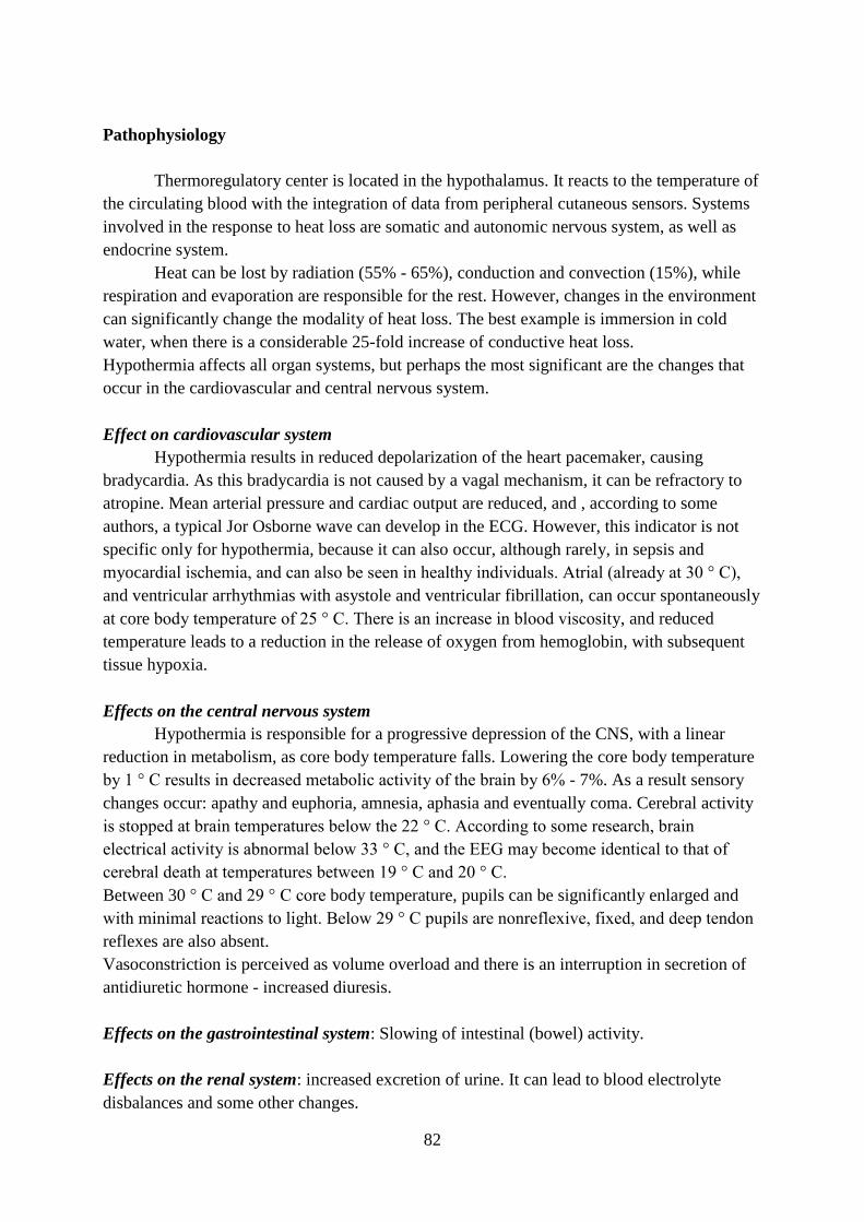
82
Pathophysiology
Thermoregulatory center is located in the hypothalamus. It reacts to the temperature of
the circulating blood with the integration of data from peripheral cutaneous sensors. Systems
involved in the response to heat loss are somatic and autonomic nervous system, as well as
endocrine system.
Heat can be lost by radiation (55% - 65%), conduction and convection (15%), while
respiration and evaporation are responsible for the rest. However, changes in the environment
can significantly change the modality of heat loss. The best example is immersion in cold
water, when there is a considerable 25-fold increase of conductive heat loss.
Hypothermia affects all organ systems, but perhaps the most significant are the changes that
occur in the cardiovascular and central nervous system.
Effect on cardiovascular system
Hypothermia results in reduced depolarization of the heart pacemaker, causing
bradycardia. As this bradycardia is not caused by a vagal mechanism, it can be refractory to
atropine. Mean arterial pressure and cardiac output are reduced, and , according to some
authors, a typical Jor Osborne wave can develop in the ECG. However, this indicator is not
specific only for hypothermia, because it can also occur, although rarely, in sepsis and
myocardial ischemia, and can also be seen in healthy individuals. Atrial (already at 30 ° C),
and ventricular arrhythmias with asystole and ventricular fibrillation, can occur spontaneously
at core body temperature of 25 ° C. There is an increase in blood viscosity, and reduced
temperature leads to a reduction in the release of oxygen from hemoglobin, with subsequent
tissue hypoxia.
Effects on the central nervous system
Hypothermia is responsible for a progressive depression of the CNS, with a linear
reduction in metabolism, as core body temperature falls. Lowering the core body temperature
by 1 ° C results in decreased metabolic activity of the brain by 6% - 7%. As a result sensory
changes occur: apathy and euphoria, amnesia, aphasia and eventually coma. Cerebral activity
is stopped at brain temperatures below the 22 ° C. According to some research, brain
electrical activity is abnormal below 33 ° C, and the EEG may become identical to that of
cerebral death at temperatures between 19 ° C and 20 ° C.
Between 30 ° C and 29 ° C core body temperature, pupils can be significantly enlarged and
with minimal reactions to light. Below 29 ° C pupils are nonreflexive, fixed, and deep tendon
reflexes are also absent.
Vasoconstriction is perceived as volume overload and there is an interruption in secretion of
antidiuretic hormone - increased diuresis.
Effects on the gastrointestinal system: Slowing of intestinal (bowel) activity.
Effects on the renal system: increased excretion of urine. It can lead to blood electrolyte
disbalances and some other changes.
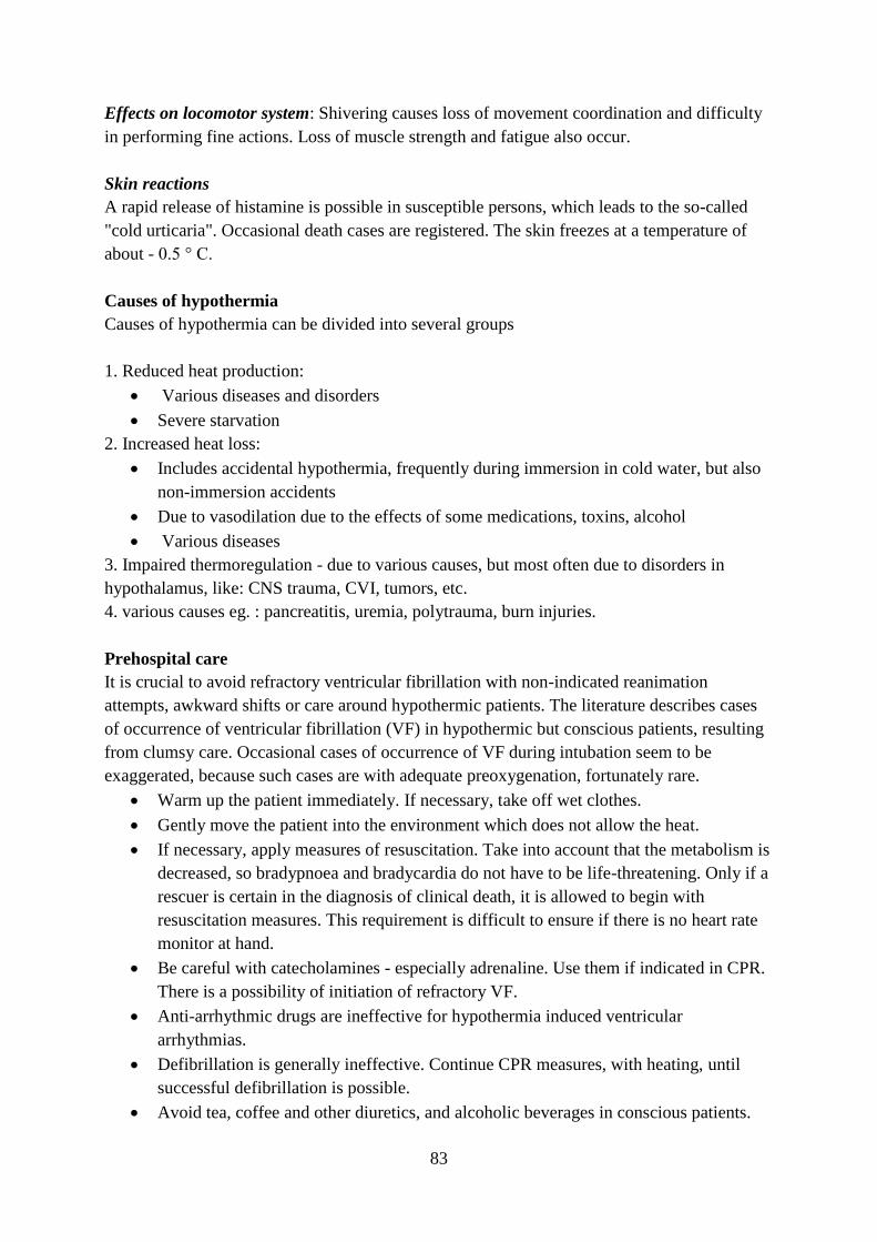
83
Effects on locomotor system: Shivering causes loss of movement coordination and difficulty
in performing fine actions. Loss of muscle strength and fatigue also occur.
Skin reactions
A rapid release of histamine is possible in susceptible persons, which leads to the so-called
"cold urticaria". Occasional death cases are registered. The skin freezes at a temperature of
about - 0.5 ° C.
Causes of hypothermia
Causes of hypothermia can be divided into several groups
1. Reduced heat production:
Various diseases and disorders
Severe starvation
2. Increased heat loss:
Includes accidental hypothermia, frequently during immersion in cold water, but also
non-immersion accidents
Due to vasodilation due to the effects of some medications, toxins, alcohol
Various diseases
3. Impaired thermoregulation - due to various causes, but most often due to disorders in
hypothalamus, like: CNS trauma, CVI, tumors, etc.
4. various causes eg. : pancreatitis, uremia, polytrauma, burn injuries.
Prehospital care
It is crucial to avoid refractory ventricular fibrillation with non-indicated reanimation
attempts, awkward shifts or care around hypothermic patients. The literature describes cases
of occurrence of ventricular fibrillation (VF) in hypothermic but conscious patients, resulting
from clumsy care. Occasional cases of occurrence of VF during intubation seem to be
exaggerated, because such cases are with adequate preoxygenation, fortunately rare.
Warm up the patient immediately. If necessary, take off wet clothes.
Gently move the patient into the environment which does not allow the heat.
If necessary, apply measures of resuscitation. Take into account that the metabolism is
decreased, so bradypnoea and bradycardia do not have to be life-threatening. Only if a
rescuer is certain in the diagnosis of clinical death, it is allowed to begin with
resuscitation measures. This requirement is difficult to ensure if there is no heart rate
monitor at hand.
Be careful with catecholamines - especially adrenaline. Use them if indicated in CPR.
There is a possibility of initiation of refractory VF.
Anti-arrhythmic drugs are ineffective for hypothermia induced ventricular
arrhythmias.
Defibrillation is generally ineffective. Continue CPR measures, with heating, until
successful defibrillation is possible.
Avoid tea, coffee and other diuretics, and alcoholic beverages in conscious patients.
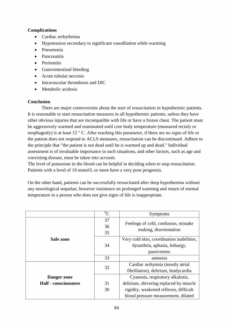
84
Complications
Cardiac arrhythmias
Hypotension secondary to significant vasodilation while warming
Pneumonia
Pancreatitis
Peritonitis
Gastrointestinal bleeding
Acute tubular necrosis
Intravascular thrombosis and DIC
Metabolic acidosis
Conclusion
There are major controversies about the start of resuscitation in hypothermic patients.
It is reasonable to start resuscitation measures in all hypothermic patients, unless they have
other obvious injuries that are incompatible with life or have a frozen chest. The patient must
be aggressively warmed and reanimated until core body temperature (measured rectaly or
esophagealy) is at least 32 ° C. After reaching this parameter, if there are no signs of life or
the patient does not respond to ACLS measures, resuscitation can be discontinued. Adhere to
the principle that "the patient is not dead until he is warmed up and dead." Individual
assessment is of invaluable importance in such situations, and other factors, such as age and
coexisting disease, must be taken into account.
The level of potassium in the blood can be helpful in deciding when to stop resuscitation.
Patients with a level of 10 mmol/L or more have a very poor prognosis.
On the other hand, patients can be successfully resuscitated after deep hypothermia without
any neurological sequelae, however insistence on prolonged warming and return of normal
temperature in a person who does not give signs of life is inappropriate.
0C Symptoms
Safe zone
37
36
35
Feelings of cold, confusion, mistake
making, disorientation
34
Very cold skin, coordination inabilities,
dysarthria, aphasia, lethargy,
passiveness
33 amnesia
Danger zone
Half - consciousness
32 Cardiac arrhytmia (mostly atrial
fibrillation), delirium, bradycardia
31
30
Cyanosis, respiratory alkalosis,
delirium, shivering replaced by muscle
rigidity, weakened reflexes, difficult
blood pressure measurement, dilated
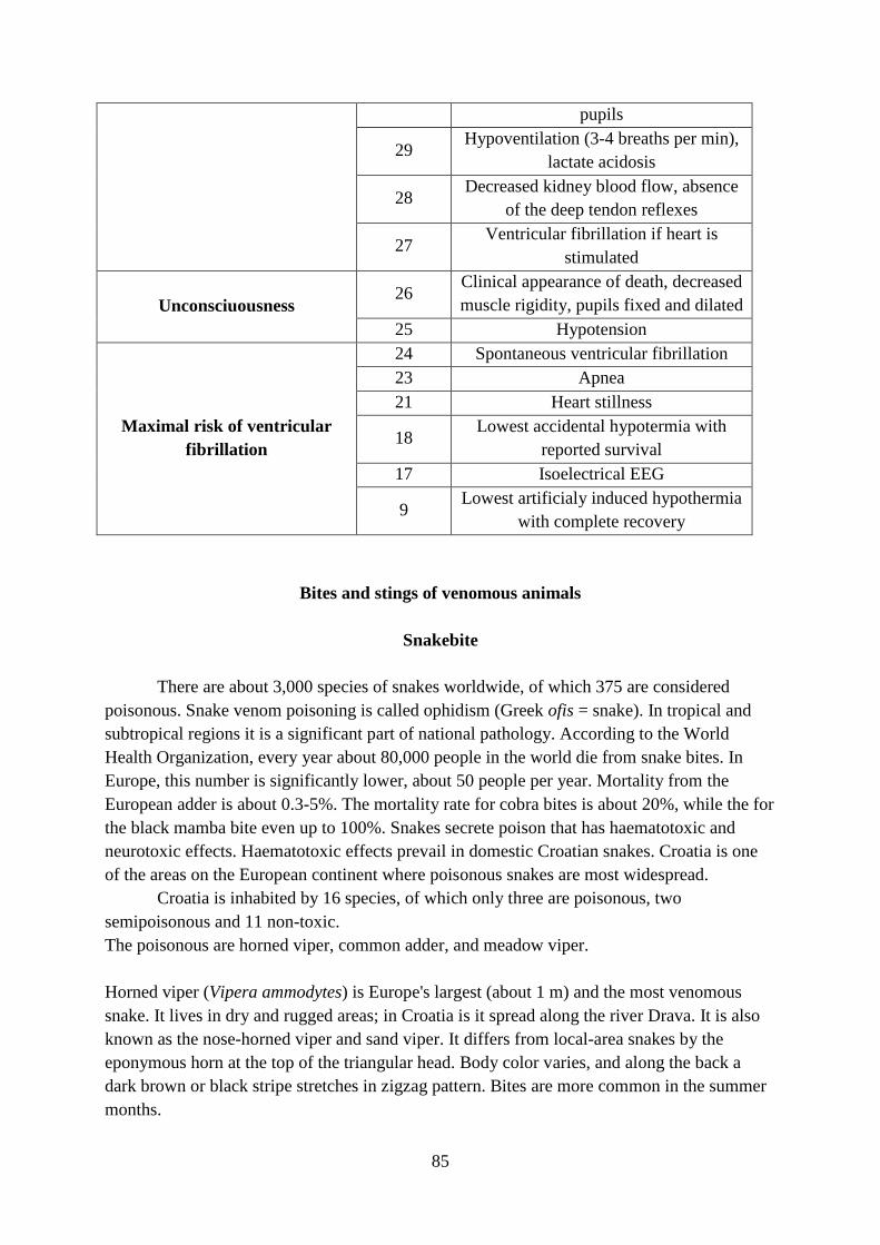
85
pupils
29 Hypoventilation (3-4 breaths per min),
lactate acidosis
28 Decreased kidney blood flow, absence
of the deep tendon reflexes
27 Ventricular fibrillation if heart is
stimulated
Unconsciuousness 26
Clinical appearance of death, decreased
muscle rigidity, pupils fixed and dilated
25 Hypotension
Maximal risk of ventricular
fibrillation
24 Spontaneous ventricular fibrillation
23 Apnea
21 Heart stillness
18 Lowest accidental hypotermia with
reported survival
17 Isoelectrical EEG
9 Lowest artificialy induced hypothermia
with complete recovery
Bites and stings of venomous animals
Snakebite
There are about 3,000 species of snakes worldwide, of which 375 are considered
poisonous. Snake venom poisoning is called ophidism (Greek ofis = snake). In tropical and
subtropical regions it is a significant part of national pathology. According to the World
Health Organization, every year about 80,000 people in the world die from snake bites. In
Europe, this number is significantly lower, about 50 people per year. Mortality from the
European adder is about 0.3-5%. The mortality rate for cobra bites is about 20%, while the for
the black mamba bite even up to 100%. Snakes secrete poison that has haematotoxic and
neurotoxic effects. Haematotoxic effects prevail in domestic Croatian snakes. Croatia is one
of the areas on the European continent where poisonous snakes are most widespread.
Croatia is inhabited by 16 species, of which only three are poisonous, two
semipoisonous and 11 non-toxic.
The poisonous are horned viper, common adder, and meadow viper.
Horned viper (Vipera ammodytes) is Europe's largest (about 1 m) and the most venomous
snake. It lives in dry and rugged areas; in Croatia is it spread along the river Drava. It is also
known as the nose-horned viper and sand viper. It differs from local-area snakes by the
eponymous horn at the top of the triangular head. Body color varies, and along the back a
dark brown or black stripe stretches in zigzag pattern. Bites are more common in the summer
months.
86
The Common European adder (Vipera berus) is the most widespread snake of Europe. Unlike
the horned viper it lives in more damp and plain areas. It withstands cold very well and
resides even in mountainous areas up to 3000 m. It has been expelled from many habitats by
the horned viper. It grows up to 90 cm. It is also known as the northern viper or crossed viper.
Its color varies depending on the environment and along the back it also possesses a wide
zigzag patterned line.
The Meadow Viper (Vipera ursinii) is the smallest (50 cm) Croatian poisonous snake. It is
said to be a small version of the Common Adder. It produces a small quantity of weak poison,
so it is not as dangerous to humans as the horned viper and common adder. In Croatia it
inhabits the Dinara mountain , but it is also present in areas along the Cetina river.
In Croatia, horned viper bites are more frequent than those of the common adder, while those
of the meadow viper are almost negligible. It often occurs that the victim does not see a snake,
just feels the sting.
Local signs and symptoms: Edema occurs locally shortly after the bite (dotted sores),
accompanied with numbness and redness, and subsequent hematoma (after 20-30 min). The
edema can spread from a finger to the whole fist, and later to the hand and hinder the
circulation. Sometimes it can extend to the shoulder and torso. Blisters filled with bloody
contents may occur at the site of the bite , along with vascular thrombosis and infection.
General symptoms and signs: headache, thirst, diarrhea, vomiting, general weakness and
numbness. Bleeding from the gums, colon, stomach or urinary tract may follow. Shock can
occur, ending with lethal outcome.
First Aid: First determine if indeed there was a venomous bite (dotted sores and clinical
picture). Calm the victim. Clean the bite wound with some disinfectant. The victim must rest.
Immobilize the bitten limb with splints, or similar means. Transport the victim to the nearest
health facility on a stretcher, if possible. If the victim has to get by herself to the doctor,
running is not recommended, but instead walking with breaks. Depending on the site of the
bite it is recommended to put a tourniquet a few inches above the bitemark and squeeze in a
manner that it would be hard to put finger underneath the tourniquet. Tourniquet should be
wide, and not be a rope or a wire. The aim is to prevent the flow of lymph, not the return of
venous blood.
What not to do: Sucking the poison from the wound is not recommended, and neither is
cutting the bite wound with a razor blade, knife or similar means, because there is a
possibility of doing severe injuries to the victim.
Treatment: Serum antiviperinum. Administer only in health institutions, where there are
means for treating allergic and anaphylactic reactions.
Prevention: It is very significant. Wear suitable clothing and footwear in areas where
87
poisonous snakes live. Snakes usually bite when surprised, therefore, banging a stick on the
rocks, breaking branches etc. announces the arrival, so that snakes can escape in time. In a
sudden encounter with a viper keep presence of mind, do not run, because it will scare the
snake and cause her attack.
The sting of the black widow spider
Spiders of the latrodectus type are spread throughout the world. Our Mediterranean
black widow spider inhabits the coastal areas of Europe and Africa, all the way to the south of
the European part of the former USSR. The spider was named after the bright red spots,
which are 13 in number (tredecim). However, the number of spots is variable, it can be up to
17, or a spider can appear even without spots. Only females are poisonous, and it is
distinguished by a body length of about 1.5 cm. The male is smaller, the average body size
about 5 mm.
The venom of the black widow is a neurotoxin, which strongly stimulates the release
of neurotransmitters. Venom is reabsorbed from the place of the bite and expands at first by
lymph ducts, and only after that by the circulatory system. A considerable amount of poison
remains at the injection site. The spider will sting in self-defense, if it is accidentally pressed
or if it climbs up the sleeve, and is in a tight position between the body and clothing. It does
not dwell in the cities. Bites occur most frequently in the summer, but can occur throughout
the year. Latrodectus appears cyclically.
Clinical presentation: The sting is very superficial and almost painless, so that 50% of people
do not register or do not pay attention to it, thinking that there was a sting of a blade of grass
or thorn. People stabbed during sleep were not awakened by the sting. Time interval from the
bite to the first signs is variable (10-60 min).
Clinical presentation:
a) minor local symptoms (most commonly),
b) strong localised pain, which lasts several hours, but without general symptoms,
c) general symptoms a few hours after the disappearance of local symptoms
d) general symptoms and generalised severe condition, the rarest.
Local findings: First, the pain occurs in regional lymph nodes close to the edema. Only the
puncture site is not changed in a significant matter. One or two dotted spots on the skin that
are raised as in urticaria can be seen. After a few hours, a bluish-purple ring, the size of a
small coin, appears around faded areas.
General symptomatology: The general condition rapidly deteriorates. Severe pain spreads
from the regional nodes, accompanied by cramps with a feelings of tightness in the abdomen
and chest. Muscle spasms and shivering frequently appear. The abdomen may be tense and
firm and mimic acute abdomen. The skin of the entire body may become sensitive to touch,
and profuse sweating can occur. The victim is often unable to walk, restless and scared and
may have a fever. A state of shock can also appear. The face may be distorted due to spasm of
masseter. Tearing from the eyes occurs. Salivation is present, but the feeling of dry mouth is
88
more common. Heart rate is initially rapid, with elevated blood pressure. As the toxin affects
the respiratory system, small bronchi become narrow while mucus secretion is profuse.
Convulsive contractions of the diaphragm and the muscles of the abdomen and chest may
develop, making it difficult to breathe. Hoarseness and speaking difficulties also occur.
Nausea and vomiting are the gastrointestinal symptoms. Urinating may be difficult due to
spasm of the sphincter of the bladder.
The main symptom is severe pain that is often described as "tearing the body by
pliers", "rolling on thorns" or "grinding bones".
The clinical presentation is more severe in children and the elderly. Paresthesia,
insomnia, dizziness, joint pain, photophobia, psychological disorders (memory loss,
confusion, hallucinations, delirium) also occur. After a few days, a rash may appear similar to
that found in scarlet fever or measles. Untreated persons suffer for about a week. Death from
a black widow bite is rare.
First aid: Incisions with a knife or similar devices or tools are not recommended, as
well as attempts at sucking out the venom. Ice cooling relieves pain at the beginning. Urgent
transport to hospital is necessary.
Treatment: Serum. Provide in medical institutions only because of the risk of
anaphylaxis.
Prevention: Avoid habitat of black widows. Wear appropriate clothing and shoes.
8. POISONING AND BURNS
Nenad Karanović, MD, PhD
Introduction and epidemiology
Acute poisoning is one of the major reasons for hospital admission. It is estimated that
different poisonings are the reason for intervention in emergency departments in 5-10% of
cases. It is known that approximately 12 million agents can cause poisoning.
Poisoning can be intentional or accidental. According to data from the US, there are
2,168,248 poisoning cases recorded annually. Adults account for one third of the cases. About
71% were accidental poisoning and in 92% the poisoning was caused by a single agent. Oral
administration was present in the majority of cases. According to British data about
intentional poisoning in adults, most of those cases are younger than 35, female, of otherwise
good health and in which this is a "call for help" and they usually want to be found or take
remedies in order to attract attention. In contrast, in the group of adults older than 55 years,
male persons are most often represented, who attempt to poison themselves due to depression
or untreatable diseases.
According to some research general intrahospital mortality due to various poisoning is
approximately 0.5%.
89
Substances that cause the greatest number of deaths are analgesics, antidepressants,
sedatives / hypnotics / antipsychotics, then different stimulants, "street drugs", medicines for
the treatment of cardiovascular disorders and alcohol. In developed countries, substances that
often cause poisoning are paracetamol, benzodiazepines, antidepressants and NSAIDs, while
in developing countries the most commonl used substances are pesticides.
Iatrogenic poisonings are caused by errors in the instructions or unrecognized drug
interactions, and have recently become a significant problem. According to some estimations,
it represents up to 1% of cases.
A very important issue that has an impact on the treatment of poisoned victims is
related to the current recommendations for the treatment of such cases. One should be aware
that all information is obtained on the basis of very limited studies conducted on animals,
people, the individual case reports and on the pharmacokinetics of drugs, known
pathophysiology and usually on agreed conclusions.
Studies on animals and healthy volunteers cannot be extrapolated to the clinical
situation, therefore, therapeutic guidelines only suggest, but do not have to be supported by
final evidence.
Furthermore, we should not forget that the operating instructions supplied with the
different drugs or agents used in households are most often outdated and do not provide
sufficient information. The same goes for textbooks, especially older ones. It is therefore
recommended to consult the relevant sites on the internet such as Toxbase
(http://www.spib.axl.co.uk) or Isabel (http://www.isabel.org.uk) or to consult the Poisoning
Control Center at the Institute for medical Research and Occupational Health, Zagreb,
Ksaverska 2. Information is available 24 hours a day on the emergency phone +385 1 234 83
42.
General guidelines for the treatment of poisoned victims before hospital admission
It is necessary to quickly and systematically orient yourself regarding the state of the poisoned
victim. The assessment is carried out with the help of mnemotechnic principle ABCD.
Assessment is repeated in certain intervals. Significant changes in the condition of a poisoned
victim can quickly occur.
Emergency measures- ABC reanimation measures:
Assess and ensure adequate airway and enable or improve ventilation.
Stabilize the cardiovascular system. Various toxins cause vasodilation, hypotension
and arrhythmias.
Pay attention to the potential for seizures and treat them.
The objectives of hospital treatment after emergency resuscitation measures and stabilization
of the condition are:
To prevent further absorption,
Speed up elimination,
Give antidote, if it exists,
Support organ systems
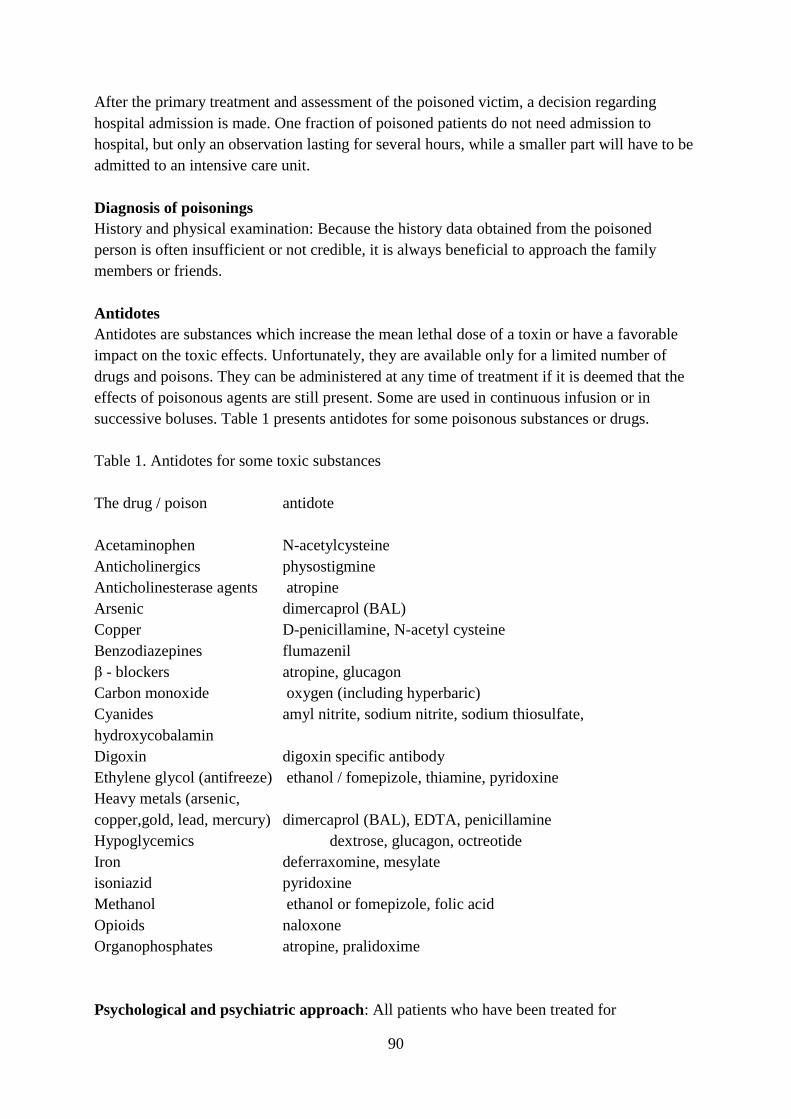
90
After the primary treatment and assessment of the poisoned victim, a decision regarding
hospital admission is made. One fraction of poisoned patients do not need admission to
hospital, but only an observation lasting for several hours, while a smaller part will have to be
admitted to an intensive care unit.
Diagnosis of poisonings
History and physical examination: Because the history data obtained from the poisoned
person is often insufficient or not credible, it is always beneficial to approach the family
members or friends.
Antidotes
Antidotes are substances which increase the mean lethal dose of a toxin or have a favorable
impact on the toxic effects. Unfortunately, they are available only for a limited number of
drugs and poisons. They can be administered at any time of treatment if it is deemed that the
effects of poisonous agents are still present. Some are used in continuous infusion or in
successive boluses. Table 1 presents antidotes for some poisonous substances or drugs.
Table 1. Antidotes for some toxic substances
The drug / poison antidote
Acetaminophen N-acetylcysteine
Anticholinergics physostigmine
Anticholinesterase agents atropine
Arsenic dimercaprol (BAL)
Copper D-penicillamine, N-acetyl cysteine
Benzodiazepines flumazenil
β - blockers atropine, glucagon
Carbon monoxide oxygen (including hyperbaric)
Cyanides amyl nitrite, sodium nitrite, sodium thiosulfate,
hydroxycobalamin
Digoxin digoxin specific antibody
Ethylene glycol (antifreeze) ethanol / fomepizole, thiamine, pyridoxine
Heavy metals (arsenic,
copper,gold, lead, mercury) dimercaprol (BAL), EDTA, penicillamine
Hypoglycemics dextrose, glucagon, octreotide
Iron deferraxomine, mesylate
isoniazid pyridoxine
Methanol ethanol or fomepizole, folic acid
Opioids naloxone
Organophosphates atropine, pralidoxime
Psychological and psychiatric approach: All patients who have been treated for
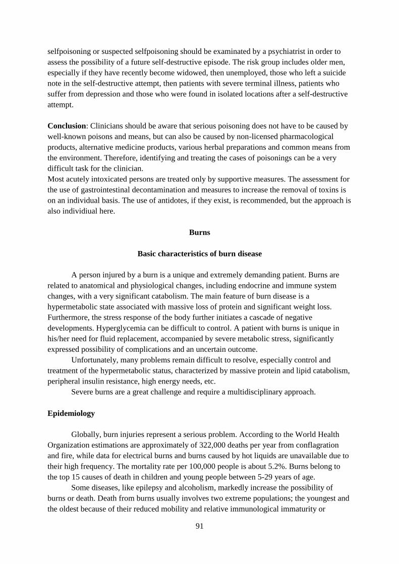
91
selfpoisoning or suspected selfpoisoning should be examinated by a psychiatrist in order to
assess the possibility of a future self-destructive episode. The risk group includes older men,
especially if they have recently become widowed, then unemployed, those who left a suicide
note in the self-destructive attempt, then patients with severe terminal illness, patients who
suffer from depression and those who were found in isolated locations after a self-destructive
attempt.
Conclusion: Clinicians should be aware that serious poisoning does not have to be caused by
well-known poisons and means, but can also be caused by non-licensed pharmacological
products, alternative medicine products, various herbal preparations and common means from
the environment. Therefore, identifying and treating the cases of poisonings can be a very
difficult task for the clinician.
Most acutely intoxicated persons are treated only by supportive measures. The assessment for
the use of gastrointestinal decontamination and measures to increase the removal of toxins is
on an individual basis. The use of antidotes, if they exist, is recommended, but the approach is
also individiual here.
Burns
Basic characteristics of burn disease
A person injured by a burn is a unique and extremely demanding patient. Burns are
related to anatomical and physiological changes, including endocrine and immune system
changes, with a very significant catabolism. The main feature of burn disease is a
hypermetabolic state associated with massive loss of protein and significant weight loss.
Furthermore, the stress response of the body further initiates a cascade of negative
developments. Hyperglycemia can be difficult to control. A patient with burns is unique in
his/her need for fluid replacement, accompanied by severe metabolic stress, significantly
expressed possibility of complications and an uncertain outcome.
Unfortunately, many problems remain difficult to resolve, especially control and
treatment of the hypermetabolic status, characterized by massive protein and lipid catabolism,
peripheral insulin resistance, high energy needs, etc.
Severe burns are a great challenge and require a multidisciplinary approach.
Epidemiology
Globally, burn injuries represent a serious problem. According to the World Health
Organization estimations are approximately of 322,000 deaths per year from conflagration
and fire, while data for electrical burns and burns caused by hot liquids are unavailable due to
their high frequency. The mortality rate per 100,000 people is about 5.2%. Burns belong to
the top 15 causes of death in children and young people between 5-29 years of age.
Some diseases, like epilepsy and alcoholism, markedly increase the possibility of
burns or death. Death from burns usually involves two extreme populations; the youngest and
the oldest because of their reduced mobility and relative immunological immaturity or
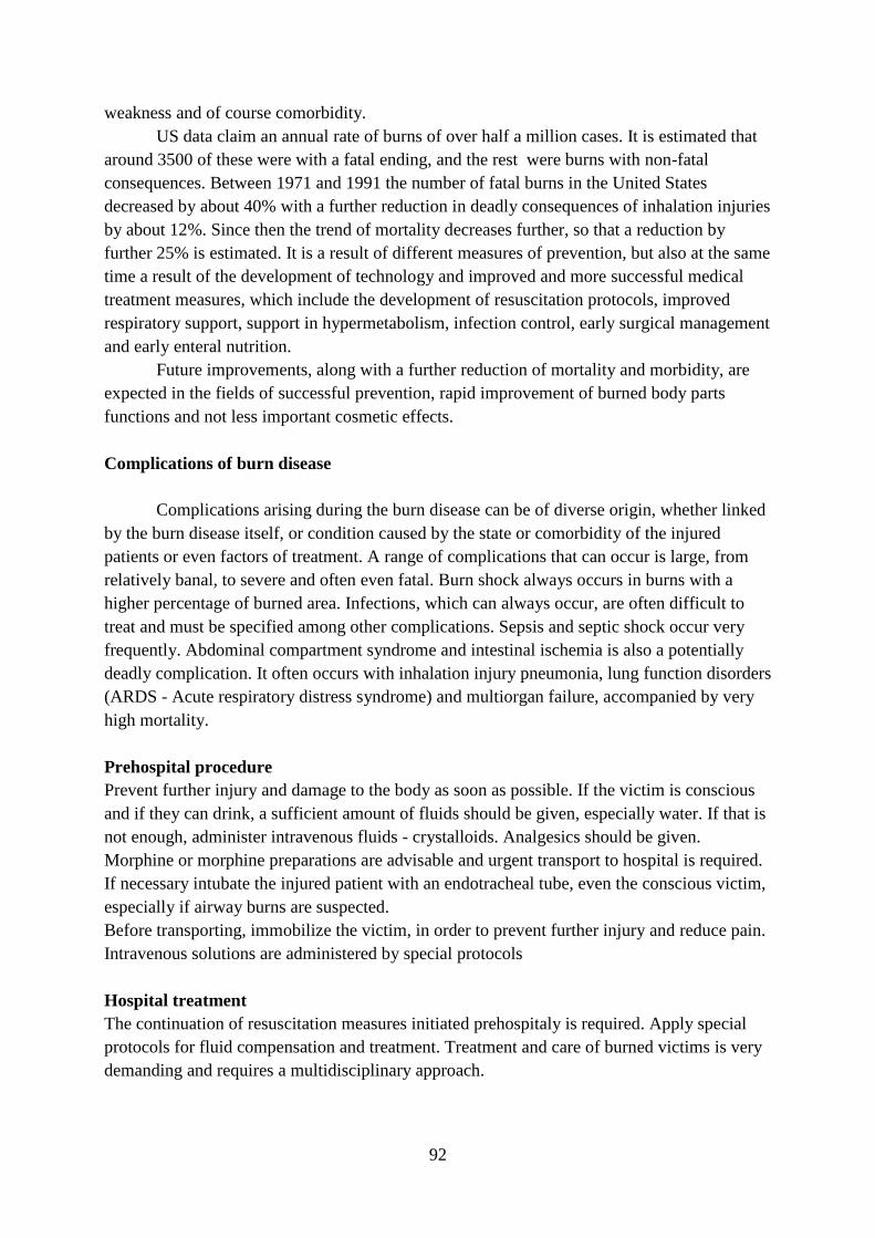
92
weakness and of course comorbidity.
US data claim an annual rate of burns of over half a million cases. It is estimated that
around 3500 of these were with a fatal ending, and the rest were burns with non-fatal
consequences. Between 1971 and 1991 the number of fatal burns in the United States
decreased by about 40% with a further reduction in deadly consequences of inhalation injuries
by about 12%. Since then the trend of mortality decreases further, so that a reduction by
further 25% is estimated. It is a result of different measures of prevention, but also at the same
time a result of the development of technology and improved and more successful medical
treatment measures, which include the development of resuscitation protocols, improved
respiratory support, support in hypermetabolism, infection control, early surgical management
and early enteral nutrition.
Future improvements, along with a further reduction of mortality and morbidity, are
expected in the fields of successful prevention, rapid improvement of burned body parts
functions and not less important cosmetic effects.
Complications of burn disease
Complications arising during the burn disease can be of diverse origin, whether linked
by the burn disease itself, or condition caused by the state or comorbidity of the injured
patients or even factors of treatment. A range of complications that can occur is large, from
relatively banal, to severe and often even fatal. Burn shock always occurs in burns with a
higher percentage of burned area. Infections, which can always occur, are often difficult to
treat and must be specified among other complications. Sepsis and septic shock occur very
frequently. Abdominal compartment syndrome and intestinal ischemia is also a potentially
deadly complication. It often occurs with inhalation injury pneumonia, lung function disorders
(ARDS - Acute respiratory distress syndrome) and multiorgan failure, accompanied by very
high mortality.
Prehospital procedure
Prevent further injury and damage to the body as soon as possible. If the victim is conscious
and if they can drink, a sufficient amount of fluids should be given, especially water. If that is
not enough, administer intravenous fluids - crystalloids. Analgesics should be given.
Morphine or morphine preparations are advisable and urgent transport to hospital is required.
If necessary intubate the injured patient with an endotracheal tube, even the conscious victim,
especially if airway burns are suspected.
Before transporting, immobilize the victim, in order to prevent further injury and reduce pain.
Intravenous solutions are administered by special protocols
Hospital treatment
The continuation of resuscitation measures initiated prehospitaly is required. Apply special
protocols for fluid compensation and treatment. Treatment and care of burned victims is very
demanding and requires a multidisciplinary approach.
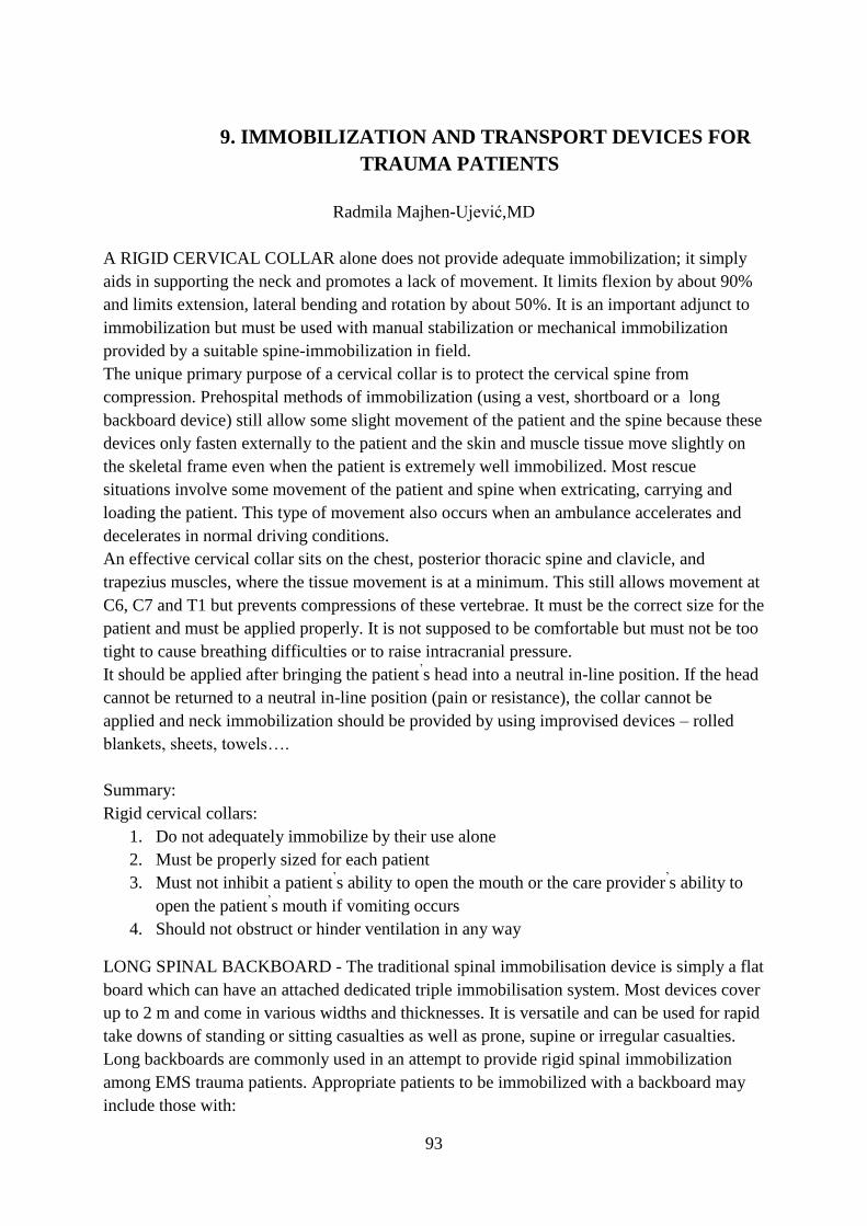
93
9. IMMOBILIZATION AND TRANSPORT DEVICES FOR
TRAUMA PATIENTS
Radmila Majhen-Ujević,MD
A RIGID CERVICAL COLLAR alone does not provide adequate immobilization; it simply
aids in supporting the neck and promotes a lack of movement. It limits flexion by about 90%
and limits extension, lateral bending and rotation by about 50%. It is an important adjunct to
immobilization but must be used with manual stabilization or mechanical immobilization
provided by a suitable spine-immobilization in field.
The unique primary purpose of a cervical collar is to protect the cervical spine from
compression. Prehospital methods of immobilization (using a vest, shortboard or a long
backboard device) still allow some slight movement of the patient and the spine because these
devices only fasten externally to the patient and the skin and muscle tissue move slightly on
the skeletal frame even when the patient is extremely well immobilized. Most rescue
situations involve some movement of the patient and spine when extricating, carrying and
loading the patient. This type of movement also occurs when an ambulance accelerates and
decelerates in normal driving conditions.
An effective cervical collar sits on the chest, posterior thoracic spine and clavicle, and
trapezius muscles, where the tissue movement is at a minimum. This still allows movement at
C6, C7 and T1 but prevents compressions of these vertebrae. It must be the correct size for the
patient and must be applied properly. It is not supposed to be comfortable but must not be too
tight to cause breathing difficulties or to raise intracranial pressure.
It should be applied after bringing the patient’s head into a neutral in-line position. If the head
cannot be returned to a neutral in-line position (pain or resistance), the collar cannot be
applied and neck immobilization should be provided by using improvised devices – rolled
blankets, sheets, towels….
Summary:
Rigid cervical collars:
1. Do not adequately immobilize by their use alone
2. Must be properly sized for each patient
3. Must not inhibit a patient’s ability to open the mouth or the care provider
’s ability to
open the patient’s mouth if vomiting occurs
4. Should not obstruct or hinder ventilation in any way
LONG SPINAL BACKBOARD - The traditional spinal immobilisation device is simply a flat
board which can have an attached dedicated triple immobilisation system. Most devices cover
up to 2 m and come in various widths and thicknesses. It is versatile and can be used for rapid
take downs of standing or sitting casualties as well as prone, supine or irregular casualties.
Long backboards are commonly used in an attempt to provide rigid spinal immobilization
among EMS trauma patients. Appropriate patients to be immobilized with a backboard may
include those with:
94
- Blunt trauma and altered level of consciousness;
- Spinal pain or tenderness;
- Neurologic complaint (e.g., numbness or motor weakness)
- Anatomic deformity of the spine
- High energy mechanism of injury and:
- Drug or alcohol intoxication
- Inability to communicate
- Distracting injury.
Patients for whom immobilization on a backboard is not necessary include those with all
of the following:
- Normal level of consciousness (GCS 15);
- No spine tenderness or anatomic abnormality;
- No neurologic findings or complaints;
- No distracting injury;
- No intoxication.
The long backboard can induce pain, patient agitation, and respiratory compromise. Further,
the backboard can decrease tissue perfusion at pressure points, leading to the development of
pressure ulcers. Low grade ulcers can appear in as few as two hours and even healthy persons
complain of pain after 30 minutes. Utilization of backboards for spinal immobilization during
transport should be judicious, so that potential benefits outweigh risks. Patients with
penetrating trauma to the head, neck or torso and no evidence of spinal injury should not be
immobilized on a backboard. Spinal precautions can be maintained by application of a rigid
cervical collar and securing the patient firmly to the EMS stretcher, and may be most
appropriate for:
- Patients who are found to be ambulatory at the scene;
- Patients who must be transported for a protracted time, particularly prior to
interfacility transfer; or
- Patients for whom a backboard is not otherwise indicated.
Two major adjustments in the previous methods are necessary when immobilizing a
small child to a long board.
• Due to the relatively large size of the child’s head, padding is needed under the torso to
elevate it and maintain the spine in neutral alignment. The padding must extend form the
lumbar area to the top of the shoulders, and to the right and left edges of the board. A folded
blanket usually works well.
• Small children are usually narrower than an adult sized board. Blanket rolls can be placed
between the child’s sides and the sides of the board to prevent lateral movement. Pediatric
immobilization devices take these differences into account, and are preferable.
Patients should be removed from backboards as soon as practical in an emergency
department.
95
SPLIT DEVICES are popular in prehospital circles as they can avoid a log roll in supine
casualties or help extricate from a vehicle.These include the aluminium orthopaedic scoop
stretcher and the Ferno 65 XL (modern scoop). However, it is designed as a transfer device
and should not be carried for any distance. Those who are not supine will still need to be
rolled. [
VACUUM MATTRESS: Perhaps the gold standard device in true spinal cord injury, the
vacuum mattress provides an individually moulded cocoon for the casualty through a double
bagged polystyrene ball system which becomes rigid when the air is removed. They come in a
range of sizes and widths and avoid the problems of local pressure areas as the force is evenly
spread out along the whole body. However, if they puncture, the valve fails or the pump is
lost, then they become of little value. Hence they should always be used with a backup device
available. They are becoming more and more popular with the ambulance service as the
device of choice, especially for pregnant women and if transport lasts longer than 30 minutes.
KENDRICK EXTRICATION DEVICE (KED)
It is an immobilization vest and it is used when spinal stabilization is indicated for a sitting
trauma patient without life-threatening conditions, usually in a vehicle extrication. Typically
used in conjunction with a cervical collar, the KED is a semi-rigid brace that secures the
head, neck and torso in an anatomically neutral position.
It is important that manual –in –line stabilisation is always performed before applying other
devices.The techniques we use to put the patients on transport devices are log-roll (LR), lift-
slide (LS), lateral trauma position (LTP)
Some other issues on immobilization and transport:
- Pregnant women in left lateral position with an angle till 30 degrees
- Suspect traumatic brain injury with head elevated till 30 degrees
- Anti-shock position with elevated legs
- If the child has no life-threatening injuries it may be better to transport the child in
their child safety seat
- Torso and legs are always fastened before head
- Removal of the helmet is necessary to assess the patient and to move the head into
neutral alignment
- Log- roll should be avoided in suspected pelvis and two long bones injury
Literature:
1. McSwain NE Jr, ed. Prehospital Trauma Life Support. 7. ed. St. Louis:Mosby JEMS
Elsevier;2011
2. Krell JM, McCoy MS, Sparto PJ, Fisher GL, Stoy WA, Hostler DP. Comparison of the
Ferno Scoop Stretcher with the long backboard for spinal immobilization.Prehosp Emerg
Care. 2006 Jan-Mar;10(1):46-51.
3. Oomens CW, Zenhorst W, Broek M, Hemmes B, Poeze M, Brink PR, Bader DL. A
numerical study to analyse the risk for pressure ulcer development on a spine board.
Clin Biomech (Bristol, Avon). 2013 Aug;28(7):736-42.
4. Conrad BP, Marchese DL, Rechtine GR, Horodyski M. Motion in the unstable
96
thoracolumbar spine when spine boarding a prone patient. J Spinal Cord Med. 2012
Jan;35(1):53-7.
5. Horodyski M, Conrad BP, Del Rossi G, DiPaola CP, Rechtine GR 2nd. Removing a
patient from the spine board: is the lift and slide safer than the log roll? J Trauma. 2011
May;70(5):1282-5;
6. Hostler D, Colburn D, Seitz SR. A comparison of three cervical immobilization devices.
Prehosp Emerg Care. 2009 Apr-Jun;13(2):256-60.
7. Peery CA, Brice J, White WD. Prehospital spinal immobilization and the
backboard quality assessment study. Prehosp Emerg Care. 2007 Jul-Sep;11(3):293-7.
8. Del Rossi G, Horodyski MH, Conrad BP, Di Paola CP, Di Paola MJ, Rechtine GR.The 6-
plus-person lift transfer technique compared with other methods of spine boarding. J Athl
Train. 2008 Jan-Mar;43(1):6-13.
9. Del Rossi G, Rechtine GR, Conrad BP, Horodyski M. Are scoop stretchers
suitable for use on spine-injured patients? Am J Emerg Med. 2010 Sep;28(7):751-6.
10. Hanson J.R., Carlin B., Sports Prehospital-Immediate Care and Spinal Injury,Br J
Sports Med. 2012;46(16):1097-1101
11. Del Rossi G, Horodyski M, Heffernan TP, Powers ME, Siders R, Brunt D, Rechtine GR.
Spine-board transfer techniques and the unstable cervical spine. Spine (Phila Pa 1976).
2004 Apr 1;29(7):E134-8.
12. National Association of EMS Physicians and the American
College of Surgeons Committee on Trauma .EMS spinal precautions and the use of
the long backboard - Position Statement , Prehosp Emerg Care 2013
13. Fattah S., Ekås G.R., The lateral trauma position: What do we know about it and how do
we use it? A cross-sectional survey of all Norwegian emergency medical services, Scand J
Trauma Resusc Emerg Med. 2011; 19: 45.
14. Ahn H., Singh J., [...], Yee A., Pre-Hospital Care Management of a Potential Spinal
Cord Injured Patient: A Systematic Review of the Literature and Evidence-Based
Guidelines ,J Neurotrauma. 2011 August; 28(8): 1341–1361.
15. Edlich RF, Mason SS, Vissers RJ, Gubler KD, Thacker JG, Pharr P, Anderson M,
Long WB 3rd. Revolutionary advances in enhancing patient comfort on patients
transported on a backboard. Am J Emerg Med. 2011 Feb;29(2):181-6.
16. Cooney D.R., Wallus H, [...], Wojcik S. Backboard time for patients receiving spinal
immobilization by emergency medical services, Int J Emerg Med. 2013; 6: 17.