Embed Size (px)
Citation preview
Available online at www.sciencedirect.com
Taiwanese Journal of Obstetrics & Gynecology 51 (2012) 100e105www.tjog-online.com
Case Report
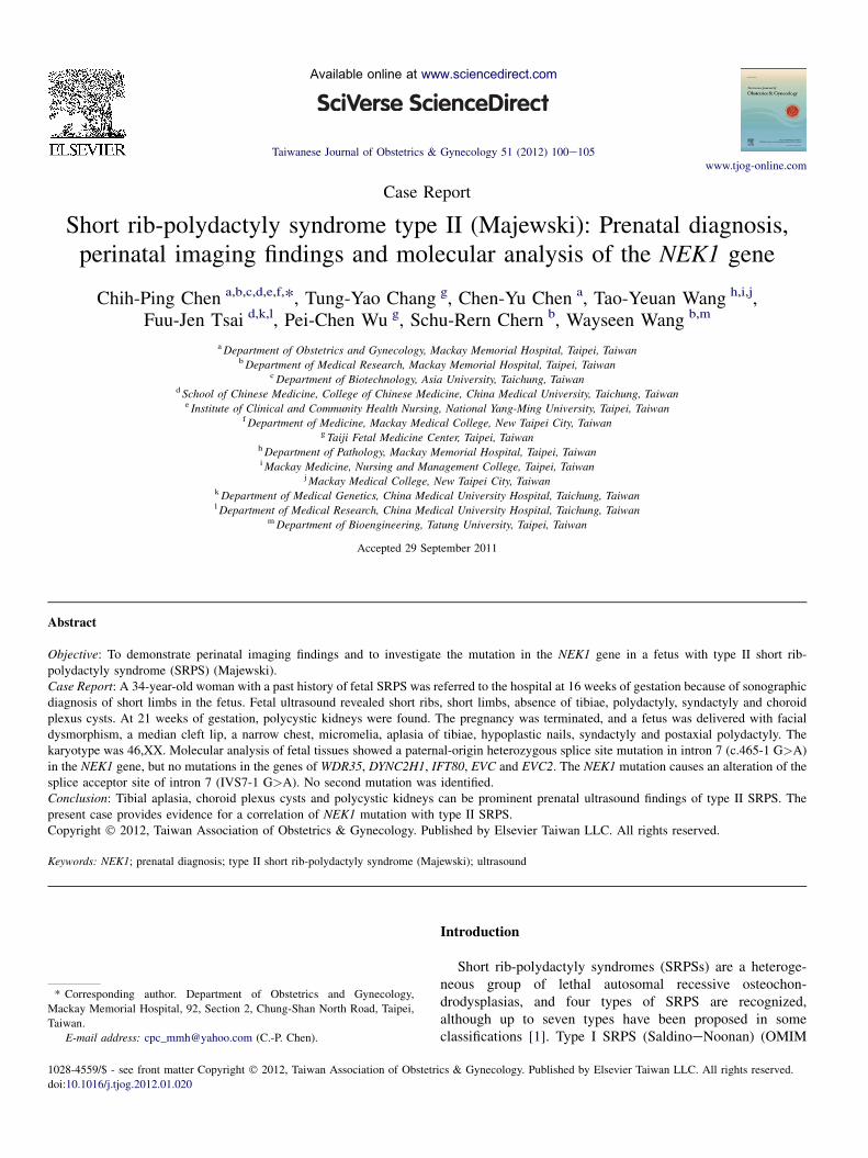
Short rib-polydactyly syndrome type II (Majewski): Prenatal diagnosis,perinatal imaging findings and molecular analysis of the NEK1 gene
Chih-Ping Chen a,b,c,d,e,f,*, Tung-Yao Chang g, Chen-Yu Chen a, Tao-Yeuan Wang h,i,j,Fuu-Jen Tsai d,k,l, Pei-Chen Wu g, Schu-Rern Chern b, Wayseen Wang b,m
aDepartment of Obstetrics and Gynecology, Mackay Memorial Hospital, Taipei, TaiwanbDepartment of Medical Research, Mackay Memorial Hospital, Taipei, Taiwan
cDepartment of Biotechnology, Asia University, Taichung, Taiwand School of Chinese Medicine, College of Chinese Medicine, China Medical University, Taichung, Taiwan
e Institute of Clinical and Community Health Nursing, National Yang-Ming University, Taipei, TaiwanfDepartment of Medicine, Mackay Medical College, New Taipei City, Taiwan
g Taiji Fetal Medicine Center, Taipei, TaiwanhDepartment of Pathology, Mackay Memorial Hospital, Taipei, TaiwaniMackay Medicine, Nursing and Management College, Taipei, Taiwan
jMackay Medical College, New Taipei City, TaiwankDepartment of Medical Genetics, China Medical University Hospital, Taichung, TaiwanlDepartment of Medical Research, China Medical University Hospital, Taichung, Taiwan
mDepartment of Bioengineering, Tatung University, Taipei, Taiwan
Accepted 29 September 2011
Abstract
Objective: To demonstrate perinatal imaging findings and to investigate the mutation in the NEK1 gene in a fetus with type II short rib-polydactyly syndrome (SRPS) (Majewski).Case Report: A 34-year-old woman with a past history of fetal SRPS was referred to the hospital at 16 weeks of gestation because of sonographicdiagnosis of short limbs in the fetus. Fetal ultrasound revealed short ribs, short limbs, absence of tibiae, polydactyly, syndactyly and choroidplexus cysts. At 21 weeks of gestation, polycystic kidneys were found. The pregnancy was terminated, and a fetus was delivered with facialdysmorphism, a median cleft lip, a narrow chest, micromelia, aplasia of tibiae, hypoplastic nails, syndactyly and postaxial polydactyly. Thekaryotype was 46,XX. Molecular analysis of fetal tissues showed a paternal-origin heterozygous splice site mutation in intron 7 (c.465-1 G>A)in the NEK1 gene, but no mutations in the genes of WDR35, DYNC2H1, IFT80, EVC and EVC2. The NEK1 mutation causes an alteration of thesplice acceptor site of intron 7 (IVS7-1 G>A). No second mutation was identified.Conclusion: Tibial aplasia, choroid plexus cysts and polycystic kidneys can be prominent prenatal ultrasound findings of type II SRPS. Thepresent case provides evidence for a correlation of NEK1 mutation with type II SRPS.Copyright � 2012, Taiwan Association of Obstetrics & Gynecology. Published by Elsevier Taiwan LLC. All rights reserved.
Keywords: NEK1; prenatal diagnosis; type II short rib-polydactyly syndrome (Majewski); ultrasound
* Corresponding author. Department of Obstetrics and Gynecology,
Mackay Memorial Hospital, 92, Section 2, Chung-Shan North Road, Taipei,
Taiwan.
E-mail address: [email protected] (C.-P. Chen).
1028-4559/$ - see front matter Copyright � 2012, Taiwan Association of Obstetri
doi:10.1016/j.tjog.2012.01.020
Introduction
Short rib-polydactyly syndromes (SRPSs) are a heteroge-neous group of lethal autosomal recessive osteochon-drodysplasias, and four types of SRPS are recognized,although up to seven types have been proposed in someclassifications [1]. Type I SRPS (SaldinoeNoonan) (OMIM
cs & Gynecology. Published by Elsevier Taiwan LLC. All rights reserved.

101C.-P. Chen et al. / Taiwanese Journal of Obstetrics & Gynecology 51 (2012) 100e105
263530) is characterized by flipper-like extremities, poly-dactyly, polycystic kidneys and pointed metaphyses. Type IISRPS (Majewski) (OMIM 263520) is characterized by poly-dactyly, micromelia, cleft lip/palate, polycystic kidneys,disproportionately short ovoid tibia and occasionally hypo-plastic epiglottis and larynx. Type III SRPS (VermaeNaum-off) (OMIM 263510) is characterized by polydactyly,micromelia, metaphyseal spurs and occasionally situs inversustotalis. Type IV SRPS (BeemereLanger) (OMIM 269860)clinically resembles type II SRPS, except there is no
Fig. 1. Prenatal ultrasound at 16 weeks of gestation shows: (A) choroid plexus cyst
and (D) and (E) polydactyly. F ¼ fibula; T ¼ tibia.
polydactyly and more normally developed tibiae. Type IIISRPS is similar but milder than type I. Both types I and III areclassified together according to current nosology and classi-fication of genetic skeletal disorders [2]. Recently, a new typeV SRPS has been suggested. Type V SRPS (OMIM 614091)is most similar to type III SRPS, but is associated withacromesomelic hypomineralization and campomelia [3,4].Because of overlap in the clinical and radiological manifes-tations in different types, it is hypothesized that the differentsubtypes may be a single genetic disorder with variable
s; (B) short ribs and a narrow chest (arrow); (C) short fibula and tibial aplasia;

102 C.-P. Chen et al. / Taiwanese Journal of Obstetrics & Gynecology 51 (2012) 100e105
expressivity. Here, we present perinatal imaging findings andmolecular investigation in a fetus with type II SRPS(Majewski).
Case report
A 34-year-old, gravida 5, para 2, woman with a past historyof fetal SRPS was referred to the hospital at 16 weeks ofgestation following sonographic diagnosis of shortening of thelimbs in the fetus. She and her husband were non-consanguineous and had two normal daughters, aged 6 yearsand 3 years, respectively. Eight years previously, she had givenbirth to a female fetus with type II SRPS (Majewski). Thefetus manifested a median cleft lip, a narrow chest, a protu-berant abdomen, ovoid short tibiae, postaxial polydactyly andmicromelia [5]. Five years previously, recurrent SRPSoccurred in her third pregnancy that resulted in a malformedmale fetus. During this pregnancy, level II ultrasound at 16weeks of gestation revealed a singleton fetus equivalent to 16weeks with short ribs, short limbs, absence of tibiae, poly-dactyly, syndactyly and choroid plexus cysts (Fig. 1). Thelengths of four limbs were less than fifth centile. At 21 weeksof gestation, polycystic kidneys were evident on prenatalultrasound (Fig. 2). The pregnancy was subsequently termi-nated. A 404-g malformed female fetus was delivered withprominent forehead, low-set ears, a short and flat nose,micrognathia, a median cleft lip, a narrow chest, micromelia,aplasia of tibiae, hypoplastic nails, syndactyly, and postaxialpolydactyly of the hands and feet (Fig. 3). The phenotype andradiological manifestations were consistent with the diagnosisof SRPS II (Majewski) (Fig. 4). Pathological examinationsshowed multiple small cysts in the kidneys (Fig. 5). The fetalkaryotype was 46,XX. Molecular analysis of the tissues of theaffected fetus showed no mutations in the genes of WDR35,DYNC2H1, IFT80, EVC and EVC2. However, there wasa paternal-origin heterozygous splice site mutation in intron 7(c.465-1 G>A) in the NEK1 gene. The mutation causes analteration of the splice acceptor site of intron 7 (IVS7-1 G>A)(Fig. 6). No second mutation could be identified in this case.
Fig. 2. Prenatal ultrasound at 21 weeks of gestation shows bilateral enlarged
echogenic kidneys (K) (arrows) consistent with polycystic kidneys.
Discussion
The present case prenatally manifested choroid plexus cystsand absence of tibiae in addition to short ribs, micromelia,enlarged echogenic kidneys and polydactyly. SRPS has beenreported to be associated with increased nuchal translucency,cystic hygroma and choroid plexus cysts on prenatal ultra-sound [6e9]. The unique aspect in the present case is tibialaplasia on prenatal ultrasound. Round hypoplastic tibia isa characteristic finding of type II SRPS. Radiological mani-festations of type II SRPS include underdeveloped mandible,short and horizontally located ribs, mesomelia, extremelyshort tibiae, rounded metaphyseal ends of long bones, preco-cious ossification of proximal femoral epiphysis, polydactyly,syndactyly and distal phalangeal hypoplasia [1].
SRPS is an autosomal recessive disorder with a recurrencerate in 25% of cases. Genetic counseling of fetal SRPS shouldinclude differential diagnosis of Jeune asphyxiating thoracicdystrophy (JATD) and Ellisevan Creveld syndrome (EvCS).SRPS, JATD and EvCS belong to ciliopathy. Ciliopathy isassociated with defects in a variety of ciliary proteins neces-sary for intraflagellar transport (IFT), primary cilia, basal bodyand centrosome. JATD (OMIM 208500) is an autosomalrecessive disorder characterized by thoracic dystrophy, chon-drodysplasia, short ribs, short long bones, inconstant poly-dactyly, trident acetabular roof and occasional involvement ofhepatic and retinal degeneration and cystic renal disease.JATD is caused by mutations of the IFT80 gene (OMIM611177) and DYNC2H1 gene (OMIM 603297). JATD and typeIII SRPS have been suggested to be variants of a single ciliarydisorder [10]. EvCS (OMIM 225500) is an autosomal reces-sive disorder characterized by short ribs, short limbs, postaxialpolydactyly of the hands, occasional polydactyly of the feet,ectodermal dysplasia such as dysplastic nails and teeth, sparsehair and an absent gingival sulcus, and congenital heart defectssuch as a common atrium, atrioventricular septal defects andpatent ductus arteriosus [11]. EvCS is caused by mutations inthe EVC gene (OMIM 604831) or EVC2 gene (OMIM607261). SRPSs share similar findings in the phenotypic andradiological manifestations with JATD and EvCS. Merrill et al[12] suggested that SRPS, JATD and EvCS comprise a familyof disorders that may be functionally related. Recently, SRPShas been found to be caused by mutations in the genes ofIFT80, DYNC2H1, NEK1 (OMIM 604588) or WDR35 (OMIM613602).
The WDR35 gene is located at 2p24.1. WDR35 is a WD40domain-containing protein and functions in intraflagellartransport [13]. Gilissen et al [13] first identified compoundheterozygous mutations in the WDR35 gene in patients withcranioectodermal dysplasia 2 (CED2;OMIM613610).Mill et al[4] later mapped the SRPS disease locus to 2p24 from twosiblings affected by type II SRPS and subsequently identified anin-frame homozygous 2,847-bp deletion spanning exon 5 of theWDR35 gene. Mill et al [4] additionally identified compoundheterozygousmutations in theWDR35 gene in an unrelated fetuswith type V SRPS. The fetus inherited a nonsense mutation ofR545X from the mother and a missense mutation of W261R

Fig. 3. The fetus at birth: (A) whole-body view; (B) characteristic facial features of prominent forehead, malformed low-set ears, a median cleft lip, a short and flat
nose and micrognathia; (C) polydactyly and syndactyly of the hands; (D) polydactyly and syndactyly of the feet with hypoplastic nails.
Fig. 4. Radiological manifestations: (A) skull with underdeveloped mandible; (B) short and horizontally located ribs; (C) precocious humeral ossification,
polydactyly, distal phalangeal hypoplasia and symphalangism; (D) precocious femoral ossification and aplasia of the tibiae.
103C.-P. Chen et al. / Taiwanese Journal of Obstetrics & Gynecology 51 (2012) 100e105

Fig. 5. Pathological manifestations: (A) polycystic kidney, hematoxylineeosin (H&E) stain 1�; (B) polycystic kidney, H&E stain 40�.
104 C.-P. Chen et al. / Taiwanese Journal of Obstetrics & Gynecology 51 (2012) 100e105
from the father. Mill et al [4] showed that the endogenousWDR35 localizes to cilia and centrosomes throughout thedeveloping embryo, and that mouse and human fibroblastslacking the WDR35 protein fail to produce cilia.
The NEK1 gene is located at 4q33. NEK1 is a mammalianprotein relative of the fungal NIMA (never in mitosis gene A)regulator [14]. Thiel et al [15] mapped the SRPS disease locus
Fig. 6. A heterozygous splice site mutation in intron 7 (c.465-1 G>A) in the NEK1
acceptor site of intron 7 (IVS7-1 G>A).
to 4q32.1-q34.3 from the affected probands of type II SRPSfrom two consanguineous families, and identified a homozy-gous R127X mutation in the NEK1 gene in an affected indi-vidual with type II SRPS and a homozygous splice sitemutation of c.869-2 A>G in NEK1 in another individual withtype II SRPS. Thiel et al [15] additionally identified a hetero-zygous 1-bp insertion (c.1640_1641insA) in the NEK1 gene,
gene in the father and the fetus. The mutation causes an alteration of the splice
105C.-P. Chen et al. / Taiwanese Journal of Obstetrics & Gynecology 51 (2012) 100e105
and a heterozygous G3916D missense mutation in theDYNC2H1 gene in the third individual with type II SRPS. Thielet al [15] found that absence of functional full-length NEK1severely reduces cilia number and alters cilia morphologyin vivo.
The IFT80 gene is located at 3q25.33. IFT80 is a proteincomponent of the intraflagellar transport complex B and isessential for the development and maintenance of motile andsensory cilia [16]. Cavalcanti et al [17] reported a homozygousmissense mutation of G241R in exon 8 of the IFT80 gene ina fetus with type III SRPS. In an Ift80 mouse model of SRPS,Rix et al [18] demonstrated defects in hedgehog signalingwithout loss or malformation of cilia and suggested that Ift80is required in hedgehog signaling, but low-level expression ofIft80 permits ciliogenesis.
The DYNC2H1 gene is located at 11q22.3. DYNC2H1 isa cytoplasmic dynein involved in retrograde transport in thecilia. Dagoneau et al [10] identified compound heterozygosityfor missense mutations of Q1537R and G2461V in theDYNC2H1 gene in a fetus with type III SRPS. Dagoneau et al[10] additionally identified compound heterozygosity fora missense mutation of T1987A inherited from the father anda frameshift mutation of 10130delT or 1-bp deletion in exon 67inherited from themother in theDYNC2H1 gene in three fetuseswith type III SRPS. Merrill et al [12] detected homozygosity fora missense mutation of R587C in the DYNC2H1 gene in fouraffected offspring with type III SRPS from first-cousin parents.Merrill et al [12] identified compound heterozygosity fora missense mutation of R2205H and a nonsense mutation ofR2838X in the DYNC2H1 gene in a patient with type III SRPS.Merrill et al [12] additionally identified compound heterozy-gosity for a substitution of consecutive basepairs in exon 5(624_625 GT>AA) resulting in a missense mutation of F209I,and for an alteration of the splice donor site of intron 33(IVS33þ1 G>T) resulting in nonsense-mediated decay in theDYNC2H1 gene in an individual with type III SRPS. A hetero-zygous G3916D missense mutation has also been observed ina patient with type II SRPS [15].
In summary, this presentation demonstrates perinatalimaging findings of a median cleft lip, tibial aplasia, choroidplexus cysts and polycystic kidneys in addition to short ribs,short limbs and polydactyly in a fetus with type II SRPS. Thepresent case provides evidence for a correlation of a mutationin the NEK1 gene with type II SRPS.
Acknowledgments
This work was supported by research grants NSC-97-2314-B-195-006-MY3 and NSC-99-2628-B-195-001-MY3 from theNational Science Council, and MMH-E-100-04 from MackayMemorial Hospital, Taipei, Taiwan.
References
[1] Lachman RS. Skeletal dysplasia. In: Taybi H, Lachman RS, editors.
Radiology of syndromes, metabolic disorders, and skeletal dysplasias.
5th ed. St Louis: Mosby; 2007. p. 1052e7.[2] Superti-Furga A, Unger S. Nosology and classification of genetic skeletal
disorders: 2006 revision. Am J Med Genet 2007;143A:1e18.
[3] Kannu P, McFarlane JH, Savarirayan R, Aftimos S. An unclassifiable
short rib-polydactyly syndrome with acromesomelic hypomineralization
and campomelia in siblings. Am J Med Genet 2007;143A:2607e11.
[4] Mill P, Lockhart PJ, Fitzpatrick E, Mountford HS, Hall EA,
Reijns MAM, et al. Human and mouse mutations in WDR35 cause short-
rib polydactyly syndromes due to abnormal ciliogenesis. Am J Hum
Genet 2011;88:508e15.
[5] Chen C-P, Chang T-Y, Tzen C-Y, Wang W. Second-trimester sonographic
detection of short rib-polydactyly syndrome type II (Majewski) following
an abnormal maternal serum biochemical screening result. Prenat Diagn
2003;23:353e5.
[6] Wu M-H, Kuo P-L, Lin S-J. Prenatal diagnosis of recurrence of short rib-
polydactyly syndrome. Am J Med Genet 1995;55:279e84.[7] Shindel B, Wise S. Recurrent short rib-polydactyly syndrome with
unusual associations. J Clin Ultrasound 1999;27:143e6.
[8] Daskalakis G, Souka AP, Kavalakis I, Haritos T, Basayiannis C,
Antsaklis P, et al. Short-rib-polydactyly syndrome presenting with
increased nuchal translucency in a high-risk family. Fetal Diagn Ther
2006;21:401e3.
[9] Taori KB, Sharbidre KG, Krishnan V, Kundargi N, Kulkarni BR,
Satkar V, et al. Diagnosis of short rib polydactyly syndrome type IV
(Beemer-Langer syndrome) with cystic hygroma: a case report. J Clin
Ultrasound 2009;37:406e9.
[10] Dagoneau N, Goulet M, Genevieve D, Sznajer Y, Martinovic J,
Smithson S, et al. DYNC2H1 mutations cause asphyxiating thoracic
dystrophy and short rib-polydactyly syndrome, type III. Am J Hum
Genet 2009;84:706e11.
[11] Chen C-P, Su Y-N, Chern S-R, Tsai F-J, Wu P-C, Chen P-T, et al. Ellis-
van Creveld syndrome: prenatal diagnosis, molecular analysis and
genetic counseling. Taiwan J Obstet Gynecol 2010;49:481e6.
[12] Merrill AE, Merriman B, Farrington-Rock C, Camacho N, Sebald ET,
Funari VA, et al. Ciliary abnormalities due to defects in the retrograde
transport protein DYNC2H1 in short-rib polydactyly syndrome. Am J
Hum Genet 2009;84:542e9.
[13] Gilissen C, Arts HH, Hoischen A, Spruijt L, Mans DA, Arts P, et al.
Exome sequencing identifies WDR35 variants involved in Sensenbrenner
syndrome. Am J Hum Genet 2010;87:418e23.
[14] Letwin K, Mizzen L, Motro B, Ben-David Y, Bernstein A, Pawson T. A
mammalian dual specificity protein kinase, Nek1, is related to the NIMA
cell cycle regulator and highly expressed in meiotic germ cells. EMBO J
1992;10:3521e31.
[15] Thiel C, Kessler K, Giessl A, Dimmler A, Shalev SA, von der Haar S,
et al. NEK1 mutations cause short-rib polydactyly syndrome type
Majewski. Am J Hum Genet 2011;88:106e14.
[16] Beales PL, Bland E, Tobin JL, Bacchelli C, Tuysuz B, Hill J, et al. IFT80,
which encodes a conserved intraflagellar transport protein, is mutated in
Jeune asphyxiating thoracic dystrophy. Nat Genet 2007;39:727e9.
[17] Cavalcanti DP, Huber C, Le Quan Sang K-H, Baujat G, Collins F,
Delezoide A-L, et al. Mutation in IFT80 in a fetus with the phenotype
of VermaeNaumoff provides molecular evidence for JeuneeVermaeNaumoff dysplasia spectrum. J Med Genet 2011;48:88e92.
[18] Rix S, Calmont A, Scambler PJ, Beales PL. An Ift80 mouse model of short
rib polydactyly syndromes shows defects in hedgehog signalling without
loss or malformation of cilia. Hum Mol Genet 2011;20:1306e14.