Embed Size (px)
Citation preview

LABORATORY DIAGNOSIS OF
MALARIA
02/19/15 1VAIDEGI.D

Clinical Diagnosis Microscopic
DiagnosisBlood smear Fluorescent Microscopy
Quantitative Buffy Coat (QBC )
202/19/15 VAIDEGI.D

Blood smear• Remains the gold standard for diagnosis• Prepare smears as soon as possible after
collecting venous blood to avoid• Changes in parasite morphology• Staining characteristics
• Take care to avoid fixing the thick smear • Risk of fixing thick when thin is fixed
with methanol if both smears on same slide
• Let alcohol on finger dry to avoid fixing thick
• Be careful if drying with heat302/19/15 VAIDEGI.D
Collection of Blood (Smears)
1.The second or third finger is usually selected and cleaned
2.Puncture at the side of the ball of the finger.
3.Gently squeeze toward the puncture site.
4.Slide must always be grasped by its edges.
5.Touch the drop of blood to the slide from below.
402/19/15 VAIDEGI.D
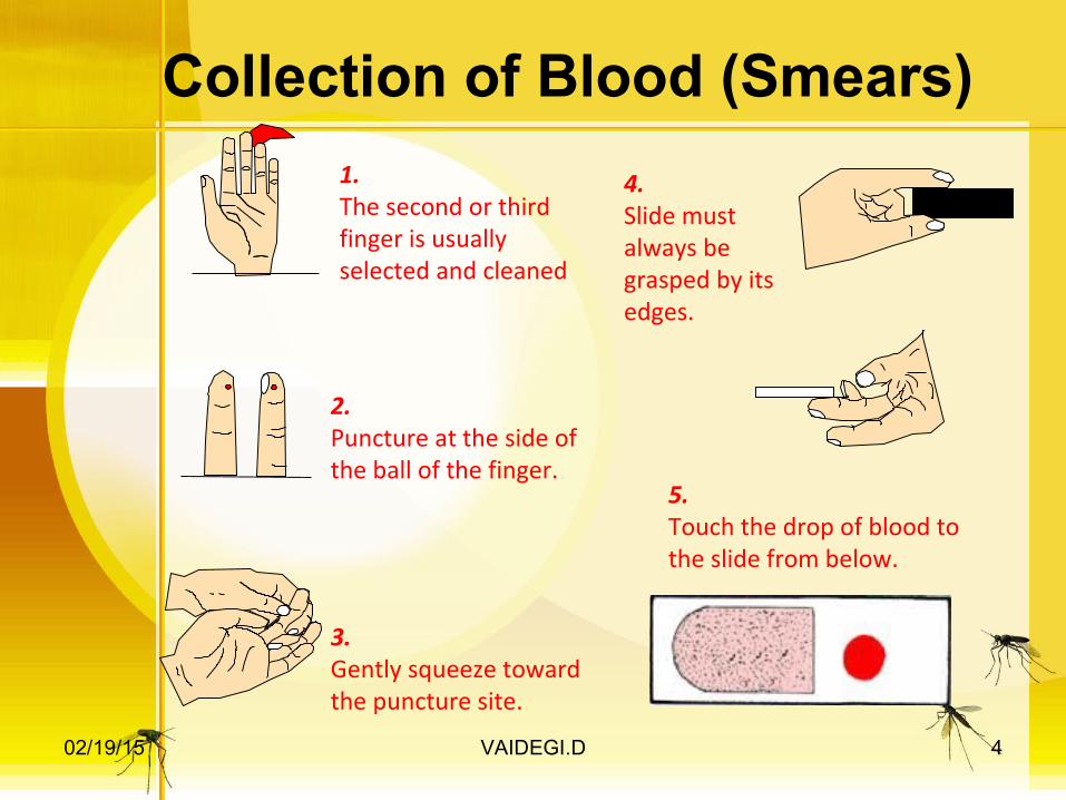
Preparing thick and thin films
2.Spread the first drop to make a 1 cm circle.
3.Touch a fresh drop of blood to the edge of another slide.
6.Wait for both to dry before fixing and staining.
5.Pull the drop of blood across the first slide in one motion.
1.Touch one drop of blood to a clean slide.
4.Carry the drop of blood to the first slide and hold at 45 degree angle.
502/19/15 VAIDEGI.D

Malaria Blood Smear
Used to determine the speciesentire thin film should be examined about 20-40 minutes for an experienced observer
Thin films:•Dry•Fix•Stain
602/19/15 VAIDEGI.D

Thick films:•Dry•Do not fix but dehemoglobinate•Stain
Staining methods:Giemsa stainLeishman's stainField’s stain
Malaria Blood Smear
702/19/15 VAIDEGI.D

Interpreting Thick and Thin FilmsTHICK FILM
•lysed RBCs•larger volume•0.25 μl blood/100 fields•more difficult to diagnose species•good screening test
THIN FILM
•fixed RBCs, single layer•smaller volume•0.005 μl blood/100 fields•good species differentiation•requires more time to read•low density infections can be missed
802/19/15 VAIDEGI.D

RING TROPHOZOITE
SCHIZONT GAMETOCYTE
BlueCytoplasm
RedChromatin
BrownPigment
Recognizing Erythrocytic Stages:Schematic Morphology
902/19/15 VAIDEGI.D

Species Differentiation on Thin Films
P. falciparum P. vivax P. ovale P. malariae
Rings
Trophozoites
Schizonts
Gametocytes
1002/19/15 VAIDEGI.D

Plasmodium falciparum
Rings: double chromatin dots; appliqué forms;multiple infections in same red cell
Gametocytes: mature (M)andimmature (I) forms (I is rarelyseen in peripheral blood)
Trophozoites: compact(rarely seen in
peripheral blood)
Schizonts: 8-24 merozoites(rarely seen in peripheral blood)
Infected erythrocytes: normal size
M I
1102/19/15 VAIDEGI.D

Plasmodium vivax
Trophozoites: ameboid; deforms the erythrocyte
Gametocytes: round-oval Schizonts: 12-24 merozoites
Rings
Infected erythrocytes: enlarged up to 2X; deformed; (Schüffner’s dots)
1202/19/15 VAIDEGI.D

Plasmodium ovaleInfected erythrocytes: moderately enlarged (11/4 X); fimbriated; oval; (Schüffner’s dots)
“malariae - like parasite in vivax - like erythrocyte”
Rings
Trophozoites: compact
Schizonts: 6-14 merozoites; dark pigment; (“rosettes”)
Gametocytes: round-oval
1302/19/15 VAIDEGI.D
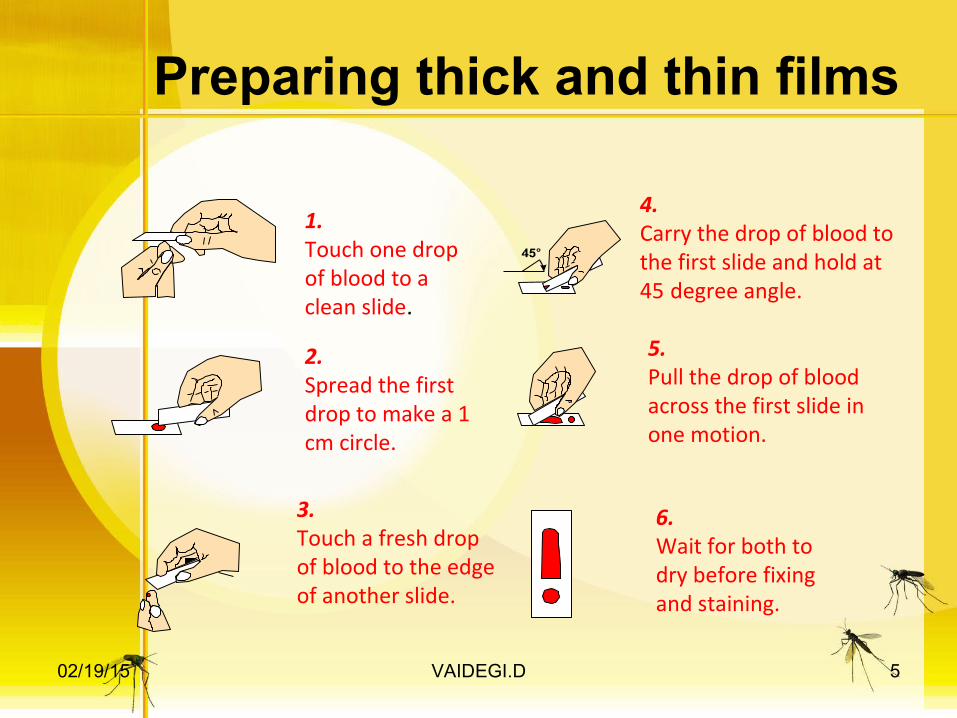
Infected erythrocytes: size normal to decreased (3/4X)
Plasmodium malariae
Trophozoite:compact
Trophozoite:typical band form
Schizont:6-12 merozoites;coarse, dark pigment
Gametocyte:round; coarse,dark pigment
1402/19/15 VAIDEGI.D

Recognizing Erythrocytic Stages
1502/19/15 VAIDEGI.D

Recognizing Erythrocytic Stages
1602/19/15 VAIDEGI.D

Calculating Parasite Density
Count the number of parasitized and nonparasitized RBCs in the same fields on thin smearCount 500-2000 RBCs
% parasitemia = # parasitized RBCs total # of RBCs
X 100
Count ≥ 200 WBCs on thick filmAssume WBC is 8000/µl (or count it)
parasites/µl = parasites counted WBC counted X WBC count/µl
1702/19/15 VAIDEGI.D

Estimating Parasite DensityAlternate Method
Count the number of asexual parasites per high-power field (HPF) on a thick blood film
1-10 parasites per 100 HPF +
11-100 parasites per 100 HPF ++
1-10 parasites per each HPF +++
> 10 parasites per each HPF ++++ 1802/19/15 VAIDEGI.D

Fluorescent MicroscopyModification of light microscopy Fluorescent dyes detect RNA and DNA that is contained in parasitesNucleic material not normally in mature RBCsKawamoto technique
Stain thin film with acridine orange (AO)Requires special equipment – fluorescent microscopeNuclei of malaria parasites floresce bright green and cytoplasm red.Staining itself is cheapSensitivities around 90%
1902/19/15 VAIDEGI.D

malaria parasites fluorescent microscope
2002/19/15 VAIDEGI.D

Quantitative Buffy Coat(QBC)Useful for screening large numbers of samplesQuick, saves timeRequires centrifuge, special stainsMalaria parasite floresce green yellow against dark red –black background.
3 main disadvantagesSpecies identification and quantification difficultHigh cost of capillaries and equipmentCan’t store capillaries for later reference
2102/19/15 VAIDEGI.D

Principle of QBC System
2202/19/15 VAIDEGI.D

Malaria SerologyAntibody detection
•Immunologic assays to detect host response •Antibodies to asexual parasites appear some days after invasion of RBCs and may persist for months•Positive test indicates past infection•Not useful for treatment decisions
•Valuable epidemiologic tool
Useful forIdentifying infective donor in transfusion-transmitted malariaInvestigating congenital malaria, esp. if mom’s smear is negativeDiagnosing, or ruling out, tropical splenomegaly syndromeRetrospective confirmation of empirically-treated non-immunes
2302/19/15 VAIDEGI.D

Malaria Antigen DetectionTarget antigens for malaria(rapid detection test) RDTCard / cassette / dipstick
HRP2HRP2 & aldolasepLDH Pf & pan pLDH Pf & PvHRP2, pLDH panHRP2, pLDH pan & pLDH Pvaldolase
"COMBO" tests
A: HRP-2 (histidine-rich protein 2) (ICT) B: pLDH (parasite lactate dehydrogenase)(Flow)C: HRP-2 (histidine-rich protein 2) (PATH)
2402/19/15 VAIDEGI.D

Steps in RDT
2502/19/15 VAIDEGI.D
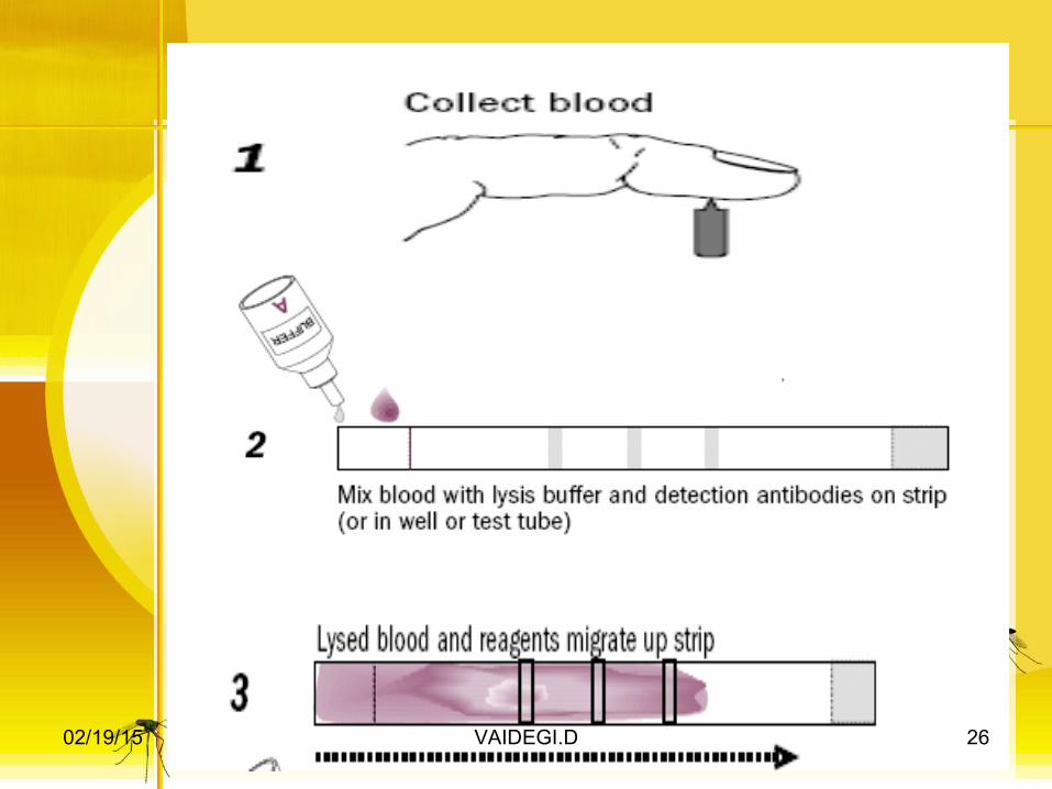
2602/19/15 VAIDEGI.D
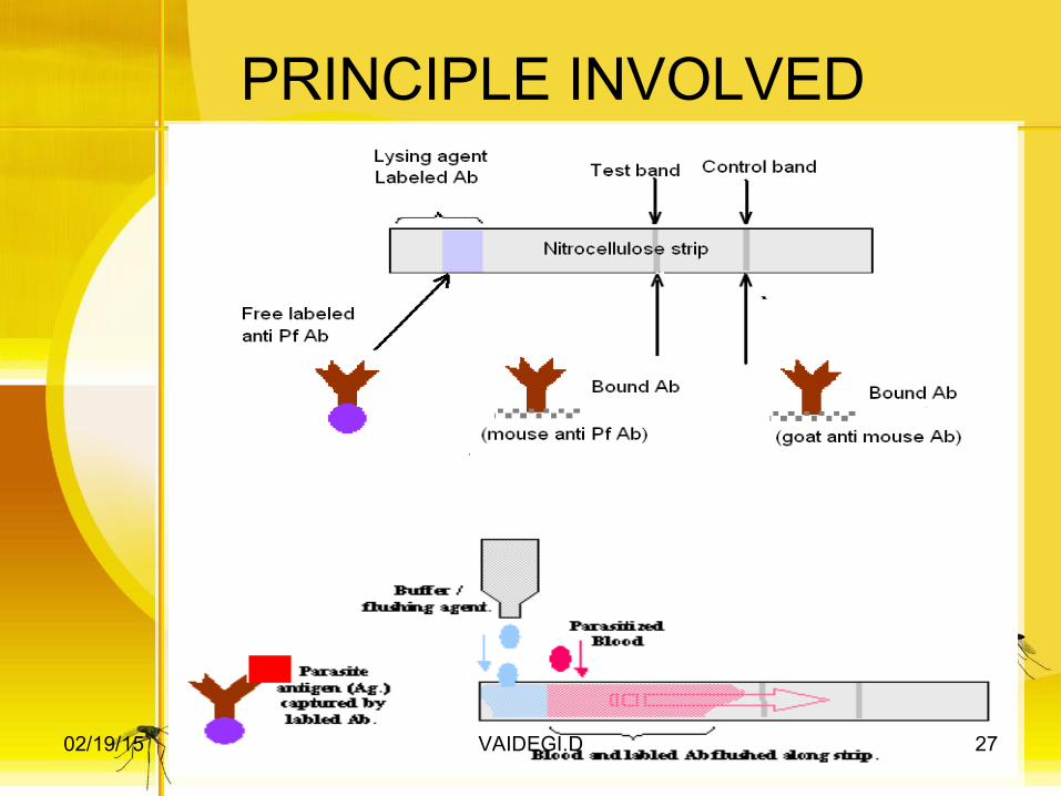
PRINCIPLE INVOLVED
2702/19/15 VAIDEGI.D

2802/19/15 VAIDEGI.D

Plastic cassette format of RDT
2902/19/15 VAIDEGI.D

Disadvantages The use of the RDT does not eliminate the need for malaria microscopy
Cannot detect mixed infectionsmay not be able to detect infections with lower parasitemiaCannot detect P. ovale and P. malariaemicroscopy is needed to quantify parasitemia
3002/19/15 VAIDEGI.D

INDIRECT FLUORESCENT ANTIBODY(IFA)
Indirect fluorescent antibody (IFA) test. The fluorescence indicates that the patient serum being tested contains antibodies that are reacting with the antigen preparation (here, Plasmodium falciparum parasites).
3102/19/15 VAIDEGI.D

ELISANot practical for routine diagnosis of acute malaria because:•Delaied development of antibody•persistence of antibodiesSerology does not detect current infection but rather measures past experienceValuable epidemiologic toolUseful for
Identifying infective donor in transfusion-transmitted malariaInvestigating congenital malaria, esp. if mom’s smear is negativeRetrospective confirmation of empirically-treated non-immunes 3202/19/15 VAIDEGI.D

Polymerase Chain Reaction (PCR)•Molecular technique to identify parasite genetic material•Uses whole blood collected in anticoagulated tube or directly onto filter paper
•Threshold of detection 5 parasites/µl •Definitive species-specific diagnosis now possible•Can identify mutations – try to correlate to drug resistance
3302/19/15 VAIDEGI.D

PCR•Parasitemia not quantifiable•May have use in epidemiologic studies•Requires specialized equipment, reagents, and training
Lane S: Molecular base pair standard (50-bp ladder). Black arrows :size of standard bands. Lane 1: P. vivax (size: 120 bp). Lane 2: P. malariae (size: 144 bp). Lane 3: P. falciparum (size: 205 bp). Lane 4: P. ovale (size: 800 bp).
3402/19/15 VAIDEGI.D

Comparison of methods for diagnosing Plasmodium infection in blood
PARAMETER MICROSCOPY PCR FLUORESCENCE Dipstick HRP-2 Dipstick pLDH, ICT-Pf/Pv
Sensitivity (parasites/micol) 50 5 50 >100 >100
Specificity All species All species P.f good, others difficult
P. falciparum P. falciparum and P.vivax good P.o and P.m only Pldh
prarasite density or parasitemia
Yes No No crude estimation
crude estimation
time for result 30-60 min 24 hr 30-60 min 20 min 20 min
skill level High High Moderate Low Low
equipment MicrocsopePCR appratus
QBC apparatus or direct fluorescence microscope
Kit only Kit only
cost /test Low High moderate/low Moderate Moderate 3502/19/15 VAIDEGI.D

3602/19/15 VAIDEGI.D


![RESEARCH Open Access Malarial parasite diversity in ... · shared between humans and several species of New World ... A difference was that Bayesian ... sporozoite [37-39], the haploid](https://img.dokumen.tips/doc/110x75/5b91b70d09d3f211298c57e8/research-open-access-malarial-parasite-diversity-in-shared-between-humans.jpg)