Embed Size (px)
Citation preview

NORMAL MICROBIAL FLORA
&
ALSO IT’S ROLE IN HUMAN DEFENSE MECHANISM
By
Shahbaz Ahmad
CONTENTS
Introduction
What is Normal Flora?
Why know about Normal Microbial Flora?
Classification of Normal Flora
Role of Normal Flora in Human Defense Mechanism
References

Introduction
In a healthy human, the internal tissues, e.g. blood, brain, muscle,
etc., are normally free of microorganisms. However, the surface
tissues, i.e., skin and mucous membranes are constantly in contact
with environmental organisms and become readily colonized by
various microbial species. The mixture of organisms regularly
found at any anatomical site is referred to as the normal flora. The
normal flora of humans consists of a few eukaryotic fungi and
protists, but bacteria are the most numerous and obvious microbial
components of the normal flora. A healthy fetus in utero is free
from microorganisms. During birth the infant in exposed to vaginal
flora. Within a few hours of birth oral and nasopharyngeal flora
develops and in a day or two resident flora of the lower intestine
appears.
In the past, the role that microorganisms played in the normal
functioning of the body was not appreciated. In the early 1900’s when Dr. Metchnikoff was credited with the discovery of the
importance of intestinal flora, other physicians felt that the colon was totally unnecessary and often surgically removed them from their patients (1) Gordon R. (1993). The colon was described as ‘a
poisonous cess-pit infecting the body with rheumatism, tuberculosis, cancer and other diseases’.
Nowadays we know that normal flora is a dynamic and complex mixture of microbes that have diverse functions including
digestion of essential nutrients, maturation of intestinal physiology, stimulation of immune system, systemic effects on blood lipids and
the inhibition of harmful bacteria.
What is Normal Flora?
The term “normal microbial flora” denotes the population of
microorganisms that inhabit the skin and mucous membranes of
healthy normal persons. It is doubtful whether a normal viral flora
exists in humans.
The skin and mucous membranes always harbor a variety of
microorganisms that can be arranged into two groups.

a. The resident flora consists of relatively fixed types of
microorganisms regularly found in a given area at a given
age; if disturbed, it promptly reestablishes itself.
b. The transient flora consists of nonpathogenic or potentially
pathogenic microorganisms that inhabit the skin or mucous
membranes for hours, days, or weeks; it is derived from the
environment, does not produce disease, and does not
establish itself permanently on the surface. (2)
http://202.120.143.134/Download/1531f207-c8b9-48d9-
8ce9-8eddfd9800e2.pdf
Although the term ‘normal flora’ is commonly used, it is really a
misnomer. Microbial flora has spatial and temporal complexity
that differs by individual, body niche, age, geographic location,
health status, diet and type of host. Even within the same
individual, the composition of the microbial flora can vary
according to changes in diet, stress, sexual behavior, medication,
hormonal changes and other host-related factors. With this caveat
in mind, the field of ‘normal flora’ can be examined for common
predominant types of flora present within body niches and shared
functional traits. (3) Lynne V. McFarland (2000)
Why know about Normal Microbial Flora?
There are many reasons to acquire knowledge of the normal human
microbial flora. Four specific examples include:
1. An understanding of the different microorganisms at
specific locations provides greater insight into the possible
infections that might result from injury to these body sites.
2. A knowledge of the normal micro-biota in an infected part
of the body gives the physician-investigator a better
perspective concerning the possible source and significance
of microorganisms isolated from an infection site.
3. A knowledge of the normal micro-biota helps the physician
investigator understand the causes and consequences of
colonization and growth by microorganisms normally
absent at a specific body site.
4. An increased awareness of the role that these normal
micro-biota play in stimulating the host immune response

can be gained. This awareness is important because the
immune system provides protection against potential
pathogens. (4) Prescott (2002)
Classification of Normal Flora
Three types of symbiotic relationships are commensalism,
mutualism, and parasitism. Within each category the association
may be either ecto-symbiotic or endo-symbiotic. In ecto-symbiosis
one organism remains outside the other. In endosymbiosis one
organism is present within the other.

Based on different parts of the human body normal flora of the
human body is classified into the following categories:-
a. Conjunctiva
b. Outer ear
c. Stomach
d. Skin
e. Urethra
f. Vagina

g. Large intestine
h. Small intestine
i. Mouth & oropharynx
j. Nose
k. Upper respiratory tract
Table Bacteria commonly found on the surfaces of the human body.
BACTERIUM Skin
Con-
junc-
tiva
Nose Pharynx Mouth Lower
GI
Ant.
ure-
thra
Vagina
Staphylococcus epidermidis (1) ++ + ++ ++ ++ + ++ ++
Staphylococcus aureus* (2) + +/- + + + ++ +/- +
Streptococcus mitis
+ ++ +/- + +
Streptococcus salivarius
++ ++
Streptococcus mutans* (3)
+ ++
Enterococcus faecalis* (4)
+/- + ++ + +
Streptococcus pneumoniae* (5)
+/- +/- + +
+/-
Streptococcus pyogenes* (6) +/- +/-
+ + +/-
+/-
Neisseria sp. (7)
+ + ++ +
+ +
Neisseria meningitidis* (8)
+ ++ +
+
Enterobacteriaceae*(Escherichia
coli) (9) +/- +/- +/- + ++ + +
Proteus sp.
+/- + + + + + +
Pseudomonas aeruginosa* (10)
+/- +/- + +/-
Haemophilus influenzae* (11)
+/- + + +
Bacteroides sp.*
++ + +/-
Bifidobacterium bifidum (12)
++
Lactobacillus sp. (13)
+ ++ ++
++
Clostridium sp.* (14)
+/- ++
Clostridium tetani (15)
+/-
Corynebacteria (16) ++ + ++ + + + + +
Mycobacteria +
+/- +/-
+ +
Actinomycetes
+ +
Spirochetes
+ ++ ++
Mycoplasmas
+ + + +/- +
++ = nearly 100 percent + = common (about 25 percent) +/- = rare (less than
5%) * = potential pathogen

Table Notes
(1) The staphylococci and corynebacteria occur at every site listed.Staphylococcus epidermidis is highly adapted to the diverse
environments of its human host. S. aureus is a potential pathogen. It is a leading cause of bacterial disease in humans. It can be transmitted from the nasal membranes of an asymptomatic carrier
to a susceptible host.
S. epidermidis. Scanning EM. CDC.
(2) Many of the normal flora are either pathogens or opportunistic pathogens, The asterisks indicate members of the normal flora a that may be considered major pathogens of humans.
S. aureus. Gram stain.
(3) Streptococcus mutans is the primary bacterium involved in plaque formation and initiation of dental caries. Viewed as an opportunistic infection, dental disease is one of the most prevalent
and costly infectious diseases in the United States.

Streptococcus mutans. Gram stain. CDC
(4) Enterococcus faecalis was formerly classified as Streptococcus faecalis.The bacterium is such a regular a component of the
intestinal flora, that many European countries use it as the standard indicator of fecal pollution, in the same way we use E. coli in the
U.S. In recent years, Enterococcus faecalis has emerged as a significant, antibiotic-resistant, nosocomial pathogen.
Vancomycin Resistant Enterococcus faecalis. Scanning E.M.
CDC
(5) Streptococcus pneumoniae is present in the upper respiratory tract of about half the population. If it invades the lower respiratory
tract it can cause pneumonia. Streptococcus pneumoniae causes 95 percent of all bacterial pneumonia.

Streptococcus pneumoniae. Direct fluorescent antibody stain.
CDC.
(6) Streptococcus pyogenes refers to the Group A, Beta-hemolytic
streptococci. Streptococci cause tonsillitis (strep throat), pneumonia, endocarditis. Some streptococcal diseases can lead to rheumatic fever or nephritis which can damage the heart and
kidney.
Streptococcus pyogenes. Gram stain.
(7) Neisseria and other Gram-negative cocci are frequent
inhabitants of the upper respiratory tract, mainly the pharynx. Neisseria meningitidis, an important cause of bacterial meningitis, can colonize as well, until the host can develop active
immunity against the pathogen.

Neisseria meningitidis. Gram stain.
(8) While E. coli is a consistent resident of the small intestine, many other enteric bacteria may reside here as well,
including Klebsiella, Enterobacter andCitrobacter. Some strains of E. coli are pathogens that cause intestinal infections, urinary
tract infections and neonatal meningitis.
E. coli. Scanning E.M. Shirley Owens. Center for Electron
Optics. Michigan State University.
(9) Pseudomonas aeruginosa is the quintessential opportunistic pathogen of humans that can invade virtually any tissue. It is a leading cause of hospital-acquired (nosocomial) Gram-negative
infections, but its source is often exogenous (from outside the host).
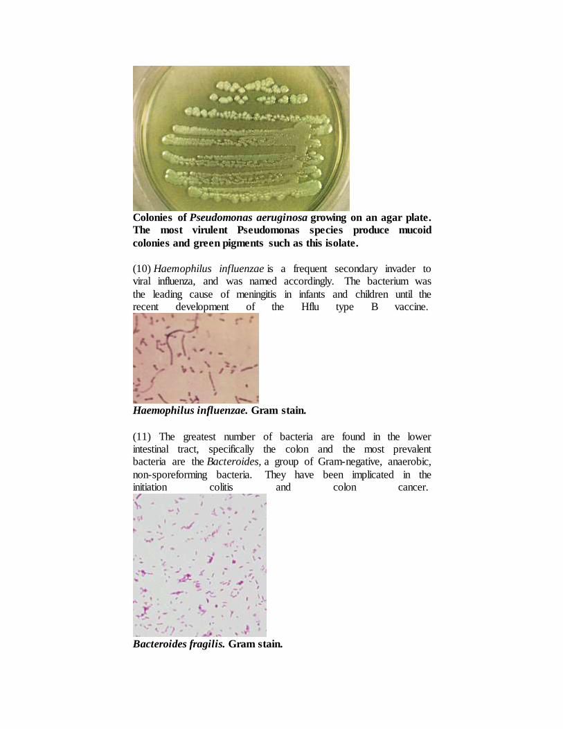
Colonies of Pseudomonas aeruginosa growing on an agar plate.
The most virulent Pseudomonas species produce mucoid
colonies and green pigments such as this isolate.
(10) Haemophilus influenzae is a frequent secondary invader to viral influenza, and was named accordingly. The bacterium was
the leading cause of meningitis in infants and children until the recent development of the Hflu type B vaccine.
Haemophilus influenzae. Gram stain.
(11) The greatest number of bacteria are found in the lower intestinal tract, specifically the colon and the most prevalent bacteria are the Bacteroides, a group of Gram-negative, anaerobic,
non-sporeforming bacteria. They have been implicated in the initiation colitis and colon cancer.
Bacteroides fragilis. Gram stain.

(12) Bifidobacteria are Gram-positive, non-sporeforming, lactic acid bacteria. They have been described as "friendly" bacteria in
the intestine of humans.Bifidobacterium bifidum is the predominant bacterial species in the intestine of breast-fed infants,
where it presumably prevents colonization by potential pathogens. These bacteria are sometimes used in the manufacture of yogurts and are frequently incorporated into probiotics.
Bifidobacterium bifidum. Gram stain
(13) Lactobacilli in the oral cavity probably contribute to acid formation that leads to dental caries. Lactobacillus
acidophilus colonizes the vaginal epithelium during child-bearing years and establishes the low pH that inhibits the growth of pathogens.
Lactobacillus species and a vaginal squaemous epithelial cell.
CDC
(14) There are numerous species of Clostridium that colonize the
bowel.Clostridium perfringens is commonly isolated from feces. Clostridium difficilemay colonize the bowel and cause
"antibiotic-induced diarrhea" or pseudomembranous colitis.

Clostridium perfringens. Gram stain.
(15) Clostridium tetani is included in the table as an example of a bacterium that is "transiently associated" with humans as a
component of the normal flora. The bacterium can be isolated from feces in 0 - 25 percent of the population. The endospores are
probably ingested with food and water, and the bacterium does not colonize the intestine.
Clostridium tetani. Gram stain.
(16) The corynebacteria, and certain related propionic acid bacteria, are consistent skin flora. Some have been implicated as a cause of acne.Corynebacterium diphtheriae, the agent of
diphtheria, was considered a member of the normal flora before the widespread use of the diphtheria toxoid, which is used to
immunize against the disease.

Corynebacterium diphtheriae. No longer a part of the normal
flora.
(5) http://textbookofbacteriology.net/normalflora.html
Role of Normal Flora in Human Defense Mechanism
The presence of Normal Flora is not essential to life, because
“germ-free” animals can be reared in the complete absence of a
normal microbial flora. Yet the resident flora of certain areas plays
a definite role in maintaining health and normal function. Members
of the resident flora in the intestinal tract synthesize vitamin K and
aid in the absorption of nutrients. On mucous membranes and skin,
the resident flora may prevent colonization by pathogens and
possible disease through “bacterial interference.” The mechanism
of bacterial interference is not clear. It may involve competition for
receptors or binding sites on host cells, competition for nutrients,
mutual inhibition by metabolic or toxic products, mutual inhibition
by antibiotic materials or bacteriocins, or other mechanisms.
Suppression of the normal flora clearly creates a partial local void
that tends to be filled by organisms from the environment or from
other parts of the body. Such organisms behave as opportunists and
may become pathogens. (2)
http://202.120.143.134/Download/1531f207-c8b9-48d9-8ce9-
8eddfd9800e2.pdf

a. Colonization resistance
Colonization resistance is the first line of defense against
invasion by exogenous, pathogenic organisms or
indigenous opportunistic organisms and the normal flora is
responsible for this formidable task. Even though the focus
here is the intestinal tract, it should be remembered that
colonization resistance plays an important role at other
body sites (oral, skin, vagina, etc.). Colonization resistance
is a dynamic phenomenon that may differ dramatically by
microbial species, type of host, diet and other host factors.
Colonization resistance has been found to be an extremely
effective natural barrier against such pathogens as C.
difficile, Salmonella, Shigella, Pseudomonas, pathogenic E.
coli strains, Candida albicans and others.
b. Production of Inhibitory End Products
Normal flora may also produce other metabolic end
products that are inhibitory to other microbes. Most notable
is hydrogen peroxide (H2O2) produced under anaerobic
conditions by several strains of normal flora. The presence
of H2O2 results in peroxidation of lipid membranes,
increased bacterial membrane permeability, destruction of
bacterial nuclear acids in bacterial strains that do not
possess catalase. The vaginal tract is usually predominantly
colonized with lactobacilli and H2O2 producing
Lactobacillus strains have been found in 75%of vagina
samples from healthy women. Vaginal colonization with
Lactobacillus strains that produce H2O2 has been shown to
be protective of infections caused by Chlamydia
trachomatis, Gardnerella vaginalis, Ureaplasma
urealyticum and the development of bacterial vaginosis. In
women with bacterial vaginosis, these strains of H2O2
producing lactobacilli are absent and, instead, high
concentrations of Gardnerella vaginalis and anaerobes are
present.

c. Production of Low pH
Another protective mechanism is the production of a low
pH environment, which may be inhibitory for certain
pathogens. The production of acids as an end product of
carbohydrate metabolism is common in many species of the
normal flora and is inhibitory against Gram-positive and
Gram-negative bacteria. Several pathogens, including
Staphylococcus aureus, Salmonella, E. coli, and Bacillus
cereus, are inhibited by acids produced by normal flora
such as lactobacilli and bifidobacteria. A common end
product of microbial fermentation is short chain fatty acids
(SCFA). The presence of these SCFA have been shown to
be inhibitory to nonindigenous bacteria. Rolfe showed in a
hamster model of C. difficile disease that SCFA levels
inhibited C. difficile growth. As newborn hamsters age,
they start to produce high levels of acetic, butyric and
propionic acids by day 16–19. Growth of C. difficile was
significantly reduced when levels of these SCFA increased.
If normal flora are disrupted, decreased levels of SCFA
result and pathogenic microbes may take advantage of this
decrease and reproduce to levels that induce disease. To
date however, the identification of which specific SCFA or
mixtures of SCFAs are responsible for the inhibition of
pathogens has not been demonstrated.
d. Competition for Nutrients
Competition for nutrients may be another mechanism for
colonization resistance. As it is extremely difficult to assess
the levels of specific nutrients in the interior of the colon,
most of the research on nutrient depletion has been done
using continuous flow culture techniques. Wilson etal.
inoculated normal flora from a mouse into a continuous
flow culture and found one or more of the flora competed
more successfully for monomeric glucose,
Nacetylglucosamine and sialic acid, resulting in
significantly reduced levels of C. difficile. Sweeney et al.
found that even small numbers of an ingested E. coli strain
F-18 could supplant established flora, as this strain utilized
an available nutrient (gluconate) more efficiently than the
other microbes present in the system. However, continuous

flow cultures are extremely dependent on culturing and
incubation parameters and the applicability of these results
is unclear.
e. Production of Inhibitory Enzymes
Normal flora may also produce extracellular enzymes that
are inhibitory or interfere with pathogen attachment.
A yeast (Saccharomyces boulardii ) has been shown to
produce a protease that destroys toxin A and toxin B
receptor sites in rabbit ileal models for C. difficile disease.
The toxins of C. difficile act by inactivating Rho proteins
that keep the cytoskeleton of the intestinal enterocyte
intact, thereby distorting the cellular morphology leading to
fluid loss and diarrhea. Rho proteins are also involved in
yeast budding processes (reproduction), thus this yeast may
produce the protease to protect itself against soil Clostridia
that may produce similar toxins to C. difficile, but in the
human, the protease may coincidentally protect the host
against infection with C. difficile. (3) Lynne V.
McFarland (2000)
f. Stimulation of the immune system
Normal flora also induces the maturation of the gut-
associated lymphoid system (GALT). The intestinal flora
provides an array of antigenic stimulants to the GALT
cells, affecting both local and systemic levels. Gnotobiotic
mice have been shown to have fewer intraepithelial
lymphocytes, plasma cells and Peyer’s patches than mice
with intact intestinal flora. When gnotobiotic mice are
immunized, the only local immune response is secretory
IgA, however, mice with intact intestinal flora also respond
with IgM and IgG. Intestinal flora may also be involved in
the development of tolerance to antigens. Herias et al. gave
germ-free rats E. coli alone or a mixture of E. coli,
Lactobacillus acidophilus and a strain of an obligate
anaerobe (Peptostreptococcus) and observed two effects.
The peptostreptococci reduced translocation rates of E. coli
and increased serum anti-E. coli antibodies. It may be that

peptostreptococci act as an immune system primer to other
bacterial antigens, thereby leading to a decrease in
translocation. Further evidence that normal flora may act as
an immune primer was found in a study using BALB:c
mice. Pulverer et al. found that normal flora releases low
molecular weight substances that interact with MALT
(mucosa associated lymphoid tissue) and these substances
appear to be essential for an adequate immune response.
Antibiotic decontamination in a mouse model resulted in a
decreased immune response. Thus, normal flora may have
an important role as an immune system primer. (3) Lynne
V. McFarland (2000) 198
g. Antibodies production
The normal flora evokes the Antibodies production. These
Antibodies cross react with pathogens having related or
shared antigens, thus raising the immune status of the host
against the invading pathogen. (6)
http://www.nios.ac.in/media/documents/dmlt/Microbiol
ogy/Lesson-07.pdf
Although normal microbiota offers some protection from
invading pathogens, they may themselves become
pathogenic and produce disease under certain
circumstances, and then are termed opportunistic
microorganisms or pathogens. These opportunistic
microorganisms are adapted to the noninvasive mode of life
defined by the limitations of the environment in which they
are living. If removed from these environmental restrictions
and introduced into the bloodstream or tissues, disease can
result. For example, streptococci of the viridans group are
the most common resident bacteria of the upper respiratory
tract. If large numbers of them are introduced into the
bloodstream (e.g., following tooth extraction or a
tonsillectomy), they may settle on deformed or prosthetic
heart valves and cause endocarditis. (4) Prescott (2002)

References
1. Gordon R. The alarming history of medicine, New York: St.
Martin’s Press, 1993 p. 164.
2. Normal microbial flora of the human body chapter 11 p 196,
196 http://202.120.143.134/Download/1531f207-c8b9-48d9-
8ce9-8eddfd9800e2.pdf
3. Lynne V. McFarland, Microbial Ecology in Health and
Disease 2000; 12: 193–207 Normal flora: diversity and
functions pages, 193, 197, 198
4. Lansing M. Prescott, Microbiology, 5th edition, 2002, p. 699,
701
5. http://textbookofbacteriology.net/normalflora.html
6. Module microbiology notes: Normal flora of the human body
page 80
http://www.nios.ac.in/media/documents/dmlt/Microbiology/Les
son-07.pdf