Embed Size (px)
Citation preview

Journal of Pharmacy and Nutrition Sciences, 2021, 11, 54-64
54
Published by SET Publisher
Journal of Pharmacy and Nutrition Sciences
ISSN (online): 1927-5951
Effect of Electronic Cigarettes on Oral Microbial Flora
T.M. Popova1, L.S. Kryvenko1, O.V. Tishchenko1, O.A. Nakonechna1, L.V. Podrigalo2, T.D. Nessonova3 and V.V. Gargin1,*
1Kharkiv National Medical University, Kharkiv, Ukraine
2Kharkiv State Academy of Physical Culture, Kharkiv, Ukraine
3Kharkiv Medical Academy of Postgraduate Education, Kharkiv, Ukraine
Article Info: Keywords: Electronic cigarette, microbiota, commensals, opportunistic microorganisms, experiment, oral mucosa. Timeline: Received: May 28, 2021 Accepted: July 17, 2021 Published: August 13, 2021 Citation: Popova TM, Kryvenko LS, Tishchenko OV, Nakonechna OA, Podrigalo LV, Nessonova TD, Gargin VV. Effect of Electronic Cigarettes on Oral Microbial Flora. J Pharm Nutr Sci 2021; 11(1): 54-64.
Abstract: Background: Despite well-documented adverse effects of tobacco consumption, cigarettes use is still rising and part of this increase is related to the popularization of alternative electronic nicotine delivery devices, such as electronic cigarettes (ECs). The aim of the performed research was to assess the effect of electronic cigarettes aerosol on the oral microbiota, using culture methods.
Methodology: 30 ten-week-old WAG rats (female 76-81 g and male 86-94 g) were randomly distributed in two groups, as follows: Group 1 – control animals (n = 10); Group 2 – EC aerosol exposed (n = 20). EC aerosol exposures were carried out by using the Boyarchuck chamber. During the study, the rat oral microbiota were collected four times: at the beginning of the experiment, on the 30th, 60th and 90th days. Microorganisms were identified using standard microbiological techniques.
Results: EC exposure to Group 2 rats resulted in a depletion of colonies commensal microbes and a greater incidence of atypical species such as Klebsiella pneumoniae, Acinetobacter lwoffii, Candida albicans compared to Group 1 on day 90. The test of independence between frequency distibution of opportunistic microbes and duration of EC exposure showed a significance for Klebsiella pneumoniae – χ2= 8.017, p=0.0456, Acinetobacter lwoffii – χ2= 36.772, p=0.0001, and Candida albicans – χ2=8.689, p=0.0337.
Conclusions: The impact of electronic cigarettes facilitated colonization of the oral cavity by opportunistic bacteria and yeast.
DOI: https://doi.org/10.29169/1927-5951.2021.11.08
*Corresponding Author Tel: +(380)990498557 E-mail: [email protected]
© 2021 Popova et al.; Licensee SET Publisher. This is an open access article licensed under the terms of the Creative Commons Attribution Non-Commercial License (http://creativecommons.org/licenses/by-nc/3.0/) which permits unrestricted, non-commercial use, distribution and reproduction in any medium, provided the work is properly cited.
Journal of Pharmacy and Nutrition Sciences, 2021, Volume 11
55
INTRODUCTION
Studies have reported about the influence of oral health on an increased risk of gastrointestinal pathology due to changes in oral microflora.
There is a long list of health risk factors associated with cigarette smoking [1]. Tobacco smoke contains a highly complex mixture of compounds that cause serious cardiovascular, respiratory, gastrointestinal tract diseases, including oral, pharyngeal, laryngeal and lung carcinomas [2, 3]. Studies have reported about the influence of oral health on an increased risk of gastrointestinal pathology due to changes of oral microflora [4, 5]. The oral microbiome refers to an ecological community of diverse bacterial species associated with human oral mucosa. Oral commensal microflora forms a bacterial biofilm preventing the attachment of harmful microorganisms to the mucosal surfaces. This microbiota participates in a wide variety of metabolic and immunological functions including differentiation of mucosa, maturation of local immune system, degradation of toxins, maintenance of the pro-inflammatory and anti-inflammatory balance, provision of “colonization resistance” and prevention of systemic diseases [6, 7]. Studies have shown that cigarette smoking contributes to the link between oral microflora dysbiosis and gastrointestinal diseases of tobacco smokers [8-10]. Despite well-documented adverse effects of tobacco consumption, cigarettes use is still rising in Ukraine and part of the increase is related to the popularization of alternative electronic nicotine delivery devices, such as electronic cigarettes (ECs). Electronic cigarette (EC) was suggested as a device free from tobacco carcinogens that may assist people in quitting smoking [11].
To our knowledge, there are studies that address the tobacco cigarettes, dokha and shisha smoking effects on oral microbiota [9, 12, 13]. However, information about the effect of EC aerosol exposure to oral bacteria remains scarce and limited to a small number of species isolated in culture media [14]. More analyses using molecular methods are still required.
The aim of this study was to assess the effect of electronic cigarettes aerosol on the oral microbiota.
2. METHODOLOGY
Experimental Design
Subjects
Thirty ten-week-old Wistar Albino Glaxo (WAG) rats (female 76-81g and male 86-94g) were obtained from
the Vivarium of National Medical University of Kharkiv. They were kept at a controlled temperature (21 ± 20C) and relative humidity (50-70%), at 12-12 hr light-dark cycle and free access to rodent chow and water ad libitum. All experimental procedures were carried out in accordance with the provisions of the European Convention for the Protection of Vertebrate Animals used for Experimental and other Scientific Purposes. The animals were randomly subdivided into two groups. Group 1 (control group) consisted of 10 intact animals (females=5 and males=5). Group 2 consisted of 20 animals (females=10 and males=10) exposed to EC aerosol.
EC aerosol was generated by using the Boyarchuck chamber operating in a one-pass mode with the aerosol feed controlled externally by a metering pump. This avoided the EC aerosol recirculation that may lead to the physical damage of respiratory epithelial cells. The functionality of the Boyarchuck chamber is based on aerosol and fresh air mixture generated via fresh air forced through a small orifice meeting a flow of incoming EC aerosol.
At the beginning of the experiment, rats were allowed a period of 2 weeks to adapt to the Boyarchuck chamber. For time adaption, all rats were exposed to the fresh airflow.
EC liquid consists of 50/50 propylene glycol/glycerin (by volume fraction), 6 mg/ml nicotine, ethyl maltol and sucralose.
The Boyarchuck chamber was turned on, and rats of Group 2 were exposed to EC aerosol for 15 min. To exposure, dilution air was delivered at 6 l/min and aerosol EC was added at 6 l/min. The EC aerosol was delivered at a rate of 1 l/min per port, a value close to the puffing flow rate.
Sample Collection
To analyze the rat oral cavity microbiota, samples were collected four times: at the beginning of the experiment, on the 30th, 60th and 90th days. The samples were collected from the rat oral cavity using swabs soaked in 0.9% NaCl sterile solution. Then swabs were inserted in test tubes with 0.95 ml of sterilized physiological solution.
Culture and Identification of Isolates
Each sample was subsequently diluted up to 107 and with the aid of sterile calibrated wire loop 0.002 ml was inoculated onto Petri dishes containing microbiological

Journal of Pharmacy and Nutrition Sciences, 2021, Volume 11
56
growth media. To detect the bacterial flora, the Petri dishes containing Blood agar, Mannitol Salt agar, Eosine Methylene Blue agar, MacConkey agar were incubated at 370C in a bacteriological chamber for 48 hours. Sabouraud dextrose agar was used for elucidating fungal members and incubated at 370C for 7 days. After incubation, the number of microbe colonies were registered as colony-forming units per ml (CFU/ml).
The bacterial isolates were examined microscopically by Gram staining technique and Lactophenol cotton blue mounts. Our Primary screening biochemical tests included: Coagulase test, Catalase test, Oxidase test, Urease test, IMVIC test (Indole, Methyl red, Voges-Proskauer and Citrate utilization tests) and germ tube test.
3. DATA ANALYSIS
The statistical analyses were performed using the STATISTICA software (StatSoftInc., USA, version 7.0). Friedman’s test was used to compare multiple dependent variables. The differences of variables amongst different groups were analyzed using Mann-Whitney U-test for comparison of two groups, or Kruskal-Wallis test for more than two groups comparison. Wilcoxon matched-pairs test was used to compare two dependent variables. Discrete variables were measured and frequency distribution tables were constructed. Data were presented as Median values (Me), lower and upper quartile, i.e. Me [Lq; Uq]. Statistical significance was accepted if p < 0.05.
4. RESULTS AND DISCUSSION
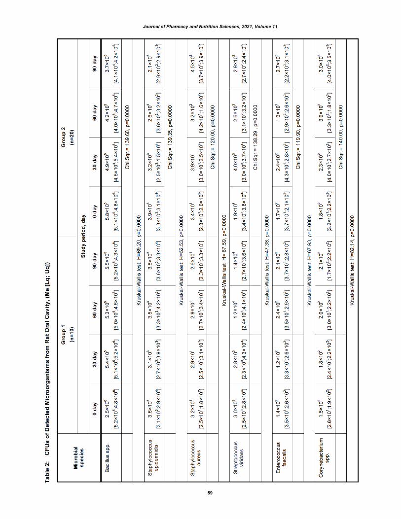
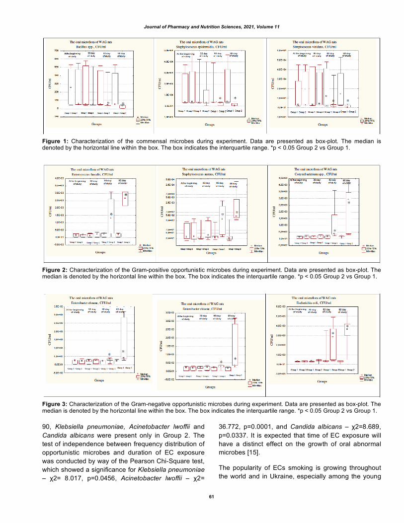
At the beginning of the experiment, in order of frequency, the detected oral Gram-positive aerobic bacteria were of the following strains: Bacillus spp., Staphylococcus spp., Streptococcus spp., Enterococcus spp., Corynebacterium spp. and Gram-negative bacteria: Enterobacter spp., Escherichia spp. Table 1 presents the distribution of bacterial species isolated from the oral cavity of WAG rats. Gram-positive and Gram-negative bacteria of all isolates comprised 89.2% and 10.8% in Group 1 and 88.5% and 11.5% in Group 2, respectively. A predominance of Gram-positive cocci and bacilli on the surfaces of the tongue and gums were detected in all WAG rats, at the beginning of the experiment. There was no statistically significant difference between Group 1 and Group 2.
During the entire observation period, no significant changes in the species composition of the microflora
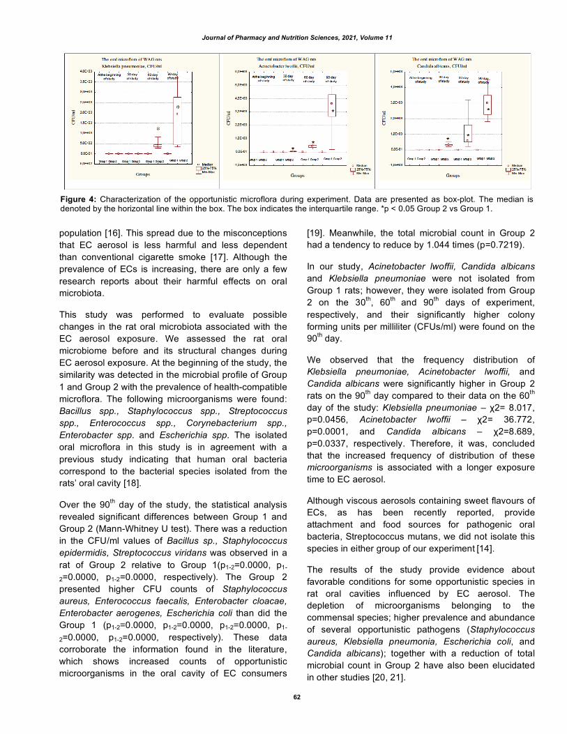
were noted in Group 1 rats (Table 2). On the 30th day of EC exposure, there was inhibition in the following commensal microbes: Bacillus sp., Staphylococcus epidermidis, Streptococcus viridans (Figure 1). At the same time, the number of colonies of the following opportunistic bacteria was increased: Staphylococcus aureus, Enterococcus faecalis, Corynebacterium spp. (Figure 2), Enterobacter cloacae, Enterobacter aerogenes, Escherichia coli (Figure 3) and Acinetobacter lwoffii, Klebsiella pneumoniae and Candida albicans appeared, which are not typical for the mouth in rats (Figure 4). On the 90th day, ECs exposure resulted in an increase in the colonies of Acinetobacter lwoffii, Klebsiella pneumoniae, Enterobacter aerogenes, Staphylococcus aureus and Candida albicans.
A test of dependence between an increasing count of isolated microorganisms and aerosol EC exposure time was conducted by way of the Friedman ANOVA Chi-Square, which showed Chi Sqr. =120.00, p =0.0000 for Staphylococcus aureus, Chi Sqr. =119.90 p =0.0000 – Enterococcus faecalis, Chi Sqr. =136.68, p =0.0000 – Enterobacter aerogenes, Chi Sqr. = 139.64, p = 0.0000 – Acinetobacter lwoffii, Chi Sqr. = 139.54, p = 0.0000 – Candida albicans. There was a statistically significant relationship between the count of CFU/ml Klebsiella pneumoniae and duration of EC aerosol exposure Z=6.16, p=0.0000 (Wilcoxon test). This suggests an association between EC smoking and an increase in the number of opportunistic microbes. On the contrary, commensal flora exhibited a significant depletion of Bacillus spp., Staphylococcus epidermidis and Streptococcus viridians during EC exposure to the Bacillus spp., Staphylococcus epidermidis, Streptococcus viridans (Friedman ANOVA Chi-Square test p =0.0000).
On the ninetieth day, the relative abundance of the oral microbiome differed between Group 1 and Group 2 (Table 2). The five most abundant genus persisted were Bacillus spp., Staphylococcus spp., Streptococcus spp., Enterococcus spp., Corynebacterium spp., which accounted for 89.2% in Group 1. The Group 2 showed significantly elevated numbers opportunistic species: Enterobacter cloacae, Enterobacter aerogenes, Escherichia coli compared with Group 1 (H=54.59, p=0.0000, H=60.30, p=0.0000 and H=87.58, p=0.0000, respectively) (Table 2). Interestingly, Acinetobacter lwoffii, Candida albicans and Klebsiella pneumoniae, were detected firstly on the 30th and 60th day of EC exposure, respectively. On day

Journal of Pharmacy and Nutrition Sciences, 2021, Volume 11
57

Journal of Pharmacy and Nutrition Sciences, 2021, Volume 11
58
Journal of Pharmacy and Nutrition Sciences, 2021, Volume 11
59

Journal of Pharmacy and Nutrition Sciences, 2021, Volume 11
60
Journal of Pharmacy and Nutrition Sciences, 2021, Volume 11
61
Figure 1: Characterization of the commensal microbes during experiment. Data are presented as box-plot. The median is denoted by the horizontal line within the box. The box indicates the interquartile range. *p < 0.05 Group 2 vs Group 1.
Figure 2: Characterization of the Gram-positive opportunistic microbes during experiment. Data are presented as box-plot. The median is denoted by the horizontal line within the box. The box indicates the interquartile range. *p < 0.05 Group 2 vs Group 1.
Figure 3: Characterization of the Gram-negative opportunistic microbes during experiment. Data are presented as box-plot. The median is denoted by the horizontal line within the box. The box indicates the interquartile range. *p < 0.05 Group 2 vs Group 1.
90, Klebsiella pneumoniae, Acinetobacter lwoffii and Candida albicans were present only in Group 2. The test of independence between frequency distribution of opportunistic microbes and duration of EC exposure was conducted by way of the Pearson Chi-Square test, which showed a significance for Klebsiella pneumoniae – χ2= 8.017, p=0.0456, Acinetobacter lwoffii – χ2=
36.772, p=0.0001, and Candida albicans – χ2=8.689, p=0.0337. It is expected that time of EC exposure will have a distinct effect on the growth of oral abnormal microbes [15].
The popularity of ECs smoking is growing throughout the world and in Ukraine, especially among the young
Journal of Pharmacy and Nutrition Sciences, 2021, Volume 11
62
population [16]. This spread due to the misconceptions that EC aerosol is less harmful and less dependent than conventional cigarette smoke [17]. Although the prevalence of ECs is increasing, there are only a few research reports about their harmful effects on oral microbiota.
This study was performed to evaluate possible changes in the rat oral microbiota associated with the EC aerosol exposure. We assessed the rat oral microbiome before and its structural changes during EC aerosol exposure. At the beginning of the study, the similarity was detected in the microbial profile of Group 1 and Group 2 with the prevalence of health-compatible microflora. The following microorganisms were found: Bacillus spp., Staphylococcus spp., Streptococcus spp., Enterococcus spp., Corynebacterium spp., Enterobacter spp. and Escherichia spp. The isolated oral microflora in this study is in agreement with a previous study indicating that human oral bacteria correspond to the bacterial species isolated from the rats’ oral cavity [18].
Over the 90th day of the study, the statistical analysis revealed significant differences between Group 1 and Group 2 (Mann-Whitney U test). There was a reduction in the CFU/ml values of Bacillus sp., Staphylococcus epidermidis, Streptococcus viridans was observed in a rat of Group 2 relative to Group 1(p1-2=0.0000, p1-
2=0.0000, p1-2=0.0000, respectively). The Group 2 presented higher CFU counts of Staphylococcus aureus, Enterococcus faecalis, Enterobacter cloacae, Enterobacter aerogenes, Escherichia coli than did the Group 1 (p1-2=0.0000, p1-2=0.0000, p1-2=0.0000, p1-
2=0.0000, p1-2=0.0000, respectively). These data corroborate the information found in the literature, which shows increased counts of opportunistic microorganisms in the oral cavity of EC consumers
[19]. Meanwhile, the total microbial count in Group 2 had a tendency to reduce by 1.044 times (p=0.7219).
In our study, Acinetobacter lwoffii, Candida albicans and Klebsiella pneumoniae were not isolated from Group 1 rats; however, they were isolated from Group 2 on the 30th, 60th and 90th days of experiment, respectively, and their significantly higher colony forming units per milliliter (CFUs/ml) were found on the 90th day.
We observed that the frequency distribution of Klebsiella pneumoniae, Acinetobacter lwoffii, and Candida albicans were significantly higher in Group 2 rats on the 90th day compared to their data on the 60th day of the study: Klebsiella pneumoniae – χ2= 8.017, p=0.0456, Acinetobacter lwoffii – χ2= 36.772, p=0.0001, and Candida albicans – χ2=8.689, p=0.0337, respectively. Therefore, it was, concluded that the increased frequency of distribution of these microorganisms is associated with a longer exposure time to EC aerosol.
Although viscous aerosols containing sweet flavours of ECs, as has been recently reported, provide attachment and food sources for pathogenic oral bacteria, Streptococcus mutans, we did not isolate this species in either group of our experiment [14].
The results of the study provide evidence about favorable conditions for some opportunistic species in rat oral cavities influenced by EC aerosol. The depletion of microorganisms belonging to the commensal species; higher prevalence and abundance of several opportunistic pathogens (Staphylococcus aureus, Klebsiella pneumonia, Escherichia coli, and Candida albicans); together with a reduction of total microbial count in Group 2 have also been elucidated in other studies [20, 21].
Figure 4: Characterization of the opportunistic microflora during experiment. Data are presented as box-plot. The median is denoted by the horizontal line within the box. The box indicates the interquartile range. *p < 0.05 Group 2 vs Group 1.
Journal of Pharmacy and Nutrition Sciences, 2021, Volume 11
63
The symbiosis between oral microorganisms is based on mutual benefits. The commensals maintain homeostasis by not allowing opportunistic and pathogenic microorganisms to adhere to the mucosa. The opportunistic species become pathogenic only after they breach the barrier of the commensals, thus causing diseases [22].
The results of the study provide evidence about favorable conditions for some opportunistic species in rat oral cavities exposed to EC aerosol. While underlying triggers for dysbiosis in Group 2 rats are unknown, the current explanation of adverse effect EC is linked with increased production of acetaldehyde, acrolein, formaldehyde, metallic nanoparticles (copper, tin, lead, chromium, nickel, and cadmium), and reactive oxygen species in consumers. The activation of the heating element and the vaporization of EC liquids are two possible sources of release of these carbonyls, metals, and reactive oxygen species from electronic cigarettes [23-25]. Tendency to depletion of total microbial count in rats of Group 2 might be caused by ethyl maltol. In one study, researchers found that ethyl maltol destabilized the cell membrane by chelating Mg2+ or Ca2+ in a pH dependent manner [26]. Thus, it is plausible that ethyl maltol acted as a potent antimicrobial agent against microbes.
Moreover, acetaldehyde and sucralose (1,6-Dichloro-1,6-dideoxy-β-D-fructofuranosyl-4-chloro-4-deoxy-α-D-galactopyranoside) accumulation rates may alter the microbiota quantitatively. High concentrations of acetaldehyde and sucralose seem to cause an increase in aerobic Gram-positive bacteria, such as Staphylococcus aureus and Corynebacterium spp. [27].
Numerous factors influent on condition of the oral cavity such as social habits [10, 28]; condition of other human systems [29, 30] their pathological and physiological processes [31, 32] even in persons with active physical lifestyle [33, 34]; changes in different tissue [35, 36]. With an increase in ECs usage among youth, based on the literature on EC effects on oral microflora and our research findings, the issue of potential longer-term deleterious consequences of EC on the health of the young population must be raised.
The present study was limited by culture-based methods. We recognize that culture-based analytical technologies cannot reveal the diversity of the oral microbiome completely; however, the present study provides a rich insight into the adverse effects of EC on rat oral microbiome which may promote further health
risks. Despite limitations, our study provides insight into the adverse effect of EC on rat oral microbiome and the health risks.
5. CONCLUSIONS
The EC aerosol exposure had a critical influence on the oral microbiota. It resulted in a higher abundance of “misplaced” microbial species and progression toward dysbiosis.
CONTRIBUTION OF EACH AUTHOR ACCORDING TO THE KEY
A - research concept and design, B - data collection, C - data analysis and interpretation, D - article writing, E - critical review of the article, F - final approval of the article.
Popova T.M.A, B, C, Kryvenko L.S.A, E, Tishchenko O.V.B,
D, Nakonechna O.A.C, E, Podrigalo L.V.E, F, Nessonova T.D.C, Gargin V.V.A, D, F
ACKNOWLEDGEMENT
This study is a part of the scientific research work “Optimization of early diagnosis, prevention and treatment of oral tissue diseases with smoking addiction”, 0120U102057, Kharkiv National Medical University”.
CONFLICT OF INTEREST
The authors declare no conflict of interest, financial or otherwise.
REFERENCES
[1] Onor IO, Stirling DL, Williams SR, et al. Clinical Effects of Cigarette Smoking: Epidemiologic Impact and Review of Pharmacotherapy Options. Int J Environ Res Public Health 2017; 14(10): 1147. https://doi.org/10.3390/ijerph14101147
[2] Messner B, Bernhard D. Smoking and cardiovascular disease: mechanisms of endothelial dysfunction and early atherogenesis. Arterioscler Thromb Vasc Biol 2014; 34(3): 509-515. https://doi.org/10.1161/ATVBAHA.113.300156
[3] Feldman C, Anderson R. Cigarette smoking and mechanisms of susceptibility to infections of the respiratory tract and other organ systems. J Infect 2013; 67(3): 169-184. https://doi.org/10.1016/j.jinf.2013.05.004
[4] Nazaryan RS, Kryvenko LS, Gargin VV: The role of nitric oxide synthase in the modulation of the immune response in atopic disease. The New Armenian Medical Journal 2017; 11(2): 52-57.
[5] Kovach I, Kravchenko L, Khotimska Y, Nazaryan R, Gargin V: Influence of ozone therapy on oral tissue in modeling of chronic recurrent aphthous stomatitis. Georgian Med News 2017; (264): 115-119.
[6] Ludwicki JK, Góralczyk K, Struciński P, et al. Hazard quotient profiles used as a risk assessment tool for PFOS and PFOA

Journal of Pharmacy and Nutrition Sciences, 2021, Volume 11
64
serum levels in three distinctive European populations. Environ Int 2015; 74: 112-118. https://doi.org/10.1016/j.envint.2014.10.001
[7] Jia G, Zhi A, Lai PFH, et al. The oral microbiota - a mechanistic role for systemic diseases. Br Dent J 2018; 224(6): 447-455. https://doi.org/10.1038/sj.bdj.2018.217
[8] Romaniuk A, Nazaryan R, Zakut Y, Popova T, Gargin V. The impact of smoking on the morphofunctional state of periodontal tissues of young organism. Inter Collegas 2021; 8(1): 47-51. https://doi.org/10.35339/ic.8.1.47-51
[9] Podrigalo LV, Iermakov SS, Jagiello W. Metabolic and Endocrine Changes Determined in Saliva of Adolescents Engaged in Computer Gaming. Biomed Res Int 2020; 2020: 1649759. https://doi.org/10.1155/2020/1649759
[10] Nazaryan R, Kryvenko L, Zakut Y, Karnaukh O, Gargin V. Application of estimated oral health indices in adolescents with tobacco addiction. Pol Merkur Lekarski 2020; 48(287): 327-330.
[11] Gathright EC, Wu WC, Scott-Sheldon LAJ. Electronic cigarette use among heart failure patients: Findings from the Population Assessment of Tobacco and Health study (Wave 1: 2013-2014). Heart Lung 2020; 49(3): 229-232. https://doi.org/10.1016/j.hrtlng.2019.11.006
[12] Vallès Y, Inman CK, Peters BA, et al. Types of tobacco consumption and the oral microbiome in the United Arab Emirates Healthy Future (UAEHFS) Pilot Study. Sci Rep 2018; 8(1): 11327. https://doi.org/10.1038/s41598-018-29730-x
[13] Shakhatreh MAK, Khabour OF, Alzoubi KH, Masadeh MM, Hussein EI, Bshara GN. Alterations in oral microbial flora induced by waterpipe tobacco smoking. Int J Gen Med 2018; 11: 47-54. https://doi.org/10.2147/IJGM.S150553
[14] Kim SA, Smith S, Beauchamp C, et al. Cariogenic potential of sweet flavors in electronic-cigarette liquids. PLoS One 2018; 13(9): e0203717. https://doi.org/10.1371/journal.pone.0203717
[15] Belda-Ferre P, Alcaraz LD, Cabrera-Rubio R, et al. The oral metagenome in health and disease. ISME J 2012; 6(1): 46-56. https://doi.org/10.1038/ismej.2011.85
[16] Rahman MA, Hann N, Wilson A, Mnatzaganian G, Worrall-Carter L. E-cigarettes and smoking cessation: evidence from a systematic review and meta-analysis. PLoS One 2015; 10(3): e0122544. https://doi.org/10.1371/journal.pone.0122544
[17] Cervellati F, Muresan XM, Sticozzi C, et al. Comparative effects between electronic and cigarette smoke in human keratinocytes and epithelial lung cells. Toxicol In Vitro 2014; 28(5): 999-1005. https://doi.org/10.1016/j.tiv.2014.04.012
[18] Duarte PM, Tezolin KR, Figueiredo LC, Feres M, Bastos MF. Microbial profile of ligature-induced periodontitis in rats. Arch Oral Biol 2010; 55(2): 142-147. https://doi.org/10.1016/j.archoralbio.2009.10.006
[19] Alanazi H, Semlali A, Chmielewski W, Rouabhia M. E-Cigarettes Increase Candida albicans Growth and Modulate its Interaction with Gingival Epithelial Cells. Int J Environ Res Public Health 2019; 16(2): 294. https://doi.org/10.3390/ijerph16020294
[20] Stewart CJ, Auchtung TA, Ajami NJ, et al. Correction: Effects of tobacco smoke and electronic cigarette vapor exposure on the oral and gut microbiota in humans: a pilot study. Peer J 2018; 6: e4693/correction-1. https://doi.org/10.7717/peerj.4693/correction-1
[21] Yu G, Phillips S, Gail MH, et al. The effect of cigarette smoking on the oral and nasal microbiota. Microbiome 2017; 5(1): 3. https://doi.org/10.1186/s40168-016-0226-6
[22] Avila M, Ojcius DM, Yilmaz O. The oral microbiota: living with a permanent guest. DNA Cell Biol 2009; 28(8): 405-411. https://doi.org/10.1089/dna.2009.0874
[23] Ballbè M, Martínez-Sánchez JM, Sureda X, et al. Cigarettes vs. e-cigarettes: Passive exposure at home measured by means of airborne marker and biomarkers. Environ Res 2014; 135: 76-80. https://doi.org/10.1016/j.envres.2014.09.005
[24] Blair SL, Epstein SA, Nizkorodov SA, Staimer N. A Real-Time Fast-Flow Tube Study of VOC and Particulate Emissions from Electronic, Potentially Reduced-Harm, Conventional, and Reference Cigarettes. Aerosol Sci Technol 2015; 49(9): 816-827. https://doi.org/10.1080/02786826.2015.1076156
[25] Hess CA, Olmedo P, Navas-Acien A, Goessler W, Cohen JE, Rule AM. E-cigarettes as a source of toxic and potentially carcinogenic metals. Environ Res 2017; 152: 221-225. https://doi.org/10.1016/j.envres.2016.09.026
[26] Schved F, Pierson MD, Juven BJ. Sensitization of Escherichia coli to nisin by maltol and ethyl maltol. Lett Appl Microbiol 1996; 22(3): 189-191. https://doi.org/10.1111/j.1472-765X.1996.tb01139.x
[27] Homann N, Tillonen J, Meurman JH, et al. Increased salivary acetaldehyde levels in heavy drinkers and smokers: a microbiological approach to oral cavity cancer. Carcinogenesis 2000; 21(4): 663-668. https://doi.org/10.1093/carcin/21.4.663
[28] Denga O, Pyndus T, Gargin V, Schneider S. Influence of metabolic syndrome on condition of microcirculatory bed of oral cavity. Georgian Med News 2017; (273): 99-104.
[29] Muriyati, Arimbi, Asnidar, Safruddin, Thahir AIA. The association between adiponectin gene polymorphism and waist circumference changes in obese/overweight adults after aerobic exercise and diet treatment. J Pharm Nutr Sci 2019; 9(5): 247-250.
[30] Ghadam OS, Fararouei M, Shahraki M, Sohrabi Z. Prevalence of iron deficiency anemia among adolescent girls in the city of Saravan. J Pharm Nutr Sci 2020; 10(2): 68-73.
[31] Al-Saadi LS, Ali A, Waly MI, Al-Zuhaibi KM. Impact of dietary patterns and nutritional status on the academic performance of Omani school students. J Pharm Nutr Sci 2020; 10(3): 74-87. https://doi.org/10.29169/1927-5951.2020.10.03.1
[32] Rukmaini, Lipoeto NI, Masrul, Effendi N. The effect of health education and mobile control application program on anemia among pregnant women. J Pharm Nutr Sci 2019; 9(6): 1-9. https://doi.org/10.29169/1927-5951.2019.09.06.1
[33] Volodchenko OA, Podrigalo LV, Iermakov SS, Żychowska MT, Jagiełło W. The Usefulness of Performing Biochemical Tests in the Saliva of Kickboxing Athletes in the Dynamic of Training. Biomed Res Int 2019; 2019: 2014347. https://doi.org/10.1155/2019/2014347
[34] Schenström A., Rönnberg S., Bodlund O. Mindfulness-Based Cognitive Attitude Training for Primary Care Staff: A Pilot Study. Complementary Health Practice Review 2006; 11: 3: 144-152. https://doi.org/10.1177/1533210106297033
[35] Lyndin M, Gluschenko N, Sikora V, et al. Morphofunctional features of articular cartilage structure. Folia Med Cracov 2019; 59(3): 81-93.
[36] Jiao Y, Hasegawa M, Inohara N. Emerging roles of immunostimulatory oral bacteria in periodontitis development. Trends Microbiol 2014; 22(3): 157-163. https://doi.org/10.1016/j.tim.2013.12.005