Embed Size (px)
Citation preview

By
Dr: Samiaa Hamdy Sadek
Lecturer of chest diseases
Assessment of small airways functions

DefinitionThe small airways are defined as those less than
2 mm in diameter.
They are a major site of pathology in many lung
diseases, not least chronic obstructive pulmonary
disease (COPD) and asthma.
They have proven relatively difficult to study due
to their relative inaccessibility to biopsy and their
small size which makes their imaging difficult.

Large V/S Small airways
Large airways Small airways
Have cartilagnous support
and mucous gland
Less cross sectional area
Turbulent flow
Resistance affected by gas
density
No surfactant lining over
the epithelium
Lack catilaginous support
and mucous gland
Larger cross sectional
area
Laminar flow
Gas density has no effect
on resistance
Surfactant lining and
hence low surface tension

Airway anatomy The first 15 generations of
airways are called the conducting airways and take no part in gas exchange, they constitute the anatomical dead space
Beyond this region lie the respiratory bronchioles which have occasional alveoli budding from them. These continue to divide until they reach the alveolar sacs with a total surface area of 70-80 m2.
The small airways occur from approximately generation 8 and include a portion of the conducting airways as well as all the acinar airways.
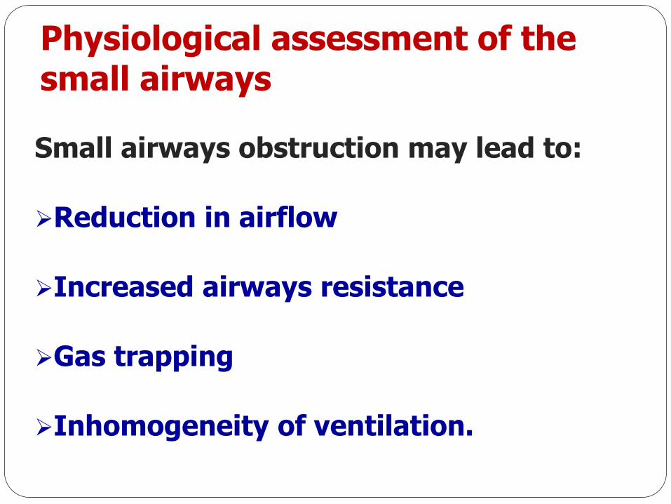
Physiological assessment of the small airways
Small airways obstruction may lead to:
Reduction in airflow
Increased airways resistance
Gas trapping
Inhomogeneity of ventilation.
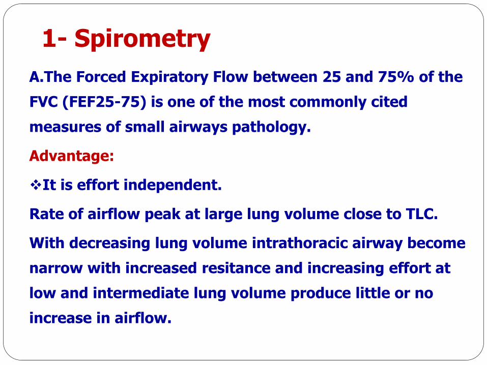
1- Spirometry
A.The Forced Expiratory Flow between 25 and 75% of the
FVC (FEF25-75) is one of the most commonly cited
measures of small airways pathology.
Advantage:
It is effort independent.
Rate of airflow peak at large lung volume close to TLC.
With decreasing lung volume intrathoracic airway become
narrow with increased resitance and increasing effort at
low and intermediate lung volume produce little or no
increase in airflow.

1- Spirometry
Disadvantages:
Poor reproducibility as it is dependent on the FVC
and therefore changes in FVC will affect the portion
of the flow-volume curve examined.
FEF25-75 is frequently normal if the FEV1/FVC
ratio is≥ 75%
There is poor correlation with other markers of
small airways disease such as gas trapping and
histological evidence of small airways inflammation
1- Spirometry
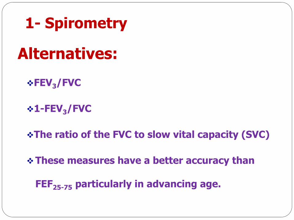
Alternatives:
FEV3/FVC
1-FEV3/FVC
The ratio of the FVC to slow vital capacity (SVC)
These measures have a better accuracy than
FEF25-75 particularly in advancing age.

2-Plethysmography
The residual volume (RV) is an important measure
of small airways dysfunction and may be raised
before the onset of abnormal spirometry in asthma
and correlates with the degree of inflammatory
changes in small airways in COPD
The residual volume/total lung capacity (RV/TLC)
ratio may be a more useful marker of gas trapping

3- Dynamic Compliance
Definition:
Change in lung volume during airflow producedbya given change in transpulmonary pressure.
Normally ratio of dynamic complianc (Cdyn)/static compliance(Cst)>0.8 even with high breathing frequeny >60 breath/minute.
In presence of uneven ventilation and small airway diseases marked decrease in Cdyn observed with increasing breathing frequency.
Physiologic principle:
In presence of uneven ventilation there are two types of alveoli.

3- Dynamic Compliance
Fast alveoli (low resistance) filled rapidly with
air, and slow alveoli(high resistance) need long
time for filling.
With increasing respiratory frequency there is
no sufficient time for filling of slow alveoli so
Cdyn decreased compared to Cst.

4- Inert gas washout
The most commonly employed technique is the
single breath nitrogen washout (SBNW) and
more recently the multiple breath nitrogen
washout (MBNW).
Other gases may be used including helium and
sulphur hexafluoride (SF6) whose physical
properties determine gas flow within the lung.

A- Single breath nitrogen washout(closing volume)
The SBNW is performed by inhaling 100%
oxygen from RV to TLC followed by a SVC
exhalation.
The exhaled volume and nitrogen concentration
is measured and the resulting trace can be
broken down into four distinct phases.

A- Single breath nitrogen washout(closing volume)
In phase I, the nitrogen concentration is close to 0% as this represents anatomical dead space. During phase II, there is a sharp rise in the expired nitrogen concentration as dead space gas mixes with resident alveolar gas. Phase III represents alveolar gas and the expired nitrogen concentration begins to plateau. Finally, in phase IV, there is a steep rise in expired N2 concentration as the most poorly ventilated areas (with little O2 mixing) empty. This is also the point at which the small airways start to and is known as the closing volume (CV).

A- Single breath nitrogen washout(closing volume)
Normally, small airways closure occurs
close to RV.
However, small airways disease may cause
premature airway collapse resulting in an
increased CV and gas trapping.
CV may be expressed as a ratio of VC and
should not exceed 10- 25% of FVC.

B-Multiple breath nitrogen washout(MBNW)
The patient inhales 100% O2 from FRC.
Follwed by exhalation with fixed tidal
volume and respiratory rate to wash out the
resident nitrogen from the lungs.
The test continues until the exhaled
nitrogen is less than 1/40th of the original
concentration (approximately 2%) for three
successive breaths.

B-Multiple breath nitrogen washout(MBNW)
This technique allows for measurement of the efficiency
of gas mixing in the whole lung through the lung clearance
index (LCI).
LCI is defined as the number of lung turnovers (FRC
equivalents) required to wash out the tracer gas to 1/40th
of the original concentration. This is calculated by
measuring the cumulative expired volume (CEV) required
to washout the resident nitrogen and dividing it by FRC:
LCI=CEV/FRC

Multiple Breath N2 Washout

Lung clearance index(LCI)
LCI =
Cumulative expired volume(CEV)/FRC
LCI = 6.25 + 0.02 x Age
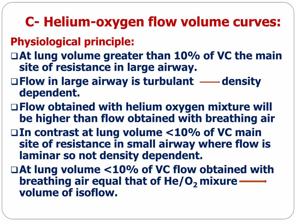
C- Helium-oxygen flow volume curves:
Physiological principle:
At lung volume greater than 10% of VC the main site of resistance in large airway.
Flow in large airway is turbulant density dependent.
Flow obtained with helium oxygen mixture will be higher than flow obtained with breathing air
In contrast at lung volume <10% of VC main site of resistance in small airway where flow is laminar so not density dependent.
At lung volume <10% of VC flow obtained with breathing air equal that of He/O2 mixurevolume of isoflow.

C- Helium-oxygen flow volume curves:
In healthy subject volume of isoflow at 10% of VC.
In small airway diseases volume of isoflow > 10% of VC.
VEmax50% He-air=VEmax50% He-air VEmax50% air/
VEmax50% air

5-Impulse oscilometry IOS
IOS is a form of forced oscillation technique where
small external pressure signals superimposed on the
natural breathing to determine a subject’s breathing
mechanics.
FOT measures respiratory impedance to this applied
forced pressure oscillations produced by a loud
speaker


Parameters measured by IOS:
The respiratory Impedance (Z) measured by IOS
is a complex quantity and consists of a real part
called respiratory Resistance (R) and an imaginary
part called respiratory Reactance (X).
Z ( f )= R ( f ) + jX ( f )
IOS also includes hallmarks such as Resonant
Frequency (Fres) and Reactance Area (AX) also
known as the “Goldman Triangle”.

a. Respiratory Resistance (R)
Resistance (R), which includes the resistance of the
proximal (central) and distal (peripheral) airways as
well as lung tissue and chest wall while these latter
resistances are usually negligible.
In healthy adult subjects, R is nearly independent
of oscillation frequency.
When an airway obstruction occurs, either central
or peripheral, R5 (Resistance at 5 Hz) is increased
above normal values.

a. Respiratory Resistance (R)
Central airway obstruction elevates R evenly
independent of oscillation frequency.
Peripheral airways obstruction is highest at
low oscillation frequencies and falls with
increasing frequency; this is called the negative
frequency-dependence of Resistance (fdR).
Resistance is measured in cmH2O/L/s or
KPa/L/s

b. Respiratory Reactance (X)
Reactance (X), includes the Inertance (I) and the
capacitance (C)
Inertance is the mass-inertive forces of the moving
air column. Inertia is the tendency of a body to
preserve its state of rest or uniform motion unless
acted upon by an external force
Capacitance (C) is the elastic properties
(compliance) of lung periphery.
Reactance is measured in cmH2O/L/s or KPa/L/s

c. Resonant Frequency
The Resonant Frequency (Fres) is the point at
which C and I are equal, therefore reactance is
zero and is measured in Hertz (1/s)
It is a suitable marker to separate low
frequency from high frequency impedance.
Respiratory system abnormalities cause Fres
value to be increased.

d. Reactance Area (AX)
The Reactance Area (AX), – the “Goldman
Triangle” - was introduced by Michael
Goldman.
AX is the integrated low frequency
respiratory reactance magnitude between 5 Hz
and Fres, and is measured in cmH2O/L or
KPa/L.
AX is a useful and sensitive index of
peripheral airway function.


Healthy adult Rrs(f) curve is almost rectilinear, frequency dependence of resistance is absent (fdr=0), which is confirmed by the following data: Rrs5=Rrs20=0.26 kPa/(L/s).

Rrs(f) and Χrs(f) curves of a 79 year-old COPD (GOLD stage III) male patient Significant increase of Rrs5 (Rrs5=0.51 kPa/(L/s)) as well as fdr,increase of ΑΧ as well as the impressively increased resonant frequency (fres=25.8 Ηz).

In bronchial asthma hyper-reactivity and inflammatory infiltration of the wall of central and peripheral airways alike in bronchial asthma, significant changes are observed also in high frequency impedance parameters and thus fdr is less impressive

A 52 year-old patient with Idiopathic Pulmonary Fibrosis Significant increase of Rrs5, whereas high frequency resistance values remain within normal significant fdr=95%. Impressive increase of Xrs5, fres, and ΑΧ.

Variable intrathoracic upper airway obstruction R increased
uniformely through all frequencies , while X, fres, and ΑΧ are
within normal.
Variable extrathoracic obstruction gives the same pattern as
peripheral airway obstruction.

Rrs may be considered within normal limits if Rrsat 5 Hz (Rrs5) is within¡1.64 SD of the predicted value. Rrs5 values between 1.64 and 2 SD above predicted may be considered minor,>2 SD moderate and >4 SD above predicted severe obstruction.
Xrs5 characterises the lung periphery, but is nonspecific as to the type of limitation. It is more negative in restrictive and obstructive lung diseases.
In normal adults, fres is usually 7–12 Hz.

Other tests
6-Exhaled nitric oxide
7-Imaging of the small airways
a) High resolution CT
b) Hyperpolarised helium magnetic resonance imaging(3He MRI)
c) Two-dimensional gamma(2-D gamma) scintigraphy
d) Single photon emission computed tomography(SPECT)
e) Positron emission tomography