Embed Size (px)
Citation preview
SENSORY & MOTOR SENSORY & MOTOR EVALUATION IN STRABISMUSEVALUATION IN STRABISMUS
DR. DEVDUTTA NAYAKDR. DEVDUTTA NAYAK

Classification of strabismusClassification of strabismus
A.A. Pseudostrabismus (false or Pseudostrabismus (false or apparent squint).apparent squint).
B. True strabismus:B. True strabismus: 1. Latent squint (heterophoria).1. Latent squint (heterophoria).
2. Manifest squint (heterotropia):2. Manifest squint (heterotropia): - non- paralytic (concomitant).- non- paralytic (concomitant). - paralytic (non- concomitant).- paralytic (non- concomitant).

CLASSIFICATIONCLASSIFICATION
1. Direction of deviation:1. Direction of deviation: - convergent (esotropia)- convergent (esotropia) - divergent (exotropia) - divergent (exotropia) - hypodeviation- hypodeviation - hyperdeviation- hyperdeviation
2. Comitancy:2. Comitancy: - comitant or non paralytic- comitant or non paralytic - incomitant or paralytic- incomitant or paralytic

3. Constancy:3. Constancy: - intermittent- intermittent - constant- constant
4. Onset:4. Onset: - childhood (congenital)- childhood (congenital) - adult (acquired)- adult (acquired)
5. Unilateral or Alternating5. Unilateral or Alternating

6. Apparent (psuedostrabismus)6. Apparent (psuedostrabismus)
Manifest (tropias)Manifest (tropias)
Latent (phorias)Latent (phorias)

Axes of the eyeAxes of the eye
Visual axis:Visual axis: Line passes from the fovea to the point of fixation Line passes from the fovea to the point of fixation (object of regard). The normal visual axes (from both (object of regard). The normal visual axes (from both eyes) intersects at the point of fixation.eyes) intersects at the point of fixation.
Optical axis:Optical axis:
It is the line passing through the centre of the It is the line passing through the centre of the cornea and meets the retina on the nasal side of the cornea and meets the retina on the nasal side of the foveafovea
Fixation axis:Fixation axis:
It is the line joining the fixation point and the It is the line joining the fixation point and the centre of rotation centre of rotation

Angle kappa is the angle between visual axis and the Angle kappa is the angle between visual axis and the anatomical (pupillary) axis. anatomical (pupillary) axis.
--
As the fovea lies just temporal to the anatomical axis, a As the fovea lies just temporal to the anatomical axis, a light shown into the cornea will cause reflex (on the visual light shown into the cornea will cause reflex (on the visual axis) just nasal to the center of the cornea in both eyes axis) just nasal to the center of the cornea in both eyes (+ve angle kappa = 5°).(+ve angle kappa = 5°).

•In high myopia the, the fovea lies nasal to the In high myopia the, the fovea lies nasal to the optical axis. So, the corneal reflex lies temporal to optical axis. So, the corneal reflex lies temporal to the center of the cornea simulating esotropia.the center of the cornea simulating esotropia.
•Negative angle kappa (myopia) leads to pseudo-Negative angle kappa (myopia) leads to pseudo-esotropia.esotropia.
•Large positive angle kappa (hypermetropia) leads Large positive angle kappa (hypermetropia) leads to pseudo-exotropia.to pseudo-exotropia.

PseudostrabismusPseudostrabismus In young infants, In young infants,
strabismus must be strabismus must be differentiated from the differentiated from the more common more common pseudostrabismuspseudostrabismus
PseudoesotropiaPseudoesotropia as a result of a broad bridge of the nose

Pseudo-deviationsPseudo-deviations
Pseudo-esotropiaPseudo-esotropia Pseudo-exotropiaPseudo-exotropia
•Epicanthic folds
Short interpupillary distance
•Negative angle kappa
•Wide interpupillary distance
•Positive angle kappa

HISTORYHISTORY
Age of onset of deviationAge of onset of deviation Is the deviation constant or intermittent?Is the deviation constant or intermittent? Is the deviation present for distance, near or both?Is the deviation present for distance, near or both? Is it unilateral or alternating?Is it unilateral or alternating? Is it present only when the patient is inattentive or Is it present only when the patient is inattentive or
fatigued?fatigued? Is it associated with trauma or physical stress?Is it associated with trauma or physical stress? Old photographsOld photographs Birth historyBirth history Is there a family history of strabismus?.Is there a family history of strabismus?. Are there any other medical problems? Are there any other medical problems? Is there a history of toxin or medication exposure?Is there a history of toxin or medication exposure?

GOALSGOALS
The goals of strabismus examination The goals of strabismus examination are to:are to:
Establishing a cause for strabismus.Establishing a cause for strabismus. Diagnosing ambylopia.Diagnosing ambylopia. Measuring the deviation.Measuring the deviation. Assessing binocular sensory status.Assessing binocular sensory status.

VISUAL ACUITYVISUAL ACUITY Visual acuity is evaluated for each eye separately and Visual acuity is evaluated for each eye separately and
together, for both distant and near visions, and with together, for both distant and near visions, and with and without glasses.and without glasses.
Easy in adults & older children >3 yrs.Easy in adults & older children >3 yrs. Special measurement techniques for younger children:Special measurement techniques for younger children:
Observation.Observation.
Optokinetic nystagmus.Optokinetic nystagmus.
Visual evoked potentials.Visual evoked potentials.
Forced choice preferential looking.Forced choice preferential looking.
Graded optotypes of special construction.Graded optotypes of special construction.
Monocular fixation.Monocular fixation.


VISUAL ACUITYVISUAL ACUITY
Recognition acuity : Lea symbols, HOTV, Snellen ChartRecognition acuity : Lea symbols, HOTV, Snellen Chart
Detection acuity : Stycar Ball testDetection acuity : Stycar Ball test
Resolution acuity : Lea PaddlesResolution acuity : Lea Paddles

Assessment of Vision Assessment of Vision Birth to 12 months – forced choice Birth to 12 months – forced choice
preferential lookingpreferential looking

Assessment of vision Assessment of vision 12 months – 2 years Cardiff Cards12 months – 2 years Cardiff Cards

Assessment of Vision - Assessment of Vision - Cardiff CardsCardiff Cards

Assessment of Vision – Assessment of Vision – occluding glassesoccluding glasses

Assessment of VisionAssessment of Vision2 – 4 years Kay Pictures2 – 4 years Kay Pictures

Assessment of VisionAssessment of Vision4 – 6 years Crowded 4 – 6 years Crowded
logMARlogMAR


MOTOR EVALUATIONMOTOR EVALUATION
Extra ocular musclesExtra ocular muscles Cover testCover test Corneal reflex test – HirschbergCorneal reflex test – Hirschberg
KrimskyKrimsky
BrucknerBruckner Dissimilar image test – Maddox rod Dissimilar image test – Maddox rod

Evaluation of MotilityEvaluation of Motility
Two principle methods of evaluating ocular motility Two principle methods of evaluating ocular motility are: are:
11. Observation of ocular ductions, which are the . Observation of ocular ductions, which are the actual monocular movements of the eye.actual monocular movements of the eye.
22. Observation of binocular ocular alignment, using . Observation of binocular ocular alignment, using cover/uncover and alternate cover testing. cover/uncover and alternate cover testing.


Monocular movements Monocular movements (Ductions)(Ductions)
A- elevation B- depression C- abduction D- adduction E–extortion F- intortion

Binocular movements Binocular movements (Versions)(Versions)
Range of eye movements examined to find out, whether Range of eye movements examined to find out, whether concomitant or paralytic squint is present. Examination concomitant or paralytic squint is present. Examination performed in nine gaze positions.performed in nine gaze positions.


Motility testsMotility testsTests versions and ductionsTests versions and ductionsGrades under/overactionGrades under/overaction
Left inferior oblique overaction Left lateral rectus underaction


Measurement of Measurement of DeviationDeviation
Hirschberg corneal reflex testHirschberg corneal reflex test Krimsky’s testKrimsky’s test Cover uncover test Cover uncover test Alternate cover testAlternate cover test Prism bar cover testPrism bar cover test Maddox rod testMaddox rod test Maddox wing testMaddox wing test

Hirschberg’s testHirschberg’s test
Amount of deviation: note location of corneal light Amount of deviation: note location of corneal light reflex reflex
1 mm = 71 mm = 7°° or 15 or 15ΔΔ
Reflex at border of pupil = 15°° Reflex at limbus = 45°°

Hirschberg’s TestHirschberg’s Test
Used as an initial screen for Used as an initial screen for strabismus.strabismus.
Reflection of the light Reflection of the light projected straight projected straight ahead and near (0.5 ahead and near (0.5 m) on the both corneas m) on the both corneas is observed.is observed.
Angle kappa has to be kept Angle kappa has to be kept in mind.in mind.
00ºº
1515ºº
4545ºº
2828ºº

Krimsky TestKrimsky Test
This test is used to centralize the corneal reflection in squinting eye as compared to the reflex in fixing eye.• Results are expressed in prism diopter (PD).• Convenient test for quick evaluation of the angle of strabismus, especially in the abnormal fixation of the squint eye and ambylopia.

Bruckner TestBruckner Test Performed by using Performed by using
direct ophthalmoscope direct ophthalmoscope to obtain a red reflex to obtain a red reflex simultaneously in both simultaneously in both eyes.eyes.
Deviated eye will have a Deviated eye will have a lighter and brighter lighter and brighter reflex than the fixing reflex than the fixing eye.eye.


Prism Cover TestPrism Cover Test
Measures Measures squint/misalignmentsquint/misalignment
Single prism/prism barSingle prism/prism bar
Primary position or in all Primary position or in all
positions of gaze positions of gaze
Denotes amount of Denotes amount of deviation in PD.deviation in PD.
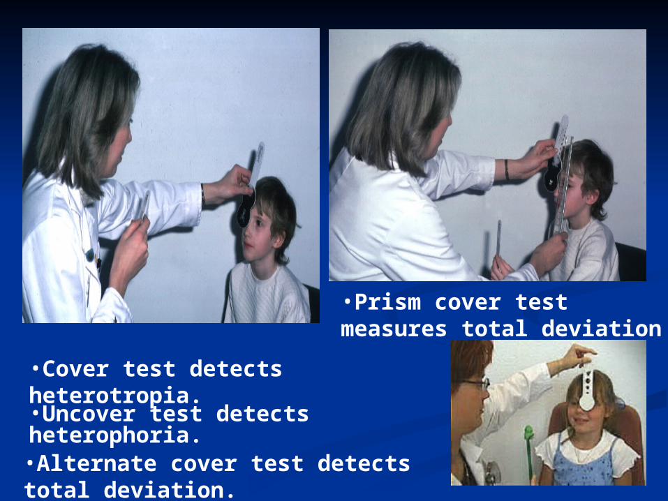
•Cover test detects heterotropia.
•Uncover test detects heterophoria.
•Alternate cover test detects total deviation.
•Prism cover test measures total deviation

Dissimilar image testsDissimilar image testsMaddox wing Maddox rod
•Dissociates eyes for near fixation (1/3 m)•Measures heterophoria
•White spot converted into red streak
•Cannot differentiate tropia from phoria

Double Maddox Rod TestDouble Maddox Rod Test
Torsional deviations can be Torsional deviations can be measured with double maddox rod measured with double maddox rod test.test.
The Maddox rods placed parallel in The Maddox rods placed parallel in both eyes; better if of different both eyes; better if of different colours.colours.
pt. asked whether the two lines pt. asked whether the two lines align exactly with each other.align exactly with each other.

Measurements of ocular Measurements of ocular misalignmentmisalignment
Synoptophore:Synoptophore: Measures angle of Measures angle of
deviation.deviation. Assesses retinal Assesses retinal
correspondence.correspondence. Checks for & measures Checks for & measures
Fusion.Fusion. Checks for & measures Checks for & measures
Stereopsis.Stereopsis.

SENSORY EVALUATIONSENSORY EVALUATION
Normal binocular vision comprises of Normal binocular vision comprises of simultaneous perception, normal fusion simultaneous perception, normal fusion amplitude, and stereoscopy.amplitude, and stereoscopy.
In the strabismus particular mechanisms In the strabismus particular mechanisms are disturbed, changing retinal are disturbed, changing retinal correspondence and frequently producing correspondence and frequently producing suppression of various degree.suppression of various degree.

Synoptophore:Synoptophore: Examination of the simultaneous Examination of the simultaneous
perception means determination of perception means determination of the angle of strabismus and retinal the angle of strabismus and retinal correspondence assessment.correspondence assessment.
Fusion amplitude evaluated with Fusion amplitude evaluated with the use of nearly the same pictures, the use of nearly the same pictures, differing in only small details.differing in only small details.
Stereoscopy examined with the use Stereoscopy examined with the use of slightly decentred special of slightly decentred special pictures. They are, therefore, pictures. They are, therefore, projected on the dispart retinal projected on the dispart retinal point, but within Panum’s area, point, but within Panum’s area, giving the impression of depth and giving the impression of depth and stereoscopy.stereoscopy.


Tests for sensory anomaliesBagolini striated glasses
a - Normal or ARCb- Diplopiac - Suppressiond - Small suppression scotoma


Tests for sensory anomaliesWorth four-dot test
a - Prior to use of glassesb - Normal c - Left suppression/ amblyopiad - Right suppression/ amblyopiae - Diplopia

4 Prism Test 4 Prism Test
A 4 diopter prism placed in front of A 4 diopter prism placed in front of one eye and the recovery is noted.one eye and the recovery is noted.
Strength of prism moves image a Strength of prism moves image a little bit in foveal area, leading to little bit in foveal area, leading to recovery movement of the other eye.recovery movement of the other eye.
Test done to rule out scotoma.Test done to rule out scotoma. Prism can be used BI, BO, BU or BD.Prism can be used BI, BO, BU or BD.

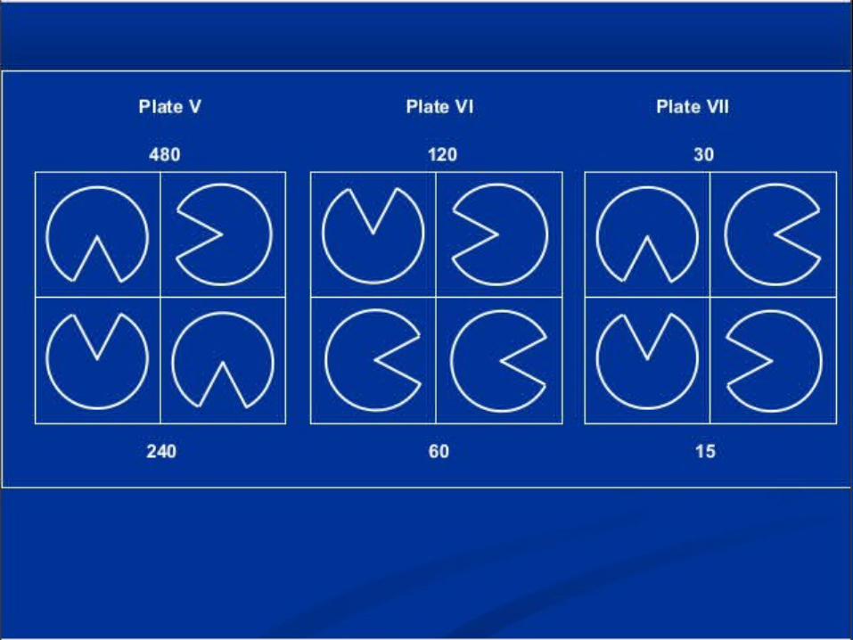
STEREOPSISSTEREOPSIS

Qualitative tests for Qualitative tests for StereopsisStereopsis: : Lang’s 2 pencil test Lang’s 2 pencil test Synoptophore Synoptophore
Quantitative tests for Quantitative tests for Stereopsis:Stereopsis: Random Dot testRandom Dot test Titmus Fly Test Titmus Fly Test TNO Test TNO Test Lang’s Stereo TestLang’s Stereo Test

Tests for stereopsisTitmus
• Red-green spectacles
TNO random dot test
• ‘Hidden’ shapes seen • Polaroid spectacles• Figures seen in 3-D
Lang
• No spectacles
Frisby
• ‘Hidden’ circle seen
• No spectacles• Shapes seen

Titmus Fly Test: Titmus Fly Test: A stereo-test.A stereo-test. Measures stereoacuity from Measures stereoacuity from
3000 secs of arc to 4o secs of 3000 secs of arc to 4o secs of arc.arc.
Fly test is for gross Fly test is for gross stereoacuity.stereoacuity.
Circle patterns consist of 4 Circle patterns consist of 4 circles; one with graded circles; one with graded disparity and can only be seen disparity and can only be seen binocularly. (800 – 40 secs of binocularly. (800 – 40 secs of arc).arc).
Animal patterns denote 400 – Animal patterns denote 400 – 100 secs of arc.100 secs of arc.