Embed Size (px)
Citation preview


The PF consists of three muscle layers: Superficial perineal layer: innervated by the pudendal nerve
Bulbocavernosus Ischiocavernosus Superficial transverse perineal External anal sphincter (EAS)
Deep urogenital diaphragm layer: innervated by pudendal nerve Compressor urethera Uretrovaginal sphincter Deep transverse perineal
Pelvic diaphragm: innervated by sacral nerve roots S3-S5 Levator ani: pubococcygeus (pubovaginalis, puborectalis),
iliococcygeus Coccygeus/ischiococcygeus Piriformis Obturator internus

2 types of muscle fibresType I slow twitch fibersType II fast twitch fibres70% PFM are slow twitch fibresThe PFM are the only transverse
load bearing muscle group in the body.

In females, supports and gives tone to the vaginal wall
Supports pelvic organs against gravity
Increase intra abdominal pressure Maintain anorectal angle. Relaxation for defecation/contraction
Prevent incontinence (urinary &
fecal)

Allow for opening of the pelvic floor to accommodate excretory functions and parturition
Reinforce urethral closure during increase of intra abdominal pressure
Has an inhibitory effect on bladder activity
Assist in unloading the spineAssists in pelvispinal stabilityContribute to sexual arousal and
performance

Diaphragmatic- The abdominal wall,
respiratory and pelvic diaphragms create a functional cylinder
This cylinder is created by muscles that must contract and become rigid in order to protect the spine and pelvis during any mechanical loading
In some patients, the pelvic diaphragm does not contract. This causes weakness of this entire support mechanism

As the abdominal muscles respiratory and pelvic diaphragms contract
The intra-abdominal pressure increases
The entire abdomen becomes more rigid, able to transmit greater mechanical loads without hurting the spine nor pelvic joints

Anatomic impairment Birth injuries Neurological
dysfunction
Psychological impairement
Motivation Sexual
abuse

Pt assessment should include Presenting symptoms in order of
importance Relevant obstetric , medical ,
gynecological and surgical h/o Investigation, and previous and current
treatment Details of voiding dysfunction/
incontinence Details from frequency / volume charts,
fluid intake

Rectal function-defecation patternObjective assessment
Digital per vaginal examination Digital per anal muscle assessment Effect of coughing and straining on vaginal
wall and organ position Six point scale- ( 0 for nil contraction, 1-
flicker, 2- weak, 3- moderate, 4- good, 5- strong)
Position – supine or standing One finger or two finger Perineometer- records vaginal pressure

PERFECTP-powerE-enduranceR –resting toneF- fast contractionEct- each contraction timeC- coordination
Other impairments ,such as pelvic floor trigger points, decreased sensation, and scars or myofascial adhesions should be noted

1.vaginal examination by the physiotherapist
2. self-examination by the patient 3. hand on perineum by the
physiotherapist 4. hand on perineum by the patient 5. observation of perineum by the
physiotherapist 6. observation of perineum by the patient –
using a mirror 7. perineometer

8.stop and start midstream 9. using the Neen Healthcare
‘Educator’ 10. using a cone in the vagina and
applying traction to the string while trying to grip the cone11. asking the partner at intercourse12. manometric and EMG biofeedback13. transperineal or labial ultrasound.

PeritronPeritron is a hand-held clinical Perineometer intended for assessing the strength of pelvic floor (PF) muscles and teaching pelvic floor exercises. In operation, air pressure in the sensor caused by a pelvic floor contraction is transferred by a tube to the Readout Unit where it is displayed in several ways .The pressure is displayed either numerically in centimetres water pressure or as a multi-range analogue bar-graph.
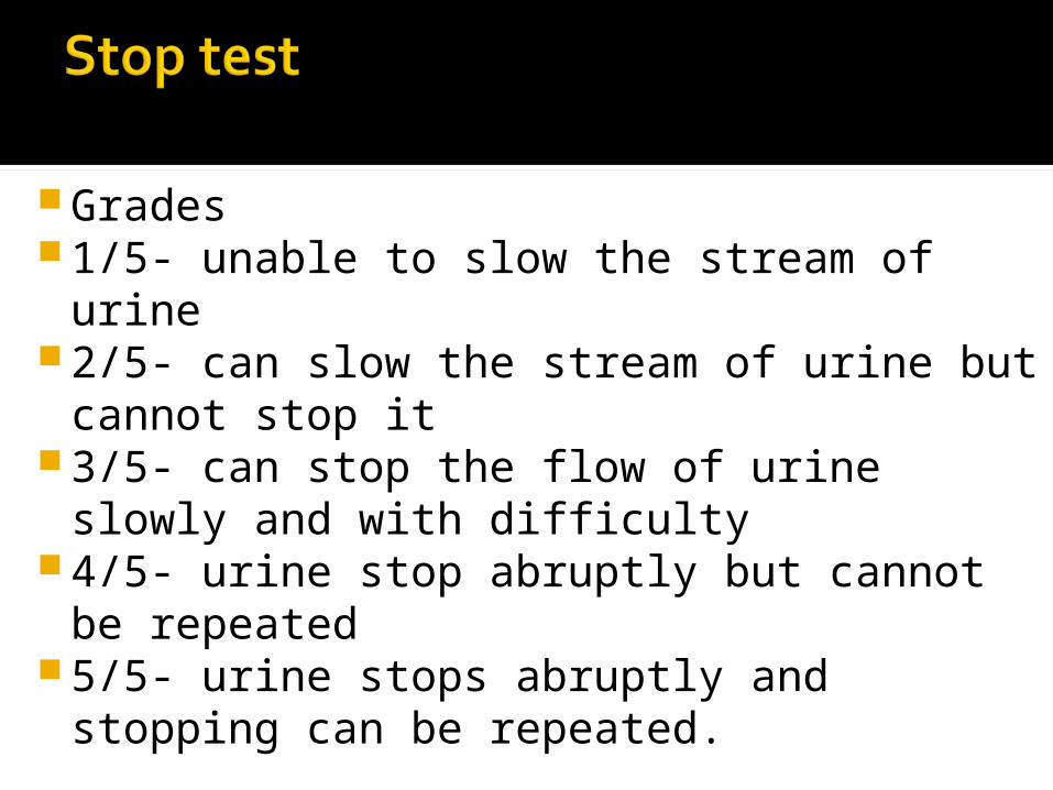
Grades 1/5- unable to slow the stream of urine2/5- can slow the stream of urine but
cannot stop it3/5- can stop the flow of urine slowly
and with difficulty4/5- urine stop abruptly but cannot be
repeated5/5- urine stops abruptly and stopping
can be repeated.

Jumping jack testPad testEMGUrodynamic assessmentPelvic floor dynamometer

PregnancyWithin 6 weeks of vaginal or cesarean
delivery and pelvic surgeryAtrophic vaginitisActive pelvic infectionsevere pelvic or vaginal painChildern and pre sexual adolescentsLack of informed concentLack of therapist training

Endurance impairmentMobility impairmentPosture impairmentCoordination impairment

Supportive dysfunctionHypertonic dysfunctionIn coordination dysfunctionVisceral dysfunction

Results from loss of strength and integrity of contractile and non contractile tissues.
This dysfunction is weakness and sagging of PFM.
Common diagnosis associated with supportive dysfunction are stress incontinence, mixed incontinence and pelvic organ prolapse.
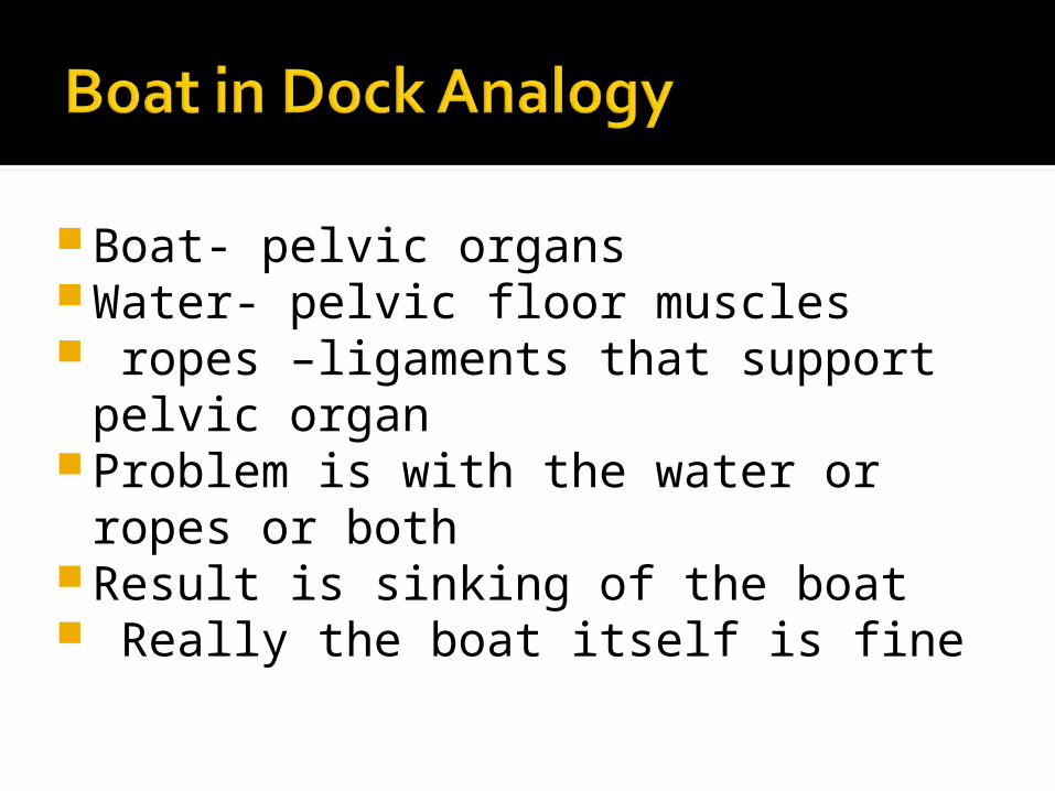
Boat- pelvic organsWater- pelvic floor muscles ropes –ligaments that support pelvic
organ Problem is with the water or ropes or
bothResult is sinking of the boat Really the boat itself is fine


Anatomic impairment of PFM and nerves in the area
Vaginal delivery Muscle atrophy due to central and
peripheral nervous system defect Decrease awareness leads to weakness Prolong intra abdominal pressure may
result in stretching of PFM or their tendons.
obesity

Impaired performance and endurance of PFM
Increased PFM length Increased connective tissue length and
muscle atrophy Impaired abdominal muscle performance Coordination of the PFM decreased Pain in PFM Mobility impairment of pelvic joint. Symptom of incontinence
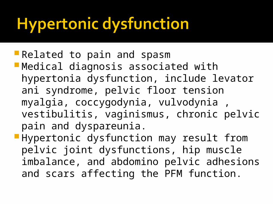
Related to pain and spasm Medical diagnosis associated with
hypertonia dysfunction, include levator ani syndrome, pelvic floor tension myalgia, coccygodynia, vulvodynia , vestibulitis, vaginismus, chronic pelvic pain and dyspareunia.
Hypertonic dysfunction may result from pelvic joint dysfunctions, hip muscle imbalance, and abdomino pelvic adhesions and scars affecting the PFM function.

Lumbo pelvic joint mobility impairments or pathology.
Injuries, such as fall onto the coccyx or pubic ramus.
Hip muscle imbalance with coordination, pain, altered tone, and muscle performance impairments.
Abdominal and perineal adhesions due to pelvic or abdominal surgery or inflammatory condition of abdomen, such as endometriosis.

Altered tone of the PFM, associated muscle of hip, buttock and trunk.
Mobility impairment of scar and connective tissue
Mobility impairment( hypermobility, hypomobility) of pelvic joints: SI joint, pubic, lumbar, hip and sacrococcygeal.
Faulty posture Pain in perineum Hypersensitivity of skin and mucosa.

Divided into neurological and non neurological Detrusor sphincter dyssynergia is a type of
incoordination resulting from neurological lesion in the spinal cord between brain stem and T10.the PFM and smooth internal sphincter contract during a bladder contraction so that urine is unable to be expelled.
Non-neurological incoordination dysfunction is characterized by absent or inappropriate patterns of timing and recruitment of the PFM.

Discuse and decrease awareness of PFM.
Pain in the pelvic or abdominal area may disrupt recruitment pattern.
PFM weaknessCoordination impairment

Pseudo- PFM dysfunction dysfunction.
It is an abnormlity in mobility or motility of the abdominopelvic visceral tissue that leads to pain and musculoskeletal impairment.
Detrusor instability

Endometriosis pelvic inflammatory disease.DysmenorrheaSurgical scars Irritable bowel syndrome Interstitial cystitis.

Weakness of the abdominal muscles, especially the oblique and transverse layers may occur in response to pain in the abdomen, causing a pendulous abdomen with poor visceral and lumbar support.
Secondary lumbo pelvic joint mobility impairment and posture impairments may result.
Altered tone or impaired muscle performance of the PFM may also occur as result of pain in the lower pelvic pain.