Embed Size (px)
DESCRIPTION
This slide gives you information regarding the Types of Palmar spaces, their contents & boundaries. Also certain aspects of Applied anatomy has been enlightened in the interest of Integrated teaching.
Citation preview

PALMAR SPACES
Dr.U. Murali. M.S;M.B.A. Prof. of Surgery D Y Patil Medical College Mauritius

Objectives
Types of Spaces
Location & Boundaries
Contents
Applied Anatomy

PALMAR SPACES
Important spaces are - The superficial pulp
spaces of the finger. The synovial tendon
sheaths of the 2nd, 3rd and 4th finger.
The ulnar bursa The radial bursa The midpalmar
space The thenar space

Other Spaces
Web spaces
Space of Parona

Sup. Pulp space Sup. Pulp space

Superficial Pulp Space
Subcutaneous space between distal phalanx and skin of terminal digit.
Proximally – closed anteriorly by fusion of fibrous flexor sheath to skin of digit at distal crease
Posteriorly fusion of deep fascia to periosteum of terminal phalanx

Contents
Subcutaneous fatty tissue
Loculated by tough fibrous septa - extending from skin to phalanx.
Sensory nerves Digital Artery

Applied Anatomy
Infection of such a space is common and serious
Commonly occurring in the thumb and index finger
Bacteria are usually introduced into the space by pinpricks or sewing needles

Felon

Whitlow
Whitlow is an infection of the pulp space of the finger, usually caused by herpes simplex type I virus (usually refer to herpetic whitlow )

Synovial Tendon Synovial Tendon Sheath Sheath

Synovial tendon sheath
The common synovial sheath for the flexor tendons is a synovial sheath in the carpal tunnel.
It contains tendons of the flexor digitorum superficialis and the flexor digitorum profundus, but not the flexor pollicis longus.

The sheath which surrounds the Flexor digitorum extends downward about half-way along the metacarpal bones, where it ends in blind diverticula around the tendons to the index, middle, and ring fingers. It is prolonged on the tendons to the little finger and usually communicates with the mucous sheath of these tendons.

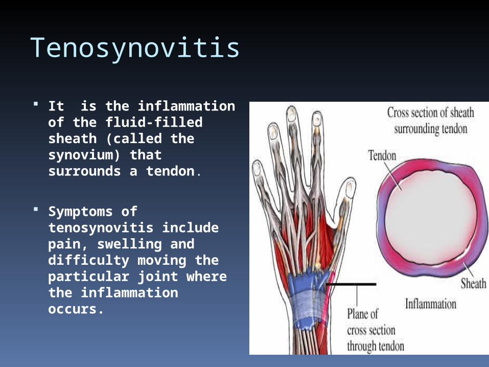
Tenosynovitis
It is the inflammation of the fluid-filled sheath (called the synovium) that surrounds a tendon.
Symptoms of tenosynovitis include pain, swelling and difficulty moving the particular joint where the inflammation occurs.

Flexor Tenosynovitis
• Finger in slight Finger in slight flexionflexion
• Fusiform swellingFusiform swelling
• Pain with extension.Pain with extension.
• Tenderness along Tenderness along tendon sheathtendon sheath

Trigger Finger
When the condition causes the finger to "stick" in a flexed position, this is called "stenosing" tenosynovitis, commonly known as "Trigger Finger”

Ulnar & Radial Ulnar & Radial Bursa Bursa

The common and pollical sheaths are frequently referred to in clinical writing as the ulnar and radial bursae, respectively.
These two sheaths project proximally a short distance above the flexor retinaculum, and they usually communicate with each other in the carpal tunnel.
Hence infection of the synovial sheaths of the thumb or little finger may spread readily into the palm and even into the forearm.

Radial BursaRadial Bursa

Radial Bursa
The synovial sheath of the tendon of flexor pollicis longus (radial bursa).
This sheath is usually separate but may be communicate with the common sheath behind the retinaculum.
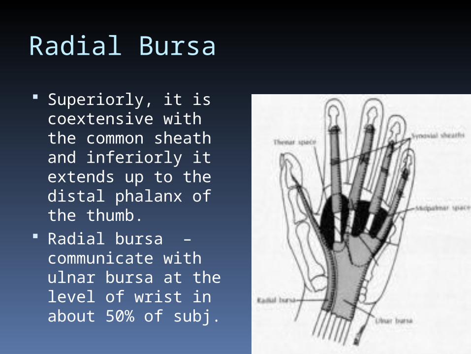
Radial Bursa
Superiorly, it is coextensive with the common sheath and inferiorly it extends up to the distal phalanx of the thumb.
Radial bursa – communicate with ulnar bursa at the level of wrist in about 50% of subj.

Infection Of Radial BursaInfection Of Radial Bursa A patient's radial bursa is a continuation of
the tendon sheath of his flexor pollicis longus, so that any infection inevitably involves both of them.
The distal phalanx of the thumb is flexed and rigid. Pt cannot extend it, although can extend other fingers normally. The hand is tender over the sheath of flexor pollicis longus, and you may be able to feel a swelling above the flexor retinaculum. If treatment is delayed, infection may spread to the ulnar bursa, or the tendon of flexor pollicis longus may slough.

Ulnar BursaUlnar Bursa

Ulnar Bursa
Common flexor synovial sheath (ulnar bursa).
The long flexor tendons of the fingers (flexor digitorum superficialis and profundus), are enclosed in a common synovial sheath while passing deep to the flexor retinaculum.

Ulnar Bursa
The sheath has a parietal layer lining the walls of the carpal tunnel, and a visceral layer closely applied to the tendons.
From the arrangement of the sheath it appears that the synovial sac has been invaginated by the tendons from its lateral side.

Ulnar Bursa
Medial part, common sheath extends distally on the tendons of little finger.
Lateral part, it stops on the middle of palm.
Distal ends of index, middle & ring finger acquire digital synovial sheaths..

Infection Of Ulnar BursaInfection Of Ulnar Bursa Infection of the ulnar bursa is the most
serious hand infection, because it contains all the flexor tendons of a patient's fingers. Pt’s whole hand is oedematous, the palm is moderately swollen, and there may be fullness immediately above the flexor retinaculum. The flexed fingers resist extension, particularly the little one, and least of all is the index.
The radial and ulnar bursa sometimes communicate with one another. So if one of them has been infected, infection may follow in the other a day or two later.

Thenar space Thenar space
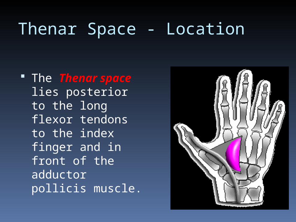
Thenar Space - Location
The Thenar space lies posterior to the long flexor tendons to the index finger and in front of the adductor pollicis muscle.

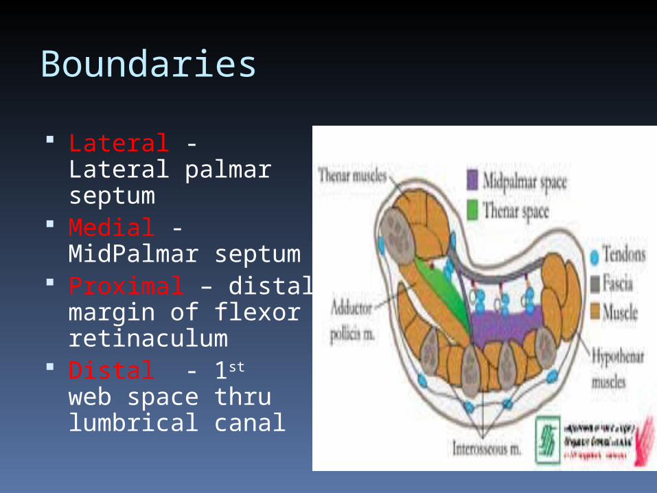
Boundaries
Anterior - Palmar aponeurosis / superficial palmar arch, flexor tendon of index finger covered with synovial sheath / tendon of FPL
Posterior – fascia covering adductor pollicis
Boundaries
Lateral - Lateral palmar septum
Medial - MidPalmar septum
Proximal – distal margin of flexor retinaculum
Distal - 1st web space thru lumbrical canal

Thenar Space(Lateral Central Palmar Space )• Contains: Tendons of
FPL / FDS&P to index finger, palmar digital nerves and vessels to thumb and radial side of index finger.
• Communicates: web of thumb and under flexor retinaculum

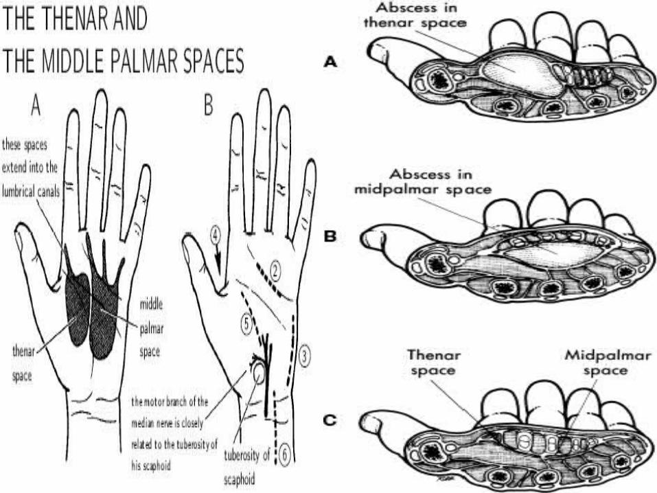
Thenar space infection
Closed space infection of the thenar space.• Pain and swelling of
thenar eminence and first web space.
• Can be from tenosynovitis of 2nd digit with rupture proximally.
• Thumb is held abducted and flexed.


Clinical Significance
The thenar space lies just superficial to the adductor pollicis muscle, forming a plane connecting the deep aspects of the radial bursa and the ulnar bursa. Abscess or space occupying lesions may spread transversely through the thenar space deep in the palm between the thumb and the carpal tunnel.

Mid – Palmar Mid – Palmar space space
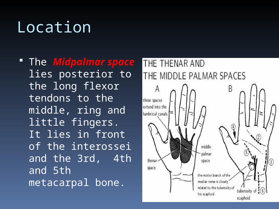
Location
The Midpalmar space lies posterior to the long flexor tendons to the middle, ring and little fingers. It lies in front of the interossei and the 3rd, 4th and 5th metacarpal bone.

Boundaries
Anterior – Palmar aponeurosis / superficial palmar arch, flexor tendons of medial 3 digits covered in ulnar bursa and medial 3 lumbricals
Posterior - Fascia covering 3rd & 4th interossei and metacarpal bones

Boundaries
Medial – Medial Palmar septum
Lateral - Midpalmar septum
Proximal – distal margin of flexor retinaculum
Distal - medial 2 web spaces thru lumbrical canals

Mid Palmar Space(Mid Central Palmar Space)
Contains: 3-5 flexor tendons, 2-4 lumbricals, superficial palmar arch, 3-5 digital vessels and nerves.
Communicates: subcutaneous tissues at webs and extends dorsal to common flexor sheaths.

Mid Palmar infection
Closed space infection of the palmar space.• Loss of normal hand
concavity.• Tenderness of
central palm.• Pain with movement
of 3rd and 4th digits.• Can be from
tenosynovitis of digits 3,4,5

Other spaces Other spaces

Web Spaces
4 Subcutaneous spaces
From its free margin – extends to level of MCP joint.
Contents - S/C fatSuperficial transverse metacarpal ligament, interosseous and lumbrical tendons, digital nerves and vessels.

Interdigital Infection
Collar button abscess due to hour glass configuration
Begins beneath palmar callus – in labourers
Incisions – 1 dorsal and 1 palmar.
Web - not incised

Space of Parona
Located in forearm Continuous with
palmar space through flexor tendons through carpal tunnel
Anterior boundary - flexor tendons covered with tendon sheath
Posterior boundary - Pronator quadratus


![Original Article A Study of Variations In The Formation of ... · of Superficial Palmar Arch In The Rural ... in his study has observed three types of SPA. ... mar spaces [8] . To](https://img.dokumen.tips/doc/110x75/5b16624a7f8b9a546d8ba7f6/original-article-a-study-of-variations-in-the-formation-of-of-superficial.jpg)