Embed Size (px)
Citation preview

Mucoid ACL Degeneration
Prof. Vladimir Bobic, MD, FRCSEdConsultant Orthopaedic Knee Surgeon Chester Knee Clinic, Chester, UK
ACL Study Group 2016 Åre, Sweden, 14 - 17 March 2016

Vladimir Bobic, ACL SG, Åre, 170316


MR I
mag
es:
Dav
e Ritch
ie,
Gla
sgow
, U
K

What is Mucoid ACL Degeneration?
Mucoid ACL degeneration is simply a biological failure or progressive structural ACL degeneration with congenital or acquired synovial tissue entrapment between ACL fibres. ACL injury does not seem to be related to progressive mucoid degeneration but may play a role in the development of ACL ganglions.
CKC UK

Mucoid ACL degeneration and ACL “ganglions” are not infrequent incidental MRI diagnosis in our practice (36 reported since 2010, approx. 50% with associated ACL cysts and 20% with tibial or femoral intraosseous cysts, almost all in 40-60 age group).
Mucoid ACL, ACL and Bone Cysts
The correlation is still poorly understood but seems to be associated with progressive structural ACL degeneration, formation of ACL ganglions and local femoral or tibial subchondral marrow oedema and intraosseous cysts (progressive degeneration but on different point on the same timeline?).
CKC UK

Mucoid ACL Degeneration MRI
CKC UK

Mucoid ACL Degeneration MRI
CKC UK

Mucoid ACL Degeneration MRI
CKC UK

Mucoid ACL & Tibial Cyst
CKC UK

Mucoid ACL Degeneration MRI
CKC UK

Mucoid ACL
Early stages appear as “degloved” ACL (missing synovial envelope and blood vessels), sometimes misdiagnosed as partially torn ACL
CKC UK

Mucoid ACL
… more often as “bare” but bulky appearance with diffuse separation and fragmentation of fibre clusters
CKC UK

Mucoid ACL + Ganglion
CKC UK
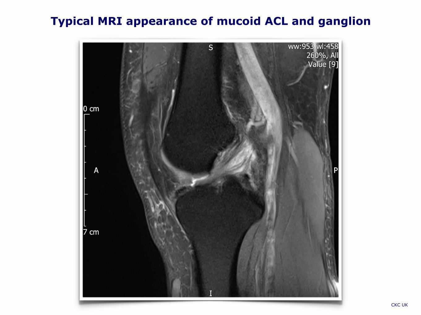
Typical MRI appearance of mucoid ACL and ganglion
CKC UK
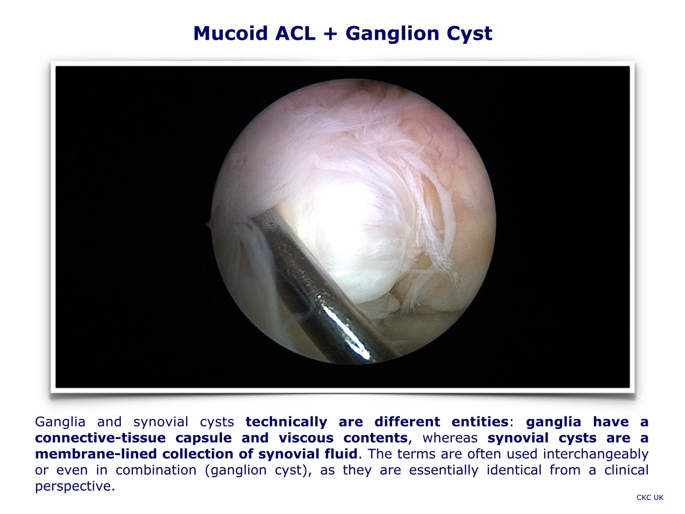
Mucoid ACL + Ganglion Cyst
Ganglia and synovial cysts technically are different entities: ganglia have a connective-tissue capsule and viscous contents, whereas synovial cysts are a membrane-lined collection of synovial fluid. The terms are often used interchangeably or even in combination (ganglion cyst), as they are essentially identical from a clinical perspective.
CKC UK

What is Known about Mucoid ACL Degeneration? only one case has been reported in the English literature up to 2004
CKC UK
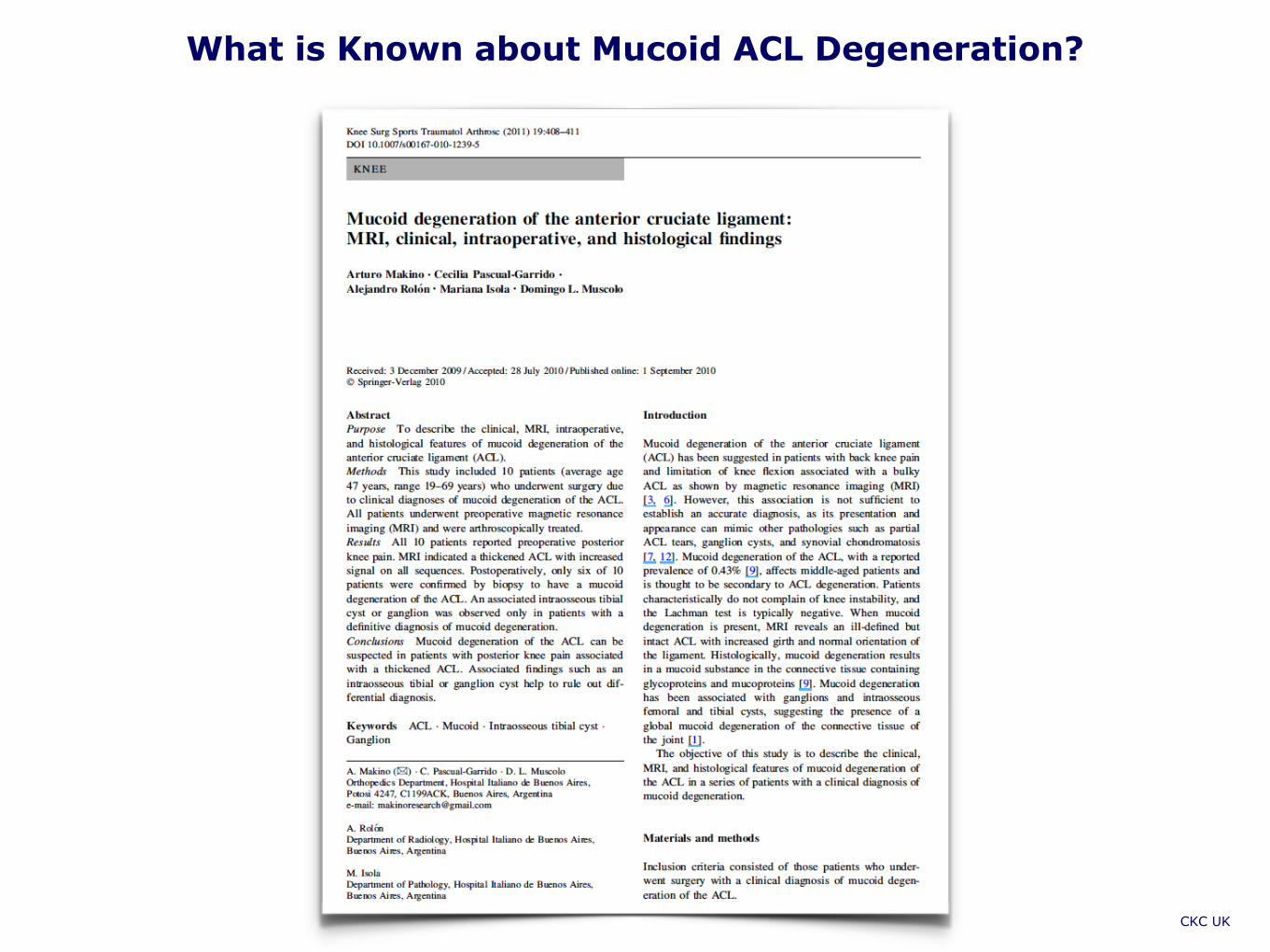
What is Known about Mucoid ACL Degeneration?
CKC UK
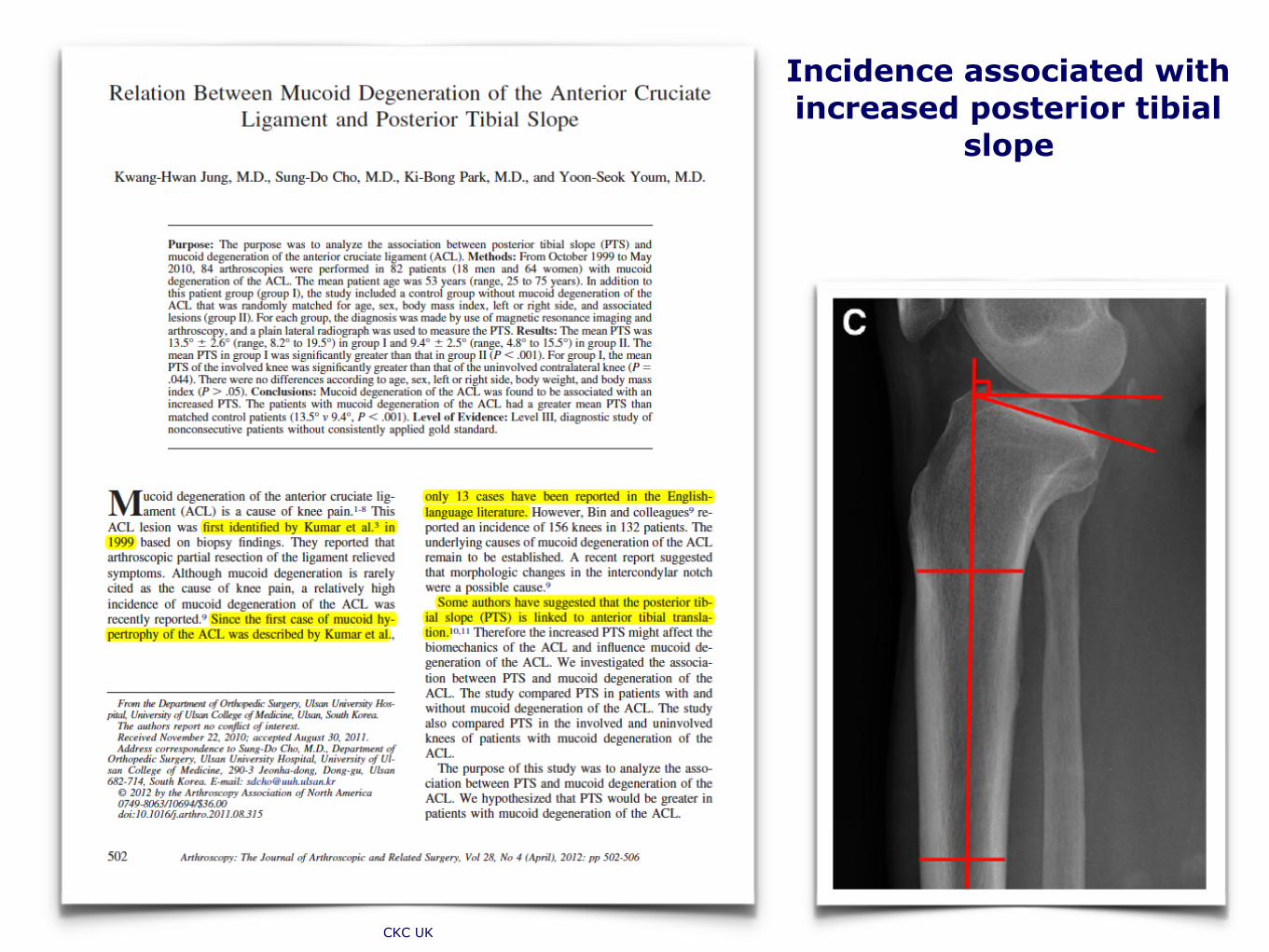
Incidence associated with increased posterior tibial
slope
CKC UK
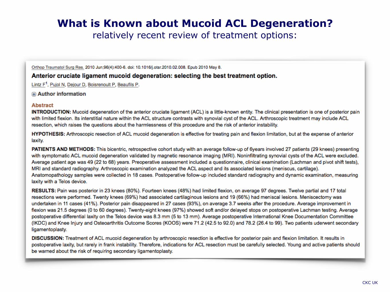
What is Known about Mucoid ACL Degeneration? relatively recent review of treatment options:
CKC UK

What is Known about Mucoid ACL Degeneration? surprisingly, not much …
CKC UK

Features of Mucoid ACL Degeneration
• Clinical Presentation: Patients usually present with medial or posterior knee pain, mechanical locking, clicking and swelling or restricted movement. Instability or giving way are rare complaints and clinical signs of ACL laxity are often absent.
• MRI: most cases are incidental and asymptomatic. The appearance can mimic acute or chronic interstitial partial tears of the ACL. The ACL is thickened and ill-defined with a “celery stalk” appearance. Its low signal longitudinal fibres are separated from each other by higher signal mucinous material, best seen on T2 weighted images. Its signal is increased on all sequences. Intact fibres are best seen on T2-weighted sequences.
• Secondary signs of ACL injury or deficiency (bone bruising and bone marrow oedema, meniscal tears, anterior subluxation of the tibia and other ligament injuries) are usually absent.
• We have observed a significant number of associated mucoid degeneration of both menisci.
• Arthroscopy: mucoid ACL often appears bare or “degloved” with clearly visible fibres. Mucoid ACL is always more voluminous than normal ACL and ligamentum mucosum is often absent.
• Histology: typically distorted collagen fibres with multifocal scanty myxomatous degeneration (generally very vague, in our practice)
CKC UK

Treatment Options: ACL “Deconstruction”
• Electrothermal debridement (beware: may induce more pain because of “electrostimulation” of remaining ACL nerves and receptors)
• ”Debulking”: arthroscopic debridement and partial excision (of torn fibres and ganglions)
• Notchplasty: makes spatial sense but may cause extended chondral damage
• Total excision: very reluctantly, if patient is desperate (may work, numerous concerns: instability, leading to meniscal and chondral deficiency, proprioceptive deficit).
• Leave alone, if not too painful and/or mechanically troublesome, do periodic FU MRIs. Seems to work as well as any of the options above.
MR Images: Albert van Kampen

Conclusion
• ACL mucoid degeneration is a relatively common cause of incidental increased MRI signal within the ACL, frequently asymptomatic
• Possibly senescent, but the causation of mucoid ACL degeneration and correlation with ganglion and bone cysts remains unclear.
• It is possible that ganglion cysts are a byproduct of mucoid degeneration of the connective tissue or a cause of herniation of synovial tissue through a defect in ACL’s synovial sheet or joint capsule.
• Discrete intraosseous ganglia were observed in 66% of other studies with intrasubstance ACL ganglia and 77% of patients with mucoid ACL degeneration.
• In our review intraosseous cysts (often at the femoral and tibial attachments of the ACL) appear to be histologically similar to soft-tissue ganglia, consisting largely of gel-like myxoid material, but they generally have no identifiable communication with the articular surface or joint cavity.
• It is possible that long-standing pressure of a ganglion cyst against an adjacent hard surface may produce an indentation through gradual erosion of the bone, creating an intraosseous ganglion.
CKC UK

Are we any wiser? Not much, but we need more work on ACL biology. Thank you.
CKC UK


![ARTICLE plasty for hypertrophic anterior ligament …ACL mucoid degeneration based on MRI criteria [1]. Patients were excluded if they had cyst-like lesions or the atrophic form of](https://img.dokumen.tips/doc/110x75/5ed58dc6e4e9005a3e7b091b/article-plasty-for-hypertrophic-anterior-ligament-acl-mucoid-degeneration-based.jpg)