Embed Size (px)
Citation preview

ORIGINAL STUDIES, REVIEWS,
AND SCHOLARLY DIALOGHYPERTHYROIDISM, OTHER CAUSES OF THYROTOXICOSIS,
AND THYROID HORMONE ACTION
Hyperthyroidism and Other Causes of Thyrotoxicosis:Management Guidelines of the American Thyroid Association
and American Association of Clinical Endocrinologists
The American Thyroid Association and American Association of Clinical EndocrinologistsTaskforce on Hyperthyroidism and Other Causes of Thyrotoxicosis
Rebecca S. Bahn (Chair),1,* Henry B. Burch,2 David S. Cooper,3 Jeffrey R. Garber,4 M. Carol Greenlee,5
Irwin Klein,6 Peter Laurberg,7 I. Ross McDougall,8 Victor M. Montori,1 Scott A. Rivkees,9
Douglas S. Ross,10 Julie Ann Sosa,11 and Marius N. Stan1
Background: Thyrotoxicosis has multiple etiologies, manifestations, and potential therapies. Appropriatetreatment requires an accurate diagnosis and is influenced by coexisting medical conditions and patient pref-erence. This article describes evidence-based clinical guidelines for the management of thyrotoxicosis that wouldbe useful to generalist and subspeciality physicians and others providing care for patients with this condition.Methods: The development of these guidelines was commissioned by the American Thyroid Association inassociation with the American Association of Clinical Endocrinologists. The American Thyroid Associationand American Association of Clinical Endocrinologists assembled a task force of expert clinicians who au-thored this report. The task force examined relevant literature using a systematic PubMed search supple-mented with additional published materials. An evidence-based medicine approach that incorporated theknowledge and experience of the panel was used to develop the text and a series of specific recommendations.The strength of the recommendations and the quality of evidence supporting each was rated according to theapproach recommended by the Grading of Recommendations, Assessment, Development, and EvaluationGroup.Results: Clinical topics addressed include the initial evaluation and management of thyrotoxicosis; man-agement of Graves’ hyperthyroidism using radioactive iodine, antithyroid drugs, or surgery; management oftoxic multinodular goiter or toxic adenoma using radioactive iodine or surgery; Graves’ disease in children,adolescents, or pregnant patients; subclinical hyperthyroidism; hyperthyroidism in patients with Graves’ophthalmopathy; and management of other miscellaneous causes of thyrotoxicosis.Conclusions: One hundred evidence-based recommendations were developed to aid in the care of patientswith thyrotoxicosis and to share what the task force believes is current, rational, and optimal medical practice.
By mutual agreement among the authors and editors of their respective journals, this work is being published jointly in Thyroid andEndocrine Practice.
*Authors are listed in alphabetical order.1Division of Endocrinology, Metabolism, and Nutrition, Mayo Clinic, Rochester, Minnesota.2Endocrinology and Metabolism Division, Walter Reed Army Medical Center, Washington, District of Columbia.3Division of Endocrinology, The Johns Hopkins University School of Medicine, Baltimore, Maryland.4Endocrine Division, Harvard Vanguard Medical Associates, Boston, Massachusetts.5Western Slope Endocrinology, Grand Junction, Colorado.6The Thyroid Unit, North Shore University Hospital, Manhassett, New York.7Department of Endocrinology, Aarhus University Hospital, Aalborg, Denmark.8Division of Nuclear Medicine, Department of Radiology and Division of Endocrinology, Department of Medicine, Stanford University
School of Medicine, Stanford, California.9Department of Pediatrics, Yale Pediatric Thyroid Center, New Haven, Connecticut.
10Massachusetts General Hospital, Boston, Massachusetts.11Divisions of Endocrine Surgery and Surgical Oncology, Yale University School of Medicine, New Haven, Connecticut.
THYROIDVolume 21, Number 6, 2011ª Mary Ann Liebert, Inc.DOI: 10.1089/thy.2010.0417
593

Introduction
Thyrotoxicosis is a condition having multiple eti-ologies, manifestations, and potential therapies. The term
‘‘thyrotoxicosis’’ refers to a clinical state that results from in-appropriately high thyroid hormone action in tissues generallydue to inappropriately high tissue thyroid hormone levels. Theterm ‘‘hyperthyroidism,’’ as used in these guidelines, is a formof thyrotoxicosis due to inappropriately high synthesis andsecretion of thyroid hormone(s) by the thyroid. Appropriatetreatment of thyrotoxicosis requires an accurate diagnosis. Forexample, thyroidectomy is an appropriate treatment for someforms of thyrotoxicosis and not for others. Additionally, betablockers may be used in almost all forms of thyrotoxicosis,whereas antithyroid drugs are useful in only some.
In the United States, the prevalence of hyperthyroidismis approximately 1.2% (0.5% overt and 0.7% subclinical);the most common causes include Graves’ disease (GD),toxic multinodular goiter (TMNG), and toxic adenoma(TA) (1). Scientific advances relevant to this topic are re-ported in a wide range of literature, including subspecialitypublications in endocrinology, pediatrics, nuclear medi-cine, and surgery, making it challenging for clinicians tokeep abreast of new developments. Although guidelinesfor the diagnosis and management of patients with hy-perthyroidism have been published previously by both theAmerican Thyroid Association (ATA) and American As-sociation of Clinical Endocrinologists (AACE), in conjunc-tion with guidelines for the treatment of hypothyroidism(1,2), both associations determined that thyrotoxicosisrepresents a priority area in need of updated evidence-based practice guidelines.
The target audience for these guidelines includes generaland subspeciality physicians and others providing care forpatients with thyrotoxicosis. In this document, we outlinewhat we believe is current, rational, and optimal medicalpractice. It is not the intent of these guidelines to replaceclinical judgment, individual decision making, or the wishesof the patient or family. Rather, each recommendation shouldbe evaluated in light of these elements in order that optimalpatient care is delivered. In some circumstances, it may beapparent that the level of care required may be best providedin centers where there is specific expertise, and that referral tosuch centers should be considered.
Methods of Development of Evidence-Based Guidelines
Administration
The ATA Executive Council and the Executive Committeeof AACE forged an agreement outlining the working rela-tionship between the two groups surrounding the develop-ment and dissemination of management guidelines for thetreatment of patients with thyrotoxicosis. A chairperson wasselected to lead the task force and this individual (R.S.B.)identified the other 11 members of the panel in consulta-tion with the ATA and the AACE boards of directors.Membership on the panel was based on clinical expertise,scholarly approach, and representation of adult and pedi-atric endocrinology, nuclear medicine, and surgery. The taskforce included individuals from both North America andEurope. In addition, the group recruited an expert on thedevelopment of evidence-based guidelines (V.M.M.) to servein an advisory capacity. Panel members declared whetherthey had any potential conflict of interest at the initialmeeting of the group and periodically during the course ofdeliberations. Funding for the guidelines was derived solelyfrom the general funds of the ATA and thus the task forcefunctioned without commercial support.
To develop a scholarly and useful document, the taskforce first developed a list of the most common causes ofthyrotoxicosis and the most important questions that apractitioner might pose when caring for a patient with aparticular form of thyrotoxicosis or special clinical condition.Two task force members were assigned to review the liter-ature relevant to each of the topics, using a systematicPubMed search for primary references and reviews supple-mented with additional published materials available beforeJune 2010, and develop recommendations based on the lit-erature and expert opinion where appropriate. A prelimi-nary document and a series of recommendations concerningall of the topics were generated by each subgroup and thencritically reviewed by the task force at large. The panelagreed recommendations would be based on consensus ofthe panel and that voting would be used if agreement couldnot be reached. Two recommendations were not unanimousand the dissenting position is noted. Task force deliberationstook place during several lengthy committee meetings,multiple telephone conference calls, and through electroniccommunication.
Table 1. Grading of Recommendations, Assessment, Development, and Evaluation System
Type of grading Definition of grades
Strength of the recommendation 1¼ strong recommendation (for or against)Applies to most patients in most circumstancesBenefits clearly outweigh the risk (or vice versa)
2¼weak recommendation (for or against)Best action may differ depending on circumstances or patient valuesBenefits and risks or burdens are closely balanced, or uncertain
Quality of the evidence þþþ¼High quality; evidence at low risk of bias, such as high quality randomizedtrials showing consistent results directly applicable to the recommendationþþ¼Moderate quality; studies with methodological flaws, showing inconsistent or
indirect evidenceþ¼Low quality; case series or unsystematic clinical observations
594 BAHN ET AL.

Rating of the recommendations
These guidelines were developed to combine the best sci-entific evidence with the experience of seasoned clinicians andthe pragmatic realities inherent in implementation. The taskforce elected to rate the recommendations according to thesystem developed by the Grading of Recommendations, As-sessment, Development, and Evaluation Group (3), with amodification in the grading of evidence (4). Although therating system we chose differs from those used in previousATA and AACE clinical practice guidelines, the approachconforms with the recently updated AACE protocol forstandardized production of clinical practice guidelines (5).The balance between benefits and risks, quality of evidence,applicability, and certainty of the baseline risk are all con-sidered in judgments about the strength of recommendations(6). Grading the quality of the evidence takes into accountstudy design, study quality, consistency of results, and di-rectness of the evidence. The strength of a recommendation isindicated by the number 1 or 2. Grade 1 indicates a strongrecommendation (for or against) that applies to most patientsin most circumstances with benefits of action clearly out-
weighing the risks and burdens (or vice versa). In contrast,Grade 2 indicates a weak recommendation or a suggestionthat may not be appropriate for every patient, depending oncontext, patient values, and preferences. The risks and bene-fits or burdens associated with a weak recommendation areclosely balanced or uncertain and the statement is generallyassociated with the phrase ‘‘we suggest’’ or ‘‘should be con-sidered.’’ The quality of the evidence is indicated by plussigns, such that þ denotes low quality evidence; þþ, mod-erate quality evidence; and þþþ, high quality evidence,based on consistency of results between studies and studydesign, limitations, and the directness of the evidence. Table 1describes the criteria to be met for each rating category.Each recommendation is preceded by a description of theevidence and, in some cases, followed by a remarks sectionincluding technical suggestions on issues such as dosing andmonitoring.
Presentation and endorsement of recommendations
The organization of the task force’s recommendations ispresented in Table 2. The page numbers and the location key
Table 2. Organization of the Task Force’s Recommendations
Location key Description Page
[A] Background 597[B] How should clinically or incidentally discovered thyrotoxicosis be evaluated
and initially managed?597
[B1] Assessment of disease severity 597[B2] Biochemical evaluation 598[B3] Determination of etiology 598[B4] Symptomatic management 599
[C] How should overt hyperthyroidism due to GD be managed? 600[D] If 131I therapy is chosen as treatment for GD, how should it be accomplished? 601
[D1] Preparation of patients with GD for 131I therapy 601[D2] Administration of 131I in the treatment of GD 601[D3] Patient follow-up after 131I therapy for GD 602[D4] Treatment of persistent Graves’ hyperthyroidism following radioactive
iodine therapy603
[E] If antithyroid drugs are chosen as initial management of GD, how should thetherapy be managed?
603
[E1] Initiation of antithyroid drug therapy for the treatment of GD 603[E2] Monitoring of patients taking antithyroid drugs 604[E3] Management of allergic reactions 604[E4] Duration of antithyroid drug therapy for GD 604
[F] If thyroidectomy is chosen for treatment of GD, how should it be accomplished? 605[F1] Preparation of patients with GD for thyroidectomy 605[F2] The surgical procedure and choice of surgeon 605[F3] Postoperative care 605
[G] How should thyroid nodules be managed in patients with GD? 606[H] How should thyroid storm be managed? 606[I] How should overt hyperthyroidism due to TMNG or TA be treated? 607[J] If 131I therapy is chosen as treatment for TMNG or TA, how should it be accomplished? 609
[J1] Preparation of patients with TMNG or TA for 131I therapy 609[J2] Evaluation of thyroid nodules prior to radioioactive iodine therapy 609[J3] Administration of radioactive iodine in the treatment of TMNG or TA 609[J4] Patient follow-up after 131I therapy for TMNG or TA 610[J5] Treatment of persistent or recurrent hyperthyroidism following 131I therapy
for TMNG or TA610
[K] If surgery is chosen, as treatment for TMNG or TA, how should it be accomplished? 610[K1] Preparation of patients with TMNG or TA for surgery 610
(continued)
HYPERTHYROIDISM MANAGEMENT GUIDELINES 595

Table 2. (Continued)
Location key Description Page
[K2] The surgical procedure and choice of surgeon 610[K3] Postoperative care 611[K4] Treatment of persistent or recurrent disease following surgery for TMNG
or TA611
[L] Is there a role for antithyroid drug therapy in patients with TMNG or TA? 611[M] Is there a role for radiofrequency, thermal or alcohol ablation in the management
of TA or TMNG?612
[N] How should GD be managed in children and adolescents? 612[N1] General approach 612
[O] If antithyroid drugs are chosen as initial management of GD in children, howshould the therapy be managed?
612
[O1] Initiation of antithyroid drug therapy for the treatment of GD in children 612[O2] Symptomatic management of Graves’ hyperthyroidism in children 613[O3] Monitoring of children taking methimazole 613[O4] Monitoring of children taking propylthiouracil 614[O5] Management of allergic reactions in children taking methimazole 614[O6] Duration of methimazole therapy in children with GD 614
[P] If radioactive iodine is chosen as treatment for GD in children, how should itbe accomplished?
615
[P1] Preparation of pediatric patients with GD for 131I therapy 615[P2] Administration of 131I in the treatment of GD in children 615[P3] Side-effects of 131I therapy in children 615
[Q] If thyroidectomy is chosen as treatment for GD in children, how should itbe accomplished?
616
[Q1] Preparation of children with GD for thyroidectomy 616[R] How should SH be managed? 617
[R1] Frequency and causes of subclinical hyperthyroidism 617[R2] Clinical significance of subclinical hyperthyroidism 617[R3] When to treat subclinical hyperthyroidism 617[R4] How to treat subclinical hyperthyroidism 618[R5] End points to be assessed to determine effective therapy of subclinical
hyperthyroidism618
[S] How should hyperthyroidism in pregnancy be managed? 619[S1] Diagnosis of hyperthyroidism in pregnancy 619[S2] Management of hyperthyroidism in pregnancy 619[S3] The role of TRAb levels measurement in pregnancy 621[S4] Postpartum thyroiditis 621
[T] How should hyperthyroidism be managed in patients with Graves’ ophthalmopathy? 622[T1] Assessment of disease activity and severity 623[T2] Prevention of GO 624[T3] Treatment of hyperthyroidism in patients with active GO of mild severity 625[T4] Treatment of hyperthyroidism in patients with active and moderate-to-severe
or sight-threatening GO625
[T5] Treatment of GD in patients with inactive GO 625[U] How should overt drug-induced thyrotoxicosis be managed? 626
[U1] Iodine-induced thyrotoxicosis 626[U2] Cytokine-induced thyrotoxicosis 627[U3] Amiodarone-induced thyrotoxicosis 627
[V] How should thyrotoxicosis due to destructive thyroiditis be managed? 628[V1] Subacute thyroiditis 628[V2] Painless thyroiditis 628[V3] Acute thyroiditis 628
[W] How should thyrotoxicosis due to unusual causes be managed? 629[W1] TSH-secreting pituitary tumors 629[W2] Struma ovarii 629[W3] Choriocarcinoma 629[W4] Thyrotoxicosis factitia 630[W5] Functional thyroid cancer metastases 630
GD, Graves’ disease; GO, Graves’ ophthalmopathy; SH, subclinical hyperthyroidism; TA, toxic adenoma; TMNG, toxic multinodulargoiter; TRAb, thyrotropin receptor antibody; TSH, thyroid-stimulating hormone.
596 BAHN ET AL.

can be used to locate specific topics and recommenda-tions. Specific recommendations are presented withinboxes in the main body of the text. Location keys can becopied into the Find or Search function in a file or Webpage to rapidly navigate to a particular section. A listingof the recommendations without text is provided asAppendix A.
The final document was approved by the ATA andAACE on March 15, 2011 and officially endorsed (in alpha-betical order) by American Academy of Otolaryngology–Head and Neck Surgery, Associazione Medici Endocrinologi,British Association of Endocrine and Thyroid Surgeons,Canadian Paediatric Endocrine Group–Groupe Canadiend’Endocrinologie Pediatrique (endorsement of pediatric sec-tion only), European Association of Nuclear Medicine, TheEndocrine Society, European Society of Endocrinology, Eu-ropean Society of Endocrine Surgeons, European ThyroidAssociation, International Association of Endocrine Sur-geons, Latin American Thyroid Society, Pediatric EndocrineSociety, Italian Endocrine Society, and Society of NuclearMedicine.
Results
[A] Background
In general, thyrotoxicosis can occur if (i) the thyroid isinappropriately stimulated by trophic factors; (ii) there isconstituitive activation of thyroid hormone synthesis andsecretion leading to autonomous release of excess thyroidhormone; (iii) thyroid stores of preformed hormone are pas-sively released in excessive amounts owing to autoimmune,infectious, chemical, or mechanical insult; or (iv) there isexposure to extra-thyroidal sources of thyroid hormone,which may be either endogenous (struma ovarii, metastaticdifferentiated thyroid cancer) or exogenous (factitious thyro-toxicosis).
Subclinical hyperthyroidism (SH) is most often causedby release of excess thyroid hormone by the gland. Thiscondition is defined as a low or undetectable serumthyroid-stimulating hormone (TSH) with values withinthe normal reference range for both triiodothyronine (T3)and free thyroxine (T4) estimates. Both overt and sub-clinical disease may lead to characteristic signs andsymptoms.
GD is an autoimmune disorder in which thyrotropin re-ceptor antibodies (TRAbs) stimulate the TSH receptor, in-creasing thyroid hormone production. The natural history ofnodular thyroid disease includes growth of establishednodules, new nodule formation, and development of au-tonomy over time (7). In TAs, autonomous hormone pro-duction can be caused by somatic activating mutations ofgenes regulating thyroid hormone systhesis. Germline mu-tations in the gene encoding the TSH receptor can causesporadic or familial nonautoimmune hyperthyroidism asso-ciated with a diffuse enlargement of the thyroid gland (8).Autonomous hormone production is caused by somatic,activating mutations of genes regulating follicular cell ac-tivities. Hormone production may progress from subclinicalto overt hyperthyroidism, and the administration of phar-macologic amounts of iodine to such patients may result iniodine-induced hyperthyroidism (9). GD is overall the mostcommon cause of hyperthyroidism in the United States
(10,11). Although toxic nodular goiter is less common thanGD, its prevalence increases with age and in the presence ofiodine deficiency. Therefore, toxic nodular goiter may actu-ally be more common than GD in older patients from regionsof iodine deficiency (12). Unlike toxic nodular goiter, whichis progressive (unless triggered by excessive iodine intake),remission of GD has been reported in up to 30% of patientswithout treatment (13).
The mechanism of hyperthyroidism in painless and sub-acute thyroiditis is inflammation of thyroid tissue with releaseof preformed hormone into the circulation. Painless thyroid-itis is the etiology of hyperthyroidism in about 10% of patients(14), occurring in the postpartum period (postpartum thy-roiditis) (15), during lithium (16), or cytokine (e.g., interferon-alpha) (17) therapy, and in 5–10% of amiodarone-treatedpatients (18). Subacute thyroiditis is thought to be causedby viral infection and is characterized by fever and thyroidpain (19).
Thyroid hormone influences almost every tissue and organsystem in the body. It increases tissue thermogenesis andbasal metabolic rate (BMR) and reduces serum cholesterollevels and systemic vascular resistance. Some of the mostprofound effects of increased thyroid hormone levels areon the cardiovascular system (20). The complications ofuntreated thyrotoxicosis include loss of weight, osteoporosis,atrial fibrillation, embolic events, and even cardiovascularcollapse and death (21,22).
The cellular actions of thyroid hormone are mediated byT3, the active form of thyroid hormone. T3 binds to nuclearreceptor proteins that function as transcription factors toregulate the expression of many genes. Nongenomic actionsof thyroid hormone also regulate important physiologic pa-rameters.
The signs and symptoms of overt and mild, or subclinical,thyrotoxicosis are similar, but differ in magnitude. Overtthyrotoxicosis, whether endogenous or exogenous, is char-acterized by excess thyroid hormones in serum and sup-pressed TSH (<0.01 mU/L). There are also measurablechanges in basal metabolic rate, cardiovascular hemody-namics, and psychiatric and neuropsychological function (23).There is only moderate correlation between the elevation inthyroid hormone concentration and clinical signs and symp-toms. Symptoms and signs that result from increased adren-ergic stimulation include tachycardia and anxiety and appearto be more pronounced in younger patients and those withlarger goiters (24).
[B] How should clinically or incidentallydiscovered thyrotoxicosis be evaluatedand initially managed?
[B1] Assessment of disease severity
The assessment of thyrotoxic manifestations, and espe-cially potential cardiovascular and neuromuscular compli-cations, is essential to formulating an appropriate treatmentplan. While it might be anticipated that the severity ofthyrotoxic symptoms is proportional to the elevation in theserum levels of free T4 and T3 estimates, in one study of 25patients with GD, the Hyperthyroid Symptom Scale did notstrongly correlate with free T4 or T3 estimates and was in-versely correlated with age (24). The importance of age as adeterminant of the prevalence and severity of hyperthyroid
HYPERTHYROIDISM MANAGEMENT GUIDELINES 597
symptoms has been recently confirmed (25). Cardiac evalu-ation may be necessary, especially in the older patient, andmay require an echocardiogram, electrocardiogram, Holtermonitor, or myocardial perfusion studies. In addition to theadministration of beta-blockers (26), specific cardiovasculartreatment may be directed toward concomitant myocardialischemia, congestive heart failure, or atrial arrhythmias (20),and anticoagulation may be necessary in patients in atrialfibrillation (27). Goiter size, obstructive symptoms, and theseverity of Graves’ ophthalmopathy (GO; the inflammatorydisease that develops in the orbit in association with auto-immune thyroid disorders can be discordant with the degreeof hyperthyroidism or hyperthyroid symptoms.
All patients with known or suspected hyperthyroidismshould undergo a comprehensive history and physicalexamination, including measurement of pulse rate, bloodpressure, respiratory rate, and body weight. In addition,thyroid size; presence or absence of thyroid tenderness,symmetry, and nodularity; pulmonary, cardiac, and neuro-muscular function (23,26,28); and presence or absence of pe-ripheral edema, eye signs, or pretibial myxedema should beassessed.
[B2] Biochemical evaluation
Serum TSH measurement has the highest sensitivity andspecificity of any single blood test used in the evaluation ofsuspected hyperthyroidism and should be used as an initialscreening test (29). However, when hyperthyroidism isstrongly suspected, diagnostic accuracy improves when botha serum TSH and free T4 are assessed at the time of the initialevaluation. The relationship between free T4 and TSH (whenthe pituitary-thyroid axis is intact) is an inverse log-linearrelationship; therefore, small changes in free T4 result in largechanges in serum TSH concentrations. Serum TSH levels areconsiderably more sensitive than direct thyroid hormonemeasurements for assessing thyroid hormone excess (30). Inovert hyperthyroidism, usually both serum free T4 and T3
estimates are elevated, and serum TSH is undetectable;however, in milder hyperthyroidism, serum T4 and free T4
estimates can be normal, only serum T3 may be elevated, andserum TSH will be <0.01 mU/L (or undectable). These labo-ratory findings have been called ‘‘T3-toxicosis’’ and may rep-resent the earliest stages of disease or that caused by anautonomously functioning thyroid nodule. As is the case withT4, total T3 measurements are impacted by protein binding.Assays for estimating free T3 are less widely validated thanthose for free T4, and therefore measurement of total T3 isfrequently preferred in clinical practice. Subclincial hyper-thyroidism is defined as a normal serum-free T4 estimate andnormal total T3 or free T3 estimate, with subnormal serumTSH concentration. Laboratory protocols that automaticallyadd free T4 estimate and T3 measurements when screeningserum TSH concentrations are low avoid the need for subse-quent blood draws.
In the absence of a TSH-producing pituitary adenoma orthyroid hormone resistance, if the serum TSH is normal, thepatient is almost never hyperthyroid. The term ‘‘euthyroidhyperthyroxinemia’’ has been used to describe a number ofentities, mostly thyroid hormone-binding protein disorders,that cause elevated total serum T4 concentrations (and fre-quently elevated total serum T3 concentrations) in the absence
of hyperthyroidism (31). These conditions include elevationsin T4 binding globulin (TBG) or transthyretin (TTR) (32), thepresence of an abnormal albumin which binds T4 with highcapacity (familial hyperthyroxinemic dysalbuminia), a simi-larly abnormal TTR, and, rarely, immunoglobulins whichdirectly bind T4 or T3. TBG excess may occur as a hereditary X-linked trait, or be acquired as a result of pregnancy or estrogenadministration, hepatitis, acute intermittent porphyuria, orduring treatment with 5-flourouracil, perphenazine, or somenarcotics. Other causes of euthyroid hyperthyroxinemia in-clude those drugs that inhibit T4 to T3 conversion, such asamiodarone (18) or high-dose propranolol (26), acute psy-chosis, extreme high altitude, and amphetamine abuse. Esti-mates of free thyroid hormone concentrations frequently alsogive erroneous results in these disorders. Spurious free T4
elevations may occur in the setting of heparin therapy. Whenfree thyroid hormone concentrations are elevated and TSH isnormal or elevated, further evaluation is necessary.
After excluding euthyroid hyperthyroxinemia, TSH-mediated hyperthyroidism should be considered. A pituitarylesion on MRI and a disproportionately high serum level ofthe alpha-subunit of the pituitary glycoprotein hormonessupport the diagnosis of a TSH-producing pituitary adenoma(33). A family history and positive result of genetic testing formutations in the T3-receptor support a diagnosis of thyroidhormone resistance (34). Rare problems with TSH assayscaused by heterophilic antibodies can cause spuriously highTSH values.
[B3] Determination of etiology
& RECOMMENDATION 1A radioactive iodine uptake should be performed when theclinical presentation of thyrotoxicosis is not diagnostic ofGD; a thyroid scan should be added in the presence ofthyroid nodularity. 1/+00
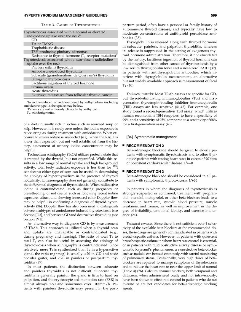
In a patient with a symmetrically enlarged thyroid, recentonset of ophthalmopathy, and moderate to severe hyper-thyroidism, the diagnosis of GD is sufficiently likely thatfurther evaluation of hyperthyroidism causation is unneces-sary. A radioactive iodine uptake (RAIU) is indicated whenthe diagnosis is in question (except during pregnancy) anddistinguishes causes of thyrotoxicosis having elevated ornormal uptake over the thyroid gland from those with near-absent uptake (Table 3). It is usually elevated in patients withGD and normal or high in toxic nodular goiter, unless therehas been a recent exposure to iodine (e.g., radiocontrast). Thepattern of RAIU in GD is diffuse unless there are coexistentnodules or fibrosis. The pattern of uptake in a patient with asingle TA generally shows focal uptake in the adenoma withsuppressed uptake in the surrounding and contralateral thy-roid tissue. The image in TMNG demonstrates multiple areasof focal increased and suppressed uptake, and if autonomy isextensive, the image may be difficult to distinguish from that ofGD (35).
The RAIU will be near zero in patients with painless,postpartum, or subacute thyroiditis, or in those with factitiousingestion of thyroid hormone or recent excess iodine intake.The radioiodine uptake may be low after exposure to iodin-ated contrast in the preceeding 1–2 months or with ingestion
598 BAHN ET AL.
of a diet unusually rich in iodine such as seaweed soup orkelp. However, it is rarely zero unless the iodine exposure isreoccurring as during treatment with amiodarone. When ex-posure to excess iodine is suspected (e.g., when the RAIU islower than expected), but not well established from the his-tory, assessment of urinary iodine concentration may behelpful.
Technetium scintigraphy (TcO4) utilizes pertechnetate thatis trapped by the thyroid, but not organified. While this re-sults in a low range of normal uptake and high backgroundactivity, total body radiation exposure is less than for 123Iscintiscans; either type of scan can be useful in determiningthe etiology of hyperthyroidism in the presence of thyroidnodularity. Ultrasonography does not generally contribute tothe differential diagnosis of thyrotoxicosis. When radioactiveiodine is contraindicated, such as during pregnancy orbreastfeeding, or not useful, such as following recent iodineexposure, ultrasound showing increased color Doppler flowmay be helpful in confirming a diagnosis of thyroid hyper-activity (36). Doppler flow has also been used to distinguishbetween subtypes of amiodarone-induced thyrotoxicosis (seeSection [U3], and between GD and destructive thyroiditis (seeSection [V1]).
An alternative way to diagnose GD is by measurementof TRAb. This approach is utilized when a thyroid scanand uptake are unavailable or contraindicated (e.g.,during pregnancy and nursing). The ratio of total T3 tototal T4 can also be useful in assessing the etiology ofthyrotoxicosis when scintigraphy is contraindicated. Sincerelatively more T3 is synthesized than T4 in a hyperactivegland, the ratio (ng/mcg) is usually >20 in GD and toxicnodular goiter, and <20 in painless or postpartum thy-roiditis (37).
In most patients, the distinction between subacuteand painless thyroiditis is not difficult. Subacute thy-roiditis is generally painful, the gland is firm to hard onpalpation, and the erythrocyte sedimentation rate (ESR) isalmost always >50 and sometimes over 100 mm/h. Pa-tients with painless thyroiditis may present in the post-
partum period, often have a personal or family history ofautoimmune thyroid disease, and typically have low tomoderate concentrations of antithyroid peroxidase anti-bodies (38).
Thyroglobulin is released along with thyroid hormonein subacute, painless, and palpation thyroiditis, whereasits release is suppressed in the setting of exogenous thy-roid hormone administration. Therefore, if not elucidatedby the history, factitious ingestion of thyroid hormone canbe distinguished from other causes of thyrotoxicosis by alow serum thyroglobulin level and a near-zero RAIU (39).In patients with antithyroglobulin antibodies, which in-terfere with thyroglobulin measurement, an alternativebut not widely available approach is measurement of fecalT4 (40).
Technical remarks: Most TRAb assays are specific for GD,but thyroid-stimulating immunoglobulins (TSI) and first-generation thyrotropin-binding inhibitor immunoglobulin(TBII) assays are less sensitive (41,42). For example, onestudy found a second-generation TBII assay, which utilizeshuman recombinant TSH receptors, to have a specificity of99% and a sensitivity of 95% compared to a sensitivity of 68%for a first-generation assay (43).
[B4] Symptomatic management
& RECOMMENDATION 2Beta-adrenergic blockade should be given to elderly pa-tients with symptomatic thyrotoxicosis and to other thyr-otoxic patients with resting heart rates in excess of 90 bpmor coexistent cardiovascular disease. 1/++0
& RECOMMENDATION 3Beta-adrenergic blockade should be considered in all pa-tients with symptomatic thyrotoxicosis. 1/+00
In patients in whom the diagnosis of thyrotoxicosis isstrongly suspected or confirmed, treatment with propran-olol, atenolol, metoprolol, or other beta-blockers leads to adecrease in heart rate, systolic blood pressure, muscleweakness, and tremor, as well as improvement in the de-gree of irritability, emotional lability, and exercise intoler-ance (24).
Technical remarks: Since there is not sufficient beta-1 selec-tivity of the available beta-blockers at the recommended do-ses, these drugs are generally contraindicated in patients withbronchospastic asthma. However, in patients with quiescentbronchospastic asthma in whom heart rate control is essential,or in patients with mild obstructive airway disease or symp-tomatic Raynaud’s phenomenon, a nonselective beta-blockersuch as nadolol can be used cautiously, with careful monitoringof pulmonary status. Occasionally, very high doses of beta-blockers are required to manage symptoms of thyrotoxicosisand to reduce the heart rate to near the upper limit of normal(Table 4) (26). Calcium channel blockers, both verapamil anddiltiazem, when administered orally and not intravenously,have been shown to effect rate control in patients who do nottolerate or are not candidates for beta-adrenergic blockingagents.
Table 3. Causes of Thyrotoxicosis
Thyrotoxicosis associated with a normal or elevatedradioiodine uptake over the necka
GDTA or TMNGTrophoblastic diseaseTSH-producing pituitary adenomasResistance to thyroid hormone (T3 receptor mutation)b
Thyrotoxicosis associated with a near-absent radioiodineuptake over the neck
Painless (silent) thyroiditisAmiodarone-induced thyroiditisSubacute (granulomatous, de Quervain’s) thyroiditisIatrogenic thyrotoxicosisFactitious ingestion of thyroid hormoneStruma ovariiAcute thyroiditisExtensive metastases from follicular thyroid cancer
aIn iodine-induced or iodine-exposed hyperthyroidism (includingamiodarone type 1), the uptake may be low.
bPatients are not uniformly clinically hyperthyroid.T3, triiodothyronine.
HYPERTHYROIDISM MANAGEMENT GUIDELINES 599

[C] How should overt hyperthyroidismdue to GD be managed?
& RECOMMENDATION 4Patients with overt Graves’ hyperthyroidism should betreated with any of the following modalities: 131I therapy,antithyroid medication, or thyroidectomy. 1/++0
Once it has been established that the patient is hyperthy-roid and the cause is GD, the patient and physician mustchoose between three effective and relatively safe initial treat-ment options: 131I therapy (radioactive iodine), antithyroiddrugs (ATD), or thyroidectomy (44). In the United States,radioactive iodine has been the therapy most preferred byphysicians. In Europe and Japan, there has been a greaterphysician preference for ATDs and/or surgery (45). The long-term quality of life (QoL) following treatment for GD wasfound to be the same in patients randomly allocated to one ofthe three treatment options (46).
Technical remarks: Once the diagnosis has been made, thetreating physician and patient should discuss each of thetreatment options, including the logistics, benefits, expectedspeed of recovery, drawbacks, potential side effects, and cost.This sets the stage for the physician to make recommenda-tions based on best clinical judgment and allows the finaldecision to incorporate the personal values and preferences ofthe patient.
Factors that favor a particular modality as treatment forGraves’ hyperthyroidism:
a. 131I: Females planning a pregnancy in the future (inmore than 4–6 months following radioiodine therapy,provided thyroid hormone levels are normal), individ-uals with comorbidities increasing surgical risk, andpatients with previously operated or externally irradi-ated necks, or lack of access to a high-volume thyroidsurgeon or contraindications to ATD use.
b. ATDs: Patients with high likelihood of remission (pa-tients, especially females, with mild disease, small goi-ters, and negative or low-titer TRAb); the elderly orothers with comorbidities increasing surgical risk orwith limited life expectancy; individuals in nursinghomes or other care facilities who may have limitedlongevity and are unable to follow radiation safetyregulations; patients with previously operated or irra-diated necks; patients with lack of access to a high-volume thyroid surgeon; and patients with moderate tosevere active GO.
c. Surgery: Symptomatic compression or large goiters(�80 g); relatively low uptake of radioactive iodine;when thyroid malignancy is documented or sus-pected (e.g., suspicious or indeterminate cytology);large nonfunctioning, photopenic, or hypofunction-ing nodule; coexisting hyperparathyroidism requir-ing surgery; females planning a pregnancy in <4–6months (i.e., before thyroid hormone levels wouldbe normal if radioactive iodine were chosen astherapy), especially if TRAb levels are particularlyhigh; and patients with moderate to severe activeGO.
Contraindications to a particular modality as treatmentfor Graves’ hyperthyroidism:
a. 131I therapy: Definite contraindications include preg-nancy, lactation, coexisting thyroid cancer, or suspicionof thyroid cancer, individuals unable to comply withradiation safety guidelines and females planning apregnancy within 4–6 months.
b. ATDs: Definite contraindications to long-term ATDtherapy include previous known major adverse reac-tions to ATDs.
c. Surgery: Factors that may mitigate against the choiceof surgery include substantial comorbidity such ascardiopulmonary disease, end-stage cancer, or other
Table 4. Beta-Adrenergic Receptor Blockade in the Treatment of Thyrotoxicosis
Drug Dosage Frequency Considerations
Propanolola 10–40 mg TID-QID Nonselective beta-adrenergic receptorblockade
Longest experienceMay block T4 to T3 conversion at high dosesPreferred agent for nursing mothers
Atenolol 25–100 mg QD or BID Relative beta� 1 selectivityIncreased compliance
Metoprolola 25–50 mg QID Relative beta� 1 selectivityNadolol 40–160 mg QD Nonselective beta-adrenergic receptor
blockadeOnce dailyLeast experience to dateMay block T4 to T3 conversion at high doses
Esmolol IV pump 50–100 mg/kg/min In intensive care unit setting of severethyrotoxicosis or storm
Each of these drugs has been approved for treatment of cardiovascular diseases, but to date none has been approved for the treatmentof thyrotoxicosis.
aAlso available in once daily preparations.T4, thyroxine.
600 BAHN ET AL.

debilitating disorders. Pregnancy is a relative contrain-dication and should only be used in this circumstance,when rapid control of hyperthyroidism is required andantithyroid medications cannot be used. Thyroidectomyis best avoided in the first and third trimesters ofpregnancy because of teratogenic effects associated withanesthetic agents and increased risk of fetal loss in thefirst trimester and increased risk of preterm labor inthe third. Optimally, thyroidectomy is performed in thelatter portion of the second trimester. Although it is thesafest time, it is not without risk (4.5%–5.5% risk ofpreterm labor) (47,48).
Factors that may impact patient preference:
a. 131I therapy: Patients choosing 131I therapy as treatmentfor GD would likely place relatively higher value ondefinitive control of hyperthyroidism, the avoidance ofsurgery, and the potential side effects of antithyroidmedications, as well as a relatively lower value on theneed for lifelong thyroid hormone replacement, rapidresolution of hyperthyroidism, and potential worseningor development of GO (49).
b. ATDs: Patients choosing antithyroid drug therapy astreatment for GD would place relatively higher valueon the possibility of remission and the avoidance oflifelong thyroid hormone treatment, the avoidance ofsurgery, and exposure to radioactivity and a relativelylower value on the avoidance of ATD side effects (seesection E), the need for continued monitoring and thepossibility of disease recurrence.
c. Surgery: Patients choosing surgery as treatment for GDwould likely place a relatively higher value on promptand definitive control of hyperthyroidism, avoidance ofexposure to radioactivity, and the potential side effects ofATDs and a relatively lower value on potential surgicalrisks and need for lifelong thyroid hormone replacement.
[D] If 131I therapy is chosen, how shouldit be accomplished?
[D1] Preparation of patients with GD for 131I therapy
& RECOMMENDATION 5Patients with GD who are at increased risk for complica-tions due to worsening of hyperthyroidism (i.e., those whoare extremely symptomatic or have free T4 estimates 2–3times the upper limit of normal) should be treated withbeta-adrenergic blockade prior to radioactive iodine ther-apy. 1/+00
& RECOMMENDATION 6Pretreatment with methimazole prior to radioactive iodinetherapy for GD should be considered in patients who are atincreased risk for complications due to worsening of hy-perthyroidism (i.e., those who are extremely symptomaticor have free T4 estimate 2–3 times the upper limit of nor-mal). 2/+00
Task force opinion was not unanimous; one person held theopinion that pretreatment with methimazole is not necessary inthis setting.
& RECOMMENDATION 7Medical therapy of any comorbid conditions should beoptimized prior to administering radioactive iodine. 1/+00
131I has been used to treat hyperthyroidism for six decades.This therapy is well tolerated and complications are rare, ex-cept for those related to ophthalmopathy (see section [T].)Thyroid storm occurs only rarely following the administra-tion of radioactive iodine (50,51). In one study of patients withthyrotoxic cardiac disease treated with radioactive iodine asthe sole modality, no clinical worsening in any of the cardinalsymptoms of thyrotoxicosis was seen (52). The frequency ofshort-term worsening of hyperthyroidism following pre-treatment with ATD therapy is not known. However, the useof methimazole (MMI) or carbimazole, the latter of which isnot marketed in the United States, before and after 131I treat-ment may be considered in patients with severe thyrotoxicosis(i.e., those who are extremely symptomatic or have free T4
estimates 2–3 times the upper limit of normal), the elderly,and those with substantial comorbidity that puts them atgreater risk for complications of worsening hyperthyroidism(53,54). The latter includes patients with cardiovascularcomplications such as atrial fibrillation, heart failure, or pul-monary hypertension and those with renal failure, infection,trauma, poorly controlled diabetes mellitus, and cerebrovas-cular or pulmonary disease (50). These comorbid conditionsshould be addressed with standard medical care and the pa-tient rendered medically stable before the administration ofradioactive iodine. In addition, beta-adrenergic blockingdrugs should be used judiciously in these patients in prepa-ration for radioiodine therapy (20,55).
One committee member felt that MMI use is not necessaryin preparation, as there is insufficient evidence for radioactiveiodine worsening either the clinical or biochemical aspects ofhyperthyroidism, and it only delays treatment with radioac-tive iodine. In addition, there is evidence that MMI pretreat-ment may reduce the efficacy of subsequent radioactiveiodine therapy (6,52,56).
Technical remarks: If given as pretreatment, MMI should bediscontinued 3–5 days before the administration of radioac-tive iodine, restarted 3–7 days later, and generally taperedover 4–6 weeks as thyroid function normalizes. Over severaldecades, there have been reports that pretreatment withlithium reduces the activity of 131I necessary for cure ofGraves’ hyperthyroidism and may prevent the thyroid hor-mone increase seen upon ATD withdrawal (57–59). However,this is not used widely, and there is insufficient evidence torecommend the practice.
[D2] Administration of 131I in the treatment of GD
& RECOMMENDATION 8Sufficient radiation should be administered in a single dose(typically 10–15 mCi) to render the patient with GD hy-pothyroid. 1/++0
& RECOMMENDATION 9A pregnancy test should be obtained within 48 hours priorto treatment in any female with childbearing potentialwho is to be treated with radioactive iodine. The treating
HYPERTHYROIDISM MANAGEMENT GUIDELINES 601

physician should obtain this test and verify a negative re-sult prior to administering radioactive iodine. 1/+00
The goal of 131I is to control hyperthyroidism by renderingthe patient hypothyroid; this treatment is very effective,provided sufficient radiation is deposited in the thyroid. Thiscan be accomplished equally well by either administering afixed activity or by calculating the activity based on the size ofthe thyroid and its ability to trap iodine (44). The first methodis simple, and there is evidence that 10 mCi (370 MBq) resultsin hypothyroidism in 69% (representing cure) at 1 year (60)and 15 mCi (450 MBq) results in hypothyroidism in 75% at 6months (61). The second method requires three unknowns tobe determined: the uptake of radioactive iodine, the size ofthe thyroid, and the quantity of radiation (mCi or Bq) to bedeposited per gram (or cc) of thyroid (e.g., activity(mCi)¼ gland weight (g)�150 mCi/g�[1/24 hour uptake on%of dose]). The activity in mCi is converted to mCi by dividingthe result by 1000. The most frequently used uptake is cal-culated at 24 hours, and the size of the thyroid is determinedby palpation or ultrasound. One study found that this esti-mate by experienced physicians is accurate compared withanatomic imaging (62); however, other investigators have notconfirmed this observation (63). There is wide variation in therecommended quantity of 131I that should be deposited (i.e.,between 50 and 200mCi/g). Historically, activities at the lowend of the spectrum have led to a higher proportion of treat-ment failures (41).
Alternately, a more detailed calculation can be made todeposit a specific number of radiation absorbed dose (rad) orGy to the thyroid. Using this approach, it is also necessary toknow the effective half-life of the 131I (44). This requires ad-ditional time and computation and, because the outcome isnot better, this method is seldom used in the United States.Evidence shows that to achieve a hypothyroid state,>150 mCi/g needs to be delivered (61,64,65). Patients who areon dialysis or who have jejunostomy or gastric feeding tubesrequire special care when being administered therapeuticdoses of radioiodine (66).
Propylthiouracil (PTU) treatment before 131I increases theradioresistance of the thyroid (51,67). Whether MMI mayhave the same effect is unclear (51). Use of higher activities of131I may offset the reduced effectiveness of 131I therapy fol-lowing antithyroid medication (53,54). A special diet is notrequired before radioactive iodine therapy, but excessiveamounts of iodine, including iodine-containing multivita-mins, should be avoided for at least 7 days. A low-iodine dietmay be useful for those with relatively low RAIU to increasethe proportion of radioactive iodine trapped.
A long-term increase in cardiovascular and cerebrovascu-lar deaths has been reported after 131I therapy, likely due tothe hyperthyroidism rather than the treatment (56). While thisstudy also found a small increase in cancer mortality, long-term studies of larger numbers of patients have not shown astatistically significant increase in cancer deaths following thistreatment (68–74). In some men, there is a modest fall in thetestosterone to luteinizing hormone (LH) ratio after 131I ther-apy that is subclinical and reversible (75). Conception shouldbe delayed for 4–6 months in women to assure stable eu-thyroidism (on thyroid hormone replacement following suc-cessful thyroid ablation) and 3–4 months in men to allow forturnover of sperm production. However, once the patient
(both genders) is euthyroid, there is no evidence of reducedfertility and offspring of treated patients show no congenitalanomalies compared to the population at large.
Technical remarks: Rendering the patient hypothyroid canbe accomplished equally well by administering either a suf-ficient fixed activity or calculating an activity based on the sizeof the thyroid and its ability to trap iodine. Fetuses exposed to131I after the 10th to 11th week of gestation may be bornathyreotic (76,77) and are also at a theoretical increased riskfor reduced intelligence and/or cancer. In breast-feedingwomen, radioactive iodine therapy should not be adminis-tered for at least 6 weeks after lactation stops to ensure that theradioactivity will no longer be actively concentrated in thebreast tissues.
& RECOMMENDATION 10The physician administering the radioactive iodine shouldprovide written advice concerning radiation safety pre-cautions following treatment. If the precautions cannot befollowed, alternative therapy should be selected. 1/+00
All national and regional radiation protection rules re-garding radioactive iodine treatment should be followed (78).In the United States, the treating physician must ensure anddocument that no adult member of the public is exposed to0.5 mSv (500 milli-roentgen equivalent in man [mrem]) whenthe patient is discharged with a retained activity of 33 mCi(1.22 GBq) or greater, or emits �7 mrem/h (70 mSv/h) at 1 m.
Technical remarks: Continuity of follow-up should be pro-vided and can be facilitated by written communication betweenthe referring physician and the treating physician, including arequest for therapy from the former and a statement from thelatter that the treatment has been administered.
[D3] Patient follow-up after 131I therapy for GD
& RECOMMENDATION 11Follow-up within the first 1–2 months after radioactiveiodine therapy for GD should include an assessment of freeT4 and total T3. If the patient remains thyrotoxic, bio-chemical monitoring should be continued at 4–6 week in-tervals. 1/+00
Most patients respond to radioactive iodine therapy with anormalization of thyroid function tests and clinical symptomswithin 4–8 weeks. Hypothyroidism may occur from 4 weekson, but more commonly between 2 and 6 months, and thetiming of thyroid hormone replacement therapy shouldbe determined by results of thyroid function tests, clinicalsymptoms, and physical examination. Transient hypothy-roidism following radioactive iodine therapy can rarely occur,with subsequent complete recovery of thyroid function orrecurrent hyperthyroidism (79). When thyroid hormonereplacement is initiated, the dose should be adjusted basedon an assessment of free T4. The required dose may be lessthan the typical full replacement, and careful titration is nec-essary owing to nonsuppressible residual thyroid function.Overt hypothyroidism should be avoided, especially in pa-
602 BAHN ET AL.

tients with active GO (see section T2). Once euthyroidismis achieved, lifelong annual thyroid function testing isrecommended.
Technical remarks: Since TSH levels may remain suppressedfor a month or longer after hyperthyroidism resolves, thelevels should be interpreted cautiously and only in concertwith free T4 and T3 estimates.
[D4] Treatment of persistent Graves’ hyperthyroidismfollowing radioactive iodine therapy
& RECOMMENDATION 12When hyperthyroidism due to GD persists after 6 monthsfollowing 131I therapy, or if there is minimal response 3 monthsafter therapy, retreatment with 131I is suggested. 2/+00
Technical remarks: Response to radioactive iodine can be as-sessed by monitoring the size of the gland, thyroid function,and clinical signs and symptoms. The goal of retreatment is tocontrol hyperthyroidism with certainty by rendering the pa-tient hypothyroid. Patients who have persistent, suppressedTSH with normal total T3 and free T4 estimates may not requireimmediate retreatment but should be monitored closely foreither relapse or development of hypothyroidism. In the smallpercentage of patients with hyperthyroidism refractory toseveral applications of 131I, surgery could be considered (80).
[E] If antithyroid drugs are chosen as initialmanagement of GD, how should the therapybe managed?
ATDs have been employed for six decades (81). The goal ofthe therapy is to render the patient euthyroid as quickly andsafely as possible. These medications do not cure Graves’hyperthyroidism. However, when given in adequate doses,they are very effective in controlling the hyperthyroidism;when they fail to achieve euthyroidism, the usual cause isnonadherence (82). The treatment might have a beneficialimmunosuppressive role, but the major effect is to reduce theproduction of thyroid hormones and maintain a euthyroidstate while awaiting a spontaneous remission.
[E1] Initiation of antithyroid drug therapy for the treatmentof GD
& RECOMMENDATION 13Methimazole should be used in virtually every patient whochooses antithyroid drug therapy for GD, except during thefirst trimester of pregnancy when propylthiouracil is pre-ferred, in the treatment of thyroid storm, and in patientswith minor reactions to methimazole who refuse radioac-tive iodine therapy or surgery. 1/++0
& RECOMMENDATION 14Patients should be informed of side effects of antithyroiddrugs and the necessity of informing the physicianpromptly if they should develop pruritic rash, jaundice,acolic stools or dark urine, arthralgias, abdominal pain,nausea, fatigue, fever, or pharyngitis. Before starting anti-thyroid drugs and at each subsequent visit, the patient
should be alerted to stop the medication immediately andcall their physician when there are symptoms suggestive ofagranulocytosis or hepatic injury. 1/+00
& RECOMMENDATION 15Prior to initiating antithyroid drug therapy for GD, wesuggest that patients have a baseline complete blood count,including white count with differential, and a liver profileincluding bilirubin and transaminases. 2/+00
In the United States, MMI and PTU are available, and insome countries, carbimazole, a precursor of MMI, is widelyused. MMI and carbimazole, which is rapidly converted toMMI in the serum (10 mg of carbimazole is metabolized toapproximately 6 mg of MMI), work in a virtually identicalfashion and will both be referred to as MMI in this text. Bothare effective as a single daily dose. At the start of MMI ther-apy, higher doses are advised (10–20 mg daily) to restoreeuthyroidism, following which the dose can be titrated to amaintenance level (generally 5–10 mg daily) (81,83). MMI hasthe benefit of once-a-day administration and a reduced risk ofmajor side effects compared to PTU. PTU has a shorter du-ration of action and is usually administered two or three timesdaily, starting with 50–150 mg three times daily, dependingon the severity of the hyperthyroidism. As the clinical find-ings and thyroid function tests return to normal, reduction toa maintenance PTU dose of 50 mg two or three times daily isusually possible. Higher doses of antithyroid medicationare sometimes administered continuously and combined withl-thyroxine in doses to maintain euthyroid levels (so-calledblock and replace therapy). However, this approach is notgenerally recommended, as it has been shown to result in ahigher rate of ATD side effects (81,84).
PTU may rarely cause agranulocytosis, whereas low dosesof MMI may be less likely to do so (85,86). PTU very infre-quently causes antineutrophil cytoplasmic antibody (ANCA)-positive small vessel vasculitis (87,88), with a risk that appearsto increase with time as opposed to other adverse effects seenwith ATDs that typically occur early in the course of treatment(89,90). PTU can cause fulminant hepatic necrosis that may befatal; liver transplantation has been necessary in some pa-tients taking PTU (91). It is for this reason that the FDA re-cently issued a safety alert regarding the use of PTU, notingthat 32 (22 adult and 10 pediatric) cases of serious liver injuryhave been associated with PTU use (92,93).
MMI hepatotoxicity is typically cholestatic, but hepatocel-lular disease may rarely be seen (94,95). Aplasia cutis of thescalp is rarely found in babies born to mothers taking MMI (96).MMI taken by the mother in the first trimester is also associatedwith a syndrome of MMI embryopathy, including choanal andesophageal atresia (97,98). Arthropathy and a lupus-like syn-drome rarely can occur with either MMI or PTU.
Technical remarks: Baseline blood tests to aid in the inter-pretation of future laboratory values should be consideredbefore initiating antithyroid drug therapy. This is suggestedin part because low white cell counts are common in patientswith autoimmune diseases and in African Americans, andabnormal liver enzymes are frequently seen in patientswith thyrotoxicosis. In addition, a baseline absolute neutro-phil count <500/mm3 or liver transaminase enzyme levelselevated more than fivefold the upper limit of normal are
HYPERTHYROIDISM MANAGEMENT GUIDELINES 603

contraindications to initiating antithyroid drug therapy. It isadvisable to provide information concerning side effects ofATDs to the patient both verbally and in writing to assuretheir comprehension, and document that this has been done.This information can be found on the UpToDate Web site (99).
[E2] Monitoring of patients taking antithyroid drugs
There is a need for periodic clinical and biochemical eval-uation of thyroid status in patients taking ATDs, and it isessential that the patient understand its importance. An as-sessment of serum free T4 should be obtained about 4 weeksafter initiation of therapy, and the dose of medication adjustedaccordingly. Serum T3 also may be monitored, since the esti-mated serum free T4 levels may normalize with persistentelevation of serum T3. Appropriate monitoring intervals areevery 4–8 weeks until euthyroid levels are achieved with theminimal dose of medication. Once the patient is euthyroid,biochemical testing and clinical evaluation can be undertakenat intervals of 2–3 months. An assessment of serum free T4 andTSH are required before treatment and at intervals afterstarting the treatment. Serum TSH may remain suppressed forseveral months after starting therapy and is therefore not agood parameter to monitor therapy early in the course.
& RECOMMENDATION 16A differential white blood cell count should be obtainedduring febrile illness and at the onset of pharyngitis in allpatients taking antithyroid medication. Routine monitor-ing of white blood counts is not recommended. 1/+00
There is no consensus concerning the utility of periodicmonitoring of white blood cell counts and liver function testsin predicting early onset of adverse reaction to the medication(100). While routine monitoring of white blood cell countsmay detect early agranulocytosis, this practice is not likely toidentify cases, as the frequency is quite low (0.2%–0.5%) andthe condition sudden in onset. Because patients are typicallysymptomatic, measuring white blood cell counts during fe-brile illnesses and at the onset of pharyngitis has been thestandard approach to monitoring. In a patient developingagranulocytosis or other serious side effects while taking ei-ther MMI or PTU, use of the other medication is absolutelycontraindicated owing to risk of cross-reactivity between thetwo medications (101).
& RECOMMENDATION 17Liver function and hepatocellular integrity should be as-sessed in patients taking propylthiouracil who experiencepruritic rash, jaundice, light-colored stool or dark urine,joint pain, abdominal pain or bloating, anorexia, nausea, orfatigue. 1/+00
Hyperthyroidism can itself cause mildly abnormal liverfunction tests, and PTU may cause transient elevations ofserum transaminases in up to one-third of patients. Significantelevations to threefold above the upper limit of normal areseen in up to 4% of patients taking PTU (102), a prevalencehigher than with MMI. As noted above, PTU can also causefatal hepatic necrosis, leading to the suggestion by some thatpatients taking this ATD have routine monitoring of theirliver function, especially during the first 6 months of therapy.
It is difficult to distinguish these abnormalities from the effectof persistent thyrotoxicosis unless they are followed prospec-tively. In patients with improving thyrotoxicosis, a rising al-kaline phosphatase with normalization of other liver functiondoes not indicate worsening hepatic toxicity. The onset of PTU-induced hepatotoxicity may be acute, difficult to appreciateclinically, and rapidly progressive. If not recognized, it can leadto liver failure and death (92,103–105). Routine monitoring ofliver function in all patients taking antithyroid medication hasnot been found to prevent severe hepatotoxicity.
Technical remarks: PTU should be discontinued if trans-aminase levels (either elevated at onset of therapy, foundincidentally or measured as clinically indicated) reach 2–3times the upper limit of normal and fail to improve within 1week with repeat testing. After discontinuing the drug, liverfunction tests should be monitored weekly until there isevidence of resolution. If resolution is not evident, promptreferral to a gastroenterologist or hepatologist is warranted.Except in cases of severe PTU-induced hepatotoxicity, MMIcan be used to control the thyrotoxicosis without ill effect(106,107).
[E3] Management of allergic reactions
& RECOMMENDATION 18Minor cutaneous reactions may be managed with concur-rent antihistamine therapy without stopping the antithy-roid drug. Persistent minor side effects of antithyroidmedication should be managed by cessation of the medi-cation and changing to radioactive iodine or surgery, orswitching to the other antithyroid drug when radioactiveiodine or surgery are not options. In the case of a seriousallergic reaction, prescribing the alternative drug is notrecommended. 1/+00
Minor allergic side effects, such as a limited, minor rash, mayoccur in up to 5% of patients taking either MMI or PTU (81).
[E4] Duration of antithyroid drug therapy for GD
& RECOMMENDATION 19If methimazole is chosen as the primary therapy for GD, themedication should be continued for approximately 12–18months, then tapered or discontinued if the TSH is normalat that time. 1/+++
& RECOMMENDATION 20Measurement of TRAb levels prior to stopping antithyroiddrug therapy is suggested, as it aids in predicting whichpatients can be weaned from the medication, with normallevels indicating greater chance for remission. 2/+00
& RECOMMENDATION 21If a patient with GD becomes hyperthyroid after complet-ing a course of methimazole, consideration should be givento treatment with radioactive iodine or thyroidectomy.Low-dose methimazole treatment for longer than 12–18months may be considered in patients not in remission whoprefer this approach. 2/+00
604 BAHN ET AL.

A patient is considered to be in remission if they have had anormal serum TSH, FT4, and T3 for 1 year after discontinuationof ATD therapy. The remission rate varies considerably be-tween geographical areas. In the United States, about 20%–30%of patients will have a lasting remission after 12–18 months ofmedication (44). The remission rate appears to be higher inEurope and Japan; a long-term European study indicated a50%–60% remission rate after 5–6 years of treatment (108). Ameta-analysis shows the remission rate in adults is not im-proved by a course of ATDs longer than 18 months (84). Alower remission rate has been described in men, smokers (es-pecially men), and those with large goiters (�80 g) (109–113).Persistently high levels of TRAb and high thyroid blood flowidentified by color Doppler ultrasound are also associated withhigher relapse rates (112,114–116), and these patients should beassessed more frequently and at shorter intervals after anti-thyroid drugs are discontinued. Conversely, patients with milddisease, small goiters, and negative TRAb have a remission rateover 50%, making the use of antithyroid medications poten-tially more favorable in this group of patients (117).
Technical remarks: When MMI is discontinued, thyroidfunction testing should continue to be monitored at 1–3-month intervals for 6–12 months to diagnose relapse early.The patient should be counseled to contact the treating phy-sician if symptoms of hyperthyroidism are recognized.
[F] If thyroidectomy is chosen for treatment of GD,how should it be accomplished?
[F1] Preparation of patients with GD for thyroidectomy
& RECOMMENDATION 22Whenever possible, patients with GD undergoing thy-roidectomy should be rendered euthyroid with methima-zole. Potassium iodide should be given in the immediatepreoperative period. 1/+00
& RECOMMENDATION 23In exceptional circumstances, when it is not possible to ren-der a patient with GD euthyroid prior to thyroidectomy, theneed for thyroidectomy is urgent, or when the patient is al-lergic to antithyroid medication, the patient should be ade-quately treated with beta-blockade and potassium iodide inthe immediate preoperative period. The surgeon and anes-thesiologist should have experience in this situation. 1/+00
Thyroid storm may be precipitated by the stress of surgery,anesthesia, or thyroid manipulation and may be prevented bypretreatment with ATDs. Whenever possible, thyrotoxic pa-tients who are undergoing thyroidectomy should be renderedeuthyroid by MMI before undergoing surgery. Preoperativepotassium iodide, saturated solution of potassium iodide(SSKI) or inorganic iodine, should be used before surgery inmost patients with GD. This treatment is beneficial as it de-creases thyroid blood flow, vascularity, and intraoperativeblood loss during thyroidectomy (118,119). In addition, rapidpreparation for emergent surgery can be facilitated by the useof corticosteroids (120).
Technical remarks: Potassium iodide can be given as 5–7drops (0.25–0.35 mL) Lugol’s solution (8 mg iodide/drop) or
1–2 drops (0.05–0.1 mL) SSKI (50 mg iodide/drop) three timesdaily mixed in water or juice for 10 days before surgery.
[F2] The surgical procedure and choice of surgeon
& RECOMMENDATION 24If surgery is chosen as the primary therapy for GD, near-totalor total thyroidectomy is the procedure of choice. 1/++0
Thyroidectomy has a high cure rate for the hyperthyroid-ism of GD. Total thyroidectomy has a nearly 0% risk of re-currence, whereas subtotal thyroidectomy may have an 8%chance of persistence or recurrence of hyperthyroidism at 5years (121).
The most common complications following near-total ortotal thyroidectomy are hypocalcemia (which can be transientor permanent), recurrent or superior laryngeal nerve injury(which can be temporary or permanent), postoperativebleeding, and complications related to general anesthesia.
& RECOMMENDATION 25If surgery is chosen as the primary therapy for GD, thepatient should be referred to a high-volume thyroid sur-geon. 1/++0
Improved patient outcome has been shown to be inde-pendently associated with high thyroidectomy surgeon vol-ume; specifically, complication rate, length of hospital stay,and cost are reduced when the operation is performed by asurgeon who conducts many thyroidectomies. A significantassociation is seen between increasing thyroidectomy volumeand improved patient outcome; the association is robust andis more pronounced with an increasing number of thyroid-ectomies (122,123).
The surgeon should be thoroughly trained in the proce-dure, have an active practice in thyroid surgery, and haveconducted a significant number of thyroidectomies with a lowfrequency of complications. There is a robust, statisticallysignificant association between increasing surgeon volumeand superior patient outcomes for thyroidectomy. Data showthat surgeons who perform more than 30 thyroid surgeriesper year have superior patient clinical and economic out-comes compared to those who perform fewer, and surgeonswho perform at least 100 per year have still better outcomes(46,123). Following thyroidectomy for GD in the hands ofhigh-volume thyroid surgeons, the rate of permanent hypo-calcemia has been determined to be <2%, and permanentrecurrent laryngeal nerve (RLN) injury occurs in <1% (124).The frequency of bleeding necessitating reoperation is 0.3%–0.7% (125). Mortality following thyroidectomy is between 1 in10,000 and 5 in 1,000,000 (126).
[F3] Postoperative care
& RECOMMENDATION 26Following thyroidectomy for GD, we suggest that serumcalcium or intact parathyroid hormone levels be measured,and that oral calcium and calcitriol supplementation beadministered based on these results. 2/+00
Successful prediction of calcium status after total thyroid-ectomy can be achieved using the slope of 6- and 12-hour
HYPERTHYROIDISM MANAGEMENT GUIDELINES 605

postoperative calcium levels or the postoperative intactparathyroid hormone (iPTH) level (127–132). Patients can bedischarged if they are asymptomatic and their serum calciumlevels are 7.8 mg/dL (1.95 mmol/L) or above and are notfalling (133). The use of ionized calcium measurements (orserum calcium corrected for albumin level) are preferred bysome, and are essential if the patient has abnormal levels ofserum proteins. Low iPTH levels (<10–15 pg/mL) in the im-mediate postoperative setting appear to predict symptomatichypocalcemia and need for calcium and calcitriol (1,25 vita-min D) supplementation (134,135).
Postoperative routine supplementation with oral cal-cium and calcitriol decreases development of hypocalcemicsymptoms and intravenous calcium requirement, allowingfor safer early discharge (136). Intravenous calcium gluconateshould be readily available and may be administered if pa-tients have worsening hypocalcemic symptoms despite oralsupplementation and/or their concomitant serum calciumlevels are falling despite oral repletion. Persistent hypocalce-mia in the postoperative period should prompt measurementof serum magnesium and possible magnesium repletion(137,138). Following discharge, serum iPTH levels should bemeasured in the setting of persistent hypocalcemia to deter-mine if permanent hypoparathyroidism is truly present orwhether ‘‘bone hunger’’ is ongoing. If the level of circulatingiPTH is appropriate for the level of serum calcium, calciumand calcitriol therapy can be tapered.
Technical remarks: Prophylactic calcium supplementationcan be accomplished with oral calcium (usually calcium car-bonate, 1250–2500 mg) four times daily, tapered by 500 mgevery 2 days, or 1000 mg every 4 days as tolerated. In addi-tion, calcitriol may be started at a dose of 0.5 mcg dailyand continued for 1–2 weeks (133) and increased or taperedaccording to the calcium and/or iPTH level. Patients can bedischarged if they are asymptomatic and have stable serumcalcium levels. Postoperative evaluation is generally con-ducted 1–2 weeks following dismissal with continuation ofsupplementation based on clinical parameters.
& RECOMMENDATION 27Antithyroid drugs should be stopped at the time of thy-roidectomy for GD, and beta-adrenergic blockers should beweaned following surgery. 1/+00
& RECOMMENDATION 28Following thyroidectomy for GD, L-thyroxine should bestarted at a daily dose appropriate for the patient’s weight(0.8 mg/lb or 1.7 mg/kg), and serum TSH measured 6–8weeks postoperatively. 1/+00
Technical remarks: Once stable and normal, TSH should bemeasured annually or more frequently if clinically indicated.
[G] How should thyroid nodules be managedin patients with GD?
& RECOMMENDATION 29If a thyroid nodule is discovered in a patient with GD, thenodule should be evaluated and managed according torecently published guidelines regarding thyroid nodules ineuthyroid individuals. 1/++0
Thyroid cancer occurs in GD with a frequency of 2% or less(139). Thyroid nodules larger than 1–1.5 cm should be eval-uated before radioactive iodine therapy. If a radioactiveiodine scan is performed, any nonfunctioning or hypo-functioning nodules should be considered for fine needle as-piration (FNA), as these may have a higher probability ofbeing malignant (46). If the cytopathology is indeterminate(suspicious) or is diagnostic of malignancy, surgery is advisedafter normalization of thyroid function with ATDs. Disease-free survival at 20 years is reported to be 99% after thyroid-ectomy for GD in patients with small (�1 cm) coexistingthyroid cancers (140).
The use of thyroid ultrasonography in all patients with GDhas been shown to identify more nodules and cancer than doespalpation and 123I scintigraphy. However, since most of thesecancers are papillary microcarcinomas with minimal clinicalimpact, further study is required before routine ultrasound(and therefore surgery) can be recommended (141,142).
Technical remarks: Both the ATA and AACE, the latter inconjunction with the European Thyroid Association and As-sociazione Medici Endocrinologi, have recently publishedupdated management guidelines for patients with thyroidnodules (143,144).
[H] How should thyroid storm be managed?
& RECOMMENDATION 30A multimodality treatment approach to patients withthyroid storm should be used, including beta-adrenergicblockade, antithyroid drug therapy, inorganic iodide, corti-costeroid therapy, aggressive cooling with acetaminophenand cooling blankets, volume resuscitation, respiratorysupport and monitoring in an intensive care unit. 1/+00
Life-threatening thyrotoxicosis or thyroid storm is a rare,occasionally iatrogenic disorder characterized by multisysteminvolvement and a high mortality rate if not immediately rec-ognized and treated aggressively (20). A high index of suspicionfor thyroid storm should be maintained in patients with thy-rotoxicosis associated with any evidence of systemic decom-pensation. Precise criteria for thyroid storm have been defined(Table 5) (21) and include tachycardia, arrhythmias, congestiveheart failure, hypotension, hyperpyrexia, agitation, delirium,psychosis, stupor and coma, as well as nausea, vomiting, di-arrhea, and hepatic failure. Precipitants of thyroid storm in apatient with previously compensated thyrotoxicosis includeabrupt cessation of antithyroid drugs, thyroid, or nonthyroidalsurgery in a patient with unrecognized or inadequately treatedthyrotoxicosis, and a number of acute illnesses unrelated tothyroid disease (145). Thyroid storm also occurs rarely follow-ing radioactive iodine therapy. Exposure to iodine from the useof iodine-containing contrast agents may be an additional factorin the development of thyroid storm in patients with illnessesunrelated to thyroid disease. Each pharmacologically accessiblestep in thyroid hormone production and action is targeted in thetreatment of patients with thyroid storm (Table 6).
Technical remarks: Treatment with inorganic iodine (SSKI/Lugol’s solution, or oral radiographic contrast) leads to rapiddecreases in both T4 and T3 levels and combined with antithy-
606 BAHN ET AL.

roid medication, results in rapid control of Graves’ hyperthy-roidism, and can aid in severely thyrotoxic patients (146). Un-fortunately, the oral radiographic contrast agents ipodate andiopanoic acid are not currently available in many countries.
[I] How should overt hyperthyroidismdue to TMNG or TA be managed?
& RECOMMENDATION 31We suggest that patients with overtly TMNG or TAbe treated with either 131I therapy or thyroidectomy. On
occasion, long-term, low-dose treatment with methimazolemay be appropriate. 2/++0
There are two effective and relatively safe treatment op-tions, 131I therapy and thyroidectomy. The decision regardingtreatment should take into consideration a number of clinicaland demographic factors, as well as patient preference. Thegoal of therapy is the rapid and durable elimination of thehyperthyroid state.
For patients with TMNG, the risk of treatment failure orneed for repeat treatment is <1% following near-total/total
Table 5. Point Scale for the Diagnosis of Thyroid Storm
Criteria Points Criteria Points
Thermoregulatory dysfunction Gastrointestinal-hepatic dysfunctionTemperature (8F) Manifestation
99.0–99.9 5 Absent 0100.0–100.9 10 Moderate (diarrhea, abdominal pain, nausea/vomiting) 10101.0–101.9 15 Severe (jaundice) 20102.0–102.9 20103.0–103.9 25� 104.0 30
Cardiovascular Central nervous system disturbanceTachycardia (beats per minute) Manifestation
100–109 5 Absent 0110–119 10 Mild (agitation) 10120–129 15 Moderate (delirium, psychosis, extreme lethargy) 20130–139 20 Severe (seizure, coma) 30� 140 25
Atrial fibrillationAbsent 0Present 10
Congestive heart failure Precipitant historyAbsent 0 StatusMild 5 Positive 0Moderate 10 Negative 10Severe 20
Scores totaled> 45 Thyroid storm25–44 Impending storm< 25 Storm unlikely
Source: Burch and Wartofsky, 1993 (21). Printed with permission.
Table 6. Thyroid Storm: Drugs and Doses
Drug Dosing Comment
Propylthiouracila 500–1000 mg load, then 250 mg Blocks new hormone synthesisevery 4 hours Blocks T4-to-T3 conversion
Methimazole 60–80 mg/day Blocks new hormone synthesisPropranolol 60–80 mg every 4 hours Consider invasive monitoring in congestive heart
failure patientsBlocks T4-to-T3 conversion in high dosesAlternate drug: esmolol infusion
Iodine (saturated solution ofpotassium iodide)
5 drops (0.25 mL or 250 mg) orallyevery 6 hours
Do not start until 1 hour after antithyroid drugsBlocks new hormone synthesisBlocks thyroid hormone release
Hydrocortisone 300 mg intravenous load, then May block T4-to-T3 conversion100 mg every 8 hours Prophylaxis against relative adrenal insufficiency
Alternative drug: dexamethasone
aMay be given intravenously.
HYPERTHYROIDISM MANAGEMENT GUIDELINES 607

thyroidectomy (147,148), compared with a 20% risk of theneed for retreatment following 131I therapy (147,149). Eu-thyroidism without the need for antithyroid drug therapy isachieved within days after surgery (147,148); after radioactiveiodine, the response is 50%–60% by 3 months, and 80% by 6months (147,149). On the other hand, the risk of hypothyroidismand the requirement for exogenous thyroid hormone therapyis 100% after near-total/total thyroidectomy. In a large studyof patients with TMNG treated with 131I, the prevalence ofhypothyroidism was 3% at 1 year and 64% at 24 years (150).Hypothyroidism was more common among patients under 50years of age (61% after 16 years), compared with those over 70years (36% after 16 years).
For patients with TA, the risk of treatment failure is <1%after surgical resection (ipsilateral thyroid lobectomy oristhmusectomy) (151), whereas following 131I there is a6%–18% risk of persistent hyperthyroidism and a 5.5% riskof recurrent hyperthyroidism (152). Typically, euthyroid-ism without the need for antithyroid drug therapy isachieved within days after surgery. There is a 75% re-sponse rate by 3 months following 131I therapy for TA(152). The prevalence of hypothyroidism is 2.3% followinglobectomy for TA (151,153), and lower after isthmu-sectomy in the unique circumstance where the TA is con-fined to the thyroid isthmus. In contrast, the prevalence ofhypothyroidism after radioactive iodine is progressive andhastened by the presence of antithyroid antibodies or anonsuppressed TSH at the time of treatment (152,154,155).A study following 684 patients with TA treated with 131Ireported a progressive increase in overt and subclinicalhypothyroidism (154). At 1 year, the investigators noted a7.6% prevalence, with 28% at 5 years, 46% at 10 years, and60% at 20 years. They observed a faster progression tohypothyroidism among patients who were older and whohad incomplete TSH suppression (correlating with onlypartial extranodular parenchymal suppression) due toprior therapy with ATDs.
In large retrospective series’ of patients with TMNG pre-senting with compressive symptoms, all patients undergo-ing total thyroidectomy had resolution of these symptomsafter treatment, whereas only 46% of patients undergoingradioactive iodine had improvement in such symptoms(156). This may be due in part to the fact that very largegoiters treated with high-activity radioactive iodine onlydecrease in size by 30%–50% (157). The nodule is rarelyeradicated in patients with TA undergoing 131I therapy,which can lead to the need for continued surveillance(152,155).
Potential complications following near-total/total thy-roidectomy include the risk of permanent hypoparathyroid-ism (<2.0%) or RLN injury (<2.0%) (158,159). There is a smallrisk of permanent RLN injury with surgery for TA (151).Following 131I therapy, there have been reports of new-onsetGD (up to 4% prevalence) (160), as well as concern for thyroidmalignancy (68) and a very minimal increase in late non-thyroid malignancy (161).
Technical remarks: Once the diagnosis has been made, thetreating physician and patient should discuss each of thetreatment options, including the logistics, benefits, expectedspeed of recovery, drawbacks, side effects, and costs. Thissets the stage for the physician to make a recommendation
based upon best clinical judgment and for the final decisionto incorporate the personal values and preferences of thepatient.
Factors that favor a particular modality as treatment forTMNG or TA:
a. 131I: Advanced patient age, significant comorbidity,prior surgery or scarring in the anterior neck, smallgoiter size, RAIU sufficient to allow therapy, and lack ofaccess to a high-volume thyroid surgeon (the latterfactor is more important for TMNG than for TA).
b. Surgery: Presence of symptoms or signs of compressionwithin the neck, concern for coexisting thyroid cancer,coexisting hyperparathyroidism requiring surgery,large goiter size (>80 g), substernal or retrosternal ex-tension, RAIU insufficient for therapy, or need for rapidcorrection of the thyrotoxic state (156).
Contraindications to a particular modality as treatment
for TMNG or TA:
a. 131I: Definite contraindications to the use of radioactiveiodine include pregnancy, lactation, coexisting thyroidcancer, individuals unable to comply with radiationsafety guidelines, and females planning a pregnancywithin 4–6 months.
b. Surgery: Factors weighing against the choice of surgeryinclude significant comorbidity such as cardiopulmo-nary disease, end-stage cancer, or other debilitatingdisorders. Pregnancy is a relative contraindication andshould only be used in this circumstance when rapidcontrol of hyperthyroidism is required and antithyroidmedications cannot be used. Thyroidectomy is bestavoided in the first and third trimesters of pregnancybecause of teratogenic effects associated with anestheticagents and increased risk of fetal loss in the first tri-mester, and increased risk of preterm labor in the third.Optimally, thyroidectomy should be performed in thelatter portion of the second trimester. Although it is thesafest time, it is not without risk (4.5%–5.5% risk ofpreterm labor) (47,48).
Factors that may impact patient preference:
a. 131I: Patients with either TMNG or TA choosing 131Itherapy would likely place relatively higher value onthe avoidance of surgery and attendant hospitaliza-tion or complications arising from either surgery oranesthesia; also, patients with TMNG would placegreater value on the possibility of remaining euthyroidafter 131I.
b. Surgery: Patients choosing surgery would likely place arelatively higher value on prompt and definitive controlof hyperthyroid symptoms and avoidance of exposureto radioactivity and a lower value on potential surgicaland anesthetic risks; patients with TA who choosesurgery would place greater value on the possibility ofachieving euthyroidism without hormone replacement,whereas patients with TMNG choosing surgery wouldplace a lower value on the certain need for lifelongthyroid hormone replacement.
608 BAHN ET AL.

[J] If 131I therapy is chosen, how shouldit be accomplished?
[J1] Preparation of patients with TMNG or TA for131I therapy
& RECOMMENDATION 32Patients with TMNG or TA who are at increased risk forcomplications due to worsening of hyperthyroidism, in-cluding the elderly and those with cardiovascular diseaseor severe hyperthyroidism, should be treated with beta-blockade prior to radioactive iodine therapy and until eu-thyroidism has been achieved. 1/+00
Medical management before 131I therapy should be tailoredto the vulnerability of the patient based on the severity of thehyperthyroidism, patient age, and comorbid conditions.Worsened chemical hyperthyroidism with increased heartrate and rare cases of supraventricular tachycardia, includingatrial fibrillation and atrial flutter, have been observed inpatients treated with 131I for either TMNG or nontoxic mul-tindoular goiter (MNG) (162–164). In susceptible patientswith pre-existing cardiac disease or in the elderly, this mayproduce significant clinical worsening (163). Therefore, theuse of beta-blockers to prevent post-treatment tachyarrhyth-mias should be considered in all patients with TMNG or TAwho are older than 60 years of age and those with cardio-vascular disease or severe hyperthyroidism (26). The decisionregarding the use of MMI pretreatment is more complex andis discussed below.
& RECOMMENDATION 33Pretreatment with methimazole prior to radioactive iodinetherapy for TMNG or TA should be considered in patientswho are at increased risk for complications due to wors-ening of hyperthyroidism, including the elderly and thosewith cardiovascular disease or severe hyperthyroidism.2/+00
Task force opinion was not unanimous; one member held theopinion that pretreatment with methimazole in patients alreadytreated with beta adrenergic blockade is not indicated in thissetting.
The minority position regarding the above recommen-dation held that pretreating TMNG patients with MMI be-fore radioactive iodine therapy is not necessary and delaysthe time to definitive treatment and cure. Beta-blockade alonewas thought to be sufficient to prevent the majority of adverseevents related to worsening of chemical hyperthyroidism thatcan occur following 131I treatment for TMNG. Young andmiddle-aged patients with TMNG or TA generally do notrequire pretreatment with ATDs (MMI) before receiving ra-dioactive iodine, but may benefit from beta-blockade ifsymptoms warrant and contraindications do not exist.
Technical remarks: If methimazole is used in prepara-tion for radioactive iodine therapy in patients withTMNG or TA, caution should be taken to avoid radio-iodine therapy when the TSH is normal or elevated toprevent direct 131I treatment of perinodular and contra-lateral normal thyroid tissue, which increases the risk ofdeveloping hypothyroidism.
[J2] Evaluation of thyroid nodules before radioactiveiodine therapy
& RECOMMENDATION 34Nonfunctioning nodules on radionuclide scintigraphy ornodules with suspicious ultrasound characteristics shouldbe managed according to recently published guidelinesregarding thyroid nodules in euthyroid individuals. 1/++0
Thorough assessment of suspicious nodules within aTMNG, according to the recently published guidelines(143,144), should be completed before selection of radioactiveiodine as the treatment of choice. The prevalence of thyroidcancer in TMNG historically has been estimated to be about3% (148). More recently, it has been estimated to be as high as9%, which is similar to the 10.6% prevalence noted in nontoxicMNG (165).
Technical remarks: Both the ATA and AACE, the latter inconjunction with the European Thyroid Association and As-sociazione Medici Endocrinologi, have recently publishedupdated management guidelines for patients with thyroidnodules (143,144).
[J3] Administration of radioactive iodine in the treatmentof TMNG or TA
& RECOMMENDATION 35For radioactive iodine treatment of TMNG, sufficient ra-diation should be administered in a single dose to alleviatehyperthyroidism. 1/++0
The goal of radioactive iodine therapy, especially in olderpatients, is elimination of the hyperthyroid state. Higher ac-tivities of 131I, even when appropriately calculated for thespecific volume or mass of hyperthyroid tissue, result in morerapid resolution of hyperthyroidism and less need for re-treatment, but a higher risk for early hypothyroidism. Onestudy showed a 64% prevalence of hypothyroidism 24 yearsafter radioactive iodine therapy for TMNG, with a higherprevalence among patients who required more than onetreatment (150). The prevalence of hypothyroidism following131I therapy is increased by normalization or elevation of TSHat the time of treatment resulting from ATD pretreatment andby the presence of antithyroid antibodies (166).
The activity of radioiodine used to treat TMNG, calculatedon the basis of goiter size to deliver 150–200 mCi per gramof tissue corrected for 24-hour RAIU, is usually higher thanthat needed to treat GD. In addition, the RAIU values forTMNG may be lower, necessitating an increase in the totaldose of radioactive iodine. Radiation safety precautionsmay be onerous if high activities of 131I are needed for largegoiters. Pretreatment with MMI to a slightly elevated TSHincreased RAIU enough to allow more efficacy from a fixedactivity (30 mCi) of 131I in a recent study of patients withTMNG (167). Use of recombinant human TSH is not indicatedin TMNG due to risk of exacerbating the patient’s hyperthy-roidism (168).
Technical remarks: Swelling of the thyroid is very rare after131I treatment. However, patients should be advised to
HYPERTHYROIDISM MANAGEMENT GUIDELINES 609
immediately report any tightening of the neck, difficultybreathing, or stridor following the administration of radio-active iodine. Any compressive symptoms, such as discom-fort, swelling, dysphagia, or hoarseness, which developfollowing radiotherapy, should be carefully assessed andmonitored, and if clinically necessary, corticosteroids can beadministered. Respiratory compromise in this setting is ex-tremely rare and requires management as any other cause ofacute tracheal compression.
& RECOMMENDATION 36For radioactive iodine treatment of TA, sufficient radiationto alleviate hyperthyroidism should be administered in asingle dose. 1/++0
Radioactive iodine administered to treat TA can be giveneither as a fixed activity (approximately 10–20 mCi) or anactivity calculated on the basis of nodule size using 150–200 mCi 131I per gram corrected for 24-hour RAIU (169). Along-term follow-up study of patients with TA, where pa-tients with small (<4 cm) nodules were administered an av-erage of 13 mCi and those with larger nodules an average of17 mCi, showed a progressive increase in hypothyroidismover time in both groups, suggesting that hypothyroidismdevelops over time regardless of activity adjustment fornodule size (154). A randomized trial of 97 patients with TAcompared the effects of high (22.5 mCi) or low (13 mCi) fixedactivity radioactive iodine, with a calculated activity that waseither high (180–200 mCi/g) or low (90–100 mCi/g) and cor-rected for 24-hour RAIU (169). This study confirmed previousreports showing an earlier disappearance of hyperthyroidismand earlier appearance of hypothyroidism with higher activ-ity treatments. Use of a calculated activity allowed for a lower131I activity to be administered for a similar efficacy in the cureof hyperthyroidism.
[J4] Patient follow-up after 131I therapy for TMNG or TA
& RECOMMENDATION 37Follow-up within the first 1–2 months after radioactiveiodine therapy for TMNG or TA should include an as-sessment of free T4, total T3 and TSH. This should be re-peated at 1–2 month intervals until stable results areobtained, then at least annually thereafter according toclinical indication. 1/+00
Radioactive iodine therapy for TMNG results in resolutionof hyperthyroidism in approximately 55% of patients at 3months and 80% of patients at 6 months, with an averagefailure rate of 15% (147–149). Goiter volume is decreased by 3months, with further reduction observed over 24 months, fora total size reduction of 40% (149). For TA, 75% of patientswere no longer hyperthyroid at 3 months, with nodule vol-ume decreased by 35% at 3 months and by 45% at 2 years(152). Risk of persistent or recurrent hyperthyroidism rangedfrom 0% to 30%, depending on the series (147–149,152). Long-term follow-up studies show a progressive risk of clinical orsubclinical hypothyroidism of about 8% by 1 year and 60% by20 years for TA (154), and an average of 3% by 1 year and 64%by 24 years for TMNG (150).
Technical remarks: If thyroid hormone therapy is necessary,the dose required may be less than full replacement due tounderlying persistent autonomous thyroid function.
[J5] Treatment of persistent or recurrent hyperthyroidismfollowing 131I therapy for TMNG or TA
& RECOMMENDATION 38If hyperthyroidism persists beyond 6 months following 131Itherapy for TMNG or TA, retreatment with radioactiveiodine is suggested. 2/+00
Technical remarks: In severe or refractory cases of persistenthyperthyroidism due to TMNG or TA, surgery may be con-sidered. As some patients with mild hyperthyroidism fol-lowing radioactive iodine administration will continue toimprove over time, use of MMI with close monitoring may beconsidered to allow control of the hyperthyroidism until theradioactive iodine is effective.
[K] If surgery is chosen, how shouldit be accomplished?
[K1] Preparation of patients with TMNG or TA for surgery
& RECOMMENDATION 39If surgery is chosen as treatment for TMNG or TA, patientswith overt hyperthyroidism should be rendered euthyroidprior to the procedure with methimazole pretreatment (inthe absence of allergy to the medication), with or withoutbeta-adrenergic blockade. Preoperative iodine should notbe used in this setting. 1/+00
Risks of surgery are increased in the presence of thyrotox-icosis. Thyrotoxic crisis during or after the operation can re-sult in extreme hypermetabolism, hyperthermia, tachycardia,hypertension, coma, or death. Therefore, prevention withcareful preparation of the patient is of paramount importance(170,171). The literature reports a very low risk of anesthesia-related mortality associated with thyroidectomy (151,172). Inpatients who wish to avoid general anesthesia, or who havesignificant comorbidities, this risk can be lowered furtherwhen cervical block anesthesia with sedation is employed bythyroid surgeons and anesthesiologists experienced in thisapproach (173). However, this technique is not widely avail-able in the U.S. Preoperative iodine therapy is not indicateddue to risk of exacerbating the hyperthyroidism (174).
[K2] The surgical procedure and choice of surgeon
& RECOMMENDATION 40If surgery is chosen as treatment for TMNG, near- total ortotal thyroidectomy should be performed. 1/++0
Recurrence can be avoided in TMNG if a near-total or totalthyroidectomy is performed initially. This procedure can beperformed with the same low rate of complications as a sub-total thyroidectomy (175–178). Reoperation for recurrent orpersistant goiter results in a 3- to 10-fold increase in risk forpermanent vocal cord paralysis or hypoparathyroidism(179,180).
610 BAHN ET AL.

& RECOMMENDATION 41Surgery for TMNG should be performed by a high-volumethyroid surgeon. 1/++0
Data regarding outcomes following thyroidectomy inelderly patients have shown conflicting results. Overall,however, studies conducted at the population level havedemonstrated significantly higher rates of postoperativecomplications, longer length of hospital stay, and higher costsamong elderly patients (122). Data showing equivalent out-comes among the elderly usually have come from high-volume centers (181). There are robust data demonstratingthat surgeon volume of thyroidectomies is an independentpredictor of patient clinical and economic outcomes (i.e.,in-hospital complications, length of stay, and total hospitalcharges) following thyroid surgery (122,123,182). There is arobust, statistically significant association between increasingsurgeon volume and superior patient outcomes for thyroid-ectomy. Data show that surgeons who perform more than 30thyroid surgeries per year have superior patient clinical andeconomic outcomes compared to those who perform fewer,and surgeons who perform at least 100 per year have stillbetter outcomes. It is for this reason that near-total or totalthyroidectomy for TMNG is best performed by a high-volumethyroid surgeon (123,181,182).
& RECOMMENDATION 42If surgery is chosen as the treatment for TA, an ipsilateralthyroid lobectomy, or isthmusectomy if the adenoma is inthe thyroid isthmus, should be performed. 1/++0
A preoperative thyroid ultrasound is useful, as it will de-tect the presence of contralateral nodularity that is suspiciousin appearance or that will necessitate future surveillance,both circumstances in which a total thyroidectomy may bemore appropriate. Lobectomy removes the TA while leavingnormal thyroid tissue, allowing residual normal thyroidfunction in the majority of patients. One large clinical seriesfor TA demonstrated no surgical deaths and low complica-tion rates (151). Patients with positive antithyroid antibodiespreoperatively have a higher risk of postoperative hypothy-roidism (166).
& RECOMMENDATION 43We suggest that surgery for TA be performed by a high-volume surgeon. 2/++0
While surgeon experience in the setting of TA is of some-what less importance than in TMNG, it remains a factor toconsider in deciding between surgery and radioactive iodine.High-volume thyroid surgeons tend to have better outcomesfollowing lobectomy than low-volume surgeons, but the dif-ferences are not statistically significant (122).
[K3] Postoperative care
& RECOMMENDATION 44Following thyroidectomy for TMNG, we suggest thatserum calcium or intact parathyroid hormone levels bemeasured, and that oral calcium and calcitriol supple-mentation be administered based on these results. 2/+00
Technical remarks: The management of hypocalcemia fol-lowing thyroidectomy for TMNG is essentially the same asthat described in section F3 for postoperative managementin GD. Severe or prolonged preoperative hyperthyroidism,and larger size and greater vascularity of the goiter (moretypically seen in GD) increases the risks of postoperativehypocalcemia.
& RECOMMENDATION 45Methimazole should be stopped at the time of surgery forTMNG or TA. Beta-adrenergic blockade should be slowlydiscontinued following surgery. 1/+00
& RECOMMENDATION 46Following surgery for TMNG, thyroid hormone replace-ment should be started at a dose appropriate for thepatient’s weight (0.8 mcg/lb or 1.7 mcg/kg) and age, withelderly patients needing somewhat less. TSH should bemeasured every 1–2 months until stable, and then annu-ally. 1/+00
Technical remarks: If a significant thyroid remnant remainsfollowing thyroidectomy, because such a remnant maydemonstrate autonomous production of thyroid hormone,immediate postoperative doses of thyroid hormone should beinitiated at somewhat less than full replacement doses andsubsequently adjusted based on thyroid function testing.
& RECOMMENDATION 47Following surgery for TA, TSH and estimated free T4 levelsshould be obtained 4–6 weeks after surgery, and thyroidhormone supplementation started if there is a persistentrise in TSH above the normal range. 1/+00
Technical remarks: After lobectomy for TA, serum calciumlevels do not need to be obtained, and calcium and calcitriolsupplements do not need to be administered.
[K4] Treatment of persistent or recurrent disease followingsurgery for TMNG or TA
& RECOMMENDATION 48Radioactive iodine therapy should be used for retreatmentof persistent or recurrent hyperthyroidism following in-adequate surgery for TMNG or TA. 1/+00
Persistent or recurrent hyperthyroidism following surgeryis indicative of inadequate surgery. As remedial thyroid sur-gery comes at significantly increased risk of hypoparathy-roidism and RLN injury, it should be avoided if possible infavor of radioactive iodine therapy (179,180). If this is not anoption, it is essential that the surgery be performed by a high-volume thyroid surgeon.
[L] Is there a role for antithyroid drug therapyin patients with TMNG or TA?
ATDs do not induce remission in patients with nodularthyroid disease. Therefore, discontinuation of treatment re-sults in relapse (117,159). However, prolonged (life-long)ATD therapy may be the best choice for some individuals
HYPERTHYROIDISM MANAGEMENT GUIDELINES 611

with limited longevity and increased surgical risk, includingresidents of nursing homes or other care facilities wherecompliance with radiation safety regulations may be difficult.
& RECOMMENDATION 49We suggest that long-term methimazole treatment ofTMNG or TA be avoided, except in some elderly or oth-erwise ill patients with limited longevity who are able tobe monitored regularly, and in patients who prefer thisoption. 2/+00
Technical remarks: Because long-term, low-dose ATD treat-ment in nodular hyperthyroidism can be difficult to regulate,frequent (every 3 months) monitoring is recommended,especially in the elderly (183).
[M] Is there a role for radiofrequency, thermal,or alcohol ablation in the managementof TA or TMNG?
Alternative techniques have been employed for the ablationof hyperfunctioning thyroid nodules; these include percuta-neous ethanol injection (PEI) under sonographic guidance, aswell as thermal and radiofrequency ablation. Data supportingthe safety and efficacy of such techniques come largely fromoutside the United States (184–186). Long-term follow-up existsto 5 years, showing that PEI is effective and safe. In a largeseries of 125 patients, Tarantino et al. demonstrated an overallcure rate (absent uptake in the nodule) of 93%, and a majorcomplication rate of 3% (184). These included transient laryn-geal nerve damage, abscess, and hematoma. All patients re-mained euthyroid (low/normal TSH and normal free T3 andfree T4 estimates) during follow-up. The average reduction inthe volume of nodules after PEI was 66%. Given the relativelack of experience with these alternative techniques, 131I ther-apy and surgery remain the mainstay of treatment. PEI or al-ternative treatments should be employed only in the very raresituation when standard therapies have failed, or are contra-indicated or refused.
[N] How should GD be managed in childrenand adolescents?
[N1] General approach
& RECOMMENDATION 50Children with GD should be treated with methimazole, 131Itherapy, or thyroidectomy. 131I therapy should be avoidedin very young children (<5 years). 131I therapy in patientsbetween 5 and 10 years of age is acceptable if the calculated131I administered activity is <10 mCi. 131I therapy in pa-tients older than 10 years of age is acceptable if the activityis >150 mCi/g of thyroid tissue. Thyroidectomy should bechosen when definitive therapy is required, the child is tooyoung for 131I, and surgery can be performed by a high-volume thyroid surgeon. 1/++0
The treatment of pediatric patients with GD varies con-siderably among institutions and practitioners. It is importantto recognize that lasting remission after ATD therapy occursin only a small minority of pediatric patients with GD,including children treated with ATDs for many years. In
determining the initial treatment approach, the patient’sage, clinical status, and likelihood of remission should beconsidered.
Because some children will go into remission, MMI therapyfor 1–2 years is still considered first-line treatment for mostchildren. However, the majority of pediatric patients with GDwill eventually require either radioactive iodine or surgery.When ATDs are used in children, only MMI should be used,except in exceptional circumstances. If clinical characteristicssuggest a low chance of remission at initial presentation,MMI, 131I, or surgery may be considered initially. If remissionis not achieved after a course of therapy with ATDs, 131I orsurgery should be considered. Alternatively, MMI therapymay be continued until the child is considered old enough forsurgery or radioactive iodine.
Properly administered, radioactive iodine is an effectivetreatment for GD in the pediatric population (187–189). 131Iis widely used in children, but still viewed as controversialby some practitioners owing primarily to concern overcancer risks (190). Although there are sparse clinical datarelating to radioactive iodine use in children with GD andsubsequent thyroid cancer (191), it is known that risks ofthyroid cancer after external irradiation are highest in chil-dren <5 years of age, and they decline with advancing age(192,193); see discussion of 131I therapy and cancer risk in[P3] below. In comparison, activities of radioactive iodineused with contemporary therapy are not known to be as-sociated with an increased risk of thyroid neoplasm inchildren.
Thyroidectomy is an effective treatment for GD, but is as-sociated with a higher complication rate in children thanadults (194,195). Thyroidectomy should be performed inthose children who are too young for radioactive iodine,provided that surgery can be performed by a high-volumethyroid surgeon, preferably with experience in conductingthyroidectomies in children.
Technical remarks: There may be circumstances in which 131Itherapy is indicated in very young children, such as when achild has developed a reaction to ATDs, proper surgical ex-pertise is not available, or the patient is not a suitable surgicalcandidate.
[O] If antithyroid drugs are chosen as initialmanagement of GD in children, how shouldthe therapy be managed?
[O1] Initiation of antithyroid drug therapy for the treatmentof GD in children
& RECOMMENDATION 51Methimazole should be used in virtually every child who istreated with antithyroid drug therapy. 1/++0
Technical remarks: MMI comes in 5 or 10 mg tablets and canbe given once daily, even in patients with severe hyperthy-roidism. Although many practitioners give MMI in divideddoses, data in adults do not support a need for such and showthat compliance with once-daily MMI therapy is superior tomultiple daily doses of PTU (83% vs. 53%) (196). The MMIdose typically used is 0.2–0.5 mg/kg daily, with a range from0.1–1.0 mg/kg daily (197–204). One approach is to prescribe
612 BAHN ET AL.

the following whole tablet or quarter to half-tablet doses: in-fants, 1.25 mg/day; 1–5 years, 2.5–5.0 mg/day; 5–10 years, 5–10 mg/day; and 10–18 years, 10–20 mg/day. With severeclinical or biochemical hyperthyroidism, doses that are 50%–100% higher than the above can be used.
When thyroid hormone levels normalize, MMI doses canbe reduced by 50% or more to maintain a euthyroid state(205). Alternatively, some physicians elect not to reduce theMMI dose and add levothyroxine to make the patient eu-thyroid, a practice referred to as ‘‘block and replace.’’ How-ever, because meta-analyses suggest a higher prevalence ofadverse events using block-and-replace regimens than dosetitration (81,84,206), and there may be dose-related compli-cations associated with MMI (207), we suggest that thispractice in general be avoided.
& RECOMMENDATION 52Pediatric patients and their caretakers should be informedof side effects of antithyroid drugs and the necessity ofstopping the medication immediately and informing theirphysician if they develop pruritic rash, jaundice, acolicstools or dark urine, arthralgias, abdominal pain, nausea,fatigue, fever, or pharyngitis. 1/+00
& RECOMMENDATION 53Prior to initiating antithyroid drug therapy, we suggest thatpediatric patients have, as a baseline, complete blood cellcount, including white blood cell count with differential,and a liver profile including bilirubin, transaminases, andalkaline phosphatase. 2/+00
PTU is associated with an unacceptable risk of hepatotox-icity in children, with a risk of liver failure of 1 in 2000–4000children taking the medication (208–210). PTU can causefulminant hepatic necrosis that may be fatal; liver transplan-tation has been necessary in some patients taking PTU (91). Itis for this reason that the FDA recently issued a safety alertregarding the use of PTU, noting that 32 (22 adult and 10pediatric) cases of serious liver injury have been associatedwith PTU use (92,93).
Because PTU-induced liver injury is of rapid onset andcan be rapidly progressive, biochemical monitoring of liverfunction tests and transaminase levels is not expected to beuseful in managing the hepatotoxicity risk in a PTU-treatedpatient (210). However, when neither prompt surgery nor131I therapy are options, and ATD therapy is necessary in apatient who has developed a minor toxic reaction to MMI,a short course of PTU use can be considered. When surgeryis the planned therapy and MMI cannot be administered, ifthe patient is not too thyrotoxic (and the hyperthyroidismis due to GD), the hyperthyroid state can be controlledbefore surgery with beta blockade and SSKI (50 mg iodide/drop) 3–7 drops (0.15–0.35 mL) by mouth, given three timesa day for 10 days before surgery). Alternatively, if thesurgery cannot be performed within a few weeks, a shortcourse of PTU may be administered with the child closelymonitored.
Technical remarks: It is advisable to provide informationconcerning side effects of ATDs to the patient in writing. Thisinformation can be found on the UpToDate Web site (99). See
Technical remarks following Recommendation 15 for a discussionregarding the utility of obtaining complete blood count and liverprofile before initiating methimazole therapy.
[O2] Symptomatic management of Graves’ hyperthyroid-ism in children
& RECOMMENDATION 54Beta adrenergic blockade is recommended for childrenexperiencing symptoms of hyperthyroidism, especiallythose with heart rates in excess of 100 beats per minute.1/+00
In children in whom the diagnosis of Graves’ hyperthy-roidism is strongly suspected or confirmed, and who areshowing significant symptoms, including, but not limited to,tachycardia, muscle weakness, tremor, or neuropsychologicalchanges, treatment with atenolol, propranolol, or metoprololleads to a decrease in heart rate and symptoms of GD. In thosewith reactive airway disease, cardioselective beta-blockerscan be used (211), with the patient monitored for exacerbationof asthma.
[O3] Monitoring of children taking methimazole
After initiation of MMI therapy, thyroid function tests(estimated free T4, total T3, TSH) are obtained monthly at first,and then every 2–4 months. Depending on the severity ofhyperthyroidism, it can take several months for elevatedthyroid hormone levels to fall into the normal range on ATDs.
& RECOMMENDATION 55Antithyroid medication should be stopped immediately, andwhite blood counts measured in children who develop fever,arthralgias, mouth sores, pharyngitis, or malaise. 1/+00
Although MMI has a better overall safety profile than PTU,MMI is associated with minor adverse events that may affectup to 20% of children (212). MMI-related adverse events in-clude allergic reactions, rashes, myalgias, and arthralgias(188,213,214), as well as hypothyroidism from overtreatment.Side effects to MMI usually occur within the first 6 months ofstarting therapy, but adverse events can occur later. In chil-dren, the risks of cholestasis and hepatocellular injury appearto be much less than that observed in adults.
Agranulocytosis has been reported in about 0.3% of adultpatients taking MMI or PTU (81,207,215). Data on the prev-alence of agranulocytosis in children are unavailable, but it isestimated to be very low. In adults, agranulocytosis is dosedependent with MMI, and rarely occurs at low doses(207,215). When agranulocytosis develops, 95% of the time itoccurs in the first 100 days of therapy (207,215). The overallrate of side effects to ATDs (both major and minor) in childrenhas been reported to be 6%–35% (214,216).
Technical remarks: While routine monitoring of white bloodcounts may occasionally detect early agranulocytosis, it is notrecommended because of the rarity of the condition and itssudden onset, which is generally symptomatic. It is for thisreason that measuring white cell counts during febrile ill-nesses and at the onset of pharyngitis has become the stan-dard approach to monitoring.
HYPERTHYROIDISM MANAGEMENT GUIDELINES 613
[O4] Monitoring of children taking propylthiouracil
& RECOMMENDATION 56When propylthiouracil is used in children, the medicationshould be stopped immediately and liver function andhepatocellular integrity assessed in children who experi-ence anorexia, pruritis, rash, jaundice, light-colored stool ordark urine, joint pain, right upper quadrant pain or ab-dominal bloating, nausea, or malaise. 1/+00
Technical remarks: PTU should be discontinued if transam-inase levels (obtained in symptomatic patients or found inci-dentally) reach 2–3 times the upper limit of normal and failto improve within a week with repeat testing. After dis-continuing the drug, liver function tests (i.e., bilirubin, alka-line phosphatase, and transaminases) should be monitoredweekly until there is evidence of resolution. If there is no ev-idence of resolution, referral to a gastroenterologist or hepa-tologist is warranted.
[O5] Management of allergic reactions in children takingmethimazole
& RECOMMENDATION 57Persistent minor cutaneous reactions to methimazoletherapy in children should be managed by concurrentantihistamine treatment or cessation of the medication andchanging to therapy with radioactive iodine or surgery. Inthe case of a serious allergic reaction to an antithyroidmedication, prescribing the other antithyroid drug is notrecommended. 1/+00
If children develop serious allergic reactions to MMI, ra-dioactive iodine or surgery should be considered becausethe risks of PTU are viewed to be greater than the risks ofradioactive iodine or surgery. PTU may be considered forshort-term therapy in this setting to control hyperthyroidismin preparation for surgery.
[O6] Duration of methimazole therapy in children with GD
& RECOMMENDATION 58If methimazole is chosen as the first-line treatment for GDin children, it should be administered for 1–2 years andthen discontinued, or the dose reduced, to assess whetherthe patient is in remission. 1/++0
The issue of how long ATDs should be used in childrenbefore considering either radioactive iodine or surgery is atopic of controversy and warrants further study. Prospectivestudies in adults show that if remission does not occur after12–18 months of therapy, there is little chance of remissionoccurring with prolonged therapy (217). In children, whenATDs are used for 1–2 years, remission rates are generally20%–30%, with remission defined as being euthyroid for 1year after cessation of therapy (187,199,214,218,219). Retro-spective studies have suggested that the chance of remissionafter 2 years of ATDs is low if the thyroid gland is large (morethan 2.5 times normal size for age), the child is young (<12years) (214,219) or not caucasian, serum TRAb levels are
above normal on therapy, or FT4 estimates are substantiallyelevated at diagnosis (>4 ng/dL; 50 pmol/L) (214). One pro-spective study suggested that likelihood of remission couldbest be predicted by the initial response to antithyroid med-ication, with achievement of euthyroid state within 3 months,suggesting higher likelihood. Younger children and thosewith high initial thyroid hormone levels were also found to beless likely to achieve remission within 2 years in the pro-spective study (214).
Remission rates in children treated with ATDs for longerthan 2 years have been reported. Although two decades agoit was suggested that 25% of children with GD go into re-mission with every 2 years of continued treatment (220),other studies of larger cohorts of pediatric patients with GDtreated with ATDs for extended periods have not revealedsimilar remission rates (213,216,221). Of 120 pediatric pa-tients treated with ATDs at one center, after 1 year of therapywith ATDs, 25% were in remission; after 2 years, 26%; after 4years, 37%; and after 4–10 years, 15%. Importantly, 30% ofthe children who went into remission eventually relapsed(213). In another large cohort of 184 medically treated chil-dren, after 1 year of therapy with ATDs, 10% were in re-mission; after 2 years, 14%; after 3 years, 20%; and after 4years, 23% (221).
Data also suggest that there are age-related differences inresponsiveness to ATDs. In one study that compared out-comes of 32 prepubertal and 68 pubertal children, remissionoccurred in only 17% of prepubertal children treated 5.9� 2.8years, compared with 30% of pubertal individuals treated2.8� 1.1 years (219). In another report, the course of GD wascompared in 7 prepubertal, 21 pubertal, and 12 postpubertalchildren (216). Remission was achieved in 10 patients (28%)with similar rates among the three groups, whereas the timeto remission tended to be longer in the small proportion ofprepubertal children (median age, 6 years) (216).
Persistence of GD in children is correlated with the per-sistence of TRAbs. A recent study found that TRAb levelsnormalized after 24 months in only 18% of pediatric pa-tients on ATDs (204). There were no data showing thatthere was normalization of TRAb levels when patients wereon ATDs for a longer time. Therefore, it appears that TRAblevels persist longer in children than in adults (204).Whereas monitoring of TRAb levels while on ATDs hasbeen shown to be useful in adult patients for predicting thelikelihood of remission or relapse of GD after stopping themedication (222), this approach has yet to be validated inchildren.
& RECOMMENDATION 59Pediatric patients with GD who are not in remissionfollowing 1–2 years of methimazole therapy should beconsidered for treatment with radioactive iodine or thy-roidectomy. 1/+00
If after stopping MMI after 1 or 2 years’ remission is notachieved, 131I or surgery should be considered, depending onthe age of the child. Alternatively, practitioners can continueMMI for extended periods, as long as adverse drug effects donot occur and the hyperthyroid state is controlled. This ap-proach can be used as a bridge to 131I therapy or surgery at alater age if remission does not occur. In selected situationswhere it might not be suitable or possible to proceed with 131I
614 BAHN ET AL.

or surgery, low-dose MMI can be continued, although thelikelihood of remission is not great.
[P] If radioactive iodine is chosen as treatmentfor GD in children, how should it be accomplished?
[P1] Preparation of pediatric patients with GD for 131Itherapy
& RECOMMENDATION 60We suggest that children with GD having total T4 levels of>20 ug/dL (260 nmol/L) or free T4 estimates >5 ng/dL(60 pmol/L) who are to receive radioactive iodine therapybe pretreated with methimazole and beta-adrenergicblockade until total T4 and/or free T4 estimates normalizebefore proceeding with radioactive iodine. 2/+00
Although the frequency of short-term worsening of hy-perthyroidism following pretreatment with ATD therapy isnot known, there are rare reports of pediatric patients withsevere hyperthyroidism who have developed thyroid stormafter receiving 131I (223).
Technical remarks: When children receiving MMI are to betreated with 131I, the medication is stopped 3–5 days beforetreatment (224). At that time, patients are placed on beta-blockers, which they continue to take until total T4 and/or freeT4 estimate levels normalize following radioactive iodinetherapy. Although some physicians restart ATDs after treat-ment with 131I (225), this practice is seldom required in chil-dren (188,189,224,226). Thyroid hormone levels in childrenbegin to fall within the first week following radioactive iodinetherapy. ATDs can complicate assessment of post-treatmenthypothyroidism, since it could be the result of the MMI ratherthan the 131I therapy.
[P2] Administration of 131I in the treatment of GDin children
& RECOMMENDATION 61If 131I therapy is chosen as treatment for GD in children,sufficient 131I should be administered in a single dose torender the patient hypothyroid. 1/++0
The goal of 131I therapy for GD is to induce hypothy-roidism, rather than euthyroidism, as lower administeredactivities of 131I result in residual, partially irradiated thy-roid tissue that is at increased risk for thyroid neoplasmdevelopment (69,227). Because of an increased risk ofthyroid nodules and cancer associated with low-level thy-roid irradiation in children (192–194,228,229), and poorremission rates with low-administered activities of 131I(61,64,65,188), it is important that larger (>150 mCi of 131Iper gram of thyroid tissue) rather than smaller activities of131I be administered to achieve hypothyroidism (230). Withlarge glands (50–80 g), higher administered activities of 131I(200–300 mCi of 131I per gram) may be needed (224). Theadministered activity of 131I to patients with very largegoiters is high, and there is a tendency to underestimate thesize of the gland (and thereby administer insufficient ra-
diation to these patients) (64). Therefore, surgery in patientswith goiters larger than 80 g may be preferable to radio-active iodine therapy.
Physicians at some centers administer a fixed dose of about15 mCi 131I to all children (226), whereas others calculate theactivity from estimation or direct measurement of gland sizeand 123I uptake (224). To assess thyroid size, particularly in thesetting of a large gland, ultrasonograhy is recommended(231). There are no data comparing outcomes of fixed versuscalculated activities in children; in adults, similar outcomeshave been reported with the two approaches (232). One po-tential advantage of calculated versus fixed dosing is that itmay be possible to use lower administered activities of 131I,especially when uptake is high and the thyroid is small. Cal-culated dosing also will help assure that an adequate ad-ministered activity is given.
When activities >150 mCi of 131I per gram of thyroid tissueare administered, hypothyroidism rates are about 95%(188,233,234). While there are reports that hyperthyroidismcan relapse in pediatric patients rendered hypothyroid with131I, this is very infrequent.
Technical remarks: Radioactive iodine is excreted by sa-liva, urine, and stool. Significant radioactivity is retainedwithin the thyroid for several days. It is therefore importantthat patients and families be informed of and adhere tolocal radiation safety recommendations following 131Itherapy. After 131I therapy, T3, T4, and/or estimated free T4
levels should be obtained every month. Because TSH levelsmay remain suppressed for several months after correctionof the hyperthyroid state, TSH determinations may not beuseful in this setting for assessing hypothyroidism. Hy-pothyroidism typically develops by 2–3 months post-treatment (224,226), at which time levothyroxine should beprescribed.
[P3] Side-effects of 131I therapy in children
Side effects of 131I therapy in children are uncommonapart from the lifelong hypothyroidism that is the goal oftherapy. Less than 10% of children complain of mild tender-ness over the thyroid in the first week after therapy; it can betreated effectively with acetaminophen or nonsteroidal anti-inflammatory agents for 24–48 hours (189,224).
If there is residual thyroid tissue in young children afterradioactive iodine treatment, there is a theoretical risk of de-velopment of thyroid cancer. Detractors of the use of 131Itherapy in children point to the increased rates of thyroidcancer and thyroid nodules observed in young childrenexposed to radiation from nuclear fallout at Hiroshima orafter the Chernobyl nuclear reactor explosion. However, thesedata do not apply directly when assessing risks of 131I ther-apy. The risk of thyroid neoplasia is greatest with exposureto low level external radiation (0.1–25 Gy; *0.09–30 mCi/g)(192,193,228,235,236), not with the higher administered ac-tivities used to treat GD. It is also important to note that iodinedeficiency and exposure to radionuclides other than 131I mayhave contributed to the increased risk of thyroid cancer inyoung children after the Chernobyl reactor explosion (192).Notably, thyroid cancer rates were not increased among 3000children exposed to 131I from the Hanford nuclear reactor sitein an iodine-replete region (237). Increased thyroid cancer
HYPERTHYROIDISM MANAGEMENT GUIDELINES 615

rates also were not seen in 6000 children who received 131I forthe purpose of diagnostic scanning (238).
There is no evidence to suggest that children or adults treatedfor GD with more than 150mCi of 131I per gram of thyroid tissuehave an increased risk of thyroid cancer directly attributable tothe radioactive iodine. While there are several studies of thisissue in adults treated with radioactive iodine for GD (see sec-tion D2), few studies have focused on populations exposed to131I for the treatment of GD in childhood or adolescence.
In one study, an analysis was carried out of 602 individualsexposed to 131I below 20 years of age in Swedish and U.S.populations (239). The average follow-up period was 10 years,and the mean administered activity of radioactive iodine tothe thyroid was 88 Gy (approximately 80mCi/g equivalent),an activity known to be associated with thyroid neoplasia andbelow that recommended for treatment of GD. Two cases ofthyroid cancer were reported compared to 0.1 cases expectedover that period of time. Effects on the development of non-thyroid cancers were not examined.
The pediatric study with the longest follow-up reported 36-year outcomes of 116 patients, treated with 131I between 1953and 1973 (240). The patients ranged in age at treatment from 3to 19 years. No patient developed thyroid cancer or leukemia.There was no increase in the rate of spontaneous abortion or inthe number of congenital anomalies in offspring. It is impor-tant to note that sample size was small; thus, the statisticalpower was inadequate to address this issue fully.
Total body radiation dose after 131I varies with age, and thesame absolute activities of 131I will result in more radiationexposure to a young child than to an adolescent or adult (241).At present, we do not have dosimetry information regarding131I use in children with GD to assess total body exposure inchildren. Using phantom modeling, it has been estimated thatat 0, 1, 5, 10, and 15 years of age, and adulthood, respectivetotal body radiation activities are 11.1, 4.6, 2.4, 1.45, 0.90, and0.85 rem (1 rem¼ 0.1 Sv) per mCi of 131I administered (241).Based on the Biological Effects of Ionizing Radiation Com-mittee VII analysis of acute, low-level radiation exposure(242), the theoretical lifetime attributable risk of all-cancerincidence and all-cancer mortality for a large population oftreated children can be estimated (Table 7).
To date, long-term studies of children treated with 131Ifor GD have not revealed an increased risk of nonthyroid
malignancies (239). If a small risk exists, a sample size of morethan 10,000 children who were treated at<10 years of age wouldbe needed to identify the risk, likely exceeding the number ofsuch treated children. Based on cancer risk projections from es-timated whole-body, low-level radiation exposure as related toage, it is theoretically possible that there may be a low risk ofmalignancies in very young children treated with 131I. Thus, werecommended above that radioactive iodine therapy be avoidedin very young children (<5 years) and that radioactive iodine beconsidered in those children between 5 and 10 years of age whenthe required activity for treatment is<10 smCi. It is important toemphasize that these recommendations are based on theoreticalconcerns and further direct study of this issue is needed. Thetheoretical risks of 131I use must therefore be weighed against theknown risks inherent in thyroidectomy or prolonged ATD usewhen choosing among the three different treatment options forGD in the pediatric age group.
The activity of radioactive iodine administered should bebased on thyroid size and uptake, and not arbitrarily reducedbecause of age in young individuals. Attempts to minimizethe radioactive iodine activity will result in undertreatmentand the possible need for additional radioactive iodine ther-apy and radiation exposure.
[Q] If thyroidectomy is chosen as treatmentfor GD in children, how should it be accomplished?
[Q1] Preparation of children with GD for thyroidectomy
& RECOMMENDATION 62Children with GD undergoing thyroidectomy should berendered euthyroid with the use of methimazole. Po-tassium iodide should be given in the immediate preop-erative period. 1/+00
Surgery is an acceptable form of therapy for GD in children.Thyroidectomy is the preferred treatment for GD in youngchildren (<5 years) when definitive therapy is required, andthe surgery can be performed by a high-volume thyroid sur-geon. In individuals with large thyroid glands (>80 g), theresponse to 131I may be poor (64,65); surgery also may bepreferable for these patients. When performed, near-total ortotal thyroidectomy is the recommended procedure (243).
Table 7. Theoretical Projections of Cancer Incidence or Cancer Mortality Related to131
I Therapy
for Hyperthyroidism as Related to Age
Lifetime attributable risk of cancer mortality
Age atexposure(year)
Total-body 131I dose(rem or rad)
Per 100,000 per0.1 Gy or SV
Per 100,000 perrad or rem
Lifetime cancerrisk for 15 mCi 131I Relative lifetime
cancer riskfor 15 mCi 131IaPer mCi Per 15 mCi Males Female Average Males Female Average Cases per 100,000 %
0 11.1 167 1099 1770 1435 110 177 143 23,884 23.9 1.961 4.6 69.0 1099 1770 1435 110 177 143 9898 9.9 1.405 2.4 36.0 852 1347 1100 85 135 110 3958 3.96 1.1610 1.45 21.8 712 1104 908 71 110 91 1975 1.97 1.0815 0.9 13.5 603 914 759 60 91 76 1024 1.02 1.0420 0.85 12.8 511 762 637 51 76 64 812 0.81 1.0340 0.85 12.8 377 507 442 38 51 44 564 0.56 1.0260 0.85 12.8 319 409 364 32 41 36 464 0.46 1.02
aUsing a gross average of dying from a spontaneous cancer of 25% data analysis by Dr. Patrick Zanzonico, Memorial Sloan KetteringCancer Center (New York, NY).
616 BAHN ET AL.

Technical remarks: MMI is typically given for 1–2 months inpreparation for thyroidectomy. Ten days before surgery, po-tassium iodide (SSKI; 50 mg iodide/drop) can be given as 3–7drops (i.e., 0.15–0.35 mL) three times daily for 10 days beforesurgery.
& RECOMMENDATION 63If surgery is chosen as therapy for GD in children, total ornear-total thyroidectomy should be performed. 1/++0
& RECOMMENDATION 64Thyroidectomy in children should be performed by high-volume thyroid surgeons. 1/++0
Surgical complication rates are higher in children than inadults, with higher rates in younger than in older children(194). Postoperatively, younger children also appear to be athigher risk for transient hypoparathyroidism than adoles-cents or adults (194).
In addition, complication rates are twofold higher whenthyroidectomy is performed by pediatric or general surgeonswho do not have extensive current experience in this proce-dure than when performed by high-volume thyroid surgeons(194). Further support for the notion that thyroidectomy forGD in children should be performed by experienced thyroidsurgeons comes from reports of institutional experienceshowing low complication rates at high-volume centers(190,244). In circumstances where local pediatric thyroidsurgery expertise is not available, referral of a child with GDto a high-volume thyroid surgery center that also has pedi-atric experience is indicated, especially for young children. Amultidisciplinary health-care team that includes pediatricendocrinologists and experienced thyroid surgeons and an-esthesiologists is optimal.
[R] How should SH be managed?
[R1] Frequency and causes of SH
SH has a prevalence of about 1% in the general population(245). In older persons, TMNG is probably the most commoncause of SH, with other etiologies of endogenous SH, in-cluding GD, solitary autonomously functioning nodules, andvarious forms of thyroiditis (246,247), the latter of whichwould be more strictly termed ‘‘subclinical thyrotoxicosis.’’Some otherwise healthy older persons may have low serumTSH levels, low normal serum levels of free T4 estimates and T3,and no evidence of thyroid or pituitary disease, suggesting analtered set point of the pituitary-thyroid axis (248,249). Thissituation can mimic SH biochemically, and it may be difficult torule out clinically, although scintigraphic studies suggestingautonomous thyroid function would favor SH. Other causes ofa suppressed TSH but normal estimated free T4 and T3 includecorticosteroid therapy, central hypothyroidism, and non-thyroidal illness.
Once SH has been detected, it is important to documentthat it is a persistent problem by repeating the serum TSH at3 or 6 months. Some reports suggest that a subnormal serumTSH may spontaneously resolve, especially if the levelsare >0.05 mU/L (250–252). Patients with GD rather than aTMNG as the cause of SH may be more likely to spontane-ously remit (253).
[R2] Clinical significance of SH
Since SH is a mild form of hyperthyroidism, deleterious ef-fects on the cardiovascular system and the skeleton mightbe expected in some patients, and subtle symptoms of thyro-toxicosis or altered cognition might also be potential problems.Regarding cardiac complications, one study found a 2.8-fold riskof atrial fibrillation in persons over age 60 years with SH (254),which has been confirmed in another population over age 65years (255). Small uncontrolled studies have shown improve-ment in cardiac parameters, with restoration of a euthyroid state(256,257) or the use of beta adrenergic blocking drugs (258).
Postmenopausal women with SH may have increased frac-ture rates even with only mildly suppressed serum TSH levels(259), as well as improvement in bone mineral density withtherapy of SH with antithyroid drugs or radioactive iodine incontrolled but nonrandomized intervention studies (260,261).There are also preliminary data suggesting an increase in boneturnover (262) and lower bone density in premenopausal wo-men with SH (263). Another uncontrolled study has shown anincrease in muscle mass and muscle strength in middle-agedwomen with SH after treatment with radioactive iodine orthyroidectomy (264). For patients receiving levothyroxine re-placement therapy, only those with a suppressed TSH had anincreased risk of cardiac or bone disease, whereas those with alow, but unsuppressed level did not (265).
One cross-sectional (266) and one longitudinal (267) studyof older individuals showed no changes in cognitive function,whereas two others suggested an association between SH anddementia in older persons (268,269). Finally, there is the po-tential risk of progression to overt hyperthyroidism if SH isleft untreated. This risk is probably somewhere between 0.5%and 1% per year (270,271).
Data on the effects of SH on mortality are conflicting. In onestudy, all-cause and cardiovascular mortality were higher in agroup of individuals with SH (serum TSH<0.5 mU/L) aged 60years and older at 1, 2, and 5 years of follow-up, but not after 10years of follow-up (271). Another study also found an increasein mortality over 4 years of follow-up among persons aged 85years and above (267), in a third study, individuals with SHand concomitant heart disease had an increase in cardiovas-cular and all-cause mortality (272). In contrast, two other lon-gitudinal population-based studies reported no increase inoverall mortality in persons with SH (255,273). A recent meta-analysis suggested that all-cause mortality risk in SH progres-sively increases with age (274), which might explain the con-flicting reports. Another meta-analysis, however, did not find astatistically significant increase in mortality in SH (275).
[R3] When to treat SH
& RECOMMENDATION 65When TSH is persistently <0.1 mU/L, treatment of SHshould be strongly considered in all individuals �65 yearsof age, and in postmenopausal women who are not onestrogens or bisphosphonates; patients with cardiac riskfactors, heart disease or osteoporosis; and individuals withhyperthyroid symptoms. 2/++0
Treatment of SH is controversial, since no controlled in-tervention studies to show benefit have been performed.However, a panel of experts determined that the evidence for
HYPERTHYROIDISM MANAGEMENT GUIDELINES 617

benefit was sufficient to warrant therapy of SH in older in-dividuals whose serum TSH level was <0.1 mU/L (276). Thiswas based primarily on the studies showing an increased rateof atrial fibrillation and altered skeletal health with a sup-pressed level of TSH described above.
There are insufficient data for or against treatment of SH inyounger persons or premenopausal women with SH and se-rum TSH <0.1 mU/L. One uncontrolled study of middle-agedpatients showed an improvement in hyperthyroid symptomswith therapy (256). Although this study did not includeyounger individuals, the task force elected to recommendtreatment of all SH patients younger than 65 years of age withpersistent TSH <0.1 mU/L and hyperthyroid symptoms.
Technical remarks: A TSH level of <0.1 mU/L on repeatedmeasurement over a 3–6-month period is considered to bepersistent, effectively ruling out transient thyroiditis as acause. The thyroid disorder underlying SH should be diag-nosed, and is most commonly TMNG, GD, or TA.
& RECOMMENDATION 66When TSH is persistently below the lower limit of normalbut �0.1 mU/L, treatment of SH should be considered inindividuals �65 years of age and in patients with cardiacdisease or symptoms of hyperthyroidism. 2/+00
Since the publication of the expert panel report discussedabove, a subsequent study showed that a higher risk of atrialfibrillation may extend to persons over age 65 years who haveserum TSH levels between 0.1 and 0.5 mU/L (where 0.5 mU/L is the lower limit of the normal range for the assay) (255).Therefore, justification for therapy in patients with this higherTSH threshold level rests with those data, as well as a meta-analysis showing a progressive increase in mortality in in-dividuals older than 60 years of age (274). In contrast, anobservational cohort study of T4-treated patients could find nosuch relationship with TSH levels between 0.04 and 0.4 mU/L.There are no data for or against treatment of individualsyounger than middle-aged with serum TSH levels between 0.1and 0.5 mU/L. In patients with symptoms of hyperthyroidism,a trial of beta-adrenergic blockers may be useful to determinewhether symptomatic therapy might suffice.
Technical remarks: A TSH level between 0.1 and 0.5 mU/L onrepeated measurement over a 3–6-month period is consideredpersistent, effectively ruling out transient thyroiditis as a cause.The thyroid disorder underlying SH with TSH persistentlywithin this range should be diagnosed to avoid treating pa-
tients with transient, functional disorders related to acute ill-ness, drugs, and other causes of low TSH. A summary of factorsto consider when deciding whether or not to treat a patient withSH is provided (Table 8).
[R4] How to treat SH
& RECOMMENDATION 67If SH is to be treated, the treatment should be based on theetiology of the thyroid dysfunction and follow the sameprinciples as outlined for the treatment of overt hyperthy-roidism. 1/+00
The treatment of SH is similar to the treatment of overt hy-perthyroidism. Radioactive iodine is appropriate for most pa-tients, especially in older patients when TMNG is a frequentcause of SH. There are no data to inform whether elderly pa-tients with SH would benefit from pretreatment with ATDs tonormalize thyroid function before radioactive iodine therapy.Given the low risk of exacerbation (51), the risks of ATD ther-apy may outweigh any potential small benefit. Long-term ATDtherapy is a reasonable alternative to radioactive iodine inpatients with GD and SH, especially in younger patients, sinceremission rates are highest in persons with mild disease (81).Some patients with SH due to GD may remit spontaneouslywithout therapy, so that continued observation without ther-apy is reasonable for younger patients with SH due to GD. Asmall subset of elderly patients with persistently low TSH andno evidence of true thyroid dysfunction can be followedwithout intervention, especially when the serum FT4 estimateand T3 levels are in the lower half of the normal range. Treat-ment with beta-adrenergic blockade may be sufficient to con-trol the cardiovascular-related morbidity from SH, especiallythat of atrial fibrillation (258).
Technical remarks: Some patients with SH due to mild GDmay remit spontaneously and may be followed withouttherapy with frequent (every 3 months) monitoring of thyroidfunction. In select patients with SH due to TMNG who havecompressive symptoms, or in whom there is concern formalignancy, surgery is also an option.
[R5] End points to be assessed to determine effectivetherapy of SH
The goal of therapy for SH is to render the patient euthyroidwith a normal TSH. Since the rationale for therapy of SH is to a
Table 8. Subclinical Hyperthyroidism: When to Treat
Factor TSH (<0.1 mU/L) TSH (0.1–0.5 mU/L)a
Age> 65 Yes Consider treatingAge< 65 with comorbidities
Heart disease Yes Consider treatingOsteoporosis Yes NoMenopausal Consider treating Consider treatingHyperthyroid symptoms Yes Consider treating
Age< 65, asymptomatic Consider treating No
aWhere 0.5 mU/L is the lower limit of the normal range.
618 BAHN ET AL.

large degree preventive, there are few end points that can beused to document that therapy has been successful. There areno studies to show that therapy prevents the onset of atrialfibrillation or decreases mortality. Several studies have shownstabilization or improvement in bone mineral density withtherapy of SH in postmenopausal women (260,261,277). Oneuncontrolled study reported an improvement in hyperthyroidsymptoms with antithyroid drug therapy of SH (256) and asecond report showed improvement in the hyperthyroidsymptoms of SH after treatment with beta-adrenergic block-ade (258).
[S] How should hyperthyroidism in pregnancybe managed?
Hyperthyroidism due to GD is common in women in thereproductive age range and both the thyrotoxicosis andtherapy of the disease may complicate the course and out-come of pregnancy. Further, normal pregnancy is accompa-nied by changes in thyroid physiology, and altered thyroidfunction testing will reflect this. In early pregnancy, physio-logical changes can mimic biochemical hyperthyroidism thatdoes not require therapy. In these guidelines, we will addressonly the most common issues related to hyperthyroidism inpregnancy, pending full guidelines on thyroid disease andpregnancy currently being developed by the ATA.
[S1] Diagnosis of hyperthyroidism in pregnancy
& RECOMMENDATION 68The diagnosis of hyperthyroidism in pregnancy should bemade using serum TSH values, and either total T4 and T3
with total T4 and T3 reference range adjusted at 1.5 timesthe nonpregnant range or free T4 and free T3 estimationswith trimester-specific normal reference ranges. 1/+00
The diagnosis of hyperthyroidism in pregnancy can bechallenging. In the vast majority of patients, the disease iscaused by a primary thyroid abnormality, and the principalfinding will be a suppressed serum TSH, with estimated se-rum free T4 and/or free T3 levels above the reference range(overt hyperthyroidism), or within the reference range (SH). Akey point is that reference ranges for thyroid function tests aredifferent during various stages of pregnancy, and for sometypes of assays, the change may be assay-dependent. GD isthe most common cause of hyperthyroidism during preg-nancy (278); nodular thyroid disease is less common. Hy-perthyroidism caused by a human chorionic gonadotropin(hCG)-producing molar pregnancy or a choriocarcinomapresents with a diffuse hyperactive thyroid similar to GD, butwithout eye signs and without serum TRAb. In these patients,serum hCG will be higher than expected, and the cause can beidentified by obstetrical investigation.
An understanding of pregnancy-related variations in thy-roid function tests is important in making the diagnosisof hyperthyroidism in pregnancy. Serum TSH levels maybe below the nonpregnant reference range in the first half ofa normal-term pregnancy (279,280), presumably the resultof stimulation of the normal thyroid by high levels of serumhCG (281). Therefore, low serum TSH levels with normalfree T4 values in early pregnancy do not indicate abnormalthyroid function. During the second half of pregnancy, the
lower limit for TSH in the nonpregnant population can beused (282).
Free T4 and T3 measured in an equilibrium dialysate oran ultrafiltrate of serum may be slightly higher (5%–10%)than nonpregnancy values around week 10 of pregnancy,corresponding to the period of high serum hCG and lowserum TSH. From this normal or slightly high level, agradual decrease occurs during pregnancy, and late thirdtrimester reference values are 10%–30% below non-pregnancy values (283).
Serum total T4 and T3 increase in early pregnancy. From thelate first trimester, they remain stable, with reference rangesclose to 1.5 times nonpregnancy ranges during the second andthird trimesters (283,284). Total T4 and T3 values may becombined with a T3 uptake test or measurements of TBG toadjust for pregnancy-associated variations in TBG. Such ‘‘freeT4 index’’ or ‘‘TBG adjusted T4’’ values may be useful for di-agnosing hyperthyroidism in pregnancy. However, trimester-specific normal reference ranges should be established foreach individual test and assay used.
Technical remarks: The reliability of automated analog-based assays for free T4 and free T3 estimations has beenquestioned for more than 25 years (285), but these estimatesare currently widely used; in many clinics, they are the stan-dard of measurement in pregnancy. Because pregnancy mayinfluence results of these assays from different manufacturersin different ways (286), method-specific reference ranges foreach trimester of pregnancy should be employed by themanufacturer (287,288).
[S2] Management of hyperthyroidism in pregnancy
& RECOMMENDATION 69Transient hCG-mediated thyrotropin suppression in earlypregnancy should not be treated with antithyroid drugtherapy. 1/+00
Once the diagnosis of hyperthyroidism is made in a preg-nant woman, attention should be focused on determining theetiology of the disorder and whether it warrants treatment.Clinical features that may indicate the presence of significanthyperthyroidism include failure to gain weight, heat intoler-ance, excessive sweating, and tachycardia, beyond that nor-mally associated with pregnancy.
The two most common types of biochemical hyper-thyroidism that occur during pregnancy are gestationalhyperthyroidism (e.g., hCG-mediated transient TSH sup-pression) and GD. Gestational hyperthyroidism is a gen-erally asymptomatic, mild biochemical hyperthyroidismthat may be observed in the first trimester of normalpregnancy. It is presumably caused by the high serum hCGof early pregnancy (281) and is not associated with adversepregnancy outcomes (289). Pregnant women having ges-tational hyperthyroidism with emesis, and particularlyhyperemesis, may develop more profound abnormalities inthyroid function, with biochemically overt hyperthyroid-ism and clinical symptoms and signs of hyperthyroidism.Complicated cases of gestational hyperthyroidism shouldbe referred to medical centers with specific expertise intreating these patients.
HYPERTHYROIDISM MANAGEMENT GUIDELINES 619

Technical remarks: There is no evidence that treatment ofgestational hyperthyroidism with ATDs is beneficial. In thesepatients, physical examination and repeat thyroid functiontests at intervals of 3–4 weeks is recommended. If the differ-ential diagnosis of the type of hyperthyroidism is unclear (i.e.,if there is suspicion of GD) or in the case of very symptomaticdisease, a trial of ATD therapy may be considered if signifi-cant clinical hyperthyroidism is evident.
& RECOMMENDATION 70Antithyroid drug therapy should be used for hyper-thyroidism due to GD that requires treatment duringpregnancy. Propylthiouracil should be used when anti-thyroid drug therapy is started during the first trimester.Methimazole should be used when antithyroid drug ther-apy is started after the first trimester. 1/+00
Untreated or insufficiently treated hyperthyroidism mayseriously complicate pregnancy (290–292), and patients withthis disorder should be treated at centers with specific ex-pertise in this area. GD as the cause of hyperthyroidism inpregnancy may be diagnosed from typical clinical findings,including the presence of GO and/or serum TRAb in a hy-perthyroid patient. Approximately 5% of patients with newlydiagnosed Graves’ hyperthyroidism are TRAb negative(43,293), especially those with milder disease.
A woman found to have GD before pregnancy and treatedwith ATD who goes into remission and is euthyroid offmedication has a low risk of recurrent hyperthyroidism dur-ing pregnancy. However, her risk of relapse (as well as the riskof postpartum thyroiditis) during the postpartum period isrelatively high (294). Antithyroid drugs have much the sameeffect on thyroid function in pregnant as in nonpregnantwomen. Both ATDs and TRAb pass the placenta and can af-fect fetal thyroid. On the other hand, T4 and T3 cross theplacenta only in limited amounts.
PTU generally has been preferred in pregnancy becauseof concerns about rare but well-documented teratogenicityassociated with MMI, namely, aplasia cutis and choanal oresophageal atresia (81). However, recent concerns aboutrare but potentially fatal PTU hepatotoxicity have led to are-examination of the role of PTU in the management ofhyperthyroidism in pregnancy (92). The U.S. Food and DrugAdministration recently recommended that PTU be reservedfor patients who are in their first trimester of pregnancy, orwho are allergic to or intolerant of MMI (92,93).
MMI and PTU both appear in breast milk in small con-centrations and studies of breast-fed infants of mothers takingATDs have demonstrated normal thyroid function and sub-sequent intellectual development (81). However, because ofthe potential for hepatic necrosis in either mother or childfrom maternal PTU use, MMI is the preferred ATD in nursingmothers.
& RECOMMENDATION 71We suggest that patients taking methimazole who decideto become pregnant obtain pregnancy testing at the earliestsuggestion of pregnancy and be switched to propylthiour-acil as soon as possible in the first trimester and changedback to methimazole at the beginning of the second trimes-ter. Similarly, we suggest that patients started on pro-
pylthiouracil during the first trimester be switched tomethimazole at the beginning of the second trimester. 2/+00
Concern is that changing back and forth between MMI andPTU might lead to poorly controlled thyroid function becauseof differences in pharmacokinetics and uncertainty aboutdose equivalency between the two drugs. This situation iscomplicated by the changing levels of TRAb in pregnancy. Ingeneral, a potency ratio of MMI to PTU of at least 20–30:1 isrecommended when changing from one drug to another, al-though there are no studies that have examined this potencyratio directly. For example, 300 mg of PTU would be roughlyequivalent to 10 to15 mg of MMI (81). Alternatively, ratherthan switching to MMI at the end of the first trimester, thepatient could remain on PTU during the second and thirdtrimesters, and have hepatic enzymes measured every 4weeks, at the same time that thyroid function is assessed.However, there are no prospective data that show that thistype of monitoring is effective in preventing fulminant PTU-related hepatotoxicity.
& RECOMMENDATION 72GD during pregnancy should be treated with the lowestpossible dose of antithyroid drugs needed to keep themother’s thyroid hormone levels slightly above the nor-mal range for total T4 and T3 values in pregnancy andthe TSH suppressed. Free T4 estimates should be kept ator slightly above the upper limit of the nonpregnantreference range. Thyroid function should be assessedmonthly, and the antithyroid drug dose adjusted as re-quired. 1/+00
Even if the mother is euthyroid during ATD therapy, thereis a risk of inducing fetal hypothyroidism during the secondand third trimesters when the fetal thyroid has begun tofunction (295,296). Thus, the dose of ATD should be kept aslow as possible. Block-replacement therapy consisting of ATDplus levothyroxine should not be used in pregnancy. If awoman receiving such therapy becomes pregnant, therapyshould be changed to an ATD alone (278).
Technical remarks: Free T4 is the parameter that has beenmost closely correlated with good fetal outcome. Serum TSHmay still be suppressed in these patients and should not beused as the sole guide in treatment, although normalization ofmaternal TSH during ATD therapy may indicate a need toreduce the dose of ATD (278).
& RECOMMENDATION 73When thyroidectomy is necessary for the treatment of hy-perthyroidism during pregnancy, the surgery should beperformed if possible during the second trimester. 1/+00
Pregnancy is a relative contraindication to thyroidectomyand should only be used in this circumstance when aggressivemedical management has not obviated the need for immedi-ate treatment of the hyperthyroidism and antithyroid medi-cations cannot be used. Thyroidectomy is best avoided in thefirst and third trimesters of pregnancy because of teratogeniceffects associated with anesthetic agents and increased risk offetal loss in the first trimester and increased risk of preterm
620 BAHN ET AL.

labor in the third. Optimally, thyroidectomy would be per-formed in the latter portion of the second trimester. Althoughit is the safest time, it is not without risk (4.5%–5.5% risk ofpreterm labor) (47,48).
Evaluation by a high-risk obstetrician is advised along withcounseling before surgery regarding risks involved (48).Thyroidectomy cures the hyperthyroid condition and is oftenfollowed by a gradual reduction in TRAb from the circulation(297). Until such remission takes place, TRAb produced by themother may stimulate the thyroid of the fetus or newborn andinduce hyperthyroidism. In the setting where the mother stillharbours high levels of TRAb after thyroidectomy, close fetalmonitoring for both cardiovascular and skeletal changes (fetalultrasound) must be established.
There are no data concerning whether SSKI or iodineshould be used to prepare pregnant patients for thyroidec-tomy. The risk of iodide therapy to the fetus is inhibition ofiodine organification, the Wolff-Chaikoff effect. The fetalthyroid gland is particularly susceptible to the inhibitory ef-fects of excess iodine at the end of gestation, and fetal goitercan occur with chronic therapy (298). However, there is noevidence that brief iodine preparation of the mother donepreoperatively to reduce thyroid blood flow and control hy-perthyroidism is harmful to the fetus.
Technical remarks: Preoperative preparation for thyroidec-tomy during the second trimester of pregnancy includes 10–14 days of iodine, along with ATD therapy and beta-blockersto control hyperthyroidism (299–301).
[S3] The role of TRAb levels measurement in pregnancy
& RECOMMENDATION 74TRAb levels should be measured when the etiology ofhyperthyroidism in pregnancy is uncertain. 1/+00
The two best indicators of the activity of GD during preg-nancy are thyroid function in the untreated patient and mea-surement of TRAb levels in the serum. TRAb measurementis useful in the diagnosis of GD in pregnant women withnewly diagnosed hyperthyroidism who do not have clinicalsigns specific for GD, keeping in mind that the diagnosticsensitivity of good assays is around 95%, and the specificity is99% (43).
& RECOMMENDATION 75Patients who were treated with radioactive iodine or thy-roidectomy for GD prior to pregnancy should have TRAblevels measured using a sensitive assay either initially at22–26 weeks of gestation, or initially during the first tri-mester and, if elevated, again at 22–26 weeks of gestation.1/+00
Measurement of TRAb levels can detect persistent TSH-receptor autoimmunity in a pregnant woman previously trea-ted with ablative therapy (radioactive iodine or thyroidectomy)for GD who is now euthyroid with or without thyroid hormonereplacement (297,302). If the mother still produces TRAb, theywill cross the placenta and may affect fetal thyroid function inthe last half of the pregnancy. Because of the slow clearance ofmaternal immunoglobulin G (IgG) from the neonatal circula-
tion, thyroid dysfunction in the child may last for severalmonths after birth. To evaluate the risk of such complications, aTRAb level should be measured in the pregnant woman eitherinitially at 22–26 weeks of gestation, or initially during the firsttrimester and, if elevated, again at 22–24 weeks of gestation. Ifthe level is high, a program of fetal and neonatal surveillancefor thyroid dysfunction should be initiated (303). While mea-suring TRAb levels only at 22–26 weeks is more cost effective,the advantage to initial measurement during the first trimesteris that this allows more time to initiate specialty consultationand, if the levels are found to be especially high at that time,intervention may be required before the third trimester. TRAbmeasurement is not necessary in a euthyroid pregnant patientpreviously found to have GD if she has an intact thyroid (i.e.,not previously treated with surgery or radioactive iodine) andis not currently taking ATDs (295,297).
& RECOMMENDATION 76Patients found to have GD during pregnancy should haveTRAb levels measured at diagnosis using a sensitive assayand, if elevated, again at 22–26 weeks of gestation. 1/+00
& RECOMMENDATION 77TRAb levels measured at 22–26 weeks of gestation shouldbe used to guide decisions regarding neonatal monitoring.1/+00
TRAb (TBII or TSI) measurement is also useful to assist in theevaluation of disease activity in a woman being treated withATDs for GD during pregnancy (297). In many patients, GDgradually remits during pregnancy. Disappearance of TRAb isan indication that ATD therapy may no longer be necessary,and that its continuation may put the fetus at risk for hypo-thyroidism. TRAb measurement also can be used during thethird trimester to assess the risk of delayed neonatal hyper-thyroidism when the mother continues to need MMI to controlhyperthyroidism up to term. After delivery, MMI delivered tothe fetus via placental passage is rapidly metabolized by theneonate, whereas the maternal TRAb disappears more slowly,with a half-life of around 3 weeks. Thus, a high level of TRAb inthe mother in late pregnancy is an indicator that the neonatemay need to be monitored for the onset of neonatal hyper-thyroidism starting a few days after birth.
Technical remarks: A sensitive TBII assay or TSI assay shouldbe used to detect TRAb during pregnancy. A summary ofTRAb measurement and management of hyperthyroidismcaused by GD during pregnancy is presented in Table 9.
[S4] Postpartum thyroiditis
& RECOMMENDATION 78In women with thyrotoxicosis after delivery, selective di-agnostic studies should be performed to distinguish post-partum thyroiditis from postpartum GD. 1/+00
Postpartum thyroid dysfunction occurs in up to 10% ofpregnancies in the United States. Postpartum thyroiditis is anautoimmune disorder unmasked in predisposed women asimmune surveillance rebounds after pregnancy. The classictriphasic pattern is thyrotoxicosis at 1–6 months postpartum,
HYPERTHYROIDISM MANAGEMENT GUIDELINES 621

followed by hypothyroidism and return to euthyroidism at 9–12 months postpartum (304,305). However, this sequence isnot observed in every patient. Among 371 cases in 13 studies,25% of patients were found to have a triphasic pattern, 43%had hypothyroidism without preceding thyrotoxicosis, and32% had thyrotoxicosis without subsequent hypothyroidism(305). In a prospective study of pregnant women, those withpositive thyroperoxidase (TPO) antibodies in the first tri-mester were 27 times more likely to develop postpartumthyroiditis than were those with negative serology (306). Inthis study, tobacco smoking and bottle feeding (maybe be-cause of higher exposure of the maternal thyroid to iodine,which is not excreted into breast milk) also increased the riskof developing thyroiditis.
Postpartum thyroiditis must be distinguished from GD torecommend proper therapy. Goiter is generally more pro-nounced in GD, and thyroid bruit or GO strongly suggest GDas well. TRAb may be measurable in patients with postpar-tum thyroiditis, but higher titers are suggestive of GD. Whenin vivo testing is required to make this distinction, 123I ortechnetium should be used rather than 131I in women who arenursing, since the shorter half-life of these agents will allowbreast milk to be pumped and discarded for several days andnursing resumed, whereas breast-feeding should not be re-sumed if 131I is given as treatment for GD (307). Total T3 to T4
ratios (ng/dL:mcg/dL) tend to be higher (>20) in patientswith GD than in those with postpartum thyroiditis.
& RECOMMENDATION 79In women with symptomatic postpartum thyrotoxicosis,the judicious use of beta-adrenergic blocking agents is re-commended. 1/+00
Treatment for postpartum thyroiditis is generally supportivein nature, with the use of beta-adrenergic blockers such aspropranolol (lowest level in breast milk) (308) or metroprolol tocontrol pulse rate and hyperadrenergic symptoms during thethyrotoxic stage. Levothyroxine therapy may be beneficial, atleast transiently, for women with symptomatic hypothyroid-ism or having TSH levels >10 mU/L (305).
Technical remarks: Because beta blockers are secreted intobreast milk in very low levels, no special monitoring is neededfor breastfed infants of mothers on these medications (308).
[T] How should hyperthyroidism be managedin patients with Graves’ ophthalmopathy?
GO is an inflammatory eye disease that develops in theorbit in association with autoimmune thyroid disorders (309).
Table 9. Summary of Recommendations Concerning Management of Graves’ Disease in Pregnancy
Timing of diagnosis Specific circumstances Recommendations
GD diagnosedduring pregnancy
Diagnosed during first trimester Begin propylthiouracila
Measure TRAb at diagnosis and, ifelevated, repeat at 22–26 weeksof gestationb
If thyroidectomy is required, it isoptimally performed during thesecond trimester
Diagnosed after first trimester Begin methimazolec
Measure TRAb at diagnosis and, ifelevated, repeat at 22–26 weeksof gestationb
If thyroidectomy is required, it isoptimally performed during thesecond trimester
GD diagnosed andtreated prior topregnancy
Currently taking methimazole Switch to propylthiouracil as soonas pregnancy is confirmed withearly testinga
Measure TRAb either initially at22–26 weeks of gestation, orinitially during the first trimesterand, if elevated, again at 22–26weeks of gestationb
In remission after stopping antithyroid medication TRAb measurement not necessaryPrevious treatment with radioiodine or surgery Measure TRAb either initially at
22–26 weeks of gestation, orinitially during the first trimesterand, if elevated, again at 22–26weeks of gestationb
aSee remarks under Recommendation 71 for discussion regarding switching from one antithyroid drug to the other during pregnancy.bIf a TRAb-positive woman becomes TRAb-negative during pregnancy, this may indicate a need to reduce or stop antithyroid drug
therapy to avoid fetal hypothyroidism. If the antithyroid drug treated mother has high TRAb values in late pregnancy this indicates a risk ofdelayed neonatal hyperthyroidism (see remarks to Recommendation 77). If the mother has undergone some type of thyroid ablation(radioactive iodine or surgery) for GD and TRAb is high, evaluate fetus carefully for hyperthyroidism in second half of pregnancy and adjustor begin antithyroid drug therapy accordingly.
cAvoid fetal hypothyroidism, especially in second half of pregnancy (see recommendation 75 for details).
622 BAHN ET AL.
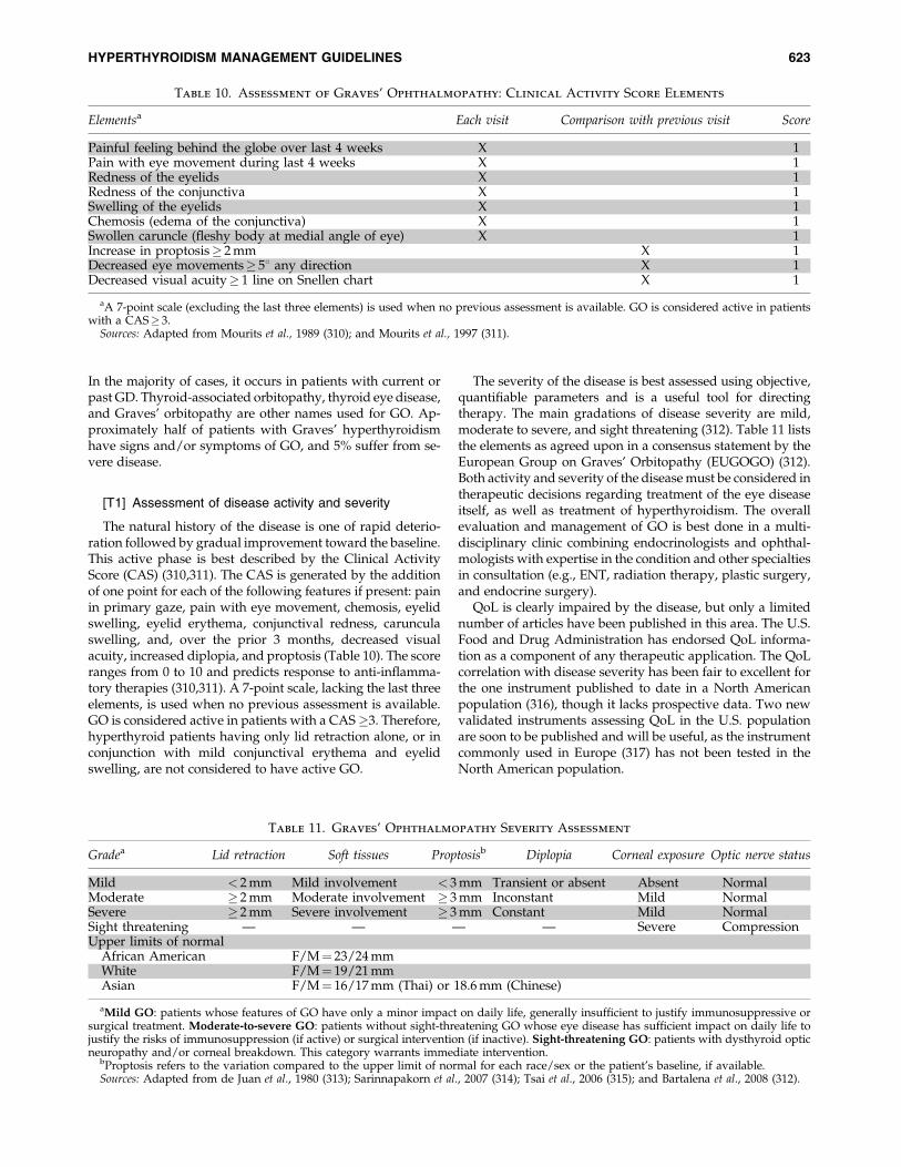
In the majority of cases, it occurs in patients with current orpast GD. Thyroid-associated orbitopathy, thyroid eye disease,and Graves’ orbitopathy are other names used for GO. Ap-proximately half of patients with Graves’ hyperthyroidismhave signs and/or symptoms of GO, and 5% suffer from se-vere disease.
[T1] Assessment of disease activity and severity
The natural history of the disease is one of rapid deterio-ration followed by gradual improvement toward the baseline.This active phase is best described by the Clinical ActivityScore (CAS) (310,311). The CAS is generated by the additionof one point for each of the following features if present: painin primary gaze, pain with eye movement, chemosis, eyelidswelling, eyelid erythema, conjunctival redness, carunculaswelling, and, over the prior 3 months, decreased visualacuity, increased diplopia, and proptosis (Table 10). The scoreranges from 0 to 10 and predicts response to anti-inflamma-tory therapies (310,311). A 7-point scale, lacking the last threeelements, is used when no previous assessment is available.GO is considered active in patients with a CAS�3. Therefore,hyperthyroid patients having only lid retraction alone, or inconjunction with mild conjunctival erythema and eyelidswelling, are not considered to have active GO.
The severity of the disease is best assessed using objective,quantifiable parameters and is a useful tool for directingtherapy. The main gradations of disease severity are mild,moderate to severe, and sight threatening (312). Table 11 liststhe elements as agreed upon in a consensus statement by theEuropean Group on Graves’ Orbitopathy (EUGOGO) (312).Both activity and severity of the disease must be considered intherapeutic decisions regarding treatment of the eye diseaseitself, as well as treatment of hyperthyroidism. The overallevaluation and management of GO is best done in a multi-disciplinary clinic combining endocrinologists and ophthal-mologists with expertise in the condition and other specialtiesin consultation (e.g., ENT, radiation therapy, plastic surgery,and endocrine surgery).
QoL is clearly impaired by the disease, but only a limitednumber of articles have been published in this area. The U.S.Food and Drug Administration has endorsed QoL informa-tion as a component of any therapeutic application. The QoLcorrelation with disease severity has been fair to excellent forthe one instrument published to date in a North Americanpopulation (316), though it lacks prospective data. Two newvalidated instruments assessing QoL in the U.S. populationare soon to be published and will be useful, as the instrumentcommonly used in Europe (317) has not been tested in theNorth American population.
Table 10. Assessment of Graves’ Ophthalmopathy: Clinical Activity Score Elements
Elementsa Each visit Comparison with previous visit Score
Painful feeling behind the globe over last 4 weeks X 1Pain with eye movement during last 4 weeks X 1Redness of the eyelids X 1Redness of the conjunctiva X 1Swelling of the eyelids X 1Chemosis (edema of the conjunctiva) X 1Swollen caruncle (fleshy body at medial angle of eye) X 1Increase in proptosis� 2 mm X 1Decreased eye movements� 58 any direction X 1Decreased visual acuity� 1 line on Snellen chart X 1
aA 7-point scale (excluding the last three elements) is used when no previous assessment is available. GO is considered active in patientswith a CAS� 3.
Sources: Adapted from Mourits et al., 1989 (310); and Mourits et al., 1997 (311).
Table 11. Graves’ Ophthalmopathy Severity Assessment
Gradea Lid retraction Soft tissues Proptosisb Diplopia Corneal exposure Optic nerve status
Mild < 2 mm Mild involvement < 3 mm Transient or absent Absent NormalModerate � 2 mm Moderate involvement � 3 mm Inconstant Mild NormalSevere � 2 mm Severe involvement � 3 mm Constant Mild NormalSight threatening — — — — Severe CompressionUpper limits of normal
African American F/M¼ 23/24 mmWhite F/M¼ 19/21 mmAsian F/M¼ 16/17 mm (Thai) or 18.6 mm (Chinese)
aMild GO: patients whose features of GO have only a minor impact on daily life, generally insufficient to justify immunosuppressive orsurgical treatment. Moderate-to-severe GO: patients without sight-threatening GO whose eye disease has sufficient impact on daily life tojustify the risks of immunosuppression (if active) or surgical intervention (if inactive). Sight-threatening GO: patients with dysthyroid opticneuropathy and/or corneal breakdown. This category warrants immediate intervention.
bProptosis refers to the variation compared to the upper limit of normal for each race/sex or the patient’s baseline, if available.Sources: Adapted from de Juan et al., 1980 (313); Sarinnapakorn et al., 2007 (314); Tsai et al., 2006 (315); and Bartalena et al., 2008 (312).
HYPERTHYROIDISM MANAGEMENT GUIDELINES 623

In the remainder of section T, we discuss the prevention ofGO and the management of hyperthyroidism in patientshaving established GO. In particular, we focus on recom-mendations regarding the concurrent use of corticosteroids inpatients choosing radioactive iodine as treatment for hyper-thyroidism (Table 12).
[T2] Prevention of GO
Current therapeutic approaches to GO, including localmeasures, corticosteroids, orbital radiation, and surgery(312), often fail to significantly improve the QoL of patientswith this debilitating condition. Therefore, efforts shouldbe made to prevent the development or progression of GOin patients with Graves’ hyperthyroidism. Identified riskfactors for GO include radioiodine therapy for hyperthy-roidism (318,319), smoking, high pretreatment T3 values(�325 ng/dL or �5 nmol/L) (319), high serum pretreatmentTRAb levels (>50% TBII inhibition or TSI >8.8 IU/Liter)(320), and hypothyroidism following radioiodine treatment(321).
& RECOMMENDATION 80Euthyroidism should be expeditiously achieved andmaintained in hyperthyroid patients with GO or risk fac-tors for the development of ophthalmopathy. 1/++0
A number of studies have suggested that development ofpersistent, untreated hypothyroidism after therapy for hy-perthyroidism plays a detrimental role in the progression ofGO. An early study noted that patients who were either hypo-or hyperthyroid had more severe GO than euthyroid patients(322). Subsequently, two cohort studies in which patients re-ceived levothyroxine therapy early after radioactive iodinewith the specific intent of preventing hypothyroidism notedthat deterioration of GO rarely occurred (0%–2%) (321,323). Arandomized study of newly diagnosed GD found that ra-dioactive iodine did not increase the risk of worsening GOcompared to therapy with MMI (RR of 0.95) in the settingwhere hypothyroidism was actively prevented by adminis-tration of thyroid hormone at 2 weeks after radioactive iodineadministration (49).
& RECOMMENDATION 81In nonsmoking patients with Graves’ hyperthyroidismwho have no clinically apparent ophthalmopathy, 131Itherapy without concurrent steroids, methimazole, orthyroidectomy should be considered equally acceptabletherapeutic options. 1/++0
Several retrospective cohort studies and randomized trialshave identified the risk of GO development or progressionafter therapy for hyperthyroidism to be between 15% and 33%.Two randomized controlled trials found that risk to be 23/150(15%) for radioactive iodine, compared with 4/148 (3%) forATDs (318) in one study, and 13/39 (33%) for radioactive io-dine compared with 4/38 (10%) for ATDs and 6/37 (16%) forsurgery (319) in the other study. In contrast, one prospectivebut nonrandomized cohort study identified no differenceamong ATD, surgery, and radioactive iodine treatment, withan overall 4.9%–7.1% frequency of GO development (324). Thehigher risk of GO worsening after radioactive iodine therapy inthe majority of studies may be related to the unique increase inTRAb levels observed following this therapy (222). Experi-mental evidence suggests that these antibodies may be directlyinvolved in GO pathogenesis (309).
There is evidence that corticosteroids given concurrentlywith radioiodine therapy may prevent worsening of GO inpatients with mild active eye disease (318). However, there isinsufficient evidence to recommend prophylactic treatmentwith corticosteroids in nonsmoking patients who do not haveclinically apparent GO. The relatively low absolute risk ofnonsmokers developing new-onset severe GO suggests thatGO prevention should not be a factor in the selection oftherapy for hyperthyroidism in this group of patients (318).
There is insufficient evidence to recommend for or againstthe use of prophylactic corticosteroids in smokers who have noevidence of GO. However, in two different studies, activesmokers who received radioactive iodine represented the groupwith the highest incidence (23%–40%) of new GO or deteriora-tion of pre-existing GO during 1 year of follow-up (49,318).
& RECOMMENDATION 82Clinicians should advise patients with GD to stop smokingand refer them to a structured smoking cessation program.
Table 12. Use of Oral Glucocorticoids for Prevention of Graves’ Ophthalmopathy Development
or Progression When Radioactive Iodine Is Used to Treat Graves’ Hyperthyroidism
RAI without glucocorticoids RAI with oral glucocorticoids
No GO (nonsmoker) Recommend Recommend againstNo GO (smoker) Insufficient data to recommend
for or againstGO present-active and mild (nonsmoker) Acceptablea Acceptablea
GO present-active and mild (smoker) Recommend against RecommendGO present-active and moderate-to-severe or
sight-threatening (smoker or nonsmoker)Recommend against Insufficient data to recommend
for or againstGO present-inactive (smoker or nonsmoker) Recommend Recommend against
Methimazole or thyroidectomy are also recommended treatment options in each of these scenarios, and they are the preferred choice oftherapy in patients with active and moderate-to-severe or sight-threatening GO.
aThe decision regarding use of concurrent glucocorticoids should be made in light of the risk-benefit ratio relative to the patient’s overallhealth. Risk factors for GO deterioration (high T3 level, high TRAb level, smoking) increase the benefit of glucocorticoids in preventing GOdeterioration. Poorly controlled diabetes, osteoporosis, psychiatric illness, high risk for infections increase the likelihood of complicationsfrom glucocorticoids.
624 BAHN ET AL.

Patients exposed to secondhand smoke should be identi-fied and advised of its negative impact. 1/++0
Smoking is the most important known risk factor for thedevelopment or worsening of GO, unrelated to type of ther-apy for GO (322), and consistent data from several studiesshow a detrimental effect of smoking on GO in patientstreated with radioactive iodine (49,318). The risk is propor-tional to the number of cigarettes smoked per day and formersmokers have significantly lower risk than current smokers,even after adjusting for lifetime cigarette consumption (325).
Technical remarks: Clinicians should consult guidelines oneffective and evidence-based approaches to aid in smokingcessation and avoidance of secondhand smoke (326,327).
[T3] Treatment of hyperthyroidism in patients with activeGO of mild severity (see Tables 10 and 11 for definitions ofdisease activity and severity)
& RECOMMENDATION 83In patients with Graves’ hyperthyroidism who have mildactive ophthalmopathy and no risk factors for deteriorationof their eye disease, 131I therapy, methimazole, and thy-roidectomy should be considered equally acceptable ther-apeutic options. 1/++0
& RECOMMENDATION 84Patients with Graves’ hyperthyroidism and mild activeophthalmopathy who have no other risk factors for dete-rioration of their eye disease and choose radioactive iodinetherapy should be considered for concurrent treatmentwith corticosteroids. 2/++0
Technical remarks: The decision whether or not to admin-ister concurrent glucocorticoids in a particular patientchoosing 131I therapy should be made in light of the risk–benefit ratio (i.e., their personal risk of worsening GO,balanced against their risk of developing glucocorticoid sideeffects). Risk factors for side effects of oral corticosteroidsinclude poorly controlled diabetes, hypertension, osteopo-rosis, psychiatric disease, and predisposition to infections.Smokers in whom the risk–benefit ratio for the concurrentuse of corticosteroids is high may be better treated withmethimazole or surgery. Besides smoking, risk factors fordeterioration of GO following radioiodine therapy includehigh pretreatment T3 values (�325 ng/dL or �5 nmol/L)(319), active and progressive GO over the preceding 3months, high serum pretreatment thyrotropin antibodylevels (>50% TBII inhibition or TSI >8.8 IU/L), and devel-opment of hypothyroidism following the treatment (321).
The recommended corticosteroid dose for GO prophylaxisis the equivalent of prednisone 0.4–0.5 mg/kg/day, started 1–3 days after radioactive iodine treatment, continued for 1month, and then tapered over 2 months (312). However, arecent retrospective cohort study suggested that lower dosesand shorter duration of oral prednisone (about 0.2 mg/kg/day for 6 weeks) may be equally effective for preventionof GO exacerbation in patients with initially mild or absenteye disease, if supported by future randomized clinicaltrials (328).
& RECOMMENDATION 85Patients with Graves’ hyperthyroidism and mild activeophthalmopathy who are smokers or have other risk fac-tors for GO and choose radioactive iodine therapy shouldreceive concurrent corticosteroids. 1/++0
A randomized study of patients having pre-existing GO ofmild severity found the relative risk for deterioration of eyedisease to be 2.2 for surgery and 1.9 for radioactive iodinecompared with ATDs, though the patients were not ran-domized with respect to their baseline GO status (319). Anearlier prospective cohort (also not randomized as to baselineGO or smoking status and in which post-treatment hypo-thyroidism was not actively prevented) identified no differ-ence in deterioration of pre-existing GO between the threemodes of therapy (324). Neither surgery nor radioactive io-dine therapy was associated with deterioration in pre-existingGO in 48 patients in another early study (329).
One large randomized controlled trial studying mainlypatients with previously treated GD showed radioactive io-dine therapy to be associated with an increased risk of GOprogression (RR of 5.8 in comparison with ATDs) and foundthat risk to be eliminated with concurrent corticosteroid ad-ministration (318).
[T4] Treatment of hyperthyroidism in patients with activeand moderate-to-severe or sight-threatening GO (see Tables10 and 11 for definitions of disease activity and severity)
& RECOMMENDATION 86Patients with Graves’ hyperthyroidism and active moderate-to-severe or sight-threatening ophthalmopathy should betreated with either methimazole or surgery. 1/+00
We are aware of no trials in patients with moderate-to-severe and active eye disease that compare hyperthyroid-ism therapies for impact on GO. However, a comparison oftwo different surgical approaches (total thyroidectomy vs.subtotal thyroidectomy) for patients with moderate-to-se-vere GO showed that the eye disease improved over 3 yearsof follow-up in all patients (330). In another series of 42patients with progressive GO treated with total thyroidec-tomy, exophthalmos was stable in 60% of cases and im-proved in the remainder (331), suggesting that surgery isnot detrimental to GO and may be associated with im-provement in some patients. Other studies suggest thatATDs may not adversely impact mild active GO, but do notaddress severe GO (318).
Technical remarks: Radioactive iodine therapy is a less de-sirable option in these patients and, if used, concurrent ste-roids should be administered.
[T5] Treatment of GD in patients with inactive GO (seeTable 10 for definition of disease inactivity)
& RECOMMENDATION 87In patients with Graves’ hyperthyroidism and inactiveophthalmopathy, we suggest that 131I therapy withoutconcurrent corticosteroids, methimazole, and thyroidec-tomy are equally acceptable therapeutic options. 2/++0
HYPERTHYROIDISM MANAGEMENT GUIDELINES 625

A series of 72 patients with inactive GO according to theCAS were treated with radioactive iodine without concurrentglucocorticoid administration (323). In those whom hypo-thyroidism was prevented by early thyroxine therapy, nodeterioration in eye disease was reported (323). Smokinghistory did not impact GO outcome in this cohort.
A recent retrospective study examined the impact of con-current oral or intravenous glucocorticoid therapy on the pre-valence of reactivation of GO after radioiodine therapy inpatients having inactive GO (332). They identified GO activa-tion in approximately 7% of patients considered at low riskwho were given no steroid prophylaxis. Despite prophylaxis,33% of patients considered at high risk who were treated withoral glucocorticoids had worsening of GO. Only intravenousglucocorticoids were effective in preventing GO reactivation.However, because of the retrospective nature of this study andthe lack of prespecified criteria for dose and route of steroid usein those considered at risk, we did not include these data in ourdeliberations regarding the above recommendation.
[U] How should overt drug-inducedthyrotoxicosis be managed?
Although numerous medications may affect thyroid func-tion or cause abnormal thyroid testing results (333), relativelyfew of these actually cause thyrotoxicosis. For those that do,three mechanisms are involved: (i) iodine-induced thyrotox-icosis; (ii) destructive thyroiditis; and (iii) induction of thyroidautoimmunity (GD or painless thyroiditis). More than onepathway has been identified for several medications. Asummary of drugs causing thyrotoxicosis, the proposedmechanism(s), approximate timing of onset, duration, andtherapeutic options is provided in Table 13.
[U1] Iodine-induced hyperthyroidism
Iodine-induced hyperthyroidism is believed to occur inpatients with underlying thyroid autonomy, especially thoseliving in areas with mild-to-moderate iodine deficiency. Inone study of 788 patients undergoing cardiac angiography,none of the 27 with a suppressed TSH at baseline developedovert hyperthyroidism, and only 2 patients with no apparentrisk factors became hyperthyroid (334). A retrospective studyfound that 7 of 28 elderly patients with hyperthyroidism had ahistory of recent iodine exposure (335), and a prospectivestudy found that 2 of 73 patients developed hyperthyroidismafter radiographic contrast (336). High iodine intake may alsobe followed by relapse of hyperthyroidism in patients withprevious GD who are in remission after ATD therapy. In asmall study of 10 patients, 2 had relapse of overt hyperthy-roidism, and 2 developed SH after stopping high iodine in-take (337).
& RECOMMENDATION 88Beta-adrenergic blocking agents alone or in combinationwith methimazole should be used to treat overt iodine-induced hyperthyroidism. 1/+00
Iodine-induced hyperthyroidism (the Jod-Basedow phe-nomenon) is usually self-limited, lasting 1–18 months(335,338). Treatment includes avoidance of additional iodineand administration of beta-blockers alone or with ATDs, de-pending on the severity of hyperthyroidism. Radioactive io-dine is not an option until the iodine load has been cleared,which may take several months depending on the length ofexposure to iodine. Surgery may be used in patients allergic orresistant to antithyroid drugs.
Table 13. Causes of Drug-Associated Thyrotoxicosis
Drug Mechanism(s)Timing of onset following
initiation of the drug Therapy
Amiodarone Iodine induced (type 1) Months to Years Supportive carea
Antithyroid drugs; perchlorateb
SurgeryThyroiditis (type 2) Often> 1 year Supportive carea
CorticosteroidsSurgery
Lithium Painless thyroiditis Often> 1 year Supportive carea
Antithyroid drugsInterferon a Painless thyroiditis; GD Months Supportive carea
Antithyroid drugs and/or radioactiveiodine (GD only)
Interleukin-2 Painless thyroiditis; GD Months Supportive carea
Antithyroid drugs and/or radioactiveiodine (GD only)
Iodinated contrast Underlying thyroidautonomy
Weeks to months Antithyroid drugs
Radioactive iodine, early Destruction 1–4 weeks Observation; if severe, administercorticosteroids
Radioactive iodinefor TMNG, late
GD 3–6 months Antithyroid drugsRepeat radioactive iodineSurgery
aSupportive care may include beta-adrenergic blockers during the thyrotoxic stage and levothyroxine if hypothyroidism develops.bNot available in the United States.
626 BAHN ET AL.

Technical remarks: Dosing of MMI for iodine-induced thy-rotoxicosis is 20–40 mg daily, given either as a daily or twice-daily dosing. There may be relative resistance to antithyroiddrugs in patients with iodine-induced hyperthyroidism. Ur-inary iodine may be monitored to assess the rate of clearanceof the iodine load.
[U2] Cytokine-induced thyrotoxicosis
& RECOMMENDATION 89Patients who develop thyrotoxicosis during therapy withinterferon-a or interleukin-2 should be evaluated to deter-mine etiology (thyroiditis vs. GD) and treated accordingly.1/+00
Interferon-a (IFN-a)- and interleukin-2-treated patientsare at increased risk for developing thyrotoxicosis, especiallythose with pre-existing thyroid autoimmunity. Thyrotoxicosisin this setting can be due to either painless thyroiditis orGD (339). In a literature review, 69% of patients with IFN-a-associated thyrotoxicosis were deemed to have GD as theetiology (340).
A meta-analysis found that 46% of patients with positivepretreatment thyroid peroxidase antibodies (TPO Ab) devel-oped thyroid dysfunction after IFN-a therapy for hepatitis Cinfection, compared to only 5% of those with negative anti-bodies (341).
[U3] Amiodarone-induced thyrotoxicosis
& RECOMMENDATION 90We suggest monitoring thyroid function tests before and at1 and 3 months following the initiation of amiodaronetherapy, and at 3–6 month intervals thereafter. 2/+00
Amiodarone is a drug frequently used in the treatment ofrefractory atrial or ventricular tachyarrhymias. Amiodarone-induced thyrotoxicosis (AIT) occurs in up to 6% of patientstaking this medication in iodine-sufficient areas of the world(18,342,343) and in up to 10% in iodine-deficient areas, such asparts of Europe (344).
& RECOMMENDATION 91We suggest testing to distinguish type 1 (iodine-induced)from type 2 (thyroiditis) varieties of amiodarone-inducedthyrotoxicosis. 1/+00
Two basic mechanisms have been identified in the devel-opment of AIT, including an iodine-induced form of hyper-thyroidism (type 1 AIT, or goitrous AIT) due to the highiodine content of amiodarone (37% by molecular weight), andtype 2 AIT, which is a destructive thyroiditis. Type 1 AITtends to occur in patients with underlying thyroid autonomyin a nodular goiter, but the term is also used when amiodar-one use is associated with GD, whereas type 2 AIT is due to adirect destructive effect of amiodarone on thyrocytes. RAIU isoccasionally measurable in type 1 AIT (particularly in regionsof iodine deficiency), but not in type 2 AIT. Increased vascularflow on color-flow Doppler ultrasound study may be seen inpatients with type 1 AIT, but not type 2 AIT. Measurement ofserum interleukin-6 levels does not reliably distinguish be-
tween the two types of AIT (345). The distinction betweentype 1 AIT and type 2 AIT is not always clear, and somepatients have elements of both types (18).
& RECOMMENDATION 92The decision to stop amiodarone in the setting of thyro-toxicosis should be determined on an individual basisin consultation with a cardiologist, based on the presenceor absence of effective alternative antiarrhythmic therapy.1/+00
The need for amiodarone discontinuation is controversialbecause (i) this drug is frequently the only medication able tocontrol cardiac arrhythmia, (ii) the effects of this fat solubledrug may persist for many months, and (iii) amiodarone mayhave T3-antagonistic properties at the cardiac level and inhibitT4 to T3 conversion, such that withdrawal may actually ag-gravate cardiac manifestations of thyrotoxicosis (18,342). Inaddition, type 2 AIT typically resolves even if amiodaronetherapy is continued.
& RECOMMENDATION 93Methimazole should be used to treat type 1 amiodarone-induced thyrotoxicosis and corticosteroids should be usedto treat type 2 amiodarone-induced thyrotoxicosis. 1/+00
& RECOMMENDATION 94Combined antithyroid drug and anti-inflammatory ther-apy should be used to treat patients with overt amiodar-one-induced thyrotoxicosis who fail to respond to singlemodality therapy, and patients in whom the type of diseasecannot be unequivocally determined. 1/+00
Type 1 AIT is best treated with MMI (40 mg daily) to pre-vent new hormone synthesis and, rarely, with added potas-sium perchlorate (250 mg four times daily; not available in theUnited States) (346). Type 2 AIT is better treated with anti-inflammatory therapy such as prednisone (40 mg daily) withimprovement occasionally seen as early as 1 week, and usu-ally within a few weeks (346).
In one study, 20 patients with AIT, including both type 1and type 2 subtypes, were treated with perchlorate for1 month to inhibit thyroid iodide transport, resulting ineuthyroidism in 12 patients (7 with type 1 AIT and 5 with type2 AIT). Corticosteroids were then given to the eight nonre-sponders, and euthyroidism was achieved in all after anaverage of approximately 6 weeks (347). When a clear dis-tinction between type 1 AIT and type 2 AIT is not possible, acombination of prednisone and methimazole should be useduntil the patient has stabilized, at which time the drugs maybe individually tapered. Thyroidectomy may be required inpatients who prove refractory to medical therapy (348).
Technical remarks: The suggested starting dose of MMI inthis setting is 40 mg once daily until the patient is euthyroid(generally 3–6 months). If high doses of MMI continue to berequired, splitting the dose may be more effective. The sug-gested dose of corticosteroids in this setting is equivalent to40 mg prednisone given once daily for 2–4 weeks, followed bya gradual taper over 2–3 months, based on the patient’s clin-ical response.
HYPERTHYROIDISM MANAGEMENT GUIDELINES 627
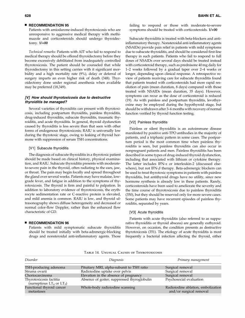
& RECOMMENDATION 95Patients with amiodarone-induced thyrotoxicosis who areunresponsive to aggressive medical therapy with methi-mazole and corticosteroids should undergo thyroidec-tomy. 1/+00
Technical remarks: Patients with AIT who fail to respond tomedical therapy should be offered thyroidectomy before theybecome excessively debilitated from inadequately controlledthyrotoxicosis. The patient should be counseled that whilethyroidectomy in this setting carries with it significant mor-bidity and a high mortality rate (9%), delay or deferral ofsurgery imparts an even higher risk of death (348). Thyr-oidectomy done under regional anesthesia when availablemay be preferred (18,349).
[V] How should thyrotoxicosis due to destructivethyroiditis be managed?
Several varieties of thyroiditis can present with thyrotoxi-cosis, including postpartum thyroiditis, painless thyroiditis,drug-induced thyroiditis, subacute thyroiditis, traumatic thy-roiditis, and acute thyroiditis. In general, thyroid dysfunctioncaused by thyroiditis is less severe than that seen with otherforms of endogenous thyrotoxicosis; RAIU is universally lowduring the thyrotoxic stage, owing to leaking of thyroid hor-mone with suppression of serum TSH concentrations.
[V1] Subacute thyroiditis
The diagnosis of subacute thyroiditis in a thyrotoxic patientshould be made based on clinical history, physical examina-tion, and RAIU. Subacute thyroiditis presents with moderate-to-severe pain in the thyroid, often radiating to the ears, jaw,or throat. The pain may begin focally and spread throughoutthe gland over several weeks. Patients may have malaise, low-grade fever, and fatigue in addition to the symptoms of thy-rotoxicosis. The thyroid is firm and painful to palpation. Inaddition to laboratory evidence of thyrotoxicosis, the eryth-rocyte sedimentation rate or C-reactive protein is elevated,and mild anemia is common. RAIU is low, and thyroid ul-trasonography shows diffuse heterogeneity and decreased ornormal color-flow Doppler, rather than the enhanced flowcharacteristic of GD.
& RECOMMENDATION 96Patients with mild symptomatic subacute thyroiditisshould be treated initially with beta-adrenergic-blockingdrugs and nonsteroidal anti-inflammatory agents. Those
failing to respond or those with moderate-to-severesymptoms should be treated with corticosteroids. 1/+00
Subacute thyroiditis is treated with beta-blockers and anti-inflammatory therapy. Nonsteroidal anti-inflammatory agents(NSAIDs) provide pain relief in patients with mild symptomsdue to subacute thyroiditis, and should be considered first-linetherapy in such patients. Patients who fail to respond to fulldoses of NSAIDs over several days should be treated insteadwith corticosteroid therapy, such as prednisone 40 mg daily for1–2 weeks followed by a gradual taper over 2–4 weeks orlonger, depending upon clinical response. A retrospective re-view of patients receiving care for subacute thyroiditis foundthat patients treated with corticosteroids had more rapid res-olution of pain (mean duration, 8 days) compared with thosetreated with NSAIDs (mean duration, 35 days). However,symptoms can recur as the dose of corticosteroid is reduced(19). As with painless and postpartum thyroiditis, levothyr-oxine may be employed during the hypothyroid stage, butshould be withdrawn after 3–6 months with recovery of normalfunction verified by thyroid function testing.
[V2] Painless thyroiditis
Painless or silent thyroiditis is an autoimmune diseasemanifested by positive anti-TPO antibodies in the majority ofpatients, and a triphasic pattern in some cases. The postpar-tum period is the most common time when painless thy-roiditis is seen, but painless thyroiditis can also occur innonpregnant patients and men. Painless thyroiditis has beendescribed in some types of drug-induced thyroid dysfunction,including that associated with lithium or cytokine therapy.The latter includes IFN-a or interleukin-2 (discussed else-where), but not IFN-b therapy. Beta-adrenergic blockers canbe used to treat thyrotoxic symptoms in patients with painlessthyroiditis, but antithyroid drugs have no utility, since newhormone synthesis is already low in these patients. Rarely,corticosteroids have been used to ameliorate the severity andthe time course of thyrotoxicosis due to painless thyroiditis(350), but they should be reserved only for more severe cases.Some patients may have recurrent episodes of painless thy-roiditis, separated by years.
[V3] Acute thyroiditis
Patients with acute thyroiditis (also referred to as suppu-rative thyroiditis or thyroid abscess) are generally euthyroid.However, on occasion, the condition presents as destructivethyrotoxicosis (351). The etiology of acute thyroiditis is mostfrequently a bacterial infection affecting the thyroid, either
Table 14. Unusual Causes of Thyrotoxicosis
Disorder Diagnosis Primary management
TSH-producing adenoma Pituitary MRI, alpha-subunit to TSH ratio Surgical removalStruma ovarii Radioiodine uptake over pelvis Surgical removalChoriocarcinoma Elevation in the absence of pregnancy Surgical removalThyrotoxicosis factitia
(surreptious LT4 or LT3)Absence of goiter; suppressed thyroglobulin Psychosocial evaluation
Functional thyroid cancermetastases
Whole-body radioiodine scanning Radioiodine ablation, embolizationand/or surgical removal
628 BAHN ET AL.

through hematogenous spread or direct extension through afistula from an infected pyriform sinus. Therapy involves sys-temic antibiotics as well as abscess drainage or removal, andexcision or occlusion of the offending pyriform sinus. Thyr-otoxicosis should be treated symptomatically with beta-block-ing agents. As in other forms of destructive thyroiditis, there isno role for antithyroid drugs.
[W] How should thyrotoxicosis due to unusualcauses be managed?
These are several unusual causes of thyrotoxicosis thatshould be considered in the differential diagnosis (Table 14).Since effective treatment depends on accurate diagnosis, it isimportant to clearly identify the etiology in every patientpresenting with thyrotoxicosis.
[W1] TSH-secreting pituitary tumors
Functional pituitary tumors secreting TSH are rare. In amulticenter review of 4400 pituitary tumors seen over a 25-year period, 43 (1%) were TSH-secreting adenomas (33). Themajority of patients present with diffuse goiter and clinicalsigns of thyrotoxicosis. In addition, serum TSH levels may beelevated or, especially in patients who have not had thyroidablation, they may be inappropriately normal. Cosecretionof either prolactin or growth hormone occurs in up to 25% ofcases; 1%–2% secrete both growth hormone and prolactin,and a similar percentage cosecrete gonadotropins. MostTSH-producing adenomas are larger than 1 cm, and ap-proximately 40% of patients have associated visual fielddeficits (352).
& RECOMMENDATION 97The diagnosis of TSH-secreting pituitary tumor should bebased on an inappropriately normal or elevated serum TSHlevel associated with elevated free T4 estimates and T3
concentrations, usually associated with the presence of apituitary tumor on MRI and the absence of a family historyor genetic testing consistent with thyroid hormone resis-tance in a thyrotoxic patient. 1/+00
Distinction between a TSH-secreting adenoma and thyroidhormone resistance is important, since thyroid function testresults are similar, yet management is quite different forthese two disorders. TSH-secreting adenomas are more likelyto have concurrent alpha-subunit elevation (not useful inpostmenopausal women due to concurrent gonadotropinelevation), a blunted TSH response to thyrotropin-releasinghormone (TRH) (when available), elevated sex-hormone-binding globulin and resting energy expenditure, and clinicalevidence of thyrotoxicosis, as well as an anatomic abnor-mality on MRI of the pituitary.
Technical remarks: Genetic testing for thyroid hormone re-sistance is commercially available and may be useful inequivocal cases, especially in those patients without familymembers available for thyroid function testing.
Surgery is generally the mainstay of therapy for TSH-pro-ducing pituitary tumors. The patient should be made euthy-roid preoperatively. Long-term ATD therapy should beavoided. Preoperative adjunctive therapy with octreotide and
dopamine agonist therapy has been examined. Treatment withoctreotide results in a >50% reduction in serum TSH values inthe majority of patients treated, and a concurrent return toeuthyroidism in most (33). A reduction in tumor size has beenobserved in 20%–50% of patients treated with octreotide(33,352), but less impressive results have been obtained withbromocriptine therapy (352). Sterotactic or conventional ra-diotherapy has also been used in cases that prove refractory tomedical therapy. For patients with TSH-producing adenomaswho are considered poor surgical candidates, primary medicaltherapy with octreotide can be considered.
& RECOMMENDATION 98Patients with TSH-secreting pituitary adenomas shouldundergo surgery performed by an experienced pituitarysurgeon. 1/+00
Technical remarks: Postoperative adjunctive therapy withoctreotide and/or external beam radiation therapy may beuseful in managing patients with persistent central hyper-thyroidism after a debulking procedure for nonresectableTSH-secreting adenomas (33).
[W2] Struma ovarii
& RECOMMENDATION 99Patients with struma ovarii should be treated initially withsurgical resection. 1/+00
Struma ovarii, defined as ectopic thyroid tissue existing as asubstantial component of an ovarian tumor, is quite rare, re-presenting <1% of all ovarian tumors. Approximately 5%–10% of patients with struma ovarii present with thyrotoxicosis(353) due to either autonomous ectopic thyroid function or thecoexistence of GD, and up to 25% of struma ovarii tumorscontain elements of papillary thyroid cancer. Patients previ-ously treated for GD may have persistent or recurrent hy-perthyroidism due to the action of TRAb on the ectopicthyroid tissue (354). Treatment of struma ovarii generally in-volves surgical removal, performed largely due to the risk ofmalignancy within the struma tissue and of curing the hy-perthyroidism. Preoperative treatment with beta-adrenergic-blocking agents and antithyroid drugs is warranted to restoreeuthyroidism before surgery.
Technical remarks: In cases of suspected metastatic malig-nant struma ovarii, radioactive iodine is generally given fol-lowing surgical removal of both the ovarian tumor and thepatient’s thyroid to facilitate delivery of isotope to any po-tential residual malignant cells.
[W3] Choriocarcinoma
& RECOMMENDATION 100Treatment of hyperthyroidism due to choriocarcinomashould include both methimazole and treatment directedagainst the primary tumor. 1/+00
Patients with choriocarcinoma, including molar pregnancyand testicular cancer, may present with thyrotoxicosis due tothe effect of tumor-derived hCG upon the TSH receptor
HYPERTHYROIDISM MANAGEMENT GUIDELINES 629

(355,356). This cross-stimulation only occurs at very high levelsof hCG, since hCG is only a weak agonist for the TSH receptor.Treatment of hyperthyroidism due to choriocarcinoma in-volves both treatment directed against the primary tumor andtreatment designed to prevent the thyroid from responding tohCG stimulation, such as with antithyroid drugs.
[W4] Thyrotoxicosis factitia
Thyrotoxicosis factitia includes all causes of thyrotoxicosisdue to the ingestion of thyroid hormone. This may includeintentional ingestion of thyroid hormone either surrepti-tiously or iatrogenically, as well as unintentional ingestioneither accidentally, such as in pediatric poisoning or phar-macy error, or through ingestion of supplements that containthyroid extracts. Historically, accidental thyroid hormoneingestion has occurred as a result of eating meat contaminatedwith animal thyroid tissue (‘‘hamburger thyrotoxicosis’’)(357). Whereas iatrogenic causes of thyrotoxicosis factitia areeasily identified, surreptitious use of thyroid hormone maypresent a diagnostic quandary. Clues to this diagnosis are anabsence of goiter, a suppressed serum thyroglobulin level,and a decreased uptake of radioactive iodine. A dis-proportionately elevated T3 level suggests that the patient maybe ingesting liothyronine or a combination T4/T3 preparation.
[W5] Functional thyroid cancer metastases
Thyrotoxicosis due to functional metastases in patientswith thyroid cancer has been described in a handful of cases.Typically, patients have either a very large primary follicularcancer or widely metastatic follicular thyroid cancer, and mayhave coexisting TRAb as the proximate cause of the thyro-toxicosis (358). More recently, thyrotoxicosis has been re-ported following multiple injections of recombinant humanTSH in patients with metastatic thyroid cancer in preparationfor imaging. In general, functioning metastasis are treatedwith radioactive iodine with the addition of ATDs as neededfor persistent hyperthyroidism. Recombinant human TSHshould be avoided in these patients.
Acknowledgments
The task force wishes to thank Ms. Bobbi Smith, ExecutiveDirector, ATA, and Ms. Sheri Slaughter, Assistant to the taskforce, for their expert help and support.
Disclosure Statement
Disclosure Information for 2 years before May 2010 and theknown future as of May 2010.
D.R. is a consultant for Abbott Laboratories and has re-ceived research grant support from Genzyme. For all otherauthors, no competing financial interests exist.
References
1. Singer PA, Cooper DS, Levy EG, Ladenson PW, BravermanLE, Daniels G, Greenspan FS, McDougall IR, Nikolai TF1995 Treatment guidelines for patients with hyperthyroid-ism and hypothyroidism. JAMA 273:808–812.
2. Baskin HJ, Cobin RH, Duick DS, Gharib H, Guttler RB,Kaplan MM, Segal RL; American Association of ClinicalEndocrinologists 2002 American Association of ClinicalEndocrinologists medical guidelines for clinical practicefor the evaluation and treatment of hyperthyroidism andhypothyroidism. Endocr Pract 6:457–469.
3. Atkins D, Best D, Briss PA, Eccles M, Falck-Ytter Y, FlottorpS, Guyatt GH, Harbour RT, Haugh MC, Henry D, Hill S,Jaeschke R, Leng G, Liberati A, Magrini N, Mason J, Mid-dleton P, Mrukowicz J, O’Connell D, Oxman AD, PhillipsB, Schunemann HJ, Edejer TT, Varonen H, Vist GE, Wil-liams JW Jr., Zaza S; GRADE Working Group 2004 Gradingquality of evidence and strength of recommendations. BMJ328:1490.
4. Guyatt G, Gutterman D, Baumann MH, Addrizzo-HarrisD, Hylek EM, Phillips B, Raskob G, Lewis SZ, SchunemannH 2006 Grading strength of recommendations and qualityof evidence in clinical guidelines: report from an americancollege of chest physicians task force. Chest 129:174–181.
5. Mechanick JI, Camacho PM, Cobin RH, Garber AJ, GarberJR, Gharib H, Petak SM, Rodbard HW, Trence DL; Amer-ican Association of Clinical Endocrinologists 2010 Ameri-can Association of Clinical Endocrinologists protocol forstandardized production of clinical practice guidelines—2010 update. Endocr Pract 16:270–283.
6. Swiglo BA, Murad MH, Schunemann HJ, Kunz R, VigerskyRA, Guyatt GH, Montori VM 2008 A case for clarity, con-sistency, and helpfulness: state-of-the-art clinical practiceguidelines in endocrinology using the grading of recom-mendations, assessment, development, and evaluationsystem. J Clin Endocrinol Metab 93:666–673.
7. Berghout A, Wiersinga WM, Smits NJ, Touber JL 1990 In-terrelationships between age, thyroid volume, thyroid no-dularity, and thyroid function in patients with sporadicnontoxic goiter. Am J Med 89:602–608.
8. Gozu HI, Lublinghoff J, Bircan R, Paschke R 2010 Geneticsand phenomics of inherited and sporadic non-autoimmunehyperthyroidism. Mol Cell Endocrinol 322:125–134.
9. Martin FI, Deam DR 1996 Hyperthyroidism in elderlyhospitalised patients. Clinical features and outcome. Med JAust 164:200–203.
10. Davis PJ, Davis FB 1974 Hyperthyroidism in patients overthe age of 60 years. Clinical features in 85 patients. Medi-cine (Baltimore) 53:161–181.
11. Tibaldi JM, Barzel US, Albin J, Surks M 1986 Thyrotoxicosisin the very old. Am J Med 81:619–622.
12. Laurberg P, Pedersen KM, Vestergaard H, Sigurdsson G1991 High incidence of multinodular toxic goitre in theelderly population in a low iodine intake area vs. high in-cidence of Graves’ disease in the young in a high iodineintake area: comparative surveys of thyrotoxicosis epide-miology in East-Jutland Denmark and Iceland. J Intern Med229:415–420.
13. Codaccioni JL, Orgiazzi J, Blanc P, Pugeat M, Roulier R,Carayon P 1988 Lasting remissions in patients treated forGraves’ hyperthyroidism with propranolol alone: a patternof spontaneous evolution of the disease. J Clin EndocrinolMetab 67:656–662.
14. Williams I, Ankrett VO, Lazarus JH, Volpe R 1983 Aetiol-ogy of hyperthyroidism in Canada and Wales. J EpidemiolCommunity Health 37:245–248.
15. Gerstein HC 1990 How common is postpartum thyroiditis?A methodologic overview of the literature. Arch Intern Med150:1397–1400.
630 BAHN ET AL.

16. Miller KK, Daniels GH 2001 Association between lithiumuse and thyrotoxicosis caused by silent thyroiditis. ClinEndocrinol (Oxf ) 55:501–508.
17. Roti E, Minelli R, Giuberti T, Marchelli S, Schianchi C,Gardini E, Salvi M, Fiaccadori F, Ugolotti G, Neri TM,Braverman LE 1996 Multiple changes in thyroid function inpatients with chronic active HCV hepatitis treated withrecombinant interferon-alpha. Am J Med 101:482–487.
18. Cohen-Lehman J, Dahl P, Danzi S, Klein I 2010 Effects ofamiodarone therapy on thyroid function. Nat Rev En-docrinology 6:34–41.
19. Fatourechi V, Aniszewski JP, Fatourechi GZ, Atkinson EJ,Jacobsen SJ 2003 Clinical features and outcome of subacutethyroiditis in an incidence cohort: Olmsted County, Min-nesota, study. J Clin Endocrinol Metab 88:2100–2105.
20. Klein I, Danzi S 2007 Thyroid disease and the heart. Cir-culation 116:1725–1735.
21. Burch HB, Wartofsky L 1993 Life-threatening hyperthy-roidism: thyroid storm. Endocrinol Metab Clin North Am22:263–277.
22. Hall P, Lundell G, Holm LE 1993 Mortality in patientstreated for hyperthyroidism with iodine-131. Acta En-docrinol (Cph) 128:230–234.
23. Trzepacz PT, McCue M, Klein I, Levey GS, Greenhouse J1988 A psychiatric and neuropsychological study of pa-tients with untreated Graves’ disease. Gen Hosp Psychiatry10:49–55.
24. Trzepacz PT, Klein I, Roberts M, Greenhouse J, Levey GS 1989Graves’ disease: an analysis of thyroid hormone levels andhyperthyroid signs and symptoms. Am J Med 87:558–561.
25. Boelaert K, Torlinska B, Holder RL, Franklyn JA 2010 Oldersubjects with hyperthyroidism present with a paucity ofsymptoms and signs: a large cross-sectional study. JCEM95:2715–2726.
26. Ventrella S, Klein I 1994 Beta-adrenergic receptor blockingdrugs in the management of hyperthyroidism. The En-docrinologist 4:391–399.
27. European Heart Rhythm Association; Heart Rhythm So-ciety, Fuster V, Ryden LE, Cannom DS, Crijns HJ, CurtisAB, Ellenbogen KA, Halperin JL, Le Heuzey JY, Kay GN,Lowe JE, Olsson SB, Prystowsky EN, Tamargo JL, Wann S,Smith SC Jr., Jacobs AK, Adams CD, Anderson JL, AntmanEM, Hunt SA, Nishimura R, Ornato JP, Page RL, Riegel B,Priori SG, Blanc JJ, Budaj A, Camm AJ, Dean V, DeckersJW, Despres C, Dickstein K, Lekakis J, McGregor K, MetraM, Morais J, Osterspey A, Zamorano JL; American Collegeof Cardiology; American Heart Association Task Force onPractice Guidelines; European Society of CardiologyCommittee for Practice Guidelines; Writing Committee toRevise the 2001 Guidelines for the Management of Patientswith Atrial Fibrillation 2006 ACC/AHA/ESC 2006Guidelines for the management of patients with atrial fi-brillation—executive summary: a report of the AmericanCollege of Cardiology/American Heart Association TaskForce on Practice Guidelines and the European Society ofCardiology Committee for Practice Guidelines (WritingCommittee to Revise the 2001 Guidelines for the Manage-ment of Patients with Atrial Fibrillation). J Am Coll Cardiol48:854–906.
28. Brennan MD, Coenen-Schimke JM, Bigelow ML, Nair KS2006 Changes in skeletal muscle protein metabolism andmyosin heavy chain isoform messenger ribonucleic acidabundance after treatment of hyperthyroidism. J Clin En-docrinol Metab 91:4650–4656.
29. de los Santos ET, Starich GH, Mazzaferri EL 1989 Sensi-tivity, specificity, and cost-effectiveness of the sensitivethyrotropin assay in the diagnosis of thyroid disease inambulatory patients. Arch Intern Med 149:526–532.
30. Spencer CA, LoPresti JS, Patel A, Guttler RB, Eigen A, ShenD, Gray D, Nicoloff JT 1990 Applications of a new chemi-luminometric thyrotropin assay to subnormal measure-ment. J Clin Endocrinol Metab 70:453–460.
31. Rajatanavi R, Braverman LE 1983 Euthyroid hyperthyrox-inemia. J Endocrinol Invest 6:493–505.
32. Rajatanavin R, Liberman C, Lawrence GD, D’ArcanguesCM, Young RA, Emerson CH 1985 Euthyroid hyperthyr-oxinemia and thyroxine-binding prealbumin excess in isletcell carcinoma. J Clin Endocrinol Metab 61:17–21.
33. Socin HV, Chanson P, Delemer B, Tabarin A, Rohmer V,Mockel J, Stevenaert A, Beckers A 2003 The changingspectrum of TSH-secreting pituitary adenomas: diagnosisand management in 43 patients. Eur J Endocrinol 148:
433–442.34. Brucker-Davis F, Skarulis MC, Grace MB, Benichou J,
Hauser P, Wiggs E, Weintraub BD 1995 Genetic and clinicalfeatures of 42 kindreds with resistance to thyroid hormone.The National Institutes of Health Prospective Study. AnnIntern Med 123:572–583.
35. Summaria V, Salvatori M, Rufini V, Mirk P, Garganese MC,Romani M 1999 Diagnostic imaging in thyrotoxicosis. Rays24:273–300.
36. Bogazzi F, Vitti P 2008 Could improved ultrasound andpower Doppler replace thyroidal radioiodine uptake toassess thyroid disease? Nat Clin Pract Endocrinol Metab4:70–71.
37. Shigemasa C, Abe K, Taniguchi S, Mitani Y, Ueda Y,Adachi T, Urabe K, Tanaka T, Yoshida A, Mashiba H1987 Lower serum free thyroxine (T4) levels in painlessthyroiditis compared with Graves’ disease despite similarserum total T4 levels. J Clin Endocrinol Metab 65:359–363.
38. Woolf PD 1980 Transient painless thyroiditis with hyper-thyroidism: a variant of lymphocytic thyroiditis? EndocrRev 1:411–420.
39. Mariotti S, Martino E, Cupini C, Lari R, Giani C, BaschieriL, Pinchera A 1982 Low serum thyroglobulin as a clue tothe diagnosis of hyperthyroidism factitia. N Engl J Med307:410–412.
40. Bouillon R, Verresen L, Staels F, Bex M, De Vos P, De RooM 1993 The measurement of fecal thyroxine in the diag-nosis of thyrotoxicosis factitia. Thyroid 3:101–103.
41. Costagliola S, Morgenthaler NG, Hoermann R, BadenhoopK, Struck J, Freitag D, Poertl S, Weglohner W, Hollidt JM,Quadbeck B, Dumont JE, Schumm-Draeger PM, BergmannA, Mann K, Vassart G, Usadel KH 1999 Second generationassay for thyrotropin receptor antibodies has superior di-agnostic sensitivity for Graves’ disease. J Clin EndocrinolMetab 84:90–97.
42. Paunkovic J, Paunkovic N 2006 Does autoantibody-nega-tive Graves’ disease exist? A second evaluation of theclinical diagnosis. Horm Met Res 38:53–56.
43. Pedersen IB, Knudsen N, Perrild H, Ovesen L, Laurberg P2001 TSH-receptor antibody measurement for differentia-tion of hyperthyroidism into Graves’ disease and multi-nodular toxic goitre: a comparison of two competitivebinding assays. Clin Endocrinol (Oxf ) 55:381–390.
44. Klein I, Becker D, Levey GS 1994 Treatment of hyperthy-roid disease. Ann Int Med 121:281–288.
HYPERTHYROIDISM MANAGEMENT GUIDELINES 631

45. Wartofsky L, Glinoer D, Solomon B, Nagataki S, Lagasse R,Nagayama Y, Izumi M 1991 Differences and similarities inthe diagnosis and treatment of Graves’ disease in Europe,Japan, and the United States. Thyroid 1:129–135.
46. Abraham-Nordling M, Torring O, Hamberger B, Lundell G,Tallstedt L, Calissendorff J, Wallin G 2005 Graves’ disease:a long-term quality-of-life follow up of patients random-ized to treatment with antithyroid drugs, radioiodine, orsurgery. Thyroid 15:1279–1286.
47. Weingold AB 1983 Appendicitis in pregnancy. Clin ObstetGynecol 26:801–809.
48. Kuy SR, Roman SA, Desai R, Sosa JA 2009 Outcomes fol-lowing thyroid and parathyroid surgery in pregnant wo-men. Arch Surg 144:399–406.
49. Traisk F, Tallstedt L, Abraham-Nordling M, Andersson T,Berg G, Calissendorff J, Hallengren B, Hedner P, Lantz M,Nystrom E, Ponjavic V, Taube A, Torring O, Wallin G,Asman P, Lundell G; Thyroid Study Group of TT 96 2009Thyroid-Associated ophthalmopathy after treatment forGraves’ hyperthyroidism with antithyroid drugs or iodine-131. J Clin Endocrinol Metab 94:3700–3707.
50. McDermott MT, Kidd GS, Dodson LE Jr., Hofeldt FD 1983Radioiodine-induced thyroid storm. Case report and liter-ature review. Am J Med 75:353–359.
51. Walter MA, Briel M, Christ-Crain M, Bonnema SJ, ConnellJ, Cooper DS, Bucher HC, Muller-Brand J, Muller B 2007Effects of antithyroid drugs on radioiodine treatment: sys-tematic review and meta-analysis of randomised controlledtrials. BMJ 334:514.
52. Delit C, Silver S, Yohalem SB, Segal RL 1961 Thyrocardiacdisease and its management with radioactive iodine I-131.JAMA 176:262–267.
53. Burch HB, Solomon BL, Cooper DS, Ferguson P, WalpertN, Howard R 2001 The effect of antithyroid drug pre-treatment on acute changes in thyroid hormone levels after(131)I ablation for Graves’ disease. J Clin Endocrinol Metab86:3016–3021.
54. Andrade VA, Gross JL, Maia AL 1999 Effect of methima-zole pretreatment on serum thyroid hormone levels afterradioactive treatment in Graves’ hyperthyroidism. J ClinEndocrinol Metab 84:4012–4016.
55. Klein I 2008 Endocrine disorders and cardiovascular dis-ease. In: Libby P et al. (eds) Braunwald’s Heart Disease: ATextbook of Cardiovascular Medicine, 8th edition. Saun-ders/Elsevier, Philadelphia, PA, pp 2033–2047.
56. Franklyn JA, Maisonneuve P, Sheppard MC, Betteridge J,Boyle P 1998 Mortality after the treatment of hyperthy-roidism with radioactive iodine. N Engl J Med 338:712–718.
57. Turner JG, Brownlie BE, Rogers TG 1976 Lithium as anadjunct to radioiodine therapy for thyrotoxicosis. Lancet1:614–615.
58. Bogazzi F, Bartalena L, Brogioni S, Scarcello G, Burelli A,Campomori A, Manetti L, Rossi G, Pinchera A, Martino E1999 Comparison of radioiodine with radioiodine pluslithium in the treatment of Graves’ hyperthyroidism. J ClinEndocrinol Metab 84:499–503.
59. Bogazzi F, Giovannetti C, Fessehatsion R, Tanda ML,Campomori A, Compri E, Rossi G, Ceccarelli C, Vitti P,Pinchera A, Bartalena L, Martino E 2010 Impact oflithium on efficacy of radioactive iodine therapy forGraves’ disease: a cohort study on cure rate, time tocure, and frequency of increased serum thyroxine afterantithyroid drug withdrawal. J Clin Endocrinol Metab95:201–208.
60. Von Hofe SE, Dorfman SG, Carretta RF, Young RL 1978The increasing incidence of hypothyroidism within oneyear after radioiodine therapy for toxic diffuse goiter. JNucl Med 19:180–184.
61. Peters H, Fischer C, Bogner U, Reiners C, Schleusener H1995 Radioiodine therapy of Graves’ hyperthyroidism:standard vs. calculated 131 iodine activity. Results from aprospective, randomized, multicentre study. Eur J Clin In-vest 25:186–193.
62. Leslie WD, Ward L, Salamon EA, Ludwig S, Rowe RC,Cowden EA 2003 A randomized comparison of radioiodinedoses in Graves’ hyperthyroidism. J Clin Endocrinol Metab88:978–983.
63. Jarløv AE, Nygaard B, Hegedus L, Hartling SG, Hansen JM1998 Observer variation in the clinical and laboratoryevaluation of patients with thyroid dysfunction and goiter.Thyroid 8:393–398.
64. Peters H, Fischer C, Bogner U, Reiners C, SchleusenerH 1996 Reduction in thyroid volume after radioiodinetherapy of Graves’ hyperthyroidism: results of a prospec-tive, randomized, multicentre study. Eur J Clin Invest26:59–63.
65. Peters H, Fischer C, Bogner U, Reiners C, Schleusener H1997 Treatment of Graves’ hyperthyroidism with radio-iodine: results of a prospective randomized study. Thyroid7:247–251.
66. Kaptein EM, Levenson H, Siegel ME, Gadallah M, AkmalM 2000 Radioiodine dosimetry in patients with end-stagerenal disease receiving continuous ambulatory peritonealdialysis therapy. J Clin Endocrinol Metab 85:3058–3064.
67. Santos RB, Romaldini JH, Ward LS 2004 Propylthiouracilreduces the effectiveness of radioiodine treatment in hy-perthyroid patients with Graves’ disease. Thyroid 14:525–530.
68. Ron E, Doody MM, Becker DV, Brill AB, Curtis RE, Gold-man MB, Harris BS 3rd, Hoffman DA, McConahey WM,Maxon HR, Preston-Martin S, Warshauer ME, Wong FL,Boice JD Jr. 1998 Cancer mortality following treatment foradult hyperthyroidism. Cooperative Thyrotoxicosis Ther-apy Follow-up Study Group. JAMA 280:347–355.
69. Sheline GE, Lindsay S, McCormack KR, Galante M 1962Thyroid nodules occurring late after treatment of thryo-toxicosis with radioiodine. J Clin Endocrinol Metab 22:8–18.
70. New England Congenital Hypothyroidism Collaborative1990 Elementary school performance of children withcongenital hypothyroidism. J Pediatr 116:27–32.
71. Franklyn JA, Sheppard MC, Maisonneuve P 2005 Thyroidfunction and mortality in patients treated for hyperthy-roidism. JAMA 294:71–80.
72. Goldman MB, Monson RR, Maloof F 1990 Cancer mortalityin women with thyroid disease. Cancer Res 50:2283–2289.
73. Holm LE, Hall P, Wiklund K, Lundell G, Berg G, Bjelk-engren G, Cederquist E, Ericsson UB, Hallquist A, LarssonLG, et al. 1991 Cancer risk after iodine-131 therapy forhyperthyroidism. J Natl Cancer Inst 83:1072–1077.
74. Metso S, Auvinen A, Huhtala H, Salmi J, Oksala H, JaatinenP 2007 Increased cancer incidence after radioiodine treat-ment for hyperthyroidism. Cancer 109:1972–1979.
75. Ceccarelli C, Canale D, Battisti P, Caglieresi C, Moschini C,Fiore E, Grasso L, Pinchera A, Vitti P 2006 Testicularfunction after 131I therapy for hyperthyroidism. Clin En-docrinol (Oxf ) 65:446–452.
76. Fisher WD, Voorhess ML, Gardner LI 1963 Congenitalhypothyroidism in infant following maternal I-131 therapy
632 BAHN ET AL.

with a review of hazards of environmental radioisotopecontamination. J Pediatr 62:132–146.
77. Berg GE, Nystrom EH, Jacobsson L, Lindberg S, LindstedtRG, Mattsson S, Niklasson CA, Noren AH, Westphal OG1998 Radioiodine treatment of hyperthyroidism in a preg-nant women. J Nucl Med 39:357–361.
78. Woodings S 2004 Radiation protection recommendationsfor I-131 thyrotoxicosis, thyroid cancer and phaeochromo-cytoma patients. Australas Phys Eng Sci Med 27:118–128.
79. Uy HL, Reasner CA, Samuels MH 1995 Pattern of recoveryof the hypothalamic-pituitary-thyroid axis following ra-dioactive iodine therapy in patients with Graves’ disease.Am J Med 99:173–179.
80. Alexander EK, Larsen PR 2002 High dose of (131)I therapyfor the treatment of hyperthyroidism caused by Graves’disease. J Clin Endocrinol Metab 87:1073–1077.
81. Cooper DS 2005 Antithyroid drugs. N Engl J Med 352:
905–917.82. Cooper DS 1985 Propylthiouracil levels in hyperthyroid
patients unresponsive to large doses. Evidence of poorpatient compliance. Ann Intern Med 102:328–331.
83. Cooper DS 2003 Antithyroid drugs in the management ofpatients with Graves’ disease: an evidence-based approachto therapeutic controversies. J Clin Endocrinol Metab 88:
3474–3481.84. Abraham P, Avenell A, Park CM, Watson WA, Bevan JS
2005 A systematic review of drug therapy for Graves’ hy-perthyroidism. Eur J Endorinol 153:489–498.
85. Andersohn F, Konzen C, Garbe E 2007 Systematic review:agranulocytosis induced by nonchemotherapy drugs. AnnIntern Med 146:657–665.
86. Meyer-Gessner M, Benker G, Lederbogen S, Olbricht T,Reinwein D 1994 Antithyroid drug-induced agranulocyto-sis: clinical experience with ten patients treated at one in-stitution and review of the literature. J Endocrinol Invest17:29–36.
87. Kang AY, Baek YH, Sohn YJ, Lee SK, Son CH, Kim K, YangDK 2006 Diffuse alveolar hemorrhage associated with an-tineutrophil cytoplasmic antibody levels in a pregnantwoman taking propylthiouracil. Korean J Intern Med21:240–243.
88. Noh JY, Yasuda S, Sato S, Matsumoto M, Kunii Y, NoguchiY, Mukasa K, Ito K, Ito K, Sugiyama O, Kobayashi H, Ni-hojima S, Okazaki M, Yokoyama S 2009 Clinical charac-teristics of myeloperoxidase antineutrophil cytoplasmicantibody-associated vasculitis caused by antithyroid drugs.J Clin Endocrinol Metab 94:2806–2811.
89. Cin MO, Gursoy A, Morris Y, Aydintug OT, Kamel N,Gullu S 2009 Prevalence and clinical significance of anti-neutrophil cytoplasmic antibody in Graves’ patients treatedwith propylthiouracil. Int J Clin Pract 63:299–302.
90. Gunton JE, Stiel J, Clifton-Bligh P, Wilmshurst E, McElduffA 2000 Prevalence of positive anti-neutrophil cytoplasmicantibody (ANCA) in patients receiving anti-thyroid medi-cation. Eur J Endocrinol 142:587.
91. Ruiz JK, Rossi GV, Vallejos HA, Brenet RW, Lopez IB,Escribano AA 2003 Fulminant hepatic failure associatedwith propylthiouracil. Ann Pharmacother 37:224–228.
92. Bahn RS, Burch HS, Cooper DS, Garber JR, Greenlee CM,Klein IL, Laurberg P, McDougall IR, Rivkees SA, Ross D,Sosa JA, Stan MN 2009 The role of propylthiouracil in themanagement of Graves’ disease in adults: report of a meet-ing jointly sponsored by the American Thyroid Associationand the Food and Drug Administration. Thyroid 19:673–674.
93. FDA U.S. Food and Drug Administration information forhealthcare professionals—propylthiouracil-induced liver fail-ure 2009 Available at www.fda.gov/Drugs/DrugSafety/PostmarketDrugSafetyInformationforPatientsandProviders/DrugSafetyInformationforHeathcareProfessionals/ucm162701.htm, accessed March 15, 2011.
94. Vilchez FJ, Toores I, Garcia-Valero A, Lopez-Tinoco C, deLos Santos A, Aguilar-Diosdado M 2006 Concomitantagranulocytosis and hepatotoxicity after treatment withcarbimazole. Ann Pharmacother 40:2059–2063.
95. Woeber KA 2002 Methimazole-induced hepatotoxicity.Endocr Pract 8:222–224.
96. Shivakumar SK, Dwarakanath S, Swaroop G, Venkatar-amana NK 2006 Aplasia cutis congenita of the scalp: ther-apeutic modalities. Neurol India 54:312–313.
97. Wolf D, Foulds N, Daya H 2006 Antenatal carbimazole andchoanal atresia: a new embryopathy. Arch OtolaryngolHead Neck Surg 132:1009–1011.
98. Clark SM, Saade GR, Snodgrass WR, Hankins GD 2006Pharmacokinetics and pharmacotherapy of thionamides inpregnancy. Ther Drug Monit 28:477–483.
99. UpToDate patient information: antithyroid drugs. Availableat www.uptodate.com/patients/content/topic.do?topicKey¼*K7dgOAIby0pAB7&source¼ see_link, accessed March16, 2011.
100. Tajiri J, Noguchi S, Murakami T, Murakami N 1990 An-tithyroid drug-induced agranulocytosis. The usefulness ofroutine white blood cell count monitoring. Arch InternMed 150:621–624.
101. Ahmed K, Rao S, Simha V 2010 ANCA-positive vascu-litis in a patient with Graves’ disease: cross-reaction be-tween propylthiouracil and methimazole. Endocr Pract9:1–11.
102. Liaw YF, Huang MJ, Fan KD, Li KL, Wu SS, Chen TJ 1993Hepatic injury during propylthiouracil therapy in patientswith hyperthyroidism. A cohort study. Ann Intern Med118:424–428.
103. Benyounes M, Sempoux C, Daumerie C, Rahier J, GeubelAP 2006 Propylthiouracyl-induced severe liver toxicity: anindication for alanine aminotransferase monitoring? WorldJ Gastroenterol 12:6232–6234.
104. Kim HJ, Kim BH, Han YS, Yang I, Kim KJ, Dong SH, KimHJ, Chang YW, Lee JI, Chang R 2001 The incidence andclinical characteristics of symptomatic propylthiouracil-induced hepatic injury in patients with hyperthyroidism: asingle-center retrospective study. Am J Gastroenterol 96:
165–169.105. Nakamura H, Noh JY, Itoh K, Fukata S, Miyauchi A,
Hamada N 2007 Comparison of methimazole and pro-pylthiouracil in patients with hyperthyroidism caused byGraves’ disease. J Clin Endocrinol Metab 92:2157–2162.
106. Weiss M, Hassin D, Bank H 1980 Propylthiouracil-inducedhepatic damage. Arch Intern Med 140:1184–1185.
107. Waseem M, Seshadri KG, Kabadi UM 1998 Successfuloutcome with methimazole and lithium combination ther-apy for propylthiouracil-induced hepatotoxicity. EndocrPract 4:197–200.
108. Mazza E, Carlini M, Flecchia D, Blatto A, Zuccarini O,Gamba S, Beninati S, Messina M 2008 Long-term follow-upof patients with hyperthyroidism due to Graves’ diseasetreated with methimazole. Comparison of usual treatmentschedule with drug discontinuation vs continuous treat-ment with low methimazole doses: a retrospective study.J Endocrinol Invest 31:866–872.
HYPERTHYROIDISM MANAGEMENT GUIDELINES 633

109. Kimball LE, Kulinskaya E, Brown B, Johnston C, Farid NR2002 Does smoking increase relapse rates in Graves’ dis-ease? J Endocrinol Invest 25:152–157.
110. Allahabadia A, Daykin J, Holder RL, Sheppard MC, GoughSC, Franklyn JA 2000 Age and gender predict the outcomeof treatment for Graves’ hyperthyroidism. J Clin En-docrinol Metab 85:1038–1042.
111. Nedrebo BG, Holm PI, Uhlving S, Sorheim JI, Skeie S, EideGE, Husebye ES, Lien EA, Aanderud S 2002 Predictors ofoutcome and comparison of different drug regimens for theprevention of relapse in patients with Graves’ disease. Eur JEndocrinol 147:583–589.
112. Orunesu E, Bagnasco M, Salmaso C, Altrinetti V, Bernas-coni D, Del Monte P, Pesce G, Marugo M, Mela GS 2004Use of an artificial neural network to predict Graves’ dis-ease outcome within 2 years of drug withdrawal. Eur J ClinInvest 34:210–217.
113. Bolanos F, Gonzalez-Ortiz M, Duron H, Sanchez C 2002Remission of Graves’ hyperthyroidism treated with me-thimazole. Rev Invest Clin 54:307–310.
114. Orgiazzi J, Madec AM 2002 Reduction of the risk of relapseafter withdrawal of medical therapy for Graves’ disease.Thyroid 12:849–853.
115. Glinoer D, de Nayer P, Bex M; Belgian Collaborative StudyGroup on Graves’ Disease 2001 Effects of l-thyroxine ad-ministration, TSH-receptor antibodies and smoking on therisk of recurrence in Graves’ hyperthyroidism treated withantithyroid drugs: a double-blind prospective randomizedstudy. Eur J Endocrinol 144:475–483.
116. Takasu N, Yamashiro K, Komiya I, Ochi Y, Sato Y, NagataA 2000 Remission of Graves’ hyperthyroidism predicted bysmooth decreases of thyroid-stimulating antibody andthyrotropin-binding inhibitor immunoglobulin during an-tithyroid drug treatment. Thyroid 10:891–896.
117. Laurberg P, Buchholtz Hansen PE, Iversen E, Eskjaer JensenS, Weeke J 1986 Goitre size and outcome of medical treat-ment of Graves’ disease. Acta Endocrinol (Cph) 111:39–43.
118. Erbil Y, Ozluk Y, Giris M, Salmaslioglu A, Issever H, Bar-baros U, Kapran Y, Ozarmagan S, Tezelman S 2007 Effect ofLugol solution on thyroid gland, blood flow and micov-essel density in patients with Graves’ disease. J Clin En-docrinol Metab 92:2182–2189.
119. Ansaldo GL, Pretolesi F, Varaldo E, Meola C, Minuto M,Borgonovo G, Derchi LE, Torre GC 2000 Doppler evalua-tion of intrathyroid arterial resistances during preoperativetreatment with Lugol’s iodide solution in patients withdiffuse toxic goiter. J Am Coll Surg 191:607–612.
120. A Baeza A, Aguayo J, Barria M, Pineda G 1991 Rapidpreoperative preparation in hyperthyroidism. Clin En-docrinol (Oxf ) 35:439–442.
121. Palit TK, Miller CC 3rd, Miltenburg DM 2000 The efficacyof thyroidectomy for Graves; disease: a meta-analysis.J Surg Res 90:161–165.
122. Sosa JA, Mehta PJ, Wang TS, Boudourakis L, Roman SA2008 A population based study of outcomes from thy-roidectomy in aging Americans: at what cost? J Am CollSurg 206:1097–1105.
123. Sosa JA, Bowman HM, Tielsch JM, Powe NR, Gordon TA,Udelsman R 1998 The importance of surgeon experience forclinical and economic outcomes from thyroidectomy. AnnSurg 228:320–330.
124. Roher HD, Goretzki PE, Hellmann P, Witte J 1999 Com-plications in thyroid surgery. Incidence and therapy.Chirurg 70:999–1010.
125. Abbas G, Dubner S, Heller KS 2001 Re-operation forbleeding after thyroidectomy and parathyroidectomy.Head Neck 23:544–546.
126. Jenkins K, Baker AB 2003 Consent and anaesthetic risk.Anaesthesia 58:962–984.
127. Husein M, Hier MP, Al-Abdulhadi K, Black M 2002 Pre-dicting calcium status post-thyroidectomy with early cal-cium levels. Otolaryngol Head Neck Surg 127:289–293.
128. Sywak MS, Palazzo FF, Yeh M, Wilkinson M, Snook K,Sidhu SB, Delbridge LW 2007 Parathyroid hormone assaypredicts hypocalcaemia after total thyroidectomy. Anz JSurg 77:667–670.
129. Wiseman JE, Mossanen M, Ituarte PH, Bath JM, Yeh MW2010 An algorithm informed by the parathyroid hormonelevel reduces hypocalcemic complications of thyroidec-tomy. World J Surg 34:532–537.
130. Sabour S, Manders E, Steward DL 2009 The role of rapidPACU parathyroid hormone in reducing post-thyroidectomyhypocalcemia. Otolaryngol Head Neck Surg 141:727–729.
131. Jumaily JS, Noordzij JP, Dukas AG, Lee SL, Bernet VJ,Payne RJ, McLeod IK, Hier MP, Black MJ, Kerr PD, RaffaelliM, Bellantone R, Lombardi CP, Dietrich MS 2010 Predictionof hypocalcemia after using 1- to 6-hour postoperativeparathyroid hormone and calcium levels: an analysis ofpooled individual patient data from 3 observational stud-ies. Head Neck 32:427–434.
132. McLeod IK, Arciero C, Noordzij JP, Stojadinovic A, PeoplesG, Melder PC, Langley R, Bernet V, Shriver CD 2006The use of rapid parathyroid hormone assay in predictingpostoperative hypocalcemia after total or completion thy-roidectomy. Thyroid 16:259–265.
133. Kaplan EL, Angelos P 2006 Thyroid disease manager. Surgeryof the thyroid gland. Available at www.thyroidmanager.org/Chapter21/21-frame.htm, accessed May 18, 2010.
134. Noordzij JP, Lee SL, Bernet VJ, Payne RJ, Cohen SM,McLeod IK, Hier MP, Black MJ, Kerr PD, Richards ML, LoCY, Raffaelli M, Bellantone R, Lombardi CP, Cohen JI,Dietrich MS 2007 Early prediction of hypocalcemia afterthyroidectomy using parathyroid hormone: an analysis ofpooled individual patient data from nine observationalstudies. J Am Coll Surg 205:748–754.
135. Cote V, Sands N, Hier MP, Black MJ, Tamilia M, MacNa-mara E, Zhang X, Payne RJ 2008 Cost savings associatedwith post-thyroidectomy parathyroid hormone levels.Otolaryngol Head Neck Surg 138:204–208.
136. Bellantone R, Lombardi CP, Raffaelli M, Boscherini M,Alesina PF, De Crea C, Traini E, Princi P 2002 Is routinesupplementation therapy (calcium and vitamin D) usefulafter total thyroidectomy? Surgery 132:1109–1113.
137. Wilson RB, Erskine C, Crowe PJ 2000 Hypomagnesemiaand hypocalcemia after thyroidectomy: prospective study.World J Surg 24:722–726.
138. Roh JL, Park CI 2006 Routine oral calcium and vitamin Dsupplements for prevention of hypocalcemia after totalthyroidectomy. Am J Surg 192:675–678.
139. Stocker DJ, Burch HB 2003 Thyroid cancer yield in patientswith Graves’ disease. Minerva Endocrinol 28:205–212.
140. Kikuchi S, Noguchi S, Yamashita H, Uchino S, KawamotoH 2006 Prognosis of small thyroid cancer in patients withGraves’ disease. Br J Surg 93:434–439.
141. Cappelli C, Pirola I, De Martino E, Agosti B, Delbarba A,Castellano M, Rosei EA 2007 The role of imaging inGraves’ disease: a cost-effectiveness analysis. Eur J Radiol65:99–103.
634 BAHN ET AL.

142. Cakir M, Arici C, Alakus H, Altunbas H, Balci MK, Kar-ayalcin U 2007 Incidental thyroid carcinoma in thyrotoxicpatients treated by surgery. Horm Res 67:96–99.
143. American Thyroid Association (ATA) Guidelines Taskforce onThyroid Nodules and Differentiated Cancer, Cooper DS, Doh-erty GM, Haugen BR, Kloos RT, Lee SL, Mandel SJ, MazzaferriEL, McIver B, Pacini F, Schlumberger M, Sherman SI, StewardDL, Tuttle RM 2009 Revised American Thyroid Associationmanagement guidelines for patients with thyroid nodules anddifferentiated thyroid cancer. Thyroid 19:1167–1214.
144. Gharib H, Papini E, Paschke R, Duick DS, Valcavi R,Hegedus L, Vitti P; AACE/AME/ETA Task Force onThyroid Nodules 2010 American Association of ClinicalEndocrinologists, Associazione Medici Endocrinologi, andEuropean Thyroid Association Medical Guidelines forclinical practice for the diagnosis and management of thy-roid nodules. Endocr Pract 16:468–475.
145. Nayak B, Burman K 2006 Thyrotoxicosis and thyroidstorm. Endocrinol Metab Clin North Am 35:663–686.
146. Roti E, Robuschi G, Gardini E, Montermini M, Salvi M,Manfredi A, Gnudi A, Braverman LE 1988 Comparison ofmethimazole, methimazole and sodium ipodate, and me-thimazole and saturated solution of potassium iodide in theearly treatment of hyperthyroid Graves’ disease. Clin En-docrinol (Oxf ) 28:305–314.
147. Erickson D, Gharib H, Li H, van Heerden JA 1998 Treat-ment of patients with toxic multinodular goiter. Thyroid8:277–282.
148. Kang AS, Grant CS, Thompson GB, van Heerden JA 2002Current treatment of nodular goiter with hyperthyroidism(Plummer’s disease): surgery versus radioiodine. Surgery132:916–923.
149. Nygaard B, Hegedus L, Ulriksen P, Nielsen KG, Hansen JM1999 Radioiodine therapy for multinodular toxic goiter.Arch Intern Med 159:1364–1368.
150. Holm LE, Lundell G, Israelsson A, Dahlqvist I 1982 In-cidence of hypothyroidism occurring long after iodine-131therapy for hyperthyroidism. J Nucl Med 23:103–107.
151. Vidal-Trecan GM, Stahl JE, Eckman MH 2004 Radioiodineor surgery for toxic thyroid adenoma: dissecting an im-portant decision. A cost-effectiveness analysis. Thyroid14:933–945.
152. Nygaard B, Hegedus L, Nielsen KG, Ulriksen P, Hansen JM1999 Long-term effect of radioactive iodine on thyroidfunction and size in patients with solitary autonomouslyfunctioning toxic thyroid nodules. Clin Endocrinol (Oxf )50:197–202.
153. Vaiman M, Nagibin A, Hagag P, Kessler A, Gavriel H 2008Hypothyroidism following partial thyroidectomy. Otolar-yngol Head Neck Surg 138:98–100.
154. Ceccarelli C, Bencivelli W, Vitti P, Grasso L, Pinchera A2005 Outcome of radioiodine-131 therapy in hyperfunc-tioning thyroid nodules: a 20 years’ retrospective study.Clin Endocrinol (Oxf ) 62:331–335.
155. Goldstein R, Hart IR 1983 Follow-up of solitary autono-mous thyroid nodules treated with 131I. N Engl J Med309:1473–1476.
156. Porterfield JR Jr., Thompson GB, Farley DR, Grant CS, Ri-chards ML 2008 Evidence-based management of toxicmultinodular goiter (Plummer’s disease). World J of Surg32:1278–1284.
157. Bonnema SJ, Bertelsen H, Mortensen J, Andersen PB,Knudsen DU, Bastholt L, Hegedus I 1999 The feasibility ofhigh dose iodine 131 as an alternative to surgery in patients
with a very large goiter; effect on thyroid function andsize and pulmonary function. J Clin Endocrinol Metab84:3636–3641.
158. Wahl RA, Rimpl I, Saalabian S, Schabram J 1998 Dif-ferentiated operative therapy of thyroid autonomy (Plum-mer’s disease). Exp Clin Endocrinol Diabetes 106:
S78–S84.159. van Soestbergen MJ, van der Vijver JC, Graafland AD 1992
Recurrence of hyperthyroidism in multinodular goiter afterlong-term drug therapy: a comparison with Graves’ dis-ease. J Endocrinol Invest 15:797–800.
160. Nygaard B, Faber J, Veje A, Hegedus L, Hansen JM 1999Transition of nodular toxic goiter to autoimmune hyper-thyroidism triggered by I131 therapy. Thyroid 9:477–481.
161. Hall P, Berg G, Bjelkengren G, Boice JD Jr., Ericsson UB,Hallquist A, Lidberg M, Lundell G, Tennvall J, Wiklund K,et al. 1992 Cancer mortality after iodine-131 therapy forhyperthyroidism. Int J Cancer 50:886–890.
162. Becker DV, Hurley JR 1971 Complications of radioiodinetreatment of hyperthyroidism. Semin Nucl Med 1:442–460.
163. Braverman L, Kloos RT, Law B Jr., Kipnes M, Dionne M,Magner J 2008 Evaluation of various doses of recombinanthuman thyrotropin in patients with multinodular goiters.Endocr Pract 14:832–839.
164. Koornstra JJ, Kerstens MN, Hoving J, Visscher KJ, SchadeJH, Gort HB, Leemhuis MP 1999 Clinical and biochemicalchanges following 131I therapy for hyperthyroidism in pa-tients not pretreated with antithyroid drugs. Neth J Med55:215–221.
165. Cerci C, Cerci SS, Eroglu E, Dede M, Kapucuoglu N, YildizM, Bulbul M 2007 Thyroid cancer in toxic and non-toxicmultinodular goiter. J Postgrad Med 53:157–160.
166. Ferrari C, Reschini E, Paracchi A 1996 Treatment of theautonomous thyroid nodule: a review. Eur J Endocrinol135:383–390.
167. Albino CC, Graf H, Sampaio AP, Vigario A, Paz-Filho GJ2008 Thiamazole as an adjuvant to radioiodine for volumereduction of multinodular goiter. Expert Opin InvestigDrugs 17:1781–1786.
168. Magner J 2008 Problems associated with the use of thyro-gen in patients with a thyroid gland. N Engl J Med 359:
1738–1739.169. Zakavi SR, Mousavi Z, Davachi B 2009 Comparison of four
different protocols of I-131 therapy for treating single toxicthyroid nodule. Nucl Med Commun 30:169–175.
170. Dobyns BM 1978 Prevention and management of hyper-thyroid storm. World J Surg 2:293–306.
171. Hamilton WF, Forrest AL, Gunn A, Peden NR, Feely J 1984Beta-adrenoceptor blockade and anaesthesia for thyroid-ectomy. Anaesthesia 39:335–342.
172. Fleisher LA 2000 Risk of anesthesia. In: Miller RD (ed)Anesthesia, 5th edition. Churchill Livingstone, Philadel-phia, PA, pp 795–823.
173. Spanknebel K, Chabot JA, DiGiorgi M, Cheung K, Lee S,Allendorf J, Logerfo P 2005 Thyroidectomy using localanesthesia: a report of 1,025 cases over 16 years. J Am CollSurg 201:375–385.
174. Siegel RD, Lee SL 1998 Toxic nodular goiter: toxic adenomaand toxic multinodular goiter. Endocrinol Metab Clin NAm 27:151–168.
175. Reeve TS, Delbridge L, Cohen A, Crummer P 1987 Totalthyroidectomy. The preferred option for multinodulargoiter. Ann Surg 203:782–786.
HYPERTHYROIDISM MANAGEMENT GUIDELINES 635

176. Mishra A, Agarwal A, Agarwal G, Mishra SK 2001 Totalthyroidectomy for benign thyroid disorders in an endemicregion. World J Surg 25:307–310.
177. Pappalardo G, Guadalaxara A, Frattaroli FM, Illomei G,Falaschi P 1998 Total compared with subtotal thyroidec-tomy in benign nodular disease: personal series and reviewof published reports. Eur J Surg 164:501–506.
178. Hisham AN, Azlina AF, Aina EN, Sarojah A 2001 Totalthyroidectomy: the procedure of choice for multinodulargoitre. Eur J Surg 167:403–405.
179. al-Suliman NN, Ryttov NF, Qvist N, Blichert-Toft M,Graversen HP 1997 Experience in a specialist thyroid sur-gery unit; a demographic study, surgical complications,and outcome. Eur J Surg 163:13–20.
180. Thomusch O, Machens A, Sekulla C, Ukkat J, Lippert H,Gastinger I, Dralle H 2000 Multivariate analysis of riskfactors for postoperative complications in benign goitersurgery: prospective multicenter study in Germany. WorldJ Surg 24:1335–1341.
181. Bliss R, Patel N, Guinea A, Reeve TS, Delbridge L 1999Age is no contraindication to thyroid surgery. Age Ageing28:363–366.
182. Sosa JA, Mehta PJ, Wang TS, Yeo HL, Roman SA 2007Racial disparities in clinical and economic outcomes fromthyroidectomy. Ann Surg 246:1083–1091.
183. Takats KI, Szabolcs I, Foldes J, Foldes I, Ferencz A, Rima-noczy E, Goth M, Dohan O, Kovacs L, Szilagyi G 1999 Theefficacy of long term thyrostatic treatment in elderly patientswith toxic nodular goiter compared to radioiodine therapywith different doses. Exp Clin Endocrinol Diabetes 107:70–74.
184. Tarantino L, Francica G, Sordelli I, Sperlongano P, Par-meggiani D, Ripa C, Parmeggiani U 2008 Percutaneousethanol injection of hyperfunctioning thyroid nodules:long-term follow-up in 125 patients. AJR Am J Roentgenol190:800–808.
185. Monzani F, Caraccio N, Goletti O, Lippolis PV, Casolaro A,Del Guerra P, Cavina E, Miccoli P 1997 Five-year follow-upof percutaneous ethanol injection for the treatment of hy-perfunctioning thyroid nodules: a study of 117 patients.Clin Endocrinol (Oxf ) 46:9–15.
186. Zingrillo M, Modoni S, Conte M, Frusciante V, Trischitta V2003 Percutaneous ethanol injection plus radioiodine ver-sus radioiodine along in the treatment of large toxic thyroidnodules. J Nucl Med 44:207–210.
187. Ma C, Kuang A, Xie J, Liu G 2008 Radioiodine treatmentfor pediatric Graves’ disease. Cochrane Database Syst Rev16:CD006294.
188. Rivkees SA, Sklar C, Freemark M 1998 Clinical review 99:the management of Graves’ disease in children, with spe-cial emphasis on radioiodine treatment. J Clin EndocrinolMetab 83:3767–3776.
189. Levy WJ, Schumacher OP, Gupta M 1988 Treatment ofchildhood Graves’ disease. A review with emphasis onradioiodine treatment. Cleve Clin J Med 55:373–382.
190. Lee JA, Grumbach MM, Clark OH 2007 The optimaltreatment for pediatric Graves’ disease is surgery. J ClinEndocrinol Metab 92:801–803.
191. Freitas JE, Swanson DP, Gross MD, Sisson JC 1979 Iodine-131: optimal therapy for hyperthyroidism in children andadolescents? J Nucl Med 20:847–850.
192. Boice JD Jr. 2006 Thyroid disease 60 years after Hiroshimaand 20 years after Chernobyl. JAMA 295:1060–1062.
193. Boice JD Jr. 2005 Radiation-induced thyroid cancer—what’snew? J Natl Cancer Inst 97:703–705.
194. Sosa JA, Tuggle CT, Wang TS, Thomas DC, Boudourakis L,Rivkees S, Roman SA 2008 Clinical and economic outcomesof thyroid and parathyroid surgery in children. J Clin En-docrinol Metab 93:3058–3065.
195. Tuggle CT, Roman SA, Wang TS, Boudourakis L, Thomas D,Udelsman R, Ann Sosa J 2008 Pediatric endocrine surgery:who is operating on our children? Surgery 144:869–877.
196. Nicholas WC, Fischer RG, Stevenson RA, Bass JD 1995Single daily dose of methimazole compared to every 8hours propylthiouracil in the treatment of hyperthyroid-ism. South Med J 88:973–976.
197. Sato H, Harada S, Yokoya S, Tanaka T, Asayama K, MoriM, Sasaki N 2007 Treatment for childhood-onset Graves’disease in Japan: results of a nationwide questionnairesurvey of pediatric endocrinologists and thyroidologists.Thyroid 17:67–72.
198. Cassio A, Corrias A, Gualandi S, Tato’ L, Cesaretti G, VoltaC, Weber G, Bona G, Cappa M, Bal M, Bellone J, CicognaniA 2006 Influence of gender and pubertal stage at diagnosison growth outcome in childhood thyrotoxicosis: results of acollaborative study. Clin Endocrinol (Oxf ) 64:53–57.
199. Dotsch J, Rascher W, Dorr HG 2003 Graves disease inchildhood: a review of the options for diagnosis andtreatment. Paediatr Drugs 5:95–102.
200. Dotsch J, Siebler T, Hauffa BP, Doeker B, Andler W, Bet-tendorf M, Heinrich U, Gohlke B, Albers N, Willgerodt H,Kiess W 2000 Diagnosis and management of juvenile hy-perthyroidism in Germany: a retrospective multicenterstudy. J Pediatr Endocrinol Metab 13:879–885.
201. Mussa GC, Corrias A, Silvestro L, Battan E, Mostert M,Mussa F, Pellegrino D 1999 Factors at onset predictive oflasting remission in pediatric patients with Graves’ diseasefollowed for at least three years. J Pediatr Endocrinol Metab12:537–541.
202. Perrild H, Gruters-Kieslich A, Feldt-Rasmussen U, Grant D,Martino E, Kayser L, Delange F 1994 Diagnosis and treat-ment of thyrotoxicosis in childhood. A European ques-tionnaire study. Eur J Endocrinol 131:467–473.
203. Slyper AH, Wyatt D, Boudreau C 2005 Effective methimazoledose for childhood Graves’ disease and use of free triiodo-thyronine combined with concurrent thyroid-stimulatinghormone level to identify mild hyperthyroidism and delayedpituitary recovery. J Pediatr Endocrinol Metab 18:597–602.
204. Smith J, Brown RS 2007 Persistence of thyrotropin (TSH)receptor antibodies in children and adolescents withGraves’ disease treated using antithyroid medication.Thyroid 17:1103–1107.
205. Abraham P, Avenell A, Watson WA, Park CM, Bevan JS2005 Antithyroid drug regimen for treating Graves’ hy-perthyroidism. Cochrane Database Syst Rev 18:CD003420.
206. Razvi S, Vaidya B, Perros P, Pearce SH 2006 What is theevidence behind the evidence-base? The premature deathof block-replace antithyroid drug regimens for Graves’disease. Eur J Endocrinol 154:783–786.
207. Cooper DS, Goldminz D, Levin AA, Ladenson PW, DanielsGH, Molitch ME, Ridgway EC 1983 Agranulocytosis as-sociated with antithyroid drugs. Effects of patient age anddrug dose. Ann Intern Med 98:26–29.
208. Rivkees SA, Mattison DR 2009 Ending propylthiouracil-induced liver failure in children. N Engl J Med 360:1574–1575.
209. Rivkees SA, Mattison DR 2009 Propylthiouracil (PTU) he-patoxicity in children and recommendations for discontin-uation of use. Int J Pediatr Endocrinol 2009:132041.
636 BAHN ET AL.

210. Eunice Kennedy Shriver National Institute of Child Healthand Human Development 2008 Conference Proceeding:Hepatic Toxicity Following Treatment for Pediatric Graves’Disease Meeting: October 28, 2008. Available at http://bpca.nichd.nih.gov/outreach/index.cfm, accessed March16, 2011.
211. Salpeter SR, Ormiston TM, Salpeter EE 2002 Cardioselec-tive beta-blockers in patients with reactive airway disease:a meta-analysis. Ann Intern Med 137:715–725.
212. Rivkees SA, Stephenson K, Dinauer C 2010 Adverse eventsassociated with methimazole therapy of graves’ disease inchildren. Int J Pediatr Endocrinol 2010:176970.
213. Hamburger JI 1985 Management of hyperthyroidism inchildren and adolescents. J Clin Endocrinol Metab 60:1019–1024.
214. Kaguelidou F, Alberti C, Castanet M, Guitteny MA, Czer-nichow P, Leger J; French Childhood Graves’ DiseaseStudy Group 2008 Predictors of autoimmune hyperthy-roidism relapse in children after discontinuation of anti-thyroid drug treatment. J Clin Endocrinol Metab 93:3817–3826.
215. Tajiri J, Noguchi S 2005 Antithyroid drug-induced agran-ulocytosis: how has granulocyte colony-stimulating factorchanged therapy? Thyroid 15:292–297.
216. Lazar L, Kalter-Leibovici O, Pertzelan A, Weintrob N, Jo-sefsberg Z, Phillip M 2000 Thyrotoxicosis in prepubertalchildren compared with pubertal and postpubertal pa-tients. J Clin Endocrinol Metab 85:3678–3682.
217. Weetman AP 2006 Graves’ hyperthyroidism: how longshould antithyroid drug therapy be continued to achieveremission? Nat Clin Pract Endocrinol Metab 2:2–3.
218. Glaser NS, Styne DM; Organization of Pediatric En-docrinologists of Northern California Collaborative Graves’Disease Study Group 2008 Predicting the likelihood of re-mission in children with Graves’ disease: a prospective,multicenter study. Pediatrics 121:e481–e488.
219. Shulman DI, Muhar I, Jorgensen EV, Diamond FB, BercuBB, Root AW 1997 Autoimmune hyperthyroidism in pre-pubertal children and adolescents: comparison of clinicaland biochemical features at diagnosis and responses tomedical therapy. Thyroid 7:755–760.
220. Lippe BM, Landaw EM, Kaplan SA 1987 Hyperthyroidismin children treated with long term medical therapy: twenty-five percent remission every two years. J Clin EndocrinolMetab 64:1241–1245.
221. Glaser NS, Styne DM 1997 Predictors of early remission ofhyperthyroidism in children. J Clin Endocrinol Metab 82:
1719–1726.222. Laurberg P, Wallin G, Tallstedt L, Abraham-Nordling M,
Lundell G, Torring O 2008 TSH-receptor autoimmunity inGraves’ disease after therapy with anti-thyroid drugs,surgery, or radioiodine: a 5-year prospective randomizedstudy. Eur J Endocrinol 158:69–75.
223. Kadmon PM, Noto RB, Boney CM, Goodwin G, GruppusoPA 2001 Thyroid storm in a child following radioactiveiodine (RAI) therapy: a consequence of RAI versus with-drawal of antithyroid medication. J Clin Endocrinol Metab86:1865–1867.
224. Rivkees SA, Cornelius EA 2003 Influence of iodine-131 doseon the outcome of hyperthyroidism in children. Pediatrics111:745–749.
225. Bonnema SJ, Bennedbaek FN, Gram J, Veje A, Marving J,Hegedus L 2003 Resumption of methimazole after 131Itherapy of hyperthyroid diseases: effect on thyroid function
and volume evaluated by a randomized clinical trial. Eur JEndocrinol 149:485–492.
226. Nebesio TD, Siddiqui AR, Pescovitz OH, Eugster EA 2002Time course to hypothyroidism after fixed-dose radio-ablation therapy of Graves’ disease in children. J Pediatr141:99–103.
227. Dobyns BM, Sheline GE, Workman JB, Tompkins EA,McConahey WM, Becker DV 1974 Malignant and benignneoplasms of the thyroid in patients treated for hyper-thyroidism: a report of the Cooperative ThyrotoxicosisTherapy Follow-up Study. J Clin Endocrinol Metab 38:
976–998.228. Boice JD Jr. 1998 Radiation and thyroid cancer: what more
can be learned? Acta Oncol 37:321–324.229. Dolphin GW 1968 The risk of thyroid cancers following
irradiation. Health Phys 15:219–228.230. Rivkees SA, Dinauer C 2007 An optimal treatment for pe-
diatric Graves’ disease is radioiodine. J Clin EndocrinolMetab 92:797–800.
231. Ueda D 1990 Normal volume of the thyroid gland in chil-dren. J Clin Ultrasound 18:455–462.
232. Kalinyak JE, McDougall IR 2003 How should the dose ofiodine-131 be determined in the treatment of Graves’ hy-perthyroidism? J Clin Endocrinol Metab 88:975–977.
233. Rivkees SA 2006 The treatment of Graves’ disease in chil-dren. J Pediatr Endocrinol Metab 19:1095–1111.
234. Rivkees SA 2007 Graves’ disease therapy in children: truthand inevitable consequences. J Pediatr Endocrinol Metab20:953–955.
235. Ron E, Lubin JH, Shore RE, Mabuchi K, Modan B, PotternLM, Schneider AB, Tucker MA, Boice JD Jr. 1995 Thyroidcancer after exposure to external radiation: a pooled anal-ysis of seven studies. Radiat Res 141:259–277.
236. Sigurdson AJ, Ronckers CM, Mertens AC, Stovall M, SmithSA, Liu Y, Berkow RL, Hammond S, Neglia JP, MeadowsAT, Sklar CA, Robison LL, Inskip PD 2005 Primary thyroidcancer after a first tumour in childhood (the ChildhoodCancer Survivor Study): a nested case-control study. Lancet365:2014–2023.
237. Davis S, Kopecky KJ, Hamilton TE, Onstad L; HanfordThyroid Disease Study Team 2004 Thyroid neoplasia, au-toimmune thyroiditis, and hypothyroidism in persons ex-posed to iodine 131 from the hanford nuclear site. JAMA292:2600–2613.
238. Dickman PW, Holm LE, Lundell G, Boice JD Jr., Hall P 2003Thyroid cancer risk after thyroid examination with 131I: apopulation-based cohort study in Sweden. Int J Cancer106:580–587.
239. Shore RE 1992 Issues and epidemiological evidence re-garding radiation-induced thyroid cancer. Radiat Res 131:
98–111.240. Read CH, Jr., Tansey MJ, Menda Y 2004 A 36 year retro-
spective analysis of the efficacy and safety of radioactiveiodine in treating young Graves’ patients. J Clin EndocrinolMetab 89:4229–4233.
241. Toohey RE, Stabin MG, Watson EE 2000 The AAPM/RSNA physics tutorial for residents: internal radiation do-simetry: principles and applications. Radiographics 20:533–546.
242. Committee to Assess Health Risks from Exposure to LowLevels of Ionizing Radiation, Board on Radiation Effects,Research Division on Earth and Life Studies, National Re-search Council of the National Academies 2006 HealthRisks from Exposure to Low Levels of Ionizing Radiation:
HYPERTHYROIDISM MANAGEMENT GUIDELINES 637

BEIR VII–Phase 2. National Academies Press, Washington,DC.
243. Miccoli P, Vitti P, Rago T, Iacconi P, Bartalena L, Bogazzi F,Fiore E, Valeriano R, Chiovato L, Rocchi R, Pinchera A 1996Surgical treatment of Graves’ disease: subtotal or totalthyroidectomy? Surgery 120:1020–1024; discussion 1024–1025.
244. Sherman J, Thompson GB, Lteif A, Schwenk WF 2nd,van Heerden J, Farley DR, Kumar S, Zimmerman D,Churchward M, Grant CS 2006 Surgical managementof Graves disease in childhood and adolescence: an insti-tutional experience. Surgery 140:1056–1061; discussion1061–1062.
245. Hollowell JG, Staehling NW, Flanders WD, Hannon WH,Gunter EW, Spencer CA, Braverman LE 2002 SerumTSH, T(4), and thyroid antibodies in the United Statespopulation (1988 to 1994): National Health and NutritionExamination Survey (NHANES III). J Clin EndocrinolMetab 87:489–499.
246. Diez JJ 2003 Hyperthyroidism in patients older than 55years: an analysis of the etiology and management. Ger-ontology 49:316–323.
247. Abraham-Nordling M, Torring O, Lantz M, Hallengren B,Ohrling H, Lundell G, Calissendorff J, Jorneskog G, WallinG 2008 Incidence of hyperthyroidism in Stockholm, Swe-den, 2003–2005. Eur J Endocrinol 158:823–827.
248. Lewis GF, Alessi CA, Imperial JG, Refetoff S 1991 Lowserum free thyroxine index in ambulating elderly is due toa resetting of the threshold of thyrotropin feedback sup-pression. J Clin Endocrinol Metab 73:843–849.
249. Mariotti S, Barbesino G, Caturegli P, Bartalena L, Sansoni P,Fagnoni F, Monti D, Fagiolo U, Franceschi C, Pinchera A1993 Complex alteration of thyroid function in healthycentenarians. J Clin Endocrinol Metab 77:1130–1134.
250. Parle JV, Franklyn JA, Cross KW, Jones SC, Sheppard MC1991 Prevalence and follow-up of abnormal thyrotrophin(TSH) concentrations in the elderly in the United Kingdom.Clin Endocrinol (Oxf ) 34:77–83.
251. Bjørndal MM, Sandmo Wilhelmsen K, Lu T, Jorde R 2008Prevalence and causes of undiagnosed hyperthyroidism inan adult healthy population. The Tromsø study. J En-docrinol Invest 31:856–860.
252. Meyerovitch J, Rotman-Pikielny P, Sherf M, Battat E, LevyY, Surks MI 2007 Serum thyrotropin measurements in thecommunity: five-year follow-up in a large network of pri-mary care physicians. Arch Intern Med 167:1533–1538.
253. Woeber KA 2005 Observations concerning the natural his-tory of subclinical hyperthyroidism. Thyroid 15:687–691.
254. Sawin CT, Geller A, Wolf PA, Belanger AJ, Baker E, Ba-charach P, Wilson PW, Benjamin EJ, D’Agostino RB 1994Low serum thyrotropin concentrations as a risk factor foratrial fibrillation in older persons. N Engl J Med 331:1249–1252.
255. Cappola AR, Fried LP, Arnold AM, Danese MD, Kuller LH,Burke GL, Tracy RP, Ladenson PW 2006 Thyroid status,cardiovascular risk, and mortality in older adults. JAMA295:1033–1041.
256. Sgarbi JA, Villaca F, Garbeline B, Villar H E, Romaldini JH2003 The effects of early antithyroid therapy for endoge-nous subclinical hyperthyroidism in clinical and heart ab-normalities. J Clin Endocrinol Metab 88:1672–1677.
257. Faber J, Wiinberg N, Schifter S, Mehlsen J 2001 Haemo-dynamic changes following treatment of subclinical andovert hyperthyroidism. Eur J Endocrinol 145:391–396.
258. Biondi B, Palmieri EA, Fazio S, Cosco C, Nocera M, Sacca L,Filetti S, Lombardi G, Perticone F 2000 Endogenous sub-clinical hyperthyroidism affects quality of life and cardiacmorphology and function in young and middle-aged pa-tients. J Clin Endocrinol Metab 85:4701–4705.
259. Bauer DC, Ettinger B, Nevitt MC, Stone KL; Study of Os-teoporotic Fractures Research Group 2001 Risk for fracturein women with low serum levels of thyroid-stimulatinghormone. Ann Intern Med 134:561–568.
260. Muddle AH, Houben AJ, Nieuwenhuijzen Kruseman AC1994 Bone metabolism during anti-thyroid drug treatmentof endogenous subclinical hyperthyroidism. Clin En-docrinol (Oxf ) 41:421–424.
261. Faber J, Jensen IW, Petersen L, Nygaard B, Hegedus L,Siersbaek-Nielsen K 1998 Normalization of serum thyro-tropin by mean of radioiodine treatment in subclinicalhyperthyroidism: effect of bone loss in postmenopausalwomen. Clin Endocrinol (Oxf ) 48:285–290.
262. Kumeda Y, Inaba M, Tahara H, Kurioka Y, Ishikawa T,Morii H, Nishizawa Y 2000 Persistent increase in boneturnover in Graves’ patients with subclinical hyperthy-roidism. J Clin Endocrinol Metab 85:4157–4161.
263. Tauchmanova L, Nuzzo V, Del Puente A, Fonderico F,Esposito-Del Puente A, Padulla S, Rossi A, Bifulco G, Lu-poli G, Lombardi G 2004 Reduced bone mass detected bybone quantitative ultrasonometry and DEXA in pre- andpostmenopausal women with endogenous subclinical hy-perthyroidism. Maturitas 48:299–306.
264. Brennan MD, Powell C, Kaufman KR, Sun PC, Bahn RS,Nair KS 2006 The impact of overt and subclinical hyper-thyroidism on skeletal muscle. Thyroid 16:375–380.
265. Flynn RW, Bonellie SR, Jung RT, MacDonald TM, MorrisAD, Leese GP 2010 Serum thyroid-stimulating hormoneconcentration and morbidity from cardiovascular diseaseand fractures in patients on long-term thyroxine therapy.J Clin Endocrinol Metab 95:186–193.
266. Roberts LM, Pattison H, Roalfe A, Franklyn J, Wilson S,Hobbs FD, Parle JV 2006 Is subclinical thyroid dysfunctionin the elderly associated with depression or cognitivedysfunction? Ann Intern Med 145:573–581.
267. Gussekloo J, van Exel E, de Craen AJ, Meinders AE, FrolichM, Westendorp RG 2004 Thyroid status, disability andcognitive function, and survival in old age. JAMA292:2591–2599.
268. Kalmijn S, Mehta KM, Pols HA, Hofman A, Drexhage HA,Breteler MM 2000 Subclinical hyperthyroidism and the riskof dementia. The Rotterdam study. Clin Endocrinol (Oxf )53:733–737.
269. Ceresini G, Lauretani F, Maggio M, Ceda GP, Morganti S,Usberti E, Chezzi C, Valcavi R, Bandinelli S, Guralnik JM,Cappola AR, Valenti G, Ferrucci L 2009 Thyroid functionabnormalities and cognitive impairment in elderly people:results of the Invecchiare in Chianti study. J Am Geriatr Soc57:89–93.
270. Sawin CT, Geller A, Kaplan MM, Bacharach P, Wilson PW,Hershman JM 1991 Low serum thyrotropin (thyroid-stimulating hormone) in older persons without hyperthy-roidism. Arch Intern Med 151:165–168.
271. Parle JV, Maisonneuve P, Sheppard MC, Boyle P, FranklynJA 2001 Prediction of all-cause and cardiovascular mortal-ity in elderly people from one low serum thyrotropin re-sult: a 10-year cohort study. Lancet 358:861–865.
272. Iervasi G, Molinaro S, Landi P, Taddei MC, Galli E, MarianiF, L’Abbate A, Pingitore A 2007 Association between in-
638 BAHN ET AL.

creased mortality and mild thyroid dysfunction in cardiacpatients. Arch Intern Med 167:1526–1532.
273. Walsh JP, Bremner AP, Bulsara MK, O’Leary P, LeedmanPJ, Feddema P, Michelangeli V 2006 Subclinical thyroiddysfunction and blood pressure: a community-based study.Clin Endocrinol (Oxf ) 65:486–491.
274. Haentjens P, Van Meerhaeghe A, Poppe K, Velkeniers B2008 Subclinical thyroid dysfunction and mortality: an es-timate of relative and absolute excess all-cause mortalitybased on time-to-event data from cohort studies. Eur JEndocrinol 159:329–341.
275. Ochs N, Auer R, Bauer DC, Nanchen D, Gussekloo J,Cornuz J, Rodondi N 2008 Meta-analysis: subclinical thy-roid dysfunction and the risk for coronary heart diseaseand mortality. Ann Intern Med 148:832–845.
276. Surks MI, Ortiz E, Daniels GH, Sawin CT, Col NF, CobinRH, Franklyn JA, Hershman, JM, Burman KD, Denke MA,Gorman C, Cooper RS, Weissman NJ 2004 Subclinicalthyroid disease: scientific review and guidelines for diag-nosis and management. JAMA 291:228–238.
277. Greenlund LJ, Nair KS, Brennan MD 2008 Changesin body composition in women following treatment ofovert and subclinical hyperthyroidism. Endocr Pract 14:
973–978.278. Laurberg P, Bournaud C, Karmisholt J, Orgiazzi J 2009
Management of Graves’ hyperthyroidism in pregnancy:focus on both maternal and foetal thyroid function, andcaution against surgical thyroidectomy in pregnancy. Eur JEndocrinol 160:1–8.
279. Burrow GN, Fisher DA, Larsen PR 1994 Maternal and fetalthyroid function. N Engl J Med 331:1072–1078.
280. Glinoer D 1997 The regulation of thyroid function inpregnancy: pathways of endocrine adaptation from physi-ology to pathology. Endocr Rev 18:404–433.
281. Hershman JM 2008 The role of human chorionic gonado-tropin as a thyroid stimulator in normal pregnancy. J ClinEndocrinol Metab 93:3305–3306.
282. Soldin OP, Tractenberg RE, Hollowell JG, Jonklaas J, JanicicN, Soldin SJ 2004 Trimester-specific changes in maternalthyroid hormone, thyrotropin, and thyroglobulin concen-trations during gestation: trends and associations acrosstrimesters in iodine sufficiency. Thyroid 14:1084–1090.
283. Mandel SJ, Spencer CA, Hollowell JG 2005 Are detectionand treatment of thyroid insufficiency in pregnancy feasi-ble? Thyroid 15:44–53.
284. Weeke J, Dybkjaer L, Granlie K, Eskjaer Jensen S, KjaerulffE, Laurberg P, Magnusson B 1982 A longitudinal study ofserum TSH, and total and free iodothyronines duringnormal pregnancy. Acta Endocrinol (Cph) 101:531–537.
285. Nelson JC, Wang R, Asher DT, Wilcox RB 2004 The natureof analogue-based free thyroxine estimates. Thyroid 14:
1030–1036.286. Lee RH, Spencer CA, Mestman JH, Miller EA, Petrovic I,
Braverman LE, Goodwin TM 2009 Free T4 immunoassaysare flawed during pregnancy. Am J Obstet Gynecol 200:
260.e1–e6.287. Gong Y, Hoffman BR 2008 Free thyroxine reference interval
in each trimester of pregnancy determined with the RocheModular E-170 electrochemiluminescent immunoassay.Clin Biochem 41:902–906.
288. Silvio R, Swapp KJ, La’ulu SL, Hansen-Suchy K, RobertsWL 2009 Method specific second-trimester reference inter-vals for thyroid-stimulating hormone and free thyroxine.Clin Biochem 42:750–753.
289. Casey BM, Dashe JS, Wells CE, McIntire DD, Leveno KJ,Cunningham FG 2006 Subclinical hyperthyroidism andpregnancy outcomes. Obstet Gynecol 107:337–341.
290. Davis LE, Lucas MJ, Hankins GD, Roark ML, CunninghamFG 1989 Thyrotoxicosis complicating pregnancy. Am JObstet Gynecol 160:63–70.
291. Millar LK, Wing DA, Leung AS, Koonings PP, MontoroMN, Mestman JH 1994 Low birth weight and preeclampsiain pregnancies complicated by hyperthyroidism. ObstetGynecol 84:946–949.
292. Kriplani A, Buckshee K, Bhargava VL, Takkar D, AmminiAC 1994 Maternal and perinatal outcome in thyrotoxicosiscomplicating pregnancy. Eur J Obstet Gynecol Reprod Bio54:159–163.
293. Vos XG, Smit N, Endert E, Tijssen JG, Wiersinga WM 2008Frequency and characteristics of TBII-seronegative patientsin a population with untreated Graves’ hyperthyroidism: aprospective study. Clin Endocrinol (Oxf ) 69:311–317.
294. Amino N, Tada H, Hidaka Y 1999 Postpartum autoimmunethyroid syndrome: a model of aggravation of autoimmunedisease. Thyroid 9:705–713.
295. Mortimer RH, Tyack SA, Galligan JP, Perry-Keene DA, TanYM 1990 Graves’ disease in pregnancy: TSH receptorbinding inhibiting immunoglobulins and maternal andneonatal thyroid function. Clin Endocrinol (Oxf ) 32:141–152.
296. Momotani N, Noh JY, Ishikawa N, Ito K 1997 Effects ofpropylthiouracil and methimazole on fetal thyroid status inmothers with Graves’ hyperthyroidism. J Clin EndocrinolMetab 82:3633–3636.
297. Laurberg P, Nygaard B, Glinoer D, Grussendorf M, Or-giazzi J 1998 Guidelines for TSH-receptor antibody mea-surements in pregnancy: results of an evidence-basedsymposium organized by the European Thyroid Associa-tion. Eur J Endocrinol 139:584–586.
298. Senior B, Chernoff HL 1971 Iodide goiter in the newborn.Pediatrics 47:510–515.
299. Geelhoed GW 1983 Surgery of the endocrine glands inpregnancy. Clin Obstet Gynecol 26:865–889.
300. Seeley BL, Burrow GN 1991 Thyrotoxicosis in pregnancy.Endocrinologist 1:409–417.
301. Chan GW, Mandel SJ 2007 Therapy insight: management ofGraves’ disease during pregnancy. Nat Clin Pract En-docrinol Metab 3:470–478.
302. Zakarija M, McKenzie JM 1983 Pregnancy-associatedchanges in the thyroid-stimulating antibody of Graves’disease and the relationship to neonatal hyperthyroidism.J Clin Endocrinol Metab 57:1036–1040.
303. Polak M, Le Gac I, Vuillard E, Guibourdenche J, Leger J,Toubert ME, Madec AM, Oury JF, Czernichow P, Luton D2004 Fetal and neonatal thyroid function in relation tomaternal Graves’ disease. Best Pract Res Clin EndocrinolMetab 18:289–302.
304. Roti E, Uberti E 2002 Post-partum thyroiditis—a clinicalupdate. Eur J Endocrinol 146:275–279.
305. Stagnaro-Green A 2002 Clinical review 152: Postpartumthyroiditis. J Clin Endocrinol Metab 87:4042–4047.
306. Kuijpens JL, Pop VJ, Vader HL, Drexhage HA, WiersingaWM 1998 Prediction of post partum thyroid dysfunction:can it be improved? Eur J Endocrinol 139:36–43.
307. Gorman CA 1999 Radioiodine and pregnancy. Thyroid 9:
721–726.308. Davanzo R, Rubert L, Oretti C 2008 Meta-variability of
advice on drugs in the breastfeeding mother: the example
HYPERTHYROIDISM MANAGEMENT GUIDELINES 639

of beta-blockers. Arch Dis Child Fetal Neonatal Ed93:F249–F250.
309. Bahn RS 2010 Graves’ ophthalmopathy. New Engl J Med362:726–738.
310. Mourits MP, Koornneef L, Wiersinga WM, Prummel MF,Berghout A, van der Gaag R 1989 Clinical criteria for theassessment of disease activity in Graves’ ophthalmopathy:a novel approach. Br J Ophthalmol 73:639–644.
311. Mourits MP, Prummel MF, Wiersinga WM, Koornneef L1997 Clinical activity score as a guide in the management ofpatients with Graves’ ophthalmopathy. Clin Endocrinol(Oxf ) 47:9–14.
312. Bartalena L, Baldeschi L, Dickinson AJ, Eckstein A,Kendall-Taylor P, Marcocci C, Mourits MP, Perros P,Boboridis K, Boschi A, Curro N, Daumerie C, Kahaly GJ,Krassas G, Lane CM, Lazarus JH, Marino M, Nardi M,Neoh C, Orgiazzi J, Pearce S, Pinchera A, Pitz S, Salvi M,Sivelli P, Stahl M, von Arx G, Wiersinga WM 2008 Con-sensus statement of the European group on Graves’ or-bitopathy (EUGOGO) on management of Graves’orbitopathy. Thyroid 18:333–346.
313. de Juan E Jr., Hurley DP, Sapira JD 1980 Racial differences innormal values of proptosis. Arch Intern Med 140:1230–1231.
314. Sarinnapakorn V, Sridama V, Sunthornthepvarakul T 2007Proptosis in normal Thai samples and thyroid patients. JMed Assoc Thai 90:679–683.
315. Tsai CC, Kau HC, Kau SC, Hsu WM 2006 Exophthalmos ofpatients with GD in Chinese of Taiwan. Eye (Lond) 20:569–573.
316. Yeatts RP 2005 Quality of life in patients with Gravesophthalmopathy. Trans Am Ophthalmol Soc 103:368–411.
317. Terwee CB, Gerding MN, Dekker FW, Prummel MF, Wier-singa WM 1998 Development of a disease specific quality oflife questionnaire for patients with Graves’ ophthalmopathy:the GO-QOL. Br J Ophthalmol 82:773–779.
318. Bartalena L, Marcocci C, Bogazzi F, Manetti L, Tanda ML,Dell’Unto E, Bruno-Bossio G, Nardi M, Bartolomei MP,Lepri A, Rossi G, Martino E, Pinchera A 1998 Relation be-tween therapy for hyperthyroidism and the course ofGraves’ ophthalmopathy. N Engl J Med 338:73–78.
319. Tallstedt L, Lundell G, Tørring O, Wallin G, Ljunggren JG,Blomgren H, Taube A 1992 Occurrence of ophthalmopathyafter treatment for Graves’ hyperthyroidism. The ThyroidStudy Group. N Engl J Med 326:1733–1738.
320. Eckstein AK, Plicht M, Lax H, Neuhauser M, Mann K,Lederbogen S, Heckmann C, Esser J, Morgenthaler NG2006 Thyrotropin receptor autoantibodies are independentrisk factors for Graves’ ophthalmopathy and help to predictseverity and outcome of the disease. J Clin EndocrinolMetab 91:3464–3470.
321. Tallstedt L, Lundell G, Blomgren H, Bring J 1994 Does earlyadministration of thyroxine reduce the development ofGraves’ ophthalmopathy after radioiodine treatment? Eur JEndocrinol 130:494–497.
322. Prummel MF, Wiersinga WM, Mourits MP, Koornneef L,Berghout A, van der Gaag R 1990 Effect of abnormal thy-roid function on the severity of Graves’ ophthalmopathy.Arch Intern Med 150:1098–1101.
323. Perros P, Kendall-Taylor P, Neoh C, Frewin S, Dickinson J2005 A prospective study of the effects of radioiodinetherapy for hyperthyroidism in patients with minimallyactive graves’ ophthalmopathy. J Clin Endocrinol Metab90:5321–5323.
324. Sridama V, DeGroot LJ 1989 Treatment of Graves’ diseaseand the course of ophthalmopathy. Am J Med 87:70–73.
325. Pfeilschifter J, Ziegler R 1996 Smoking and endocrineophthalmopathy: impact of smoking severity and currentvs lifetime cigarette consumption. Clin Endocrinol (Oxf )45:477–481.
326. Eisenberg MJ, Filion KB, Yavin D, Belisle P, Mottillo S, JosephL, Gervais A, O’Loughlin J, Paradis G, Rinfret S, Pilote L 2008Pharmacotherapies for smoking cessation: a meta-analysis ofrandomized controlled trials. CMAJ 179:135–144.
327. Fiore MC, Jaen CR 2008 A clinical blueprint to acceleratethe elimination of tobacco use. JAMA 299:2083–2085.
328. Lai A, Sassi L, Compri E, Marino F, Sivelli P, Piantanida E,Tanda ML, Bartalena L 2010 Lower dose prednisone pre-vents radioiodine-associated exacerbation of initially mildor absent Graves’ orbitopathy: a retrospective cohort study.J Clin Endocrinol Metab 95:1333–1337.
329. Fernandez Sanchez JR, Rosell Pradas J, Carazo Martinez O,Torres Vela E, Escobar Jimenez F, Garbin Fuentes I, VaraThorbeck R 1993 Graves’ ophthalmopathy after subtotal thy-roidectomy and radioiodine therapy. Br J Surg 80:1134–1136.
330. Jarhult J, Rudberg C, Larsson E, Selvander H, Sjovall K,Winsa B, Rastad J, Karlsson FA; TEO Study Group 2005Graves’ disease with moderate-severe endocrine ophthal-mopathy-long term results of a prospective, randomizedstudy of total or subtotal thyroid resection. Thyroid15:1157–1164.
331. Domosławski P, qukienczuk T, Forkasiewicz Z, BalcerzakW, Bednarz W, Dawiskiba1 J, Krawczyk Z, Wojtczak B,Olewinski R, Podhorska-Okołow M 2007 Influence of totalthyroidectomy on orbital opthalmopathy and levels of an-tithyroid antibodies in patients with Graves’ disease. PolskiPrzeglad Chirurgiczny 79:303–312.
332. Vannucchi G, Campi I, Covelli D, Dazzi D, Curro N, Si-monetta S, Ratiglia R, Beck-Peccoz P, Salvi M 2009 Graves’orbitopathy activation after radioactive iodine therapy withand without steroid prophylaxis. J Clin Endocrinol Metab94:3381–3386.
333. Surks MI, Sievert R 1995 Drugs and thyroid function. NEngl J Med 333:1688–1694.
334. Hintze G, Blombach O, Fink H, Burkhardt U, Kobberling J1999 Risk of iodine-induced thyrotoxicosis after coronaryangiography: an investigation in 788 unselected subjects.Eur J Endocrinol 140:264–267.
335. Martin FI, Tress BW, Colman PG, Deam DR 1993 Iodine-induced hyperthyroidism due to nonionic contrast radi-ography in the elderly. Am J Med 95:78–82.
336. Conn JJ, Sebastian MJ, Deam D, Tam M, Martin FI 1996 Aprospective study of the effect of nonionic contrast mediaon thyroid function. Thyroid 6:107–110.
337. Roti E, Gardini E, Minelli R, Bianconi L, Salvi M, GavaruzziG, Braverman LE 1993 Effects of chronic iodine adminis-tration on thyroid status in euthyroid subjects previouslytreated with antithyroid drugs for Graves’ hyperthyroid-ism. J Clin Endocrinol Metab 76:928–932.
338. Fradkin JE, Wolff J 1983 Iodide-induced thyrotoxicosis.Medicine (Baltimore) 62:1–20.
339. Carella C, Mazziotti G, Amato G, Braverman LE, Roti E2004 Clinical review 169: Interferon-alpha-related thyroiddisease: pathophysiological, epidemiological, and clinicalaspects. J Clin Endocrinol Metab 89:3656–3661.
340. Prummel MF, Laurberg P 2003 Interferon-alpha and auto-immune thyroid disease. Thyroid 13:547–551.
341. Koh LK, Greenspan FS, Yeo PP 1997 Interferon-alpha in-duced thyroid dysfunction: three clinical presentations anda review of the literature. Thyroid 7:891–896.
640 BAHN ET AL.
342. Basaria S, Cooper DS 2005 Amiodarone and the thyroid.Am J Med 118:706–714.
343. Iudica-Souza C, Burch HB 1999 Amiodarone-induced thy-roid dysfunction. Endocrinologist 9:216–227.
344. Martino E, Safran M, Aghini-Lombardi F, Rajatanavin R,Lenziardi M, Fay M, Pacchiarotti A, Aronin N, Macchia E,Haffajee C, et al. 1984 Enviromental iodine intake andthyroid dysfunction during chronic amiodarone therapy.Ann Int Med 101:28–34.
345. Eaton SE, Euinton HA, Newman CM, Weetman AP, BennetWM 2002 Clinical experience of amiodarone-induced thy-rotoxicosis over a 3-year period: role of colour-flowDoppler sonography. Clin Endocrinol (Oxf ) 56:33–38.
346. Bartalena L, Brogioni S, Grasso L, Bogazzi F, Burelli A,Martino E 1996 Treatment of amiodarone-induced thyro-toxicosis, a difficult challenge: results of a prospectivestudy. J Clin Endocrinol Metab 81:2930–2933.
347. Erdogan MF, Gulec S, Tutar E, Baskal N, Erdogan G 2003 Astepwise approach to the treatment of amiodarone-inducedthyrotoxicosis. Thyroid 13:205–209.
348. Houghton SG, Farley DR, Brennan MD, van Heerden JA,Thompson GB, Grant CS 2004 Surgical management ofamiodarone-associated thyrotoxicosis: Mayo Clinic experi-ence. World J Surg 28:1083–1087.
349. Williams M, Lo Gerfo P 2002 Thyroidectomy using localanesthesia in critically ill patients with amiodarone-in-duced thyrotoxicosis: a review and description of thetechnique. Thyroid 12:523–525.
350. Nikolai TF, Coombs GJ, McKenzie AK, Miller RW, Weir GJJr. 1982 Treatment of lymphocytic thyroiditis with sponta-neously resolving hyperthyroidism (silent thyroiditis).Arch Intern Med 142:2281–2283.
351. Sicilia V, Mezitis S 2006 A case of acute suppurative thy-roiditis complicated by thyrotoxicosis. J Endocrinol Invest29:997–1000.
352. Beck-Peccoz P, Brucker-Davis F, Persani L, Smallridge RC,Weintraub BD 1996 Thyrotropin-secreting pituitary tu-mors. Endocr Rev 17:610–638.
353. Ross DS 1998 Syndromes of thyrotoxicosis with low ra-dioactive iodine uptake. Endocrinol Metab Clin North Am27:169–185.
354. Kung AW, Ma JT, Wang C, Young RT 1990 Hyperthyr-oidism during pregnancy due to coexistence of strumaovarii and Graves’ disease. Postgrad Med J 66:132–133.
355. Hershman JM 1999 Human chorionic gonadotropin and thethyroid: hyperemesis gravidarum and trophoblastic tu-mors. Thyroid 9:653–657.
356. Goodarzi MO, Van Herle AJ 2000 Thyrotoxicosis in a malepatient associated with excess human chorionic gonado-tropin production by germ cell tumor. Thyroid 10:611–619.
357. Hedberg CW, Fishbein DB, Janssen RS, Meyers B, McMil-len JM, MacDonald KL, White KE, Huss LJ, Hurwitz ES,Farhie JR, et al. 1987 An outbreak of thyrotoxicosis causedby the consumption of bovine thyroid gland in groundbeef. N Engl J Med 316:993–998.
358. Kasagi K, Takeuchi R, Miyamoto S, Misaki T, Inoue D,Shimazu A, Mori T, Konishi J 1994 Metastatic thyroidcancer presenting as thyrotoxicosis: report of three cases.Clin Endocrinol (Oxf ) 40:429–434.
Address correspondence to:Rebecca S. Bahn, M.D.
Division of Endocrinology, Metabolism, and NutritionMayo Clinic
200 First St. SWRochester, MN 55905
E-mail: [email protected]
(Appendix follows?)
HYPERTHYROIDISM MANAGEMENT GUIDELINES 641

Appendix A. Hyperthyroidism Management Guidelines of the American Thyroid Associationand American Association of Clinical Endocrinologists: Summary of Recommendations
[A] Background[B] How should clinically or incidentally discovered thyrotoxicosis be evaluated and initially managed?
Recommendation 1 A radioactive iodine uptake should be performed when the clinical presentation ofthyrotoxicosis is not diagnostic of GD; a thyroid scan should be added in the presenceof thyroid nodularity. 1/+00
Recommendation 2 Beta-adrenergic blockade should be given to elderly patients with symptomatic thyrotoxi-cosis and to other thyrotoxic patients with resting heart rates in excess of 90 bpm orcoexistent cardiovascular disease. 1/++0
Recommendation 3 Beta-adrenergic blockade should be considered in all patients with symptomatic thyrotox-icosis. 1/+00
[C] How should overt hyperthyroidism due to GD be managed?Recommendation 4 Patients with overt Graves’ hyperthyroidism should be treated with any of the following
modalities: 131I therapy, antithyroid medication, or thyroidectomy. 1/++0
[D] If 131I therapy is chosen, as treatment for GD, how should it be accomplished?Recommendation 5 Patients with GD who are at increased risk for complications due to worsening of
hyperthyroidism (i.e., those who are extremely symptomatic or have free T4 estimates 2–3times the upper limit of normal) should be treated with beta-adrenergic blockade prior toradioactive iodine therapy. 1/+00
Recommendation 6* Pretreatment with methimazole prior to radioactive iodine therapy for GD should beconsidered in patients who are at increased risk for complications due to worsening ofhyperthyroidism (i.e., those who are extremely symptomatic or have free T4 estimate 2–3times the upper limit of normal). 2/+00
Recommendation 7 Medical therapy of any comorbid conditions should be optimized prior to administeringradioactive iodine. 1/+00
Recommendation 8 Sufficient radiation should be administered in a single dose (typically 10–15 mCi) to renderthe patient with GD hypothyroid. 1/++0
Recommendation 9 A pregnancy test should be obtained within 48 hours prior to treatment in any female withchildbearing potential who is to be treated with radioactive iodine. The treating physicianshould obtain this test and verify a negative result prior to administering radioactiveiodine. 1/+00
Recommendation 10 The physician administering the radioactive iodine should provide written advice concerningradiation safety precautions following treatment. If the precautions cannot be followed,alternative therapy should be selected. 1/+00
Recommendation 11 Follow-up within the first 1–2 months after radioactive iodine therapy for GD should includean assessment of free T4 and total T3. If the patient remains thyrotoxic, biochemicalmonitoring should be continued at 4–6 week intervals. 1/+00
Recommendation 12 When hyperthyroidism due to GD persists after 6 months following 131I therapy, or if there isminimal response 3 months after therapy, retreatment with 131I is suggested. 2/+00
[E] If antithyroid drugs are chosen as initial management of GD, how should the therapy be managed?Recommendation 13 Methimazole should be used in virtually every patient who chooses antithyroid drug therapy
for GD, except during the first trimester of pregnancy when propylthiouracil is preferred,in the treatment of thyroid storm, and in patients with minor reactions to methimazole whorefuse radioactive iodine therapy or surgery. 1/++0
Recommendation 14 Patients should be informed of side effects of antithyroid drugs and the necessity of informingthe physician promptly if they should develop pruritic rash, jaundice, acolic stools or darkurine, arthralgias, abdominal pain, nausea, fatigue, fever, or pharyngitis. Before startingantithyroid drugs and at each subsequent visit, the patient should be alerted to stop themedication immediately and call their physician when there are symptoms suggestive ofagranulocytosis or hepatic injury. 1/+00
Recommendation 15 Prior to initiating antithyroid drug therapy for GD, we suggest that patients have a baselinecomplete blood count, including white count with differential, and a liver profile includingbilirubin and transaminases. 2/+00
Recommendation 16 A differential white blood cell count should be obtained during febrile illness and at the onsetof pharyngitis in all patients taking antithyroid medication. Routine monitoring of whiteblood counts is not recommended. 1/+00
Recommendation 17 Liver function and hepatocellular integrity should be assessed in patients takingpropylthiouracil who experience pruritic rash, jaundice, light colored stool or dark urine,joint pain, abdominal pain or bloating, anorexia, nausea, or fatigue. 1/+00
Recommendation 18 Minor cutaneous reactions may be managed with concurrent antihistamine therapy withoutstopping the antithyroid drug. Persistent minor side effects of antithyroid medicationshould be managed by cessation of the medication and changing to radioactive iodine orsurgery, or switching to the other antithyroid drug when radioactive iodine or surgery are
*Task force opinion was not unanimous; one person held the opinion that pretreatment with methimazole is not necessary in this setting.
642 BAHN ET AL.
not options. In the case of a serious allergic reaction, prescribing the alternative drug is notrecommended. 1/+00
Recommendation 19 If methimazole is chosen as the primary therapy for GD, the medication should be continuedfor approximately 12–18 months, then tapered or discontinued if the TSH is normal at thattime. 1/+++
Recommendation 20 Measurement of TRAb levels prior to stopping antithyroid drug therapy is suggested, as itaids in predicting which patients can be weaned from the medication, with normal levelsindicating greater chance for remission. 2/+00
Recommendation 21 If a patient with GD becomes hyperthyroid after completing a course of methimazole,consideration should be given to treatment with radioactive iodine or thyroidectomy. Low-dose methimazole treatment for longer than 12–18 months may be considered in patientsnot in remission who prefer this approach. 2/+00
[F] If thyroidectomy is chosen for treatment of GD, how should it be accomplished?Recommendation 22 Whenever possible, patients with GD undergoing thyroidectomy should be rendered
euthyroid with methimazole. Potassium iodide should be given in the immediatepreoperative period. 1/+00
Recommendation 23 In exceptional circumstances, when it is not possible to render a patient with GD euthyroidprior to thyroidectomy, the need for thyroidectomy is urgent, or when the patient is allergicto antithyroid medication, the patient should be adequately treated with beta-blockade andpotassium iodide in the immediate preoperative period. The surgeon and anesthesiologistshould have experience in this situation. 1/+00
Recommendation 24 If surgery is chosen as the primary therapy for GD, near-total or total thyroidectomy is theprocedure of choice. 1/++0
Recommendation 25 If surgery is chosen as the primary therapy for GD, the patient should be referred to a high-volume thyroid surgeon. 1/++0
Recommendation 26 Following thyroidectomy for GD, we suggest that serum calcium or intact parathyroidhormone levels be measured, and that oral calcium and calcitriol supplementation beadministered based on these results. 2/+00
Recommendation 27 Antithyroid drugs should be stopped at the time of thyroidectomy for GD, and beta-adrenergic blockers should be weaned following surgery. 1/+00
Recommendation 28 Following thyroidectomy for GD, L-thyroxine should be started at a daily dose appropriatefor the patient’s weight (0.8 mg/lb or 1.7 mg/kg), and serum TSH measured 6–8 weekspostoperatively. 1/+00
[G] How should thyroid nodules be managed in patients with GD?Recommendation 29 If a thyroid nodule is discovered in a patient with GD, the nodule should be evaluated and
managed according to recently published guidelines regarding thyroid nodules ineuthyroid individuals. 1/++0
[H] How should thyroid storm be managed?Recommendation 30 A multimodality treatment approach to patients with thyroid storm should be used,
including beta-adrenergic blockade, antithyroid drug therapy, inorganic iodide, cortico-steroid therapy, aggressive cooling with acetaminophen and cooling blankets, volumeresuscitation, respiratory support and monitoring in an intensive care unit. 1/+00
[I] How should overt hyperthyroidism due to TMNG or TA be treated?Recommendation 31 We suggest that patients with overtly TMNG or TA be treated with either 131I therapy or
thyroidectomy. On occasion, long term, low-dose treatment with methimazole may beappropriate. 2/++0
[J] If 131I therapy is chosen as treatment for TMNG or TA, how should it be accomplished?Recommendation 32 Patients with TMNG or TA who are at increased risk for complications due to worsening of
hyperthyroidism, including the elderly and those with cardiovascular disease or severehyperthyroidism, should be treated with beta-blockade prior to radioactive iodine therapyand until euthyroidism has been achieved. 1/+00
Recommendation 33{ Pretreatment with methimazole prior to radioactive iodine therapy for TMNG or TA shouldbe considered in patients who are at increased risk for complications due to worsening ofhyperthyroidism, including the elderly and those with cardiovascular disease or severehyperthyroidism. 2/+00
Recommendation 34 Nonfunctioning nodules on radionuclide scintigraphy or nodules with suspicious ultrasoundcharacteristics should be managed according to recently published guidelines regardingthyroid nodules in euthyroid individuals. 1/++0
Recommendation 35 For radioactive iodine treatment of TMNG, sufficient radiation should be administered in asingle dose to alleviate hyperthyroidism. 1/++0
Recommendation 36 For radioactive iodine treatment of TA, sufficient radiation to alleviate hyperthyroidismshould be administered in a single dose. 1/++0
Recommendation 37 Follow-up within the first 1–2 months after radioactive iodine therapy for TMNG or TAshould include an assessment of free T4, total T3 and TSH. This should be repeated at 1–2month intervals until stable results are obtained, then at least annually thereafter accordingto clinical indication. 1/+00
{Task force opinion was not unanimous; one member held the opinion that pretreatment with methimazole in patients already treated withbeta adrenergic blockade is not indicated in this setting.
HYPERTHYROIDISM MANAGEMENT GUIDELINES 643

Recommendation 38 If hyperthyroidism persists beyond 6 months following 131I therapy for TMNG or TA,retreatment with radioactive iodine is suggested. 2/+00
[K] If surgery is chosen for treatment of TMNG or TA, how should it be accomplished?Recommendation 39 If surgery is chosen as treatment for TMNG or TA, patients with overt hyperthyroidism
should be rendered euthyroid prior to the procedure with methimazole pretreatment (inthe absence of allergy to the medication), with or without beta-adrenergic blockade.Preoperative iodine should not be used in this setting. 1/+00
Recommendation 40 If surgery is chosen as treatment for TMNG, near- total or total thyroidectomy should beperformed. 1/++0
Recommendation 41 Surgery for TMNG should be performed by a high-volume thyroid surgeon. 1/++0Recommendation 42 If surgery is chosen as the treatment for TA, an ipsilateral thyroid lobectomy, or
isthmusectomy if the adenoma is in the thyroid isthmus, should be performed. 1/++0Recommendation 43 We suggest that surgery for TA be performed by a high-volume surgeon. 2/++0Recommendation 44 Following thyroidectomy for TMNG, we suggest that serum calcium or intact parathyroid
hormone levels be measured, and that oral calcium and calcitriol supplementation beadministered based on these results. 2/+00
Recommendation 45 Methimazole should be stopped at the time of surgery for TMNG or TA. Beta-adrenergicblockade should be slowly discontinued following surgery. 1/+00
Recommendation 46 Following surgery for TMNG, thyroid hormone replacement should be started at a doseappropriate for the patient’s weight (0.8 mcg/lb or 1.7 mcg/kg) and age, with elderlypatients needing somewhat less. TSH should be measured every 1–2 months until stable,and then annually. 1/+00
Recommendation 47 Following surgery for TA, TSH and estimated free T4 levels should be obtained 4–6 weeksafter surgery, and thyroid hormone supplementation started if there is a persistent rise inTSH above the normal range. 1/+00
Recommendation 48 Radioactive iodine therapy should be used for retreatment of persistent or recurrenthyperthyroidism following inadequate surgery for TMNG or TA. 1/+00
[L] Is there a role for antithyroid drug therapy in patients with TMNG or TA?Recommendation 49 We suggest that long-term methimazole treatment of TMNG or TA be avoided, except in
some elderly or otherwise ill patients with limited longevity who are able to be monitoredregularly, and in patients who prefer this option. 2/+00
[M] Is there a role for radiofrequency, thermal or alcohol ablation in the management of TA or TMNG?[N] How should GD be managed in children and adolescents?
Recommendation 50 Children with GD should be treated with methimazole, 131I therapy, or thyroidectomy. 131Itherapy should be avoided in very young children (<5 years). 131I therapy in patientsbetween 5 and 10 years of age is acceptable if the calculated 131I administered activityis< 10 mCi. 131I therapy in patients older than 10 years of age is acceptable if the activityis> 150 uCi/g of thyroid tissue. Thyroidectomy should be chosen when definitive therapyis required, the child is too young for 131I, and surgery can be performed by a high-volumethyroid surgeon. 1/++0
[O] If antithyroid drugs are chosen as initial management of GD in children, how should the therapy be managed?Recommendation 51 Methimazole should be used in virtually every child who is treated with antithyroid drug
therapy. 1/++0Recommendation 52 Pediatric patients and their caretakers should be informed of side effects of antithyroid drugs
and the necessity of stopping the medication immediately and informing their physician ifthey develop pruritic rash, jaundice, acolic stools or dark urine, arthralgias, abdominalpain, nausea, fatigue, fever, or pharyngitis. 1/+00
Recommendation 53 Prior to initiating antithyroid drug therapy, we suggest that pediatric patients have, as abaseline, complete blood cell count, including white blood cell count with differential, anda liver profile including bilirubin, transaminases, and alkaline phosphatase. 2/+00
Recommendation 54 Beta adrenergic blockade is recommended for children experiencing symptoms of hyperthy-roidism, especially those with heart rates in excess of 100 beats per minute. 1/+00
Recommendation 55 Antithyroid medication should be stopped immediately, and white blood counts measured inchildren who develop fever, arthralgias, mouth sores, pharyngitis, or malaise. 1/+00
Recommendation 56 When propylthiouracil is used in children, the medication should be stopped immediatelyand liver function and hepatocellular integrity assessed in children who experienceanorexia, pruritis, rash, jaundice, light-colored stool or dark urine, joint pain, right upperquadrant pain or abdominal bloating, nausea or malaise. 1/+00
Recommendation 57 Persistent minor cutaneous reactions to methimazole therapy in children should be managedby concurrent antihistamine treatment or cessation of the medication and changing totherapy with radioactive iodine or surgery. In the case of a serious allergic reaction to anantithyroid medication, prescribing the other antithyroid drug is not recommended. 1/+00
Recommendation 58 If methimazole is chosen as the first-line treatment for GD in children, it should beadministered for 1–2 years and then discontinued, or the dose reduced, to assess whetherthe patient is in remission. 1/++0
644 BAHN ET AL.
Recommendation 59 Pediatric patients with GD who are not in remission following 1–2 years of methimazole ther-apy should be considered for treatment with radioactive iodine or thyroidectomy. 1/+00
[P] If radioactive iodine is chosen as treatment for GD in children, how should it be accomplished?Recommendation 60 We suggest that children with GD having total T4 levels of> 20 ug/dL (200 nmol/L) or free
T4 estimates> 5 ng/dL (60 pmol/L) who are to receive radioactive iodine therapy bepretreated with methimazole and beta-adrenergic blockade until total T4 and/or free T4
estimates normalize before proceeding with radioactive iodine. 2/+00Recommendation 61 If 131I therapy is chosen as treatment for GD in children, sufficient 131I should be administered
in a single dose to render the patient hypothyroid. 1/++0
[Q] If thyroidectomy is chosen as treatment for GD in children, how should it be accomplished?Recommendation 62 Children with GD undergoing thyroidectomy should be rendered euthyroid with the use
of methimazole. Potassium iodide should be given in the immediate preoperative period.1/+00
Recommendation 63 If surgery is chosen as therapy for GD in children, total or near-total thyroidectomy should beperformed. 1/++0
Recommendation 64 Thyroidectomy in children should be performed by high-volume thyroid surgeons. 1/++0
[R] How should SH be managed?Recommendation 65 When TSH is persistently< 0.1 mU/L, treatment of SH should be strongly considered in all
individuals� 65 years of age, and in postmenopausal women who are not on estrogens orbisphosphonates; patients with cardiac risk factors, heart disease or osteoporosis; andindividuals with hyperthyroid symptoms. 2/++0
Recommendation 66 When TSH is persistently below the lower limit of normal but> 0.1 mU/L, treatment of SHshould be considered in individuals� 65 years of age and in patients with cardiac diseaseor symptoms of hyperthyroidism. 2/+00
Recommendation 67 If SH is to be treated, the treatment should be based on the etiology of the thyroid dysfunctionand follow the same principles as outlined for the treatment of overt hyperthyroidism.1/+00
[S] How should hyperthyroidism in pregnancy be managed?Recommendation 68 The diagnosis of hyperthyroidism in pregnancy should be made using serum TSH values,
and either total T4 and T3 with total T4 and T3 reference range adjusted at 1.5 times thenonpregnant range or free T4 and free T3 estimations with trimester-specific normalreference ranges. 1/+00
Recommendation 69 Transient hCG-mediated thyrotropin suppression in early pregnancy should not be treatedwith antithyroid drug therapy. 1/+00
Recommendation 70 Antithyroid drug therapy should be used for hyperthyroidism due to GD that requirestreatment during pregnancy. Propylthiouracil should be used when antithyroid drugtherapy is started during the first trimester. Methimazole should be used when antithyroiddrug therapy is started after the first trimester. 1/+00
Recommendation 71 We suggest that patients taking methimazole who decide to become pregnant obtainpregnancy testing at the earliest suggestion of pregnancy and be switched topropylthiouracil as soon as possible in the first trimester and changed back to methimazoleat the beginning of the second trimester. Similarly, we suggest that patients started onpropylthiouracil during the first trimester be switched to methimazole at the beginning ofthe second trimester. 2/+00
Recommendation 72 GD during pregnancy should be treated with the lowest possible dose of antithyroid drugsneeded to keep the mother’s thyroid hormone levels slightly above the normal range fortotal T4 and T3 values in pregnancy and the TSH suppressed. Free T4 estimates should bekept at or slightly above the upper limit of the nonpregnant reference range. Thyroidfunction should be assessed monthly, and the antithyroid drug dose adjusted as required.1/+00
Recommendation 73 When thyroidectomy is necessary for the treatment of hyperthyroidism during pregnancy,the surgery should be performed if possible during the second trimester. 1/+00
Recommendation 74 TRAb levels should be measured when the etiology of hyperthyroidism in pregnancy isuncertain. 1/+00
Recommendation 75 Patients who were treated with radioactive iodine or thyroidectomy for GD prior topregnancy should have TRAb levels measured using a sensitive assay either initially at 22–26 weeks of gestation,or initially during the first trimester and, if elevated, again at 22–26weeks of gestation. 1/+00
Recommendation 76 Patients found to have GD during pregnancy should have TRAb levels measured at diagnosisusing a sensitive assay and, if elevated, again at 22–26 weeks of gestation. 1/+00
Recommendation 77 TRAb levels measured at 22–26 weeks of gestation should be used to guide decisionsregarding neonatal monitoring. 1/+00
Recommendation 78 In women with thyrotoxicosis after delivery, selective diagnostic studies should be performedto distinguish postpartum thyroiditis from postpartum GD. 1/+00
Recommendation 79 In women with symptomatic postpartum thyrotoxicosis, the judicious use of beta-adrenergicblocking agents is recommended. 1/+00
HYPERTHYROIDISM MANAGEMENT GUIDELINES 645

[T] How should hyperthyroidism be managed in patients with Graves’ ophthalmopathy?Recommendation 80 Euthyroidism should be expeditiously achieved and maintained in hyperthyroid patients
with Graves’ ophthalmopathy or risk factors for the development of ophthalmopathy.1/++0
Recommendation 81 In nonsmoking patients with Graves’ hyperthyroidism who have no clinically apparentophthalmopathy, 131I therapy without concurrent steroids, methimazole or thyroidectomyshould be considered equally acceptable therapeutic options. 1/++0
Recommendation 82 Clinicians should advise patients with GD to stop smoking and refer them to a structuredsmoking cessation program. Patients exposed to secondhand smoke should be identifiedand advised of its negative impact. 1/++0
Recommendation 83 In patients with Graves’ hyperthyroidism who have mild active ophthalmopathy and no riskfactors for deterioration of their eye disease, 131I therapy, methimazole, and thyroidectomyshould be considered equally acceptable therapeutic options. 1/++0
Recommendation 84 Patients with Graves’ hyperthyroidism and mild active ophthalmopathy who have no otherrisk factors for deterioration of their eye disease and choose radioactive iodine therapyshould be considered for concurrent treatment with corticosteroids. 2/++0
Recommendation 85 Patients with Graves’ hyperthyroidism and mild active ophthalmopathy who are smokers orhave other risk factors for Graves’ ophthalmopathy and choose radioactive iodine therapyshould receive concurrent corticosteroids. 1/++0
Recommendation 86 Patients with Graves’ hyperthyroidism and active moderate-to-severe or sight-threateningophthalmopathy should be treated with either methimazole or surgery. 1/+00
Recommendation 87 In patients with Graves’ hyperthyroidism and inactive ophthalmopathy, we suggest that 131Itherapy without concurrent corticosteroids, methimazole, and thyroidectomy are equallyacceptable therapeutic options. 2/++0
[U] How should overt drug-induced thyrotoxicosis be managed?Recommendation 88 Beta-adrenergic blocking agents alone or in combination with methimazole should be used to
treat overt iodine-induced hyperthyroidism. 1/+00Recommendation 89 Patients who develop thyrotoxicosis during therapy with interferon-a or interleukin-2 should
be evaluated to determine etiology (thyroiditis vs. GD) and treated accordingly. 1/+00Recommendation 90 We suggest monitoring thyroid function tests before and at 1 and 3 months following the
initiation of amiodarone therapy, and at 3–6-month intervals thereafter. 2/+00Recommendation 91 We suggest testing to distinguish type 1 (iodine-induced) from type 2 (thyroiditis) varieties of
amiodarone-induced thyrotoxicosis. 1/+00Recommendation 92 The decision to stop amiodarone in the setting of thyrotoxicosis should be determined on an
individual basis in consultation with a cardiologist, based on the presence or absence ofeffective alternative antiarrhythmic therapy. 1/+00
Recommendation 93 Methimazole should be used to treat type 1 amiodarone-induced thyrotoxicosis andcorticosteroids should be used to treat type 2 amiodarone-induced thyrotoxicosis. 1/+00
Recommendation 94 Combined antithyroid drug and anti-inflammatory therapy should be used to treat patientswith overt amiodarone-induced thyrotoxicosis who fail to respond to single modality therapy,and patients in whom the type of disease cannot be unequivocally determined. 1/+00
Recommendation 95 Patients with amiodarone-induced thyrotoxicosis who are unresponsive to aggressivemedical therapy with methimazole and corticosteroids should undergo thyroidectomy.1/+00
[V] How should thyrotoxicosis due to destructive thyroiditis be managed?Recommendation 96 Patients with mild symptomatic subacute thyroiditis should be treated initially with beta-
adrenergic-blocking drugs and nonsteroidal anti-inflammatory agents. Those failing torespond or those with moderate-to-severe symptoms should be treated with corticoste-roids. 1/+00
[W] How should thyrotoxicosis due to unusual causes be managed?Recommendation 97 The diagnosis of TSH-secreting pituitary tumor should be based on an inappropriately
normal or elevated serum TSH level associated with elevated free T4 estimates and T3
concentrations, usually associated with the presence of a pituitary tumor on MRI and theabsence of a family history or genetic testing consistent with thyroid hormone resistance ina thyrotoxic patient. 1/+00
Recommendation 98 Patients with TSH-secreting pituitary adenomas should undergo surgery performed by anexperienced pituitary surgeon. 1/+00
Recommendation 99 Patients with struma ovarii should be treated initially with surgical resection. 1/+00Recommendation 100 Treatment of hyperthyroidism due to choriocarcinoma should include both methimazole and
treatment directed against the primary tumor. 1/+00
646 BAHN ET AL.

This article has been cited by:
1. Geraldo Medeiros-Neto , João H. Romaldini , Marcos Abalovich . 2011. Highlights of the Guidelines on the Management ofHyperthyroidism and Other Causes of ThyrotoxicosisHighlights of the Guidelines on the Management of Hyperthyroidism andOther Causes of Thyrotoxicosis. Thyroid 21:6, 581-584. [Citation] [Full Text] [PDF] [PDF Plus]
2. Shunichi Yamashita , Nobuyuki Amino , Young Kee Shong . 2011. The American Thyroid Association and American Associationof Clinical Endocrinologists Hyperthyroidism and Other Causes of Thyrotoxicosis Guidelines: Viewpoints from Japan andKoreaThe American Thyroid Association and American Association of Clinical Endocrinologists Hyperthyroidism and OtherCauses of Thyrotoxicosis Guidelines: Viewpoints from Japan and Korea. Thyroid 21:6, 577-580. [Citation] [Full Text] [PDF][PDF Plus]
3. Elizabeth N. Pearce , James V. Hennessey , Michael T. McDermott . 2011. New American Thyroid Association and AmericanAssociation of Clinical Endocrinologists Guidelines for Thyrotoxicosis and Other Forms of Hyperthyroidism: Significant Progressfor the Clinician and a Guide to Future ResearchNew American Thyroid Association and American Association of ClinicalEndocrinologists Guidelines for Thyrotoxicosis and Other Forms of Hyperthyroidism: Significant Progress for the Clinician anda Guide to Future Research. Thyroid 21:6, 573-576. [Citation] [Full Text] [PDF] [PDF Plus]
4. George J. Kahaly , Luigi Bartalena , Laszlo Hegedüs . 2011. The American Thyroid Association/American Association of ClinicalEndocrinologists Guidelines for Hyperthyroidism and Other Causes of Thyrotoxicosis: A European PerspectiveThe AmericanThyroid Association/American Association of Clinical Endocrinologists Guidelines for Hyperthyroidism and Other Causes ofThyrotoxicosis: A European Perspective. Thyroid 21:6, 585-591. [Citation] [Full Text] [PDF] [PDF Plus]