Embed Size (px)
Citation preview

Welcome to the Mini Residency in Oral Implantology
Indiarsquos Most Extensive Single Day Implant Course
- Dr Aman Singh MClinDent BDS
Welcome to the Odontos Academy for Clinical Dental Studies Mini
Residency in Oral Implantology
ODONTOS ACADEMY ISO9001 Certified Only ACADEMY in INDIA which
trains you to Perfection in Dentistry Started in 2011 We have Trained 1500 Students across the country 300 being at Zirakpur Center An Academy aimed at Excellence We believe a BDS is as good as MDS if he or she has
the zeal to learn and work
ODONTOS ACADEMY Only Academy in North India with Laser and CAD
CAM Sensors for accurate measurement of Cavity cuttings and crown preparation that helps you meet CanadianAustralian Standards
Supported by 7 Clinics in India Odontos is fastest emerging Dental Speciality in country
ODONTOS ACADEMY Awarded Prestigious Presidentrsquos award for excellence
in Medicine 2012 Nominated for the Prestigious Presidentrsquos award for
excellence in Medicine 2011 Most awarded Clinic in North India Awards and Nominations include 1 Excellence Award- CNBC TV18 2 New Idea Award- Lead Medical Chicago USA 3 Empanelment with ShareCare New York USA
What we will Cover Today Introduction and History Neurovascular Considerations Implant Surfaces How to decide the Implant Length and Diameter Osseointegration and Bioscience of Implant Surface Dental Implant Surface enhancement Implant stability Immediate Loading- Biomechanical Aspects Biological Reactions to Dental Implants Realistic discussion on Longevity of a Dental Implant
Introduction History Linkow - ldquofather of modern implantologyrdquo Placed Worlds First Dental Implant in 1952 Branemark ndash Gave the concept of
Osteointegration by placing Titanium Implants in Rabbit Femur He founded worlds first company in 1978 to manufacture and commercialize Dental Implant
Today there are 337 Companies manufacturing dental implants
lengh amp diameter Lengh
Varies between 6 to 45mm
Depends on bone characterstics in the insertion location
Diameter
o Varies between 25mm to 55mm
o 33mm to 5mm is the preferred and most commonly used
Biomaterials used Cp titanium (commercially pure titanium)
Titanium alloy (titanium-6aluminum-4vanadium)
(Ti-6Al-4V)
Zirconium
Hydroxyapatite (HA) one type of calcium
phosphate ceramic material
Biomaterial used Pure(CP) titanium
lightweight
biocompatible
corrosion resistant (dynamic inert oxide layer)
strong amp low-priced
Implant design (root-form) Cylindrical Implant Threaded Implant
Implant surface Increased pitch (number of threads per unit length
)and increased depth between individual threads allows for improved contact area between bone and implant
Moderately rough surfaces with 15microm improved contact area between bone and implant surface
Reactive implant surface by Oxide layer acid etching or HA coating enhanced osseointegration
How it works Taking a titanium post and inserting it under the gum
or deep within the jaw bone The bone accepts and osseointegrates with the
titanium rod merging into the bone in a similar manner as to how a natural tooth root is enclosed within the bone
Once the bone has completely fused with the titanium an artificial tooth can be secured into the rod
As rod is implanted in the gum so its impossible to come out so secure then other means
Types Endosteal Subperiosteal Transosteal Endosteal - During endosteal implants o the gum is opened up then a hole is drilled within the
bone o Titanium screws and cylinders are then inserted within the
jawbone o Once the bone has healed the teeth can be secured in
place
Subperiosteal implants A less common screws are placed on top of the bone but under the
gum line This method is typically only used for patients who
have minimal bone height and are unable or unwilling to wear dentures
Transosteal implants Use even less than subperiosteal implants drilling completely through the lower jaw then
bolting a metal plate into the bottom of the mouth The titanium then goes through the bone
skin under the chin is opened resulting scarring around the neck area and unnecessary recovery time
High failure rate
Types of Prosthesis Removable implant prosthesis Fixed implant prosthesis Removable implant - o Rod itself is not removable but the tooth that screws into the
rod is o This form of prosthesis includes an artificial white tooth with a
plastic pink gum to appear realistic o Tooth snaps into the metal rod and is typically removed at
night Advantages bull Easy to remove for repairs bull Can cover a wider area for
multiple missing teeth for a lower cost
Fixed implant prosthesis o Stays in place all the time o Either due to permanently being screwed into the
metal rod or because the implant has been cemented in place
Advantages o More secure than removable implants o Can be cleaned and treated like normal teeth
Procedure o Surgical procedure (for 3-9 months) o First surgery- insert titanium post in the bone or gum of mouth o Patient sedated gum is cut holes are drilled o titanium cylinder placed cylinder covered by stitched(self dissolving) metal cylinder osseointegrate with bone(2-6 month) o swelling bruising pain and minor bleeding around the
gum area is expected o Pain reliever and antibiotics given for
pain and further infection
During the procedure After the bone gets merged with metal second surgery
is done gum is reopened expose previously implanted
metal rod abutment attached who would rather not have two surgeries the
abutment placed within the gum during the first(bone is still healing teeth is not placed yet)
Imaging is done before and after dental implants placement to assess bone characterstics at the site of insertion
High resolution CT imaging (0625 mm slices) Assessment of analytical damage
DATA MEASURED o Bone type o Bone thickness o Density surrounding the tip and parrallel section of
microimplant
Advantages
oFeels and chews like real teeth oDoesnrsquot alter neighbouring teeth oCompletely secure after healing oBetter for long-term oral health oLooks identical to real teeth oCan be used for one tooth or several oEasy to care oHigh success rate of around 95 o bone stabilization amp maintenance
Disadvantages
o Expensive
risk of screw loosening
risk of fixture failure
length of treatment time
need for multiple surgeries
challenging esthetics
What is involved with getting a dental implant Only patients who need a replacement tooth will be
benefited to correct cosmetic problems such as having
discoloured or missing teeth those who have lost teeth due to gingivitis eligible for
dental implants patients should be of adult age( as children and
teenagers still have their jaw bones growing) NOT FOR CHILDREN amp
TEENAGERS
WHAT IS INVOLVED WITH GETTING A DENTAL IMPLANT Tooth implants cost is quite high ranging from INR 12000 to INR 30000 per implant price depend on certain factors such as where the tooth
is being implanted if a tooth is being placed in the upper jaw cost more
than a tooth being placed in the lower jaw (sinus areas are affected making the surgery much more complicated)
multiple teeth missing the price of implants can rise to as much as INR 3 to 5 lakhs
Risk bull Infection at or around the implantation area bull Injuries to the surrounding teeth bull Nerve damage bull Pain numbness or tingling feeling in the gums
mouth chin or neck area bull Sinus problems especially if the implants are being
placed in the upper jaw
What can be expected after a dental implant 95 dental implanting surgeries are successful 5 of failures - due to the bone failing to fuse with the
metal patients practicing bad habits lead to complications
resulting in a failure smoking If a patient must smoke using an electronic cigarette is
encouraged as this prevents smoke from damaging the implant area
Avoid chewing hard items such as pens pencils ice or hard candy
What can be expected after dental implants
Patients should visit their dentist every six months
after the surgery to ensure that bone is healthy
The dentist SHOULD CHECK periodically the healthy
teeth so that they can be preserved
Patients should be advised to use interdental brush
Who would benefit from dental implant Individuals who have trouble eating or chewing due to
lack of teeth Any adult who is experiencing speech problems due to
missing teeth Individuals missing one or more teeth due to injuries or
tooth decay Adults who are developing premature wrinkles or
sunken cheeks due to missing teeth Patients who would like to have a tooth
added without damaging neighboring teeth
Neuro-Vascular Considerations The anatomy of the intrabony course of the inferior
alveolar nerve (IAN) is very important for dentists neurologist radiologists and pathologists to aid in diagnosis treatment planning surgery and the application of local anesthesia (Polland et al 2001)
Due to increase in number of Implants that are being placed worldwide nowadays knowledge of course of inferior alveolar nerve becomes of great importance
Neuro-Vascular Considerations The nerve descends medial to the lateral pterygoid muscle and then at its lower margin passes between the sphenomandibular ligament and the mandibular ramus to enter mandibular canal by the mandibular foramen
Classification of the topography of the IAN (A = the nerve has a course near the apices of the teeth B = the main trunk is low down in the body C = the main trunk is low down in the body of the mandible with several smaller trunks to the molar teeth
Neuro-Vascular Considerations
Below the lateral pterygoid muscle it is accompanied by the inferior alveolar artery a branch of maxillary artery The artery also enters the canal In the canal the IAN lies downward and forward usualy below the tip of the teeth until below the first and second premolars at this point it divides into incicive and mental branches as the terminal branches It continues forward in the canal or in a plexiform distrubition and giving off branches to the first premolar canine and incisor teeth and associated labial gingiva Just before entering the mandibular canal the IAN gives off mylohyoid branch which pierces the sphenomandibular ligament and occurs a shallow groove on the medial surface of the mandible It passes below the origin of mylohyoid muscle to lie on the surface of the muscle (Standring et al 2005Snell 2011)
The mandibular foramen placed on midway between the ventral and dorsal magrin of ascending ramus of mandible nearly 1 cm above the occlusal surface of the lower teeth The small triangular lingula guards the anterior border of the mandibular foramen and provides attachments for he sphenomandibular ligament from which the mandible swings
In many cases there is a single nerve which runs a few millimeters below the roots of teeth nearly equal number of the nerve lies much lower in the mandible to continue near the lower border of the bone or sometimes it is plexiform The nerve can lie on the lingual or buccal side of the mandible (Standring et al 2005 Snell 2011) The MN a branch of the IAN when emerges through the mental foramen and then divides into three branches that supply the skin of the chin and mucous membrane of the lower lip and gum Two of them pass upward and forward nearby the mucosal surface of the lower lip The third one passes through the intermingled fibers of platysma and depressor anguli oris muscles to harvest the skin of the lower lip and chin As the MN is one of the two terminal branches of the IAN it is understandable why onersquos chin and lover lip on the affected side lose sensation as well(Standring et al 2005 Snell 2011)
The MN is significant during surgical procedures of the chin area such as genioplasty and mandibular anterior segmented osteotomy (Westermark et al1998 Seo et al 2005 Gilbert amp Dickerson 1981) and it can also be damaged during dental procedures such as dental implant surgery orthodontic treatment and endodontic treatment Mental neuropathy also may be caused by systemic diseases and tumors (Bodner et al 1989 Klokkevold et al 1989 Chand et al 1997)
A relatively common problem is the use of an inappropriate attachment depth or path during the insertion of dental implant fixtures which may injure the IAN and MN The incidence of permanent sensory disturbance to the lower lip after dental implant insertion in the mental foramen region is reportedly 7 to 10 (Wismeijer et al 1997 Mardinger et al 2000) Complications such as loss of lip and chin sensation may result in lip biting impaired speech and diminished salivary retention deficits that have a significant impact on a casesrsquo activities of daily living (Deeb et al 2000 Smiler 1993)
Nerve Morphology The nerve trunk is surrounded of four connective tissue sheaths These are the mesoneurium epineurium perineurium and endoneurium from the outside inward (Polland et al 2001) In 1943 Seddon described a triple classification of mechanical nerve injuries to characterize the morphophysiologic types Seddonrsquos classification includes neuropraxia axonotmesis and neurotmesis and is based on the time course and completeness of sensory recovery (Seddon 1943)
What to do if the Implant is too Close to the Nerve
65 year-old female patient admitted to Ludhiana Mediways Hospital Department of Oral and Maxillofacial Surgery with missing teeth in mandible As she couldnrsquot use removable partial denture we evaluated posterior mandibular area But mandibular posterior bone height was inadequate for implant placement A preoperative panoramic radiograph (Fig 2) and computerized tomograhic (CT) scan revealed only 5 mm of bone between the alveolar crest and the inferior alveolar canal
What to do if the Implant is too Close to the Nerve
What to do if the Implant is too Close to the Nerve
Nerve Lateralization or Nerve Repositioning Is the way
What to do if the Implant is too Close to the Nerve
The surgical procedure was performed under local anesthesia A full thickness mucoperiosteal flap was elevated to the inferior border of the mandible For performing inferior alveolar nerve lateralization the corticotomy started 4 mm distal to the mental foramen A small round bur in a straight hand piece with high torque and copious amount of water irrigation was used to prepare the corticotomy site To remove the trabecular bone and gain access to the neurovascular bundle only hand instruments (small curettes) were used The IAN was mobilized from its position After the nerve was completely released from the canal and before starting to drill half a rubber piston from a dental anaesthetic cartridge or a piece of membrane was inserted between the nerve bundle and the bone where the drill was expected to reach At left and right second molar region we placed 475x12 mm Ankylos implant
What to do if the Implant is too Close to the Nerve
Bio Materials as Implant Machined Surface- Branemark- 1969
Bio Materials as Implant Sand blasted Implant
Bio Materials as Implant Acid Etched Implant
Bio Materials as Implant Acid Etched- Sand Blasted Implant
Bio Materials as Implant Anodized Implant
Bio Materials as Implant Anodized Implant
How to Decide Implant Size Sizes of implants have biomechanical and clinical significance There are two
biomechanical patterns 1) The longer the implant is the greater integration with bony tissue it features
This allows heavier functional load on the implant and surrounding osseous tissue
2) Larger implants diameter promotes better load distribution in surrounding bone tissue and higher strength
Thus size diameter and length of the implant are to be as great as practicable from the points of view of both biomechanical and clinical effectiveness
However size of the implant is significantly constrained by jaw dimensions as well as other anatomical structures of maxillo-facial area In addition to ensure adequate osseogenesis the implant is to be all round surrounded with bone which thickness is over 075-10 mm
Thus from biological and clinical points of view the implant dimensions are to be small enough to be all round surrounded with a bone mass which provides adequate osseogenesis
How to Decide Implant Size
The distance between the two teeth can be determined by the gap width Depending on the tooth shape the gap width is 10 mm (2x0 5 mm) smaller than the distance at the bone level
Our Implant Cases
SINUS LIFT SURGERY
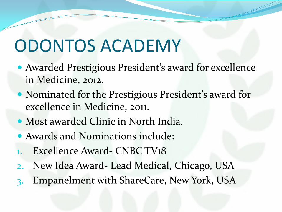
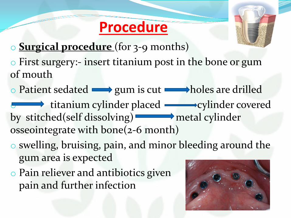
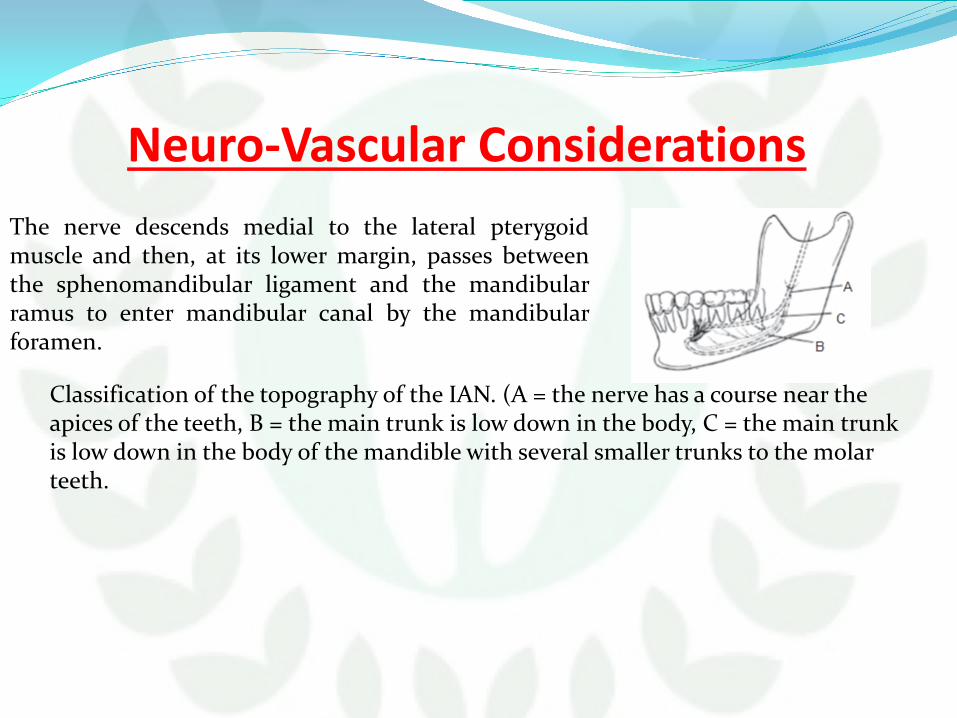

What to do if the Implant is too Close to the Nerve
What to do if the Implant is too Close to the Nerve
Nerve Lateralization or Nerve Repositioning Is the way
What to do if the Implant is too Close to the Nerve
The surgical procedure was performed under local anesthesia A full thickness mucoperiosteal flap was elevated to the inferior border of the mandible For performing inferior alveolar nerve lateralization the corticotomy started 4 mm distal to the mental foramen A small round bur in a straight hand piece with high torque and copious amount of water irrigation was used to prepare the corticotomy site To remove the trabecular bone and gain access to the neurovascular bundle only hand instruments (small curettes) were used The IAN was mobilized from its position After the nerve was completely released from the canal and before starting to drill half a rubber piston from a dental anaesthetic cartridge or a piece of membrane was inserted between the nerve bundle and the bone where the drill was expected to reach At left and right second molar region we placed 475x12 mm Ankylos implant
What to do if the Implant is too Close to the Nerve
The surgical procedure was performed under local anesthesia A full thickness mucoperiosteal flap was elevated to the inferior border of the mandible For performing inferior alveolar nerve lateralization the corticotomy started 4 mm distal to the mental foramen A small round bur in a straight hand piece with high torque and copious amount of water irrigation was used to prepare the corticotomy site To remove the trabecular bone and gain access to the neurovascular bundle only hand instruments (small curettes) were used The IAN was mobilized from its position After the nerve was completely released from the canal and before starting to drill half a rubber piston from a dental anaesthetic cartridge or a piece of membrane was inserted between the nerve bundle and the bone where the drill was expected to reach At left and right second molar region we placed 475x12 mm Ankylos implant
What to do if the Implant is too Close to the Nerve
Bio Materials as Implant Machined Surface- Branemark- 1969
Bio Materials as Implant Sand blasted Implant
Bio Materials as Implant Acid Etched Implant
Bio Materials as Implant Acid Etched- Sand Blasted Implant
Bio Materials as Implant Anodized Implant
Bio Materials as Implant Anodized Implant
How to Decide Implant Size Sizes of implants have biomechanical and clinical significance There are two
biomechanical patterns 1) The longer the implant is the greater integration with bony tissue it features
This allows heavier functional load on the implant and surrounding osseous tissue
2) Larger implants diameter promotes better load distribution in surrounding bone tissue and higher strength
Thus size diameter and length of the implant are to be as great as practicable from the points of view of both biomechanical and clinical effectiveness
However size of the implant is significantly constrained by jaw dimensions as well as other anatomical structures of maxillo-facial area In addition to ensure adequate osseogenesis the implant is to be all round surrounded with bone which thickness is over 075-10 mm
Thus from biological and clinical points of view the implant dimensions are to be small enough to be all round surrounded with a bone mass which provides adequate osseogenesis
How to Decide Implant Size
The distance between the two teeth can be determined by the gap width Depending on the tooth shape the gap width is 10 mm (2x0 5 mm) smaller than the distance at the bone level
Our Implant Cases
SINUS LIFT SURGERY

What to do if the Implant is too Close to the Nerve
The surgical procedure was performed under local anesthesia A full thickness mucoperiosteal flap was elevated to the inferior border of the mandible For performing inferior alveolar nerve lateralization the corticotomy started 4 mm distal to the mental foramen A small round bur in a straight hand piece with high torque and copious amount of water irrigation was used to prepare the corticotomy site To remove the trabecular bone and gain access to the neurovascular bundle only hand instruments (small curettes) were used The IAN was mobilized from its position After the nerve was completely released from the canal and before starting to drill half a rubber piston from a dental anaesthetic cartridge or a piece of membrane was inserted between the nerve bundle and the bone where the drill was expected to reach At left and right second molar region we placed 475x12 mm Ankylos implant
What to do if the Implant is too Close to the Nerve
Bio Materials as Implant Machined Surface- Branemark- 1969
Bio Materials as Implant Sand blasted Implant
Bio Materials as Implant Acid Etched Implant
Bio Materials as Implant Acid Etched- Sand Blasted Implant
Bio Materials as Implant Anodized Implant
Bio Materials as Implant Anodized Implant
How to Decide Implant Size Sizes of implants have biomechanical and clinical significance There are two
biomechanical patterns 1) The longer the implant is the greater integration with bony tissue it features
This allows heavier functional load on the implant and surrounding osseous tissue
2) Larger implants diameter promotes better load distribution in surrounding bone tissue and higher strength
Thus size diameter and length of the implant are to be as great as practicable from the points of view of both biomechanical and clinical effectiveness
However size of the implant is significantly constrained by jaw dimensions as well as other anatomical structures of maxillo-facial area In addition to ensure adequate osseogenesis the implant is to be all round surrounded with bone which thickness is over 075-10 mm
Thus from biological and clinical points of view the implant dimensions are to be small enough to be all round surrounded with a bone mass which provides adequate osseogenesis
How to Decide Implant Size
The distance between the two teeth can be determined by the gap width Depending on the tooth shape the gap width is 10 mm (2x0 5 mm) smaller than the distance at the bone level
Our Implant Cases
SINUS LIFT SURGERY

What to do if the Implant is too Close to the Nerve
The surgical procedure was performed under local anesthesia A full thickness mucoperiosteal flap was elevated to the inferior border of the mandible For performing inferior alveolar nerve lateralization the corticotomy started 4 mm distal to the mental foramen A small round bur in a straight hand piece with high torque and copious amount of water irrigation was used to prepare the corticotomy site To remove the trabecular bone and gain access to the neurovascular bundle only hand instruments (small curettes) were used The IAN was mobilized from its position After the nerve was completely released from the canal and before starting to drill half a rubber piston from a dental anaesthetic cartridge or a piece of membrane was inserted between the nerve bundle and the bone where the drill was expected to reach At left and right second molar region we placed 475x12 mm Ankylos implant
What to do if the Implant is too Close to the Nerve
Bio Materials as Implant Machined Surface- Branemark- 1969
Bio Materials as Implant Sand blasted Implant
Bio Materials as Implant Acid Etched Implant
Bio Materials as Implant Acid Etched- Sand Blasted Implant
Bio Materials as Implant Anodized Implant
Bio Materials as Implant Anodized Implant
How to Decide Implant Size Sizes of implants have biomechanical and clinical significance There are two
biomechanical patterns 1) The longer the implant is the greater integration with bony tissue it features
This allows heavier functional load on the implant and surrounding osseous tissue
2) Larger implants diameter promotes better load distribution in surrounding bone tissue and higher strength
Thus size diameter and length of the implant are to be as great as practicable from the points of view of both biomechanical and clinical effectiveness
However size of the implant is significantly constrained by jaw dimensions as well as other anatomical structures of maxillo-facial area In addition to ensure adequate osseogenesis the implant is to be all round surrounded with bone which thickness is over 075-10 mm
Thus from biological and clinical points of view the implant dimensions are to be small enough to be all round surrounded with a bone mass which provides adequate osseogenesis
How to Decide Implant Size
The distance between the two teeth can be determined by the gap width Depending on the tooth shape the gap width is 10 mm (2x0 5 mm) smaller than the distance at the bone level
Our Implant Cases
SINUS LIFT SURGERY

What to do if the Implant is too Close to the Nerve
Bio Materials as Implant Machined Surface- Branemark- 1969
Bio Materials as Implant Sand blasted Implant
Bio Materials as Implant Acid Etched Implant
Bio Materials as Implant Acid Etched- Sand Blasted Implant
Bio Materials as Implant Anodized Implant
Bio Materials as Implant Anodized Implant
How to Decide Implant Size Sizes of implants have biomechanical and clinical significance There are two
biomechanical patterns 1) The longer the implant is the greater integration with bony tissue it features
This allows heavier functional load on the implant and surrounding osseous tissue
2) Larger implants diameter promotes better load distribution in surrounding bone tissue and higher strength
Thus size diameter and length of the implant are to be as great as practicable from the points of view of both biomechanical and clinical effectiveness
However size of the implant is significantly constrained by jaw dimensions as well as other anatomical structures of maxillo-facial area In addition to ensure adequate osseogenesis the implant is to be all round surrounded with bone which thickness is over 075-10 mm
Thus from biological and clinical points of view the implant dimensions are to be small enough to be all round surrounded with a bone mass which provides adequate osseogenesis
How to Decide Implant Size
The distance between the two teeth can be determined by the gap width Depending on the tooth shape the gap width is 10 mm (2x0 5 mm) smaller than the distance at the bone level
Our Implant Cases
SINUS LIFT SURGERY
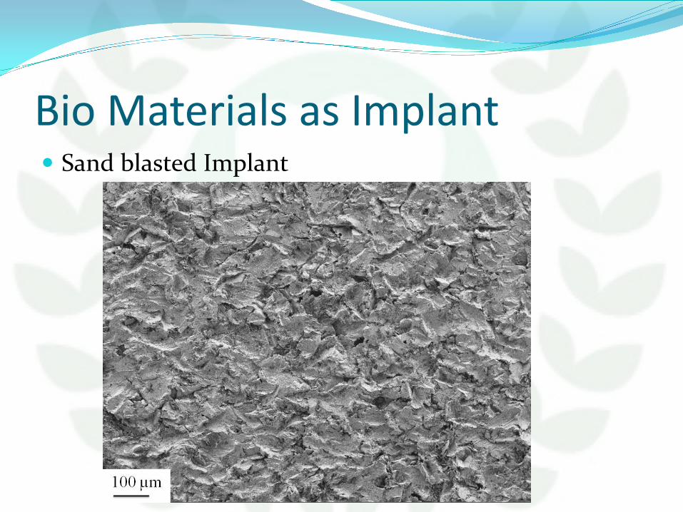
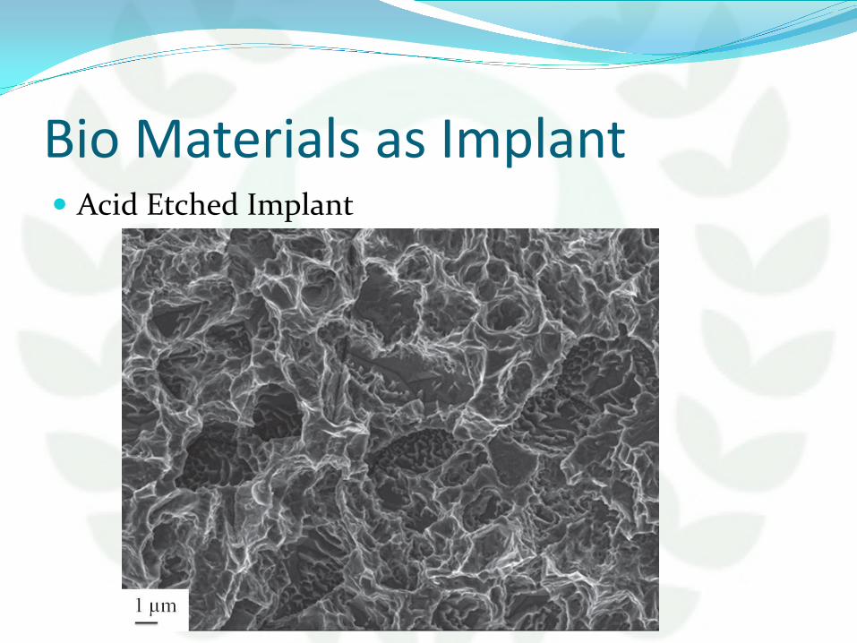
Bio Materials as Implant Anodized Implant
Bio Materials as Implant Anodized Implant
How to Decide Implant Size Sizes of implants have biomechanical and clinical significance There are two
biomechanical patterns 1) The longer the implant is the greater integration with bony tissue it features
This allows heavier functional load on the implant and surrounding osseous tissue
2) Larger implants diameter promotes better load distribution in surrounding bone tissue and higher strength
Thus size diameter and length of the implant are to be as great as practicable from the points of view of both biomechanical and clinical effectiveness
However size of the implant is significantly constrained by jaw dimensions as well as other anatomical structures of maxillo-facial area In addition to ensure adequate osseogenesis the implant is to be all round surrounded with bone which thickness is over 075-10 mm
Thus from biological and clinical points of view the implant dimensions are to be small enough to be all round surrounded with a bone mass which provides adequate osseogenesis
How to Decide Implant Size
The distance between the two teeth can be determined by the gap width Depending on the tooth shape the gap width is 10 mm (2x0 5 mm) smaller than the distance at the bone level
Our Implant Cases
SINUS LIFT SURGERY
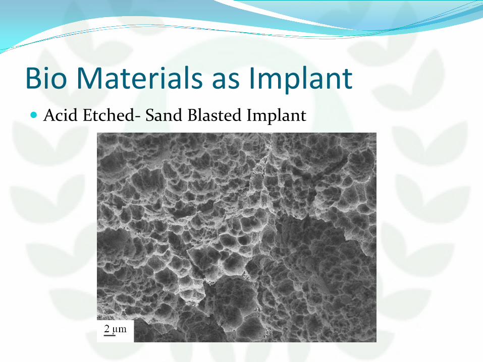
Bio Materials as Implant Anodized Implant
How to Decide Implant Size Sizes of implants have biomechanical and clinical significance There are two
biomechanical patterns 1) The longer the implant is the greater integration with bony tissue it features
This allows heavier functional load on the implant and surrounding osseous tissue
2) Larger implants diameter promotes better load distribution in surrounding bone tissue and higher strength
Thus size diameter and length of the implant are to be as great as practicable from the points of view of both biomechanical and clinical effectiveness
However size of the implant is significantly constrained by jaw dimensions as well as other anatomical structures of maxillo-facial area In addition to ensure adequate osseogenesis the implant is to be all round surrounded with bone which thickness is over 075-10 mm
Thus from biological and clinical points of view the implant dimensions are to be small enough to be all round surrounded with a bone mass which provides adequate osseogenesis
How to Decide Implant Size
The distance between the two teeth can be determined by the gap width Depending on the tooth shape the gap width is 10 mm (2x0 5 mm) smaller than the distance at the bone level
Our Implant Cases
SINUS LIFT SURGERY

How to Decide Implant Size
The distance between the two teeth can be determined by the gap width Depending on the tooth shape the gap width is 10 mm (2x0 5 mm) smaller than the distance at the bone level
Our Implant Cases
SINUS LIFT SURGERY
The distance between the two teeth can be determined by the gap width Depending on the tooth shape the gap width is 10 mm (2x0 5 mm) smaller than the distance at the bone level
Our Implant Cases
SINUS LIFT SURGERY

Our Implant Cases
SINUS LIFT SURGERY
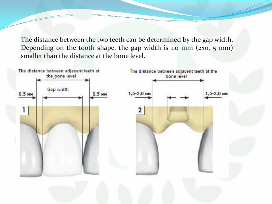
SINUS LIFT SURGERY

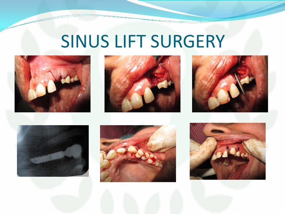
Clinical Aspects of Surgery
contents General principles of implant surgery
Patient preparation Implant site preparation One stage versus two stage implant surgeries
Two stage ldquosubmergedrdquo implant placement Flap designs incisions and reflection Implant site preparation Flap closure and suturing Post operative care Second stage exposure surgery
57
One stage ldquonon-submergedrdquo implant placement Flap designs incisions and elevation Implant site preparation Flap closure and suturing Postoperative care
Conclusion
58
General principles of implant surgery
Patient preparation
Implant site preparation
One stage Vs two stage implant surgery
59
Patient preparation 1 Explanation of risks and benefits to the patient
2 Written Informed consent
3 Local or General Anesthesia depending on patientrsquos
needs
60
Basic principles of implant therapy 1 Implants must be sterile and made of a biocompatible material (eg titanium)
2 Implant site preparation should be performed under sterile conditions
3 Implant site preparation should be completed with an atraumatic surgical technique
that avoids overheating of the bone during preparation of the recipient site
4 Implants should be placed with good initial stability
5 Implants should be allowed to heal without loading or micro-movement (ie
undisturbed healing period to allow for osseointegration) for 2 to 4 or 4 to 6 months
depending on the bone density bone maturation and implant stability
61
Surgical site preparation 1 Patient drape
2 Rinsing or swabbing the mouth with chlorhexidine gluconate
for 1 to 2 minutes immediately before the procedure
3 Atraumatic implant site preparation
4 Avoid damage to bone or vital structures
5 Copious irrigation to avoid heating and debris removal
6 The implant must be placed in healthy bone
7 The surgical site should be kept aseptic
62
Operative requirements 1 Good operating light
2 Good high volume suction
3 A dental chair which can be adjusted by foot controls
4 A surgical drilling unit which can deliver relatively high speeds (up to 3000 rpm) and low drilling
speeds (down to about 10 rpm) with good control of torque
5 An irrigation system for keeping bone cool during the drilling process
6 The appropriate surgical instrumentation for the implant system being used and the surgical
procedure
7 Sterile drapes gowns gloves suction tubing etc
8 The appropriate number and design of implants planned plus an adequate stock to meet
unexpected eventualities during surgery
63
Operative requirements 9 The surgical stent
10 The complete radiographs including tomographs
11 A trained assistant
12 A third person to act as a get things in between to and from the
sterile and non-sterile environment
13 Light handles should be autoclaved or covered with sterile aluminum foil
14 The instrument tray and any other surfaces which are to be used are covered in sterile drapes
64
One stage VS two stage technique
65
One stage technique In the one-stage approach the
implant or the abutment
emerges through the
mucoperiosteumgingival
tissue at the time of implant
placement
66
Advantages of one stage Easier Mucogingival management around the implant Patient management is simplified because a second
stage exposure surgery is not necessary
67
Two stage technique
In the two-stage approach the top of the implant
and cover screw are completely covered with the
flap closure
Implants are allowed to heal without loading or
micro movement for a period of time to allow for
osseointegration
The implant must be surgically exposed following
an undisturbed healing period
68
In areas with dense cortical bone and good initial implant support the implants are left to heal undisturbed for a period of 2 to 4 months whereas in areas of loose trabecular bone grafted sites and sites with lesser implant stability implants may be allowed to heal for periods of 4 to 6 months or more
Longer healing periods are indicated for implants placed in less dense bone or when there is less initial implant stability (ie slight looseness caused by limited bone-to-implant contact) regardless of jaw or specific anatomic location
In the second-stage (exposure) surgery the implant is uncovered and a healing abutment is connected to allow emergence of the implantabutment through the soft tissues thus facilitating access to the implant from the oral cavity
The restorative dentist then proceeds with the prosthodontic aspects of the implant therapy (impressions and fabrication of prosthesis) after soft tissue healing
69
Advantages of 2nd stage surgery Situations that require simultaneous bone augmentation procedures at
the time of implant placement because membranes can be covered by
primary flap closure which will minimize postoperative exposure
Prevents movement of the implant by the patient who may
inadvertently bite on the healing abutment during the healing period
(one-stage protocol)
Mucogingival tissues can be augmented if desired at the second-stage
surgery in a two-stage protocol
70
Two stage ldquosubmergedrdquo implant placement The first stage ends by
Suturing So the implant remains submerged and isolated from the oral
cavity Mandible implants ndash 2 to 4 months Maxillary implants ndash 4 to 6 months
Longer periods ndash
less dense bone Less initial implant stability
Shorter periods ndash More dense bone Altered surface microtopography
71
In second stage The implant is uncovered and a healing abutment is
connected to allow emergence of the implant through the soft tissue thus facilitating access to the implant from the oral cavity
72
Two stage ldquosubmergedrdquo implant placement Flap design incisions and elevation
Vary slightly depending on the location and objective of the
planned surgery Crestal
The incision is made from along the crest of the ridge bisecting the existing zone of keratinized mucosa
Adv Easy to manage results in less bleeding less edema faster healing
Suturing placed generally do not interfere with the healing Remote
The incision is made some distance from the planned osteotomy site
Layer suturing is indicated to minimize the bone graft exposure
73
Incisions
74
Implant site preparation A mucoperiosteal (full-thickness) flap is reflected
up to or slightly beyond the level of the
mucogingival junction exposing the alveolar ridge
of the implant surgical sites
Elevated flaps may be sutured to the buccal mucosa
or the opposing teeth to keep the surgical site open
during the surgery
The bone at the implant site(s) must be thoroughly
debrided of all granulation tissue
75
Once the flaps are reflected and the bone is prepared (ie all
granulation tissue removed and knife-edge ridges flattened) the
implant osteotomy site can be prepared
A series of drills are used to prepare the osteotomy site precisely
and incrementally for an implant
A surgical guide or stent is inserted checked for proper
positioning and used throughout the procedure to direct the
proper implant placement
76
77
Tissue management f or a two-stage implant placement A Crestal incision made along the crest of the ridge bisecting the existing zone of keratinized mucosa B Full-thickness flap is raised buccally and lingually to the level of the mucogingival junction A narrow sharp ridge can be surgically reducedcontoured to provide a reasonably f lat bed f or the implant C Implant is placed in the prepared osteotomy site D Tissue approximation to achieve primary flap closure without tension
78
Implant site preparation Sequence of drills used for standard-diameter (40-mm) implant site osteotomy preparation round 2-mm twist pilot 3-mm twist and countersink Bone tap (not shown here) is an optional drill that is sometimes used in dense bone before implant placement
79
A series of drills are used to prepare the osteotomy site precisely and incrementally for an implant A surgical guide or stent is inserted checked for proper positioning and used throughout the procedure to direct the proper implant placement
80
Round bur A small round bur (or spiral drill) is used to mark the
implant site(s) The surgical guide is removed and the initial marks are checked for their appropriate buccal-lingual and mesial-distal location as well as the positions relative to each other and adjacent teeth
Slight modifications may be necessary to adjust spatial relationships and to avoid minor ridge defects Any changes should be compared to the prosthetically-driven surgical guide positions
Each marked site is then prepared to a depth of 1 to 2 mm with a round drill breaking through the cortical bone and creating a starting point for the 2-mm twist drill
81
Round bur spiral drill
82
2MM twist drill
83
Twist drills (To Enlarge the Osteotomy Site to required diameter)
84
Pilot drill
85
Guide pins
86
Depth gauge
87
Counter sink drill
88
Bone tap
89
As the final step in preparing the osteotomy site in dense
cortical bone a tapping procedure may be necessary
With self-tapping implants being almost universal there
is less need for a tapping procedure in most sites
However in dense cortical bone or when placing longer
implants into moderately dense bone it is prudent to
tap the bone (create threads in the osteotomy site)
before implant placement to facilitate implant insertion
and to reduce the risk of implant binding
90
It is better to allow the threaded implant to ldquocutrdquo
its own path into the osteotomy site
Bone tapping and implant insertion are both done
at very slow speeds (eg 20 to 40 rpm) All other
drills in the sequence are used at higher speeds
(800 to 1500 rpm)
It is important to create a recipient site that is very
accurate in size and angulation
91
In partially edentulous cases limited jaw opening or proximity to
adjacent teeth may prevent appropriate positioning of the drills in
posterior edentulous areas
In fact implant therapy may be contraindicated in some patients
because of a lack of inter occlusal clearance lack of interdental space
or a lack of access for the instrumentation
Therefore a combination of longer drills and shorter drills with or
without extensions may be necessary
Anticipating these needs before surgery facilitates the procedure and
improves the results
92
When wide-diameter drills are used for implant site
preparation it is advisable to reduce the drilling speed
according to the manufacturers guidelines to prevent
overheating the bone
Copious external irrigation is critical In the case of wide
diameter implants a specific pilot drill is often indicated as
a transition between each of the subsequent wider drills
93
Implant site preparation (osteotomy ) for a 40-mm diameter 10 mm length screw-type threaded (external hex) implant in a subcrestal position A Initial marking or preparation of the implant site with a round bur B Use of a 2-mm twist drill to establish depth and align the implant C Guide pin is placed in the osteotomy site to confirm position and angulation D Pilot drill is used to increase the diameter of the coronal aspect of the osteotomy site
94
E Final drill used is the 3- mm twist drill to finish preparation of the osteotomy site F Countersink drill is used to widen the entrance of the recipient site and allow for the subcrestal placement of the implant collar and cover screw G Implant is inserted into the prepared osteotomy site with a handpiece or handheld driver note In systems that use an implant mount it would be removed prior to placement of the cover screw H Cover screw is placed and soft tissues are closed and sutured
95
96
97
Wrench Ratchet Fits on top of fixture mount amp used to tighten fixture after placement
98
Implant fixtures
99
Cover screw
100
Flap closure and suturing
101
Once the implants are inserted and the cover screws secured the surgical sites should be
thoroughly irrigated with sterile saline to remove debris and clean the wound
Proper closure of the flap over the implant(s) is essential
One of the most important aspects of flap management is achieving good approximation
and primary closure of the tissues in a tension free manner
This is achieved by incising the periosteum (innermost layer of full-thickness flap)
which is non-elastic
Once the periosteum is released the flap becomes very elastic and is able to be stretched
over the implant(s) without tension
102
One suturing technique that consistently provides the desired result is
a combination of alternating horizontal mattress and interrupted
sutures
Horizontal mattress sutures evert the wound edges and approximate
the inner connective tissue surfaces of the flap to facilitate closure and
wound healing
Interrupted sutures help to bring the wound edges together
counterbalancing the eversion caused by the horizontal mattress
sutures
103
Post operative care Simple implant surgery in a healthy patient usually
does not require antibiotic therapy
However patients can be premedicated with
antibiotics (eg amoxicillin 500 mg three times a
day [tid]) starting 1 hour before the surgery and
continuing for 1 week postoperatively if the surgery
is extensive if it requires bone augmentation or if
the patient is medically compromised
Postoperative swelling is likely after flap surgery 104
This is particularly true when the periosteum has been incised
(released)
As a preventive measure patients should apply an ice pack to the area
intermittently for 20 minutes (on and off) over the first 24 to 48 hours
Chlorhexidine gluconate oral rinses can be prescribed to facilitate
plaque control especially in the days after surgery when oral hygiene is
typically poorer Adequate pain medication should be prescribed (eg
ibuprofen 600 to 800 mg tid)
105
Patients should be instructed to maintain a relatively soft diet after surgery
Then as soft tissue healing progresses they can gradually return to a normal diet
Patients should also refrain from tobacco and alcohol use at least 1 week before and several weeks after surgery
Provisional restorations whether fixed or removable should be checked and adjusted so that impingement on the surgical area is avoided
106
Second stage exposure surgery For implants placed using a two-stage ldquosubmergedrdquo
protocol a second-stage exposure surgery is necessary
after the prescribed healing period
Thin soft tissue with an adequate amount of
keratinized attached gingiva along with good oral
hygiene ensures healthier peri-implant soft tissues
and better clinical results
107
Objectives of second stage technique 1 To expose the submerged implant without damaging the
surrounding bone
2 To control the thickness of the soft tissue surrounding the implant
3 To preserve or create attached keratinized tissue around the implant
4 To facilitate oral hygiene
5 To ensure proper abutment seating
6 To preserve soft tissue aesthetics
108
Simple circular ldquopunchrdquo incision In areas with sufficient zones of keratinized tissue the
gingiva covering the head of the implant can be exposed
with a circular or ldquopunchrdquo incision
Alternatively a crestal incision through the middle of the
keratinized tissue and full-thickness flap reflection can be
used to expose implants
This latter approach may be necessary when bone has
grown over the implant and needs to be removed
109
Clinical view of stage two implant exposure surgery in a case with adequate keratinized tissue A Simple circular ldquopunchrdquo incision used to expose implant when sufficient keratinized tissue is present around the implant(s) B Implant exposed C Healing abutment attached D Final restoration in place achieving an esthetic result with a good zone of keratinized tissue 110
Clinical v iew of stage two implant exposure surgery in a case with inadequate keratinized tissue A Two endosseous implants were placed 4 months previously and are ready to be exposed B Two vertical incisions are connected by crestal incision C Buccal partial thickness flap is sutured to the periosteum apical to the emerging implants D Gingival tissue coronal to the cover screws is excised using the gingivectomy technique E Cover screws are removed and heads of the implants are cleared F Abutments are placed Visual inspection ensures intimate contact between the abutments and the implants
111
G Healing at 2 to 3 weeks after second-stage surgery H Four months after the final restoration Note the healthy band of keratinized attached gingiv a around the implants
112
Post operative care remind the patient of the need for good oral hygiene
around the implant and adjacent teeth rinse can be used to enhance oral hygiene for the
initial few weeks after implant exposure oral hygiene procedures to avoid dislodging any
repositioned or grafted soft tissues any direct pressure or movement directed toward the
soft tissue from a provisional prosthesis can delay healing and should be avoided
113
Impressions for the final prosthesis fabrication can begin about 2 to 6 weeks after implant exposure surgery depending on healing and maturation of soft tissues
114
One stage ldquonon-submergedrdquo implant placement
115
In the one-stage implant surgical approach a second implant exposure surgery is not needed because the implant is exposed (per gingival) from the time of implant placement
In the standard (classic) implant protocol the implants are left unloaded and undisturbed for a period similar to that for implants placed in the two-stage approach (ie in areas with dense cortical bone and good initial
implant support the implants are left to heal undisturbed for a period of 2 to 4 months
whereas in areas of loose trabecular bone grafted sites andor minimal implant support they may be allowed to heal for periods of 4 to 6 months or more)
116
In the one-stage surgical approach the implant or the healing abutment protrudes about 2 to 3 mm from the bone crest and the flaps are adapted around the implantabutment
117
Flap design incisions and elevation The flap design for the one-stage surgical approach is
always a crestal incision bisecting the existing keratinized tissue
Facial and lingual flaps in posterior areas should be carefully thinned before total reflection to minimize the soft tissue thickness (if needed or desired)
The soft tissue is not thinned in anterior or other esthetic areas of the mouth to maintain tissue height and to minimize metallic implant components from showing through tissue
118
Implant site preparation The primary difference is that the coronal aspect of the
implant or the healing abutment (two-stage implant) is placed about 2 to 3 mm above the bone crest and the soft tissues are approximated around the implantimplant abutment
119
Flap closure and suturing The keratinized edges of the flap are sutured with
single interrupted sutures around the implant Depending on the clinicians preference the wound
may be sutured with resorbable or nonresorbable sutures
When keratinized tissue is abundant scalloping around the implant(s) provides better flap adaptation
However if minimal keratinized tissue exists in an area tissues should remain thick and soft tissue augmentation may be indicated
120
Post operative care The postoperative care for one-stage surgical approach
is similar to that for the two-stage surgical approach except that the cover screw or healing abutment is exposed to the oral cavity
Patients are advised to avoid chewing in the area of the implant
Prosthetic appliances should not be used if direct chewing forces can be transmitted to the implant particularly in the early healing period (first 4 to 8 weeks)
121
conclusion It is essential to understand and follow basic
guidelines to achieve osseointegration predictably Fundamentals must be followed for implant placement
and implant exposure surgery These fundamentals apply to all implant systems
122
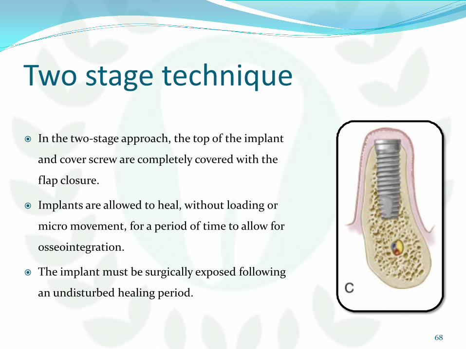
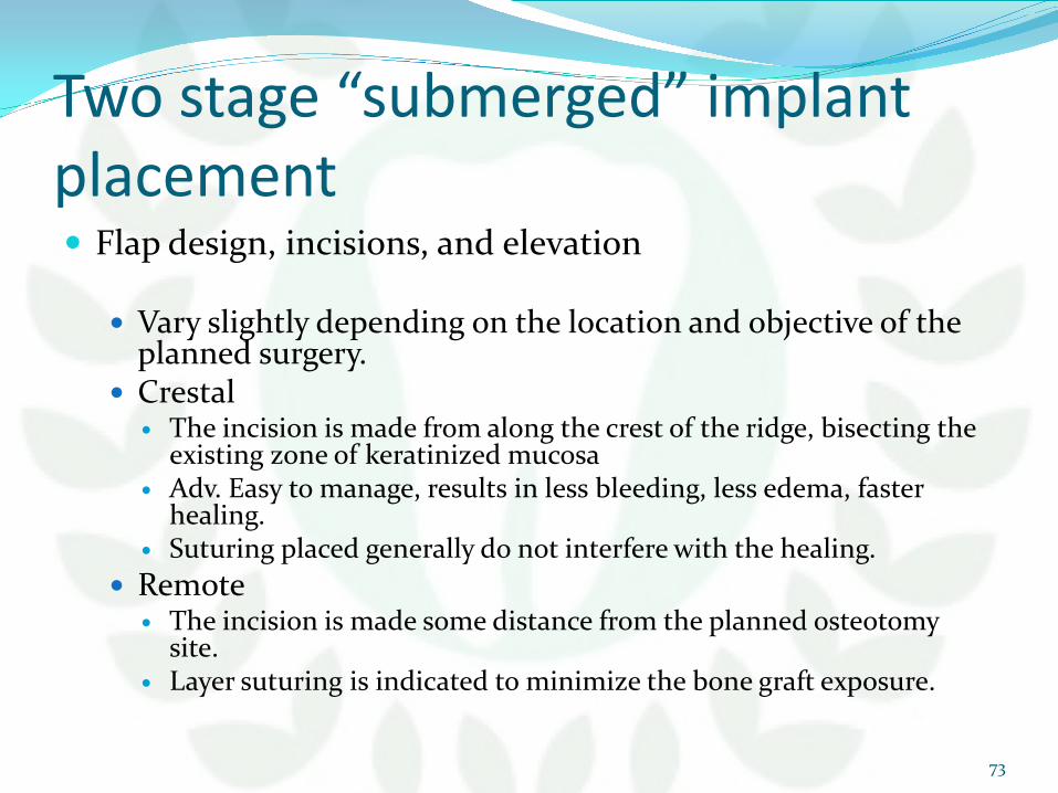
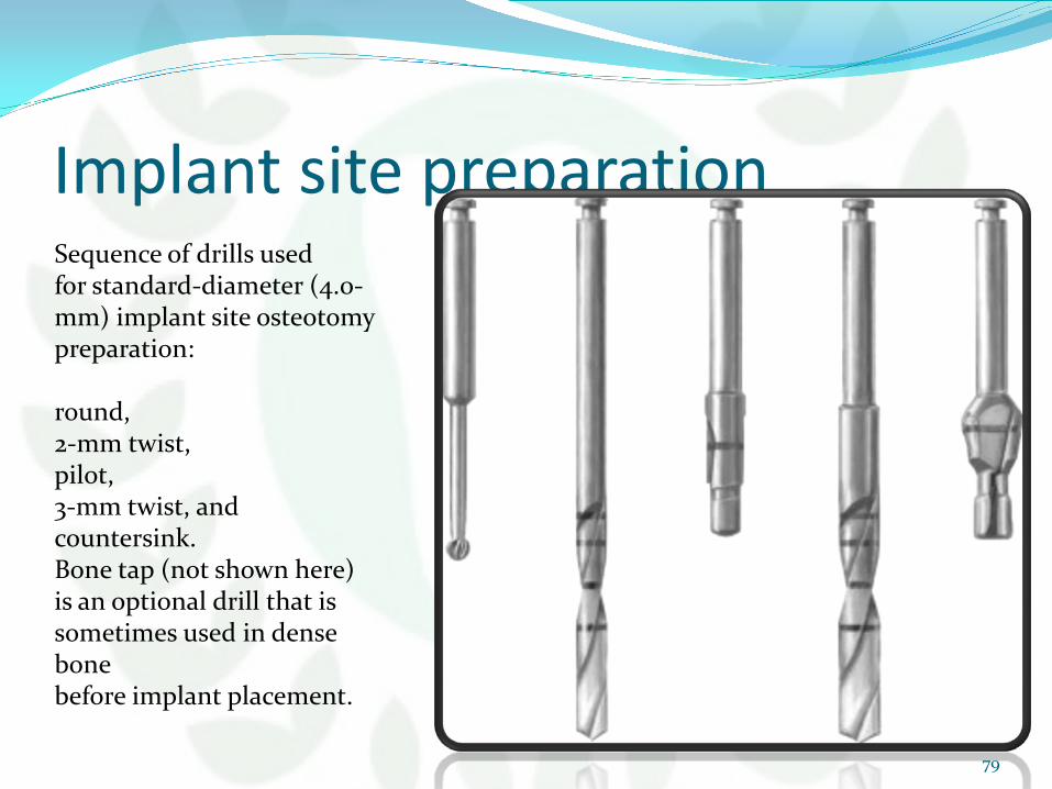
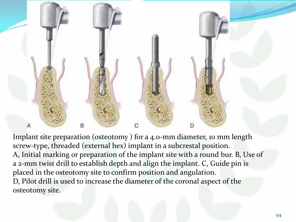
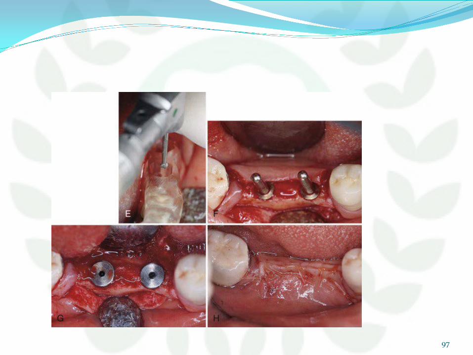
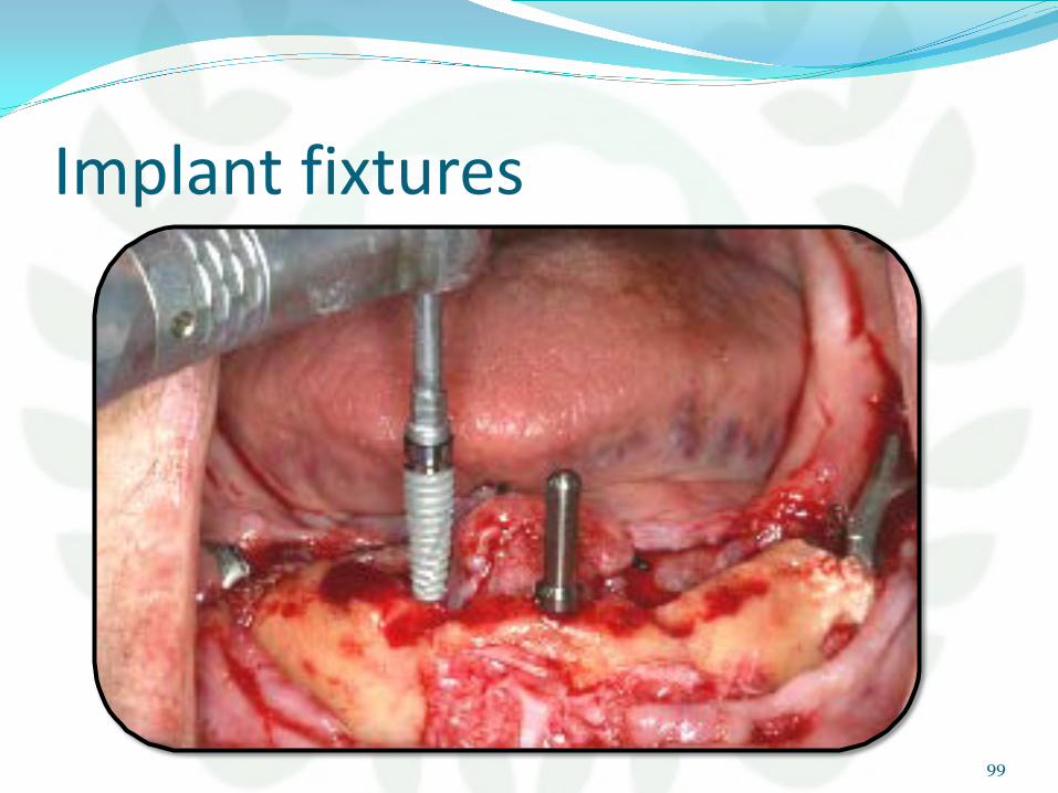
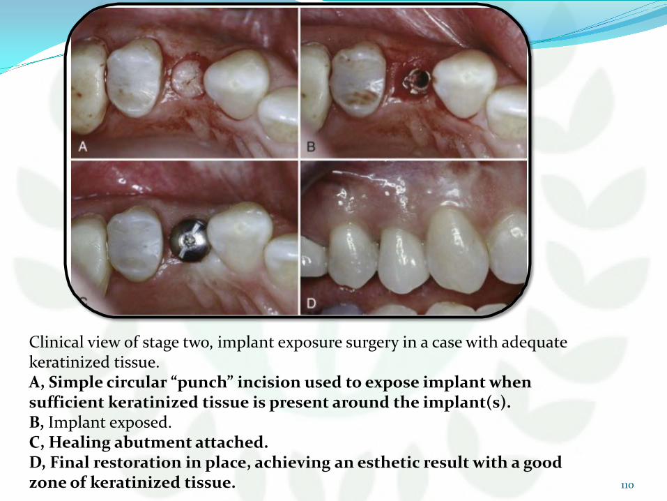
conclusion It is essential to understand and follow basic
guidelines to achieve osseointegration predictably Fundamentals must be followed for implant placement
and implant exposure surgery These fundamentals apply to all implant systems
122