Embed Size (px)
Citation preview
Human Embryology II

Embryonic period – the third week of development
The 2nd stage of gastrulation Germ layer initial differentiation and axial organ formation Primitive cardiovascular system formation Subsequent chorion development Allantois appearance Folding
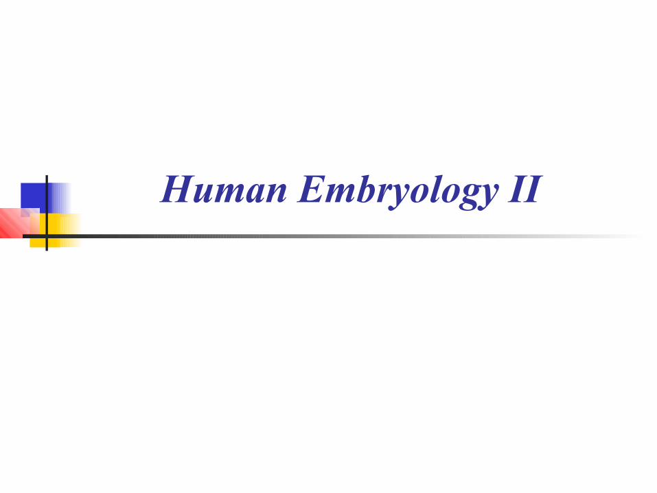
The second stage of gastrulation
results in
- trilaminar embryonic disk formation occurs
- on the 14th to 15th day of development
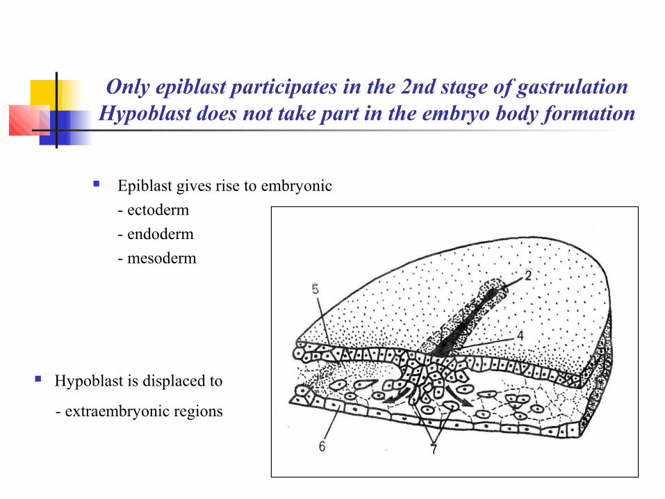
Only epiblast participates in the 2nd stage of gastrulationHypoblast does not take part in the embryo body formation
Epiblast gives rise to embryonic
- ectoderm
- endoderm
- mesoderm
Hypoblast is displaced to
- extraembryonic regions

Primitive streak is the key structure of the 2nd stage of gastrulation
Epiblastic cells at the disk cranial end
- proliferate
- migrate along the disk margins
- converge at the disk caudal end
- turn back to the disk cranial end
towards the midline
⇓ primitive streak ⇒
cranial end
caudal end

Primitive streak anterior portion thickens to form the primitive knot or Hensen’s nodule
Primitive groove
- develops in the primitive streak
- is continuous with the primitive pit
in the primitive knot
⇔
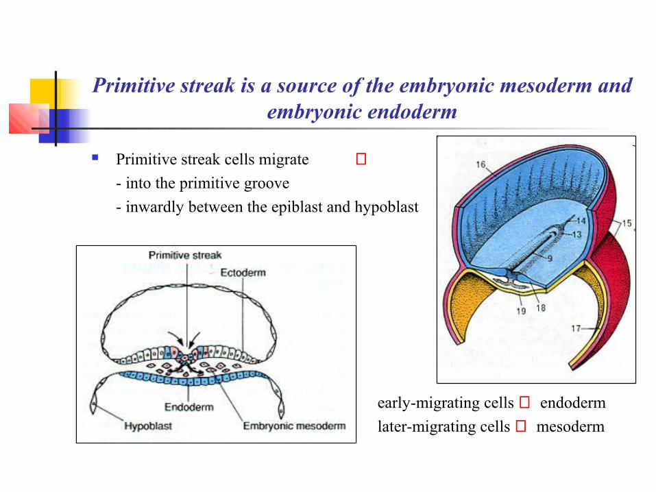
Primitive streak is a source of the embryonic mesoderm and embryonic endoderm
Primitive streak cells migrate ⇒
- into the primitive groove
- inwardly between the epiblast and hypoblast
early-migrating cells ⇒ endoderm
later-migrating cells ⇒ mesoderm
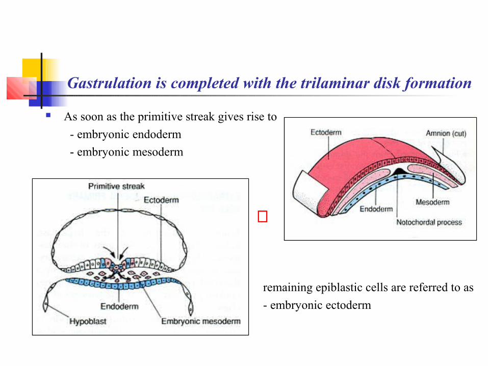
Gastrulation is completed with the trilaminar disk formation
As soon as the primitive streak gives rise to
- embryonic endoderm
- embryonic mesoderm
remaining epiblastic cells are referred to as
- embryonic ectoderm
⇔

Duplication of the primitive streak results in twinning
Удвоение
⇐ duplication of the primitive streak
⇐ monochorial monoamniotic twins
~30% ~4%~70%

Conjoint twins (~1% of monozygotic twins) result from the primitive streak duplication
partial duplication of the primitive streak (Y-shaped)
⇓
complete duplication of the primitive streak but incomplete duplication of the germ layers
⇓
bifurcation of the spinal cord and vertebral column fusion of soft tissues (Siamese twins)

Siamese twins
may be separated surgically
⇒
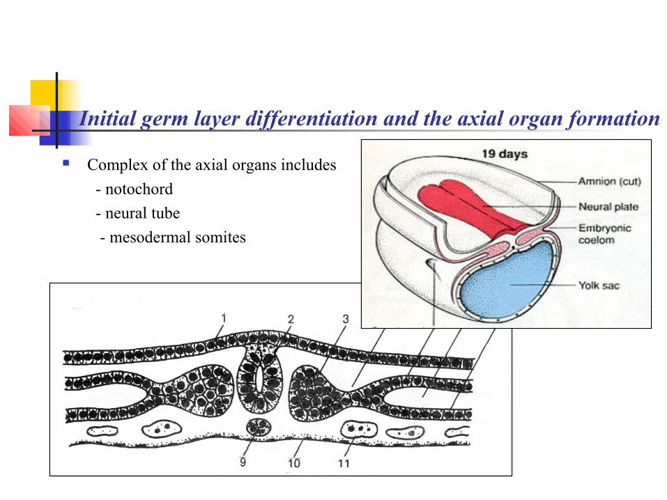
Initial germ layer differentiation and the axial organ formation
Complex of the axial organs includes
- notochord
- neural tube
- mesodermal somites

Notochord is the first to appear concurrently with mesoderm
Primitive pit
- extends into the primitive knot
- forms the notochordal canal Primitive knot cells
- migrate through the canal
- give rise to the notochord
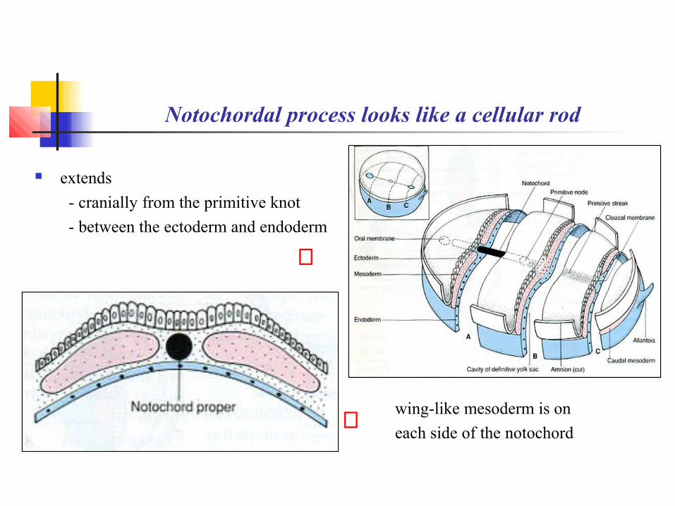
Notochordal process looks like a cellular rod
extends
- cranially from the primitive knot
- between the ectoderm and endoderm
wing-like mesoderm is on
each side of the notochord⇐
⇒
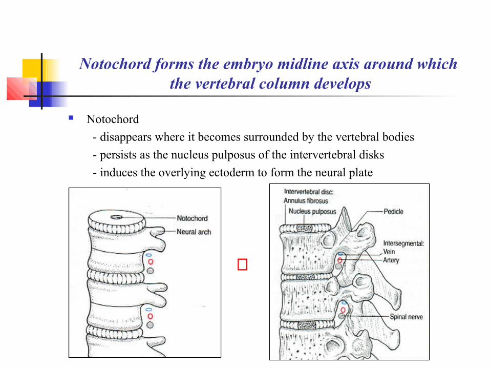
Notochord forms the embryo midline axis around which the vertebral column develops
Notochord
- disappears where it becomes surrounded by the vertebral bodies
- persists as the nucleus pulposus of the intervertebral disks
- induces the overlying ectoderm to form the neural plate
⇒

Neurulation or the neural tube formation is induced by the notochord with the adjacent mesoderm
Stages of the neural tube development neural plate (15 – 16 days) neural groove and neural folds (18 – 21 days) neural tube (23 – 25 day)
⇔

Neuroectoderm includes the neural tube and neural crest
Neural tube⇓
BrainSpinal cordRetinaOlfactory epithelium
Neural crest⇓
Neural gangliaPia mater and arachnoidSkin melanocytesAdrenal medullaThyroid gland C-cells

Surface ectoderm remains after the neural tube separation
Gives rise to
- skin epidermis
- sweat and sebaceous glands
- nails and hair
- mammary glands
- salivary glands
- tooth enamel
- oral cavity epithelium
- corneal epithelium
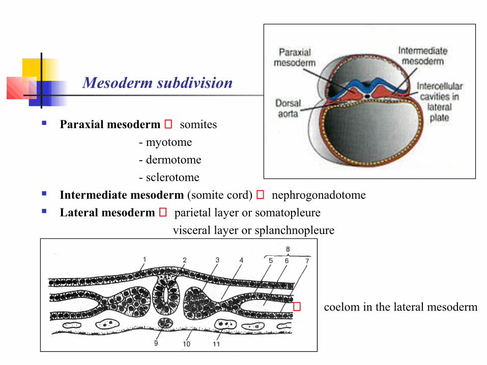
Mesoderm subdivision
Paraxial mesoderm ⇒ somites
- myotome
- dermotome
- sclerotome Intermediate mesoderm (somite cord) ⇒ nephrogonadotome Lateral mesoderm ⇒ parietal layer or somatopleure
visceral layer or splanchnopleure
⇐ coelom in the lateral mesoderm

Subsequent mesoderm differentiation
Myotome ⇒ skeletal muscles Dermatome ⇒ skin dermis Sclerotome ⇒ bones and cartilages
Nephrogonadotome ⇒ kidney and gonads Coelom ⇒ - pericardial
- pleural
- peritoneal cavities Somatopleure ⇒ mesothelium Splanchnopleure ⇒ - mesothelium
- myocardium
- epicardium
- adrenal cortex
⇒
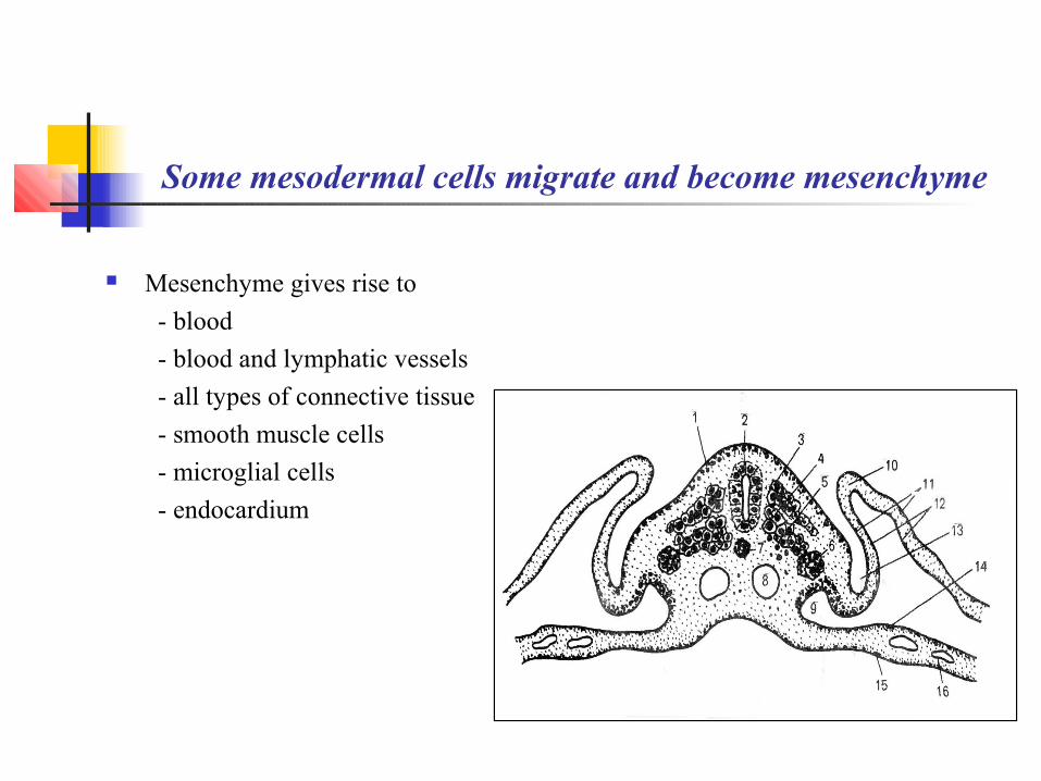
Some mesodermal cells migrate and become mesenchyme
Mesenchyme gives rise to
- blood
- blood and lymphatic vessels
- all types of connective tissue
- smooth muscle cells
- microglial cells
- endocardium
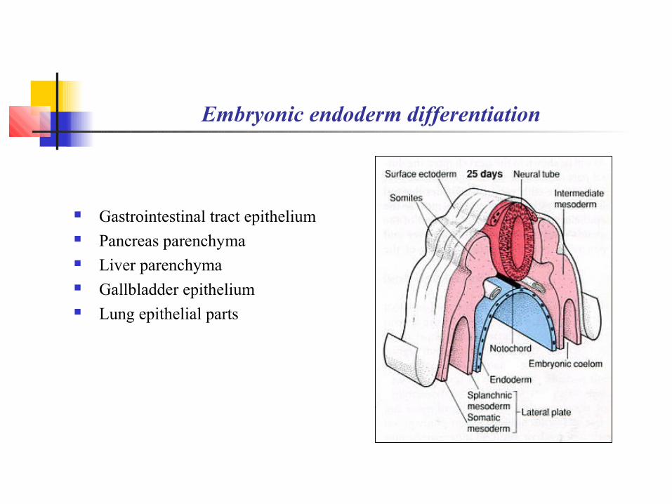
Embryonic endoderm differentiation
Gastrointestinal tract epithelium Pancreas parenchyma Liver parenchyma Gallbladder epithelium Lung epithelial parts
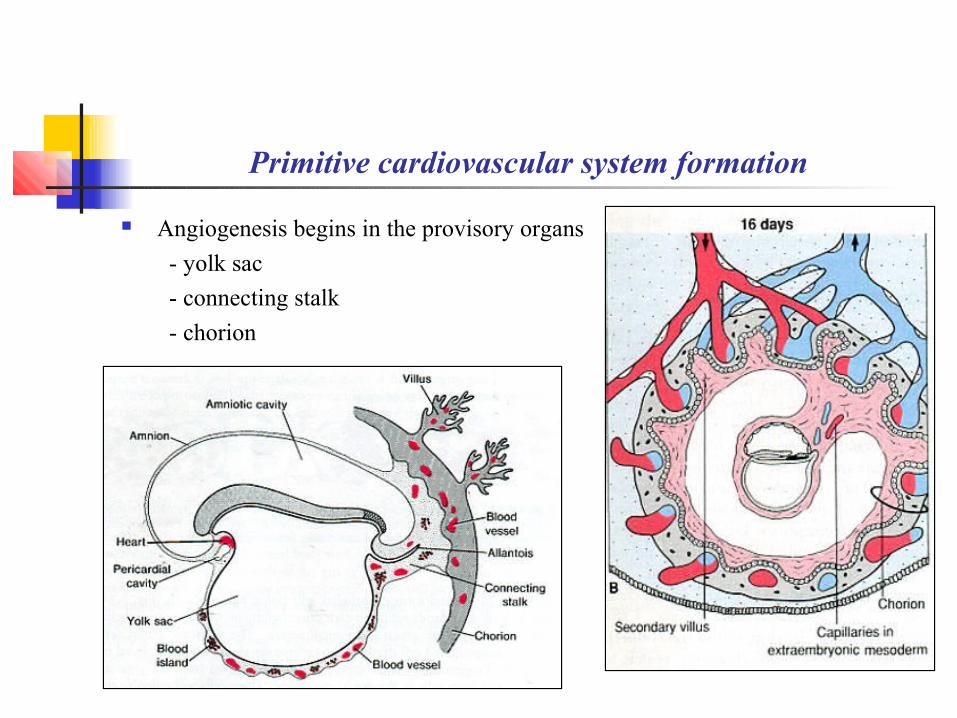
Primitive cardiovascular system formation
Angiogenesis begins in the provisory organs
- yolk sac
- connecting stalk
- chorion
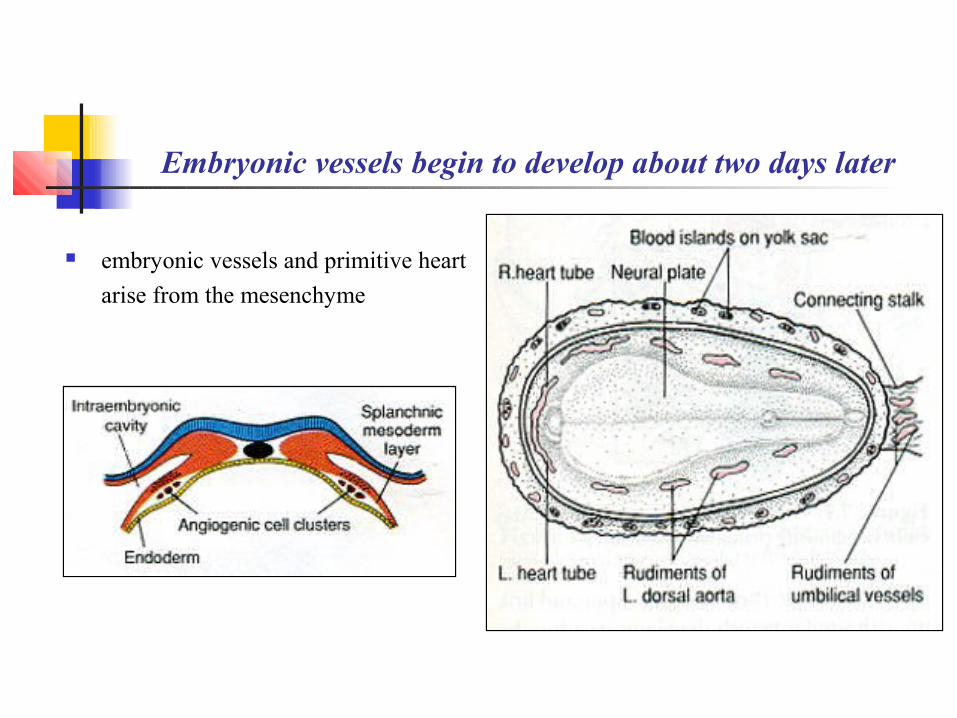
Embryonic vessels begin to develop about two days later
embryonic vessels and primitive heart
arise from the mesenchyme
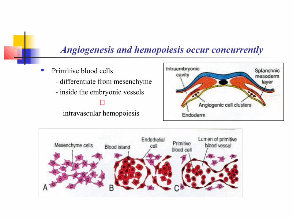
Angiogenesis and hemopoiesis occur concurrently
Primitive blood cells
- differentiate from mesenchyme
- inside the embryonic vessels
⇓ intravascular hemopoiesis
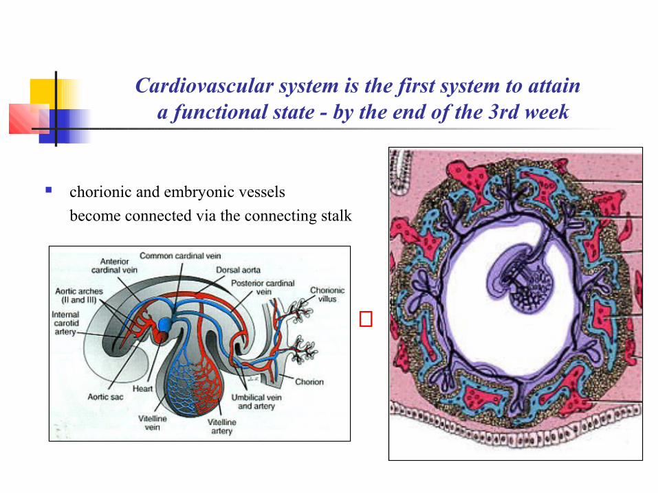
Cardiovascular system is the first system to attain a functional state - by the end of the 3rd week
сhorionic and embryonic vessels
become connected via the connecting stalk
⇔
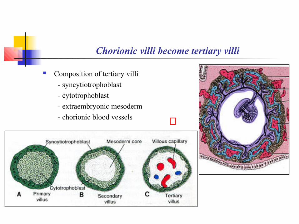
Chorionic villi become tertiary villi
Composition of tertiary villi
- syncytiotrophoblast
- cytotrophoblast
- extraembryonic mesoderm
- chorionic blood vessels ⇓
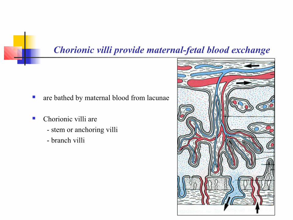
Chorionic villi provide maternal-fetal blood exchange
are bathed by maternal blood from lacunae
Chorionic villi are
- stem or anchoring villi
- branch villi
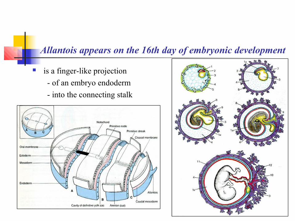
Allantois appears on the 16th day of embryonic development
is a finger-like projection
- of an embryo endoderm
- into the connecting stalk
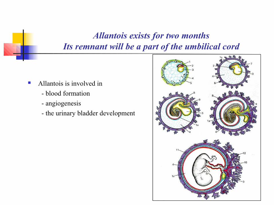
Allantois exists for two monthsIts remnant will be a part of the umbilical cord
Allantois is involved in
- blood formation
- angiogenesis
- the urinary bladder development

Folding – the body fold formation
begins on the 21st day of development
There are two pairs of folds
- longitudinal folds
- transversal folds
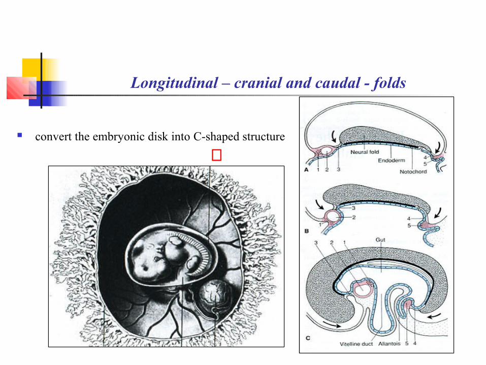
Longitudinal – cranial and caudal - folds
convert the embryonic disk into C-shaped structure
⇓
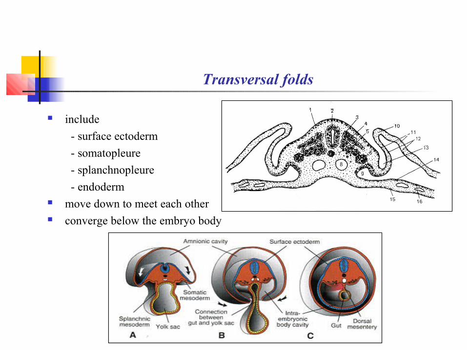
Transversal folds
include
- surface ectoderm
- somatopleure
- splanchnopleure
- endoderm move down to meet each other converge below the embryo body

Transversal fold results
embryonic disk is converted to
a cylinder-like structure
⇔
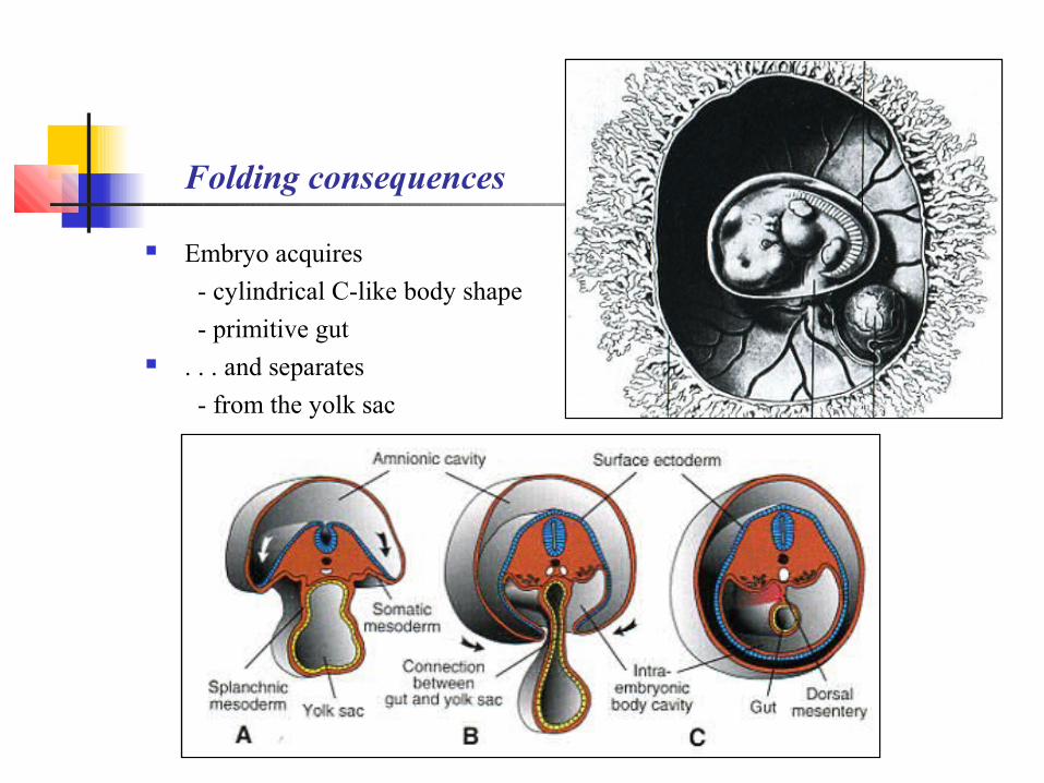
Folding consequences
Embryo acquires
- cylindrical C-like body shape
- primitive gut . . . and separates
- from the yolk sac

Embryonic period from the 4th to the 8th weeks
All tissues and organs differentiate, develop,
and begin to function
The period is the most critical period of embryogenesis because
any disturbances may give rise to congenital malformations
7th week embryo
⇔
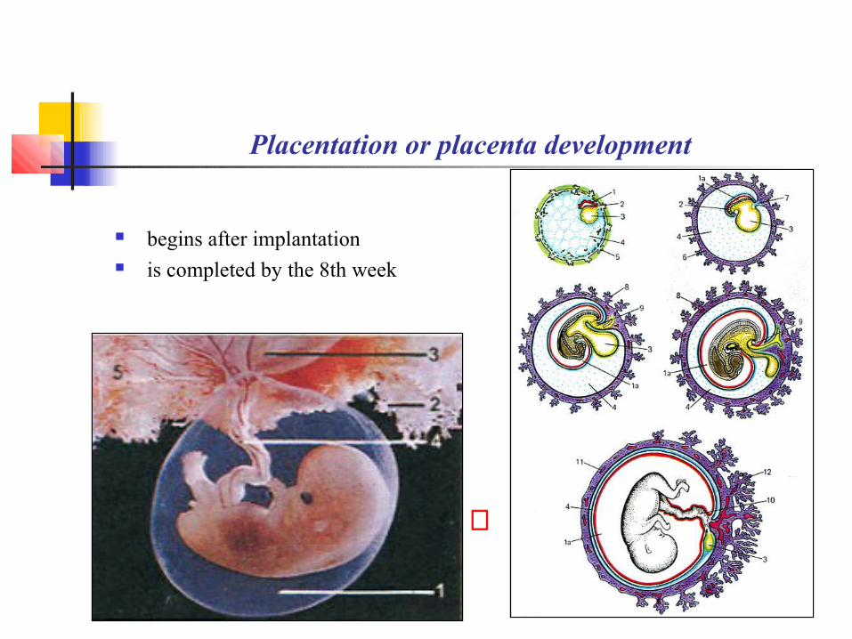
Placentation or placenta development
begins after implantation is completed by the 8th week
⇔
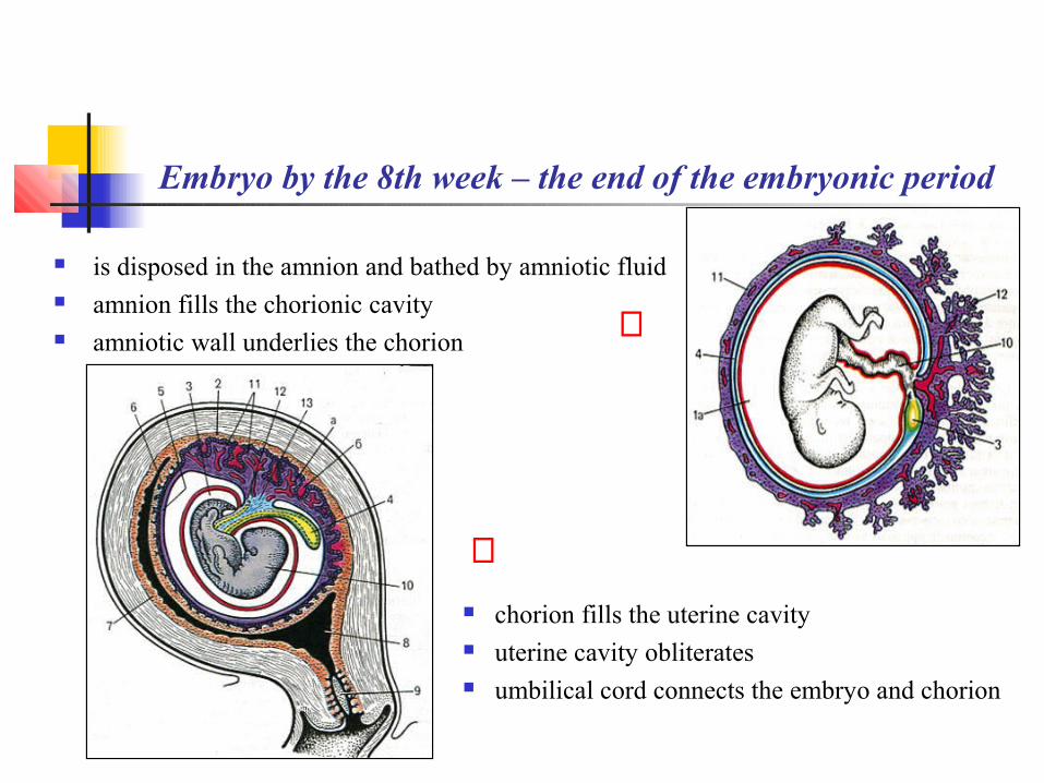
Embryo by the 8th week – the end of the embryonic period
is disposed in the amnion and bathed by amniotic fluid amnion fills the chorionic cavity amniotic wall underlies the chorion
chorion fills the uterine cavity uterine cavity obliterates umbilical cord connects the embryo and chorion
⇒
⇐
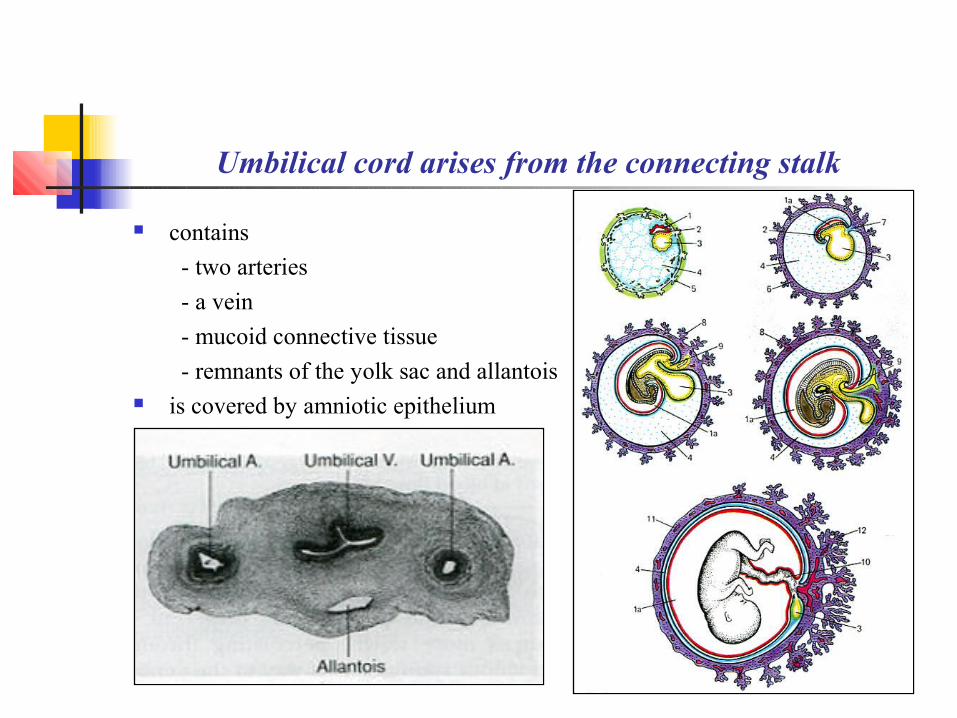
Umbilical cord arises from the connecting stalk
contains
- two arteries
- a vein
- mucoid connective tissue
- remnants of the yolk sac and allantois is covered by amniotic epithelium

Umbilical cord functions to connect
fetal cardiovascular system with chorionic vessels

Endometrium in pregnancy is called the decidua graviditas
Decidua basalis
- underlies the implantation site Decidua capsularis
- covers the implantation site Decidua parietalis
- remaining endometrium

Endometrium by the 8th week of development
Decidua basalis
- takes part in placenta formation Decidua capsularis
- fuses with decidua parietalis
when the uterine cavity obliterates

Chorion by the 8th week of development is subdivided into
Smooth chorion
- almost lacks villi
- is associated with the decidua capsularis
Villous chorion
- possesses large and branched villi
- is associated with the decidua basalis
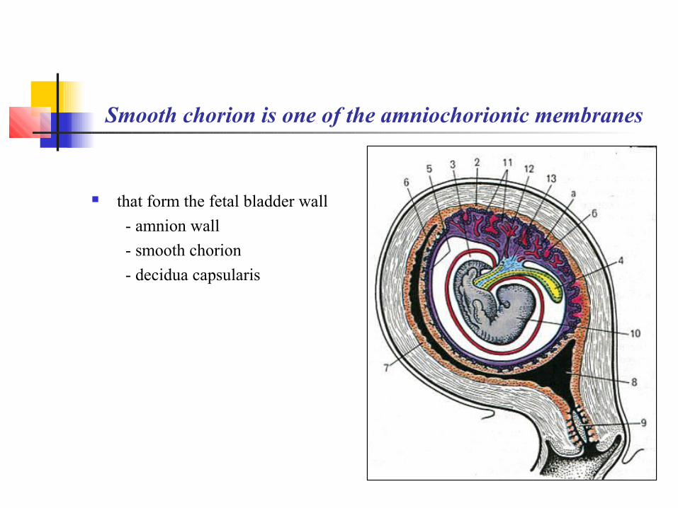
Smooth chorion is one of the amniochorionic membranes
that form the fetal bladder wall
- amnion wall
- smooth chorion
- decidua capsularis
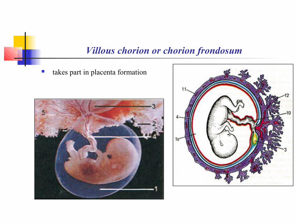
Villous chorion or chorion frondosum
takes part in placenta formation

Placenta is a combined organ
is formed by
- maternal body – the decidua basalis
- fetal body – the villous chorion
Two placental parts are involved in
the maternal-fetal circulation exchange ⇒
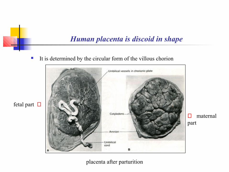
Human placenta is discoid in shape
It is determined by the circular form of the villous chorion
fetal part ⇒
⇐ maternal part
placenta after parturition

Fetal part of placenta
Chorionic plate Tertiary villi
Umbilical cord is attached to the fetal surface Amniotic epithelium surrounds the umbilical cord
and covers the fetal placenta part
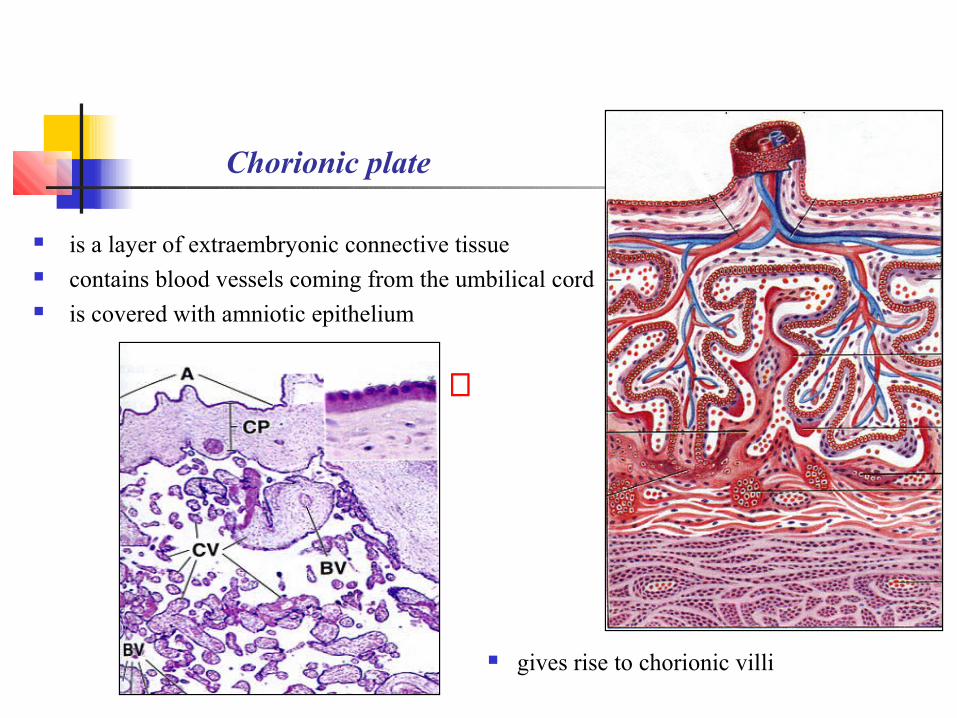
Chorionic plate
is a layer of extraembryonic connective tissue contains blood vessels coming from the umbilical cord is covered with amniotic epithelium
gives rise to chorionic villi
⇐
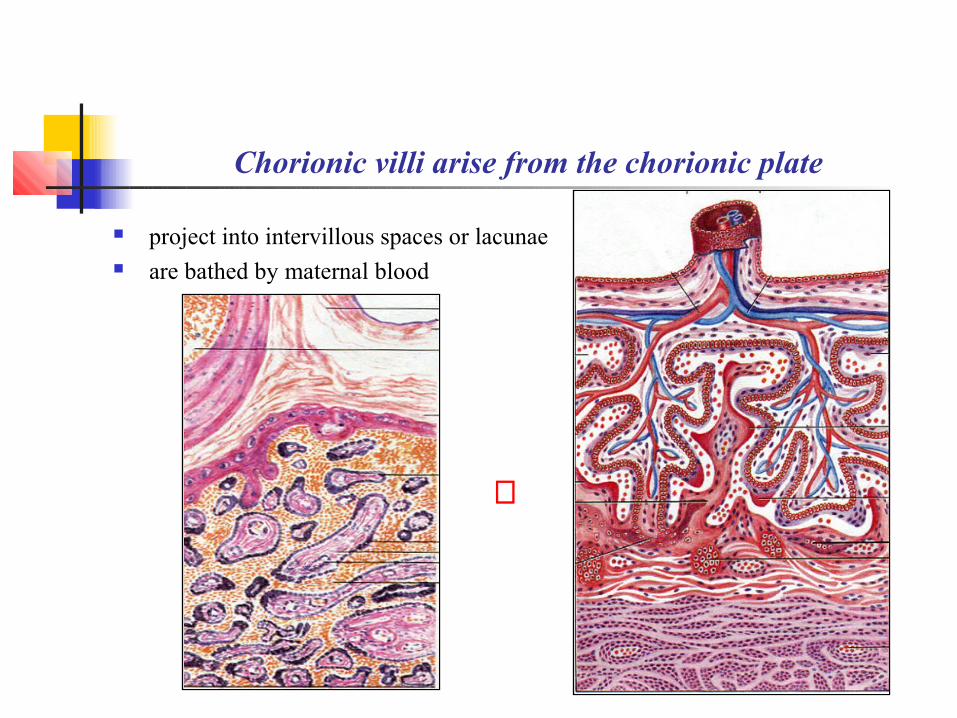
Chorionic villi arise from the chorionic plate
project into intervillous spaces or lacunae are bathed by maternal blood
⇔
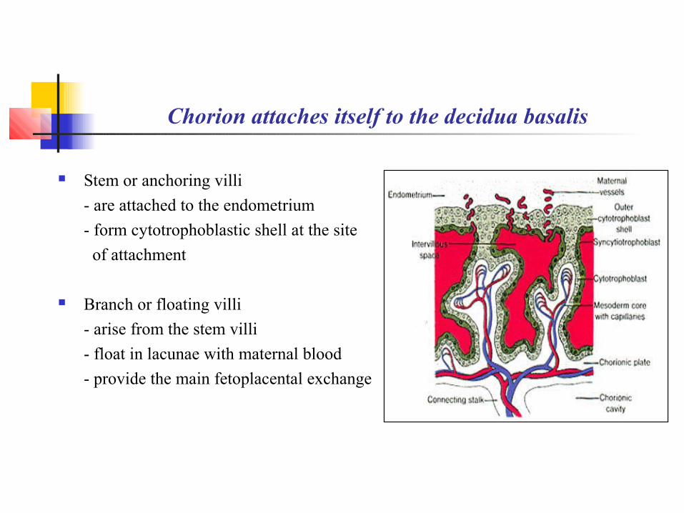
Chorion attaches itself to the decidua basalis
Stem or anchoring villi
- are attached to the endometrium
- form cytotrophoblastic shell at the site
of attachment
Branch or floating villi
- arise from the stem villi
- float in lacunae with maternal blood
- provide the main fetoplacental exchange
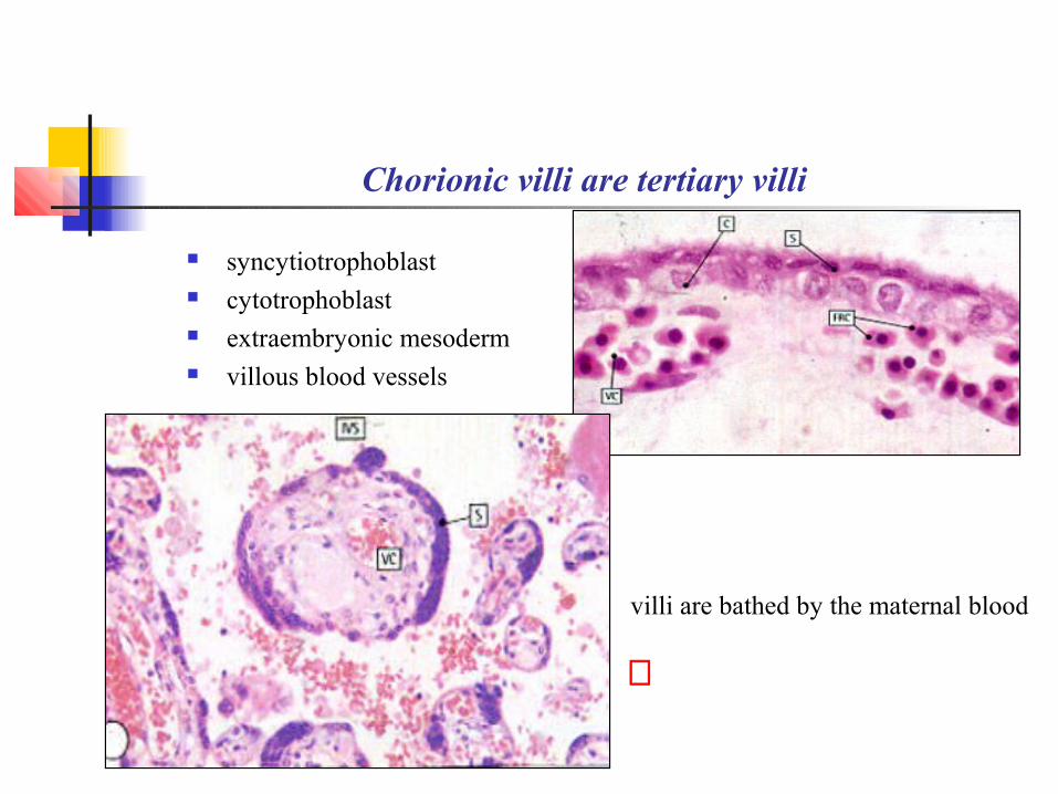
Chorionic villi are tertiary villi
syncytiotrophoblast cytotrophoblast extraembryonic mesoderm villous blood vessels
villi are bathed by the maternal blood
⇐

Fibrinoid material arises from the decidua basalis necrosis
results from the syncytiotrophoblast enzyme activity contains fibrin and immunoglobulins covers villi and the endometrium separates the fetal tissues from maternal tissues takes part in immune defence
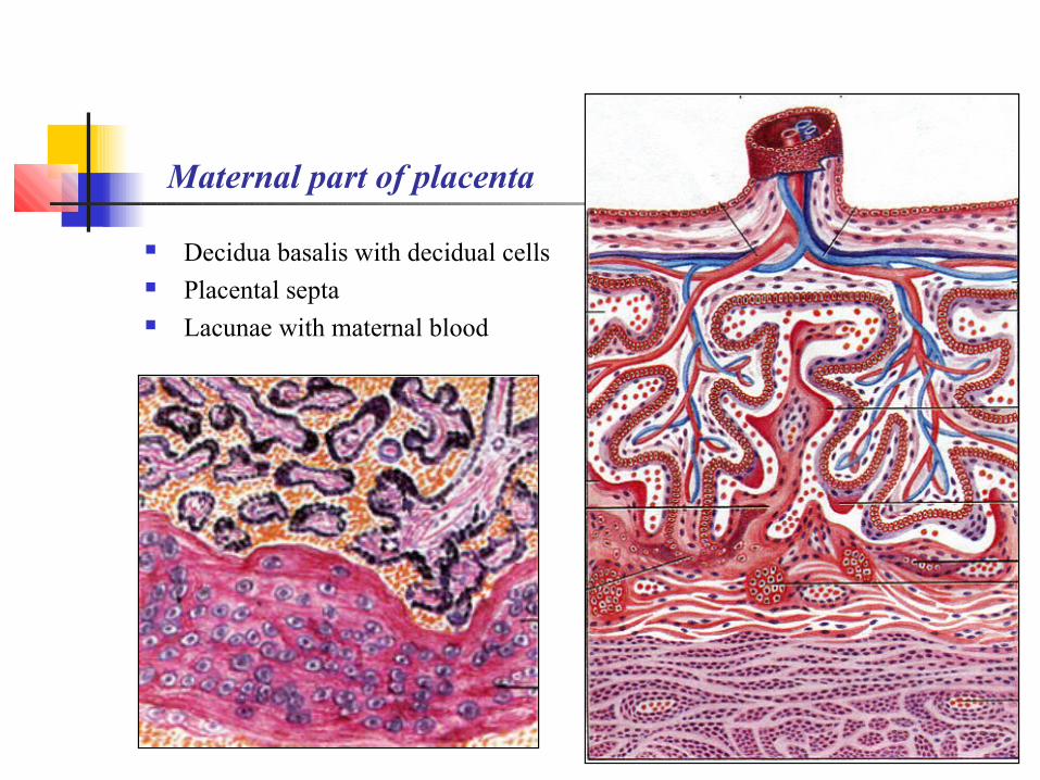
Maternal part of placenta
Decidua basalis with decidual cells Placental septa Lacunae with maternal blood

Decidua basalis
is a layer of the lamina propria connective tissue contains ruptured blood vessels and gland remnants
is underlined by the decidual plate
- remains after parturition
- is involved in the endometrium regeneration
⇔
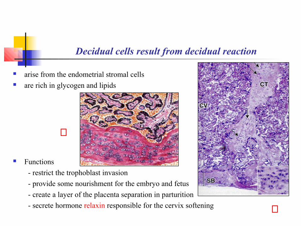
Decidual cells result from decidual reaction
arise from the endometrial stromal cells are rich in glycogen and lipids
Functions
- restrict the trophoblast invasion
- provide some nourishment for the embryo and fetus
- create a layer of the placenta separation in parturition
- secrete hormone relaxin responsible for the cervix softening ⇑
⇒

Placental septa are wedge-like areas of the endometrium
project from the decidua basalis to the chorionic plate
(never attach themselves) divide placenta into 15 to 20 lobules – cotyledons
Cotyledon includes
- two or more stem villi
- numerous branch villi
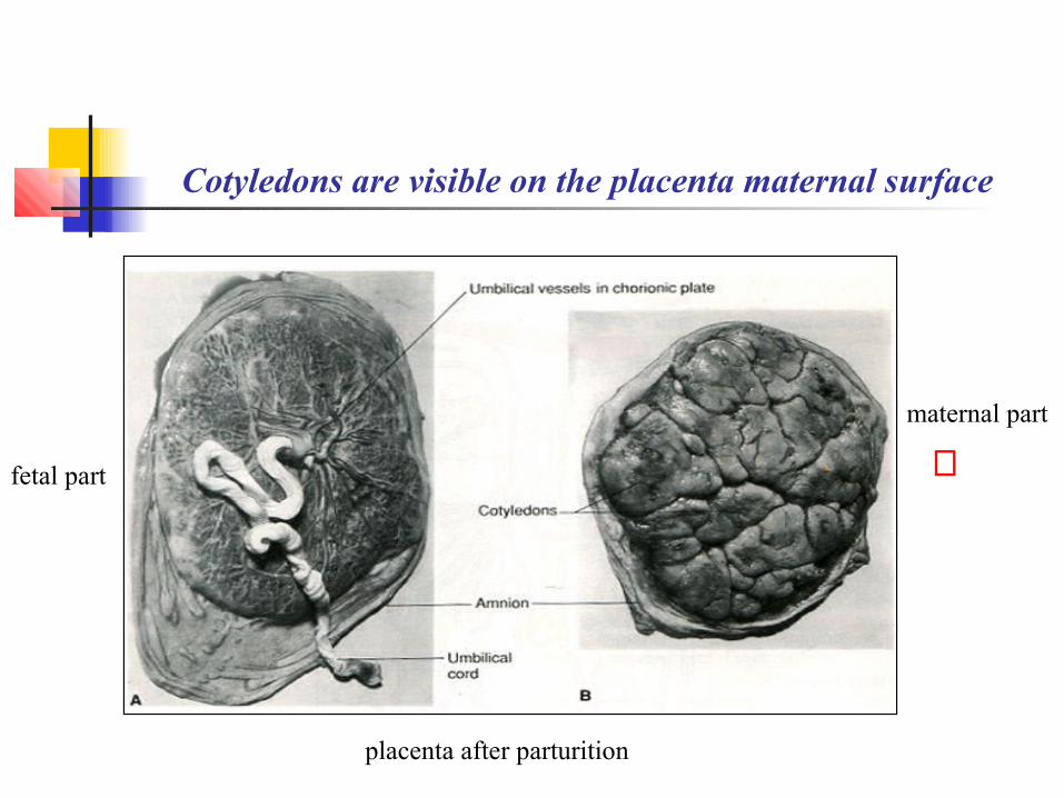
Cotyledons are visible on the placenta maternal surface
⇐maternal part
fetal part
placenta after parturition
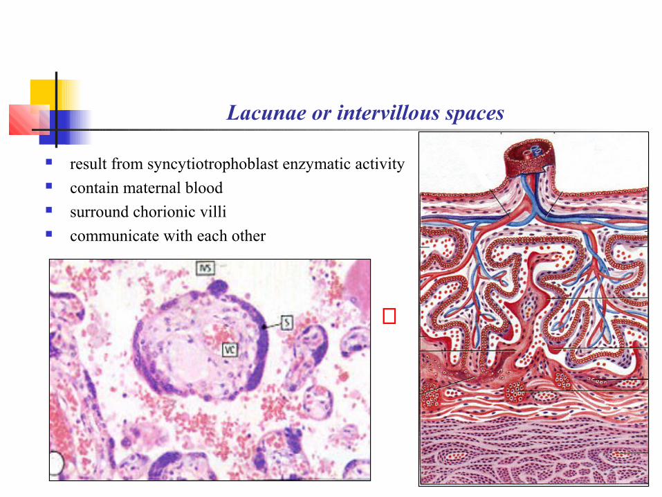
Lacunae or intervillous spaces
result from syncytiotrophoblast enzymatic activity contain maternal blood surround chorionic villi communicate with each other
⇔
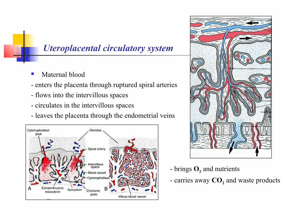
Uteroplacental circulatory system
Maternal blood
- enters the placenta through ruptured spiral arteries
- flows into the intervillous spaces
- circulates in the intervillous spaces
- leaves the placenta through the endometrial veins
- brings O2 and nutrients
- carries away CO2 and waste products
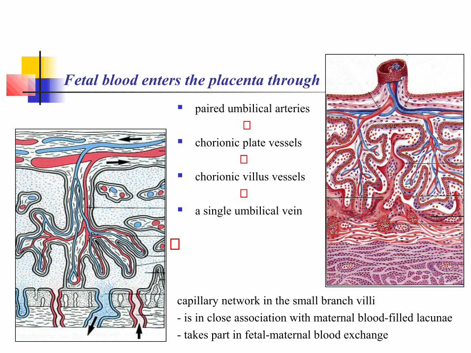
Fetal blood enters the placenta through
paired umbilical arteries
⇓ chorionic plate vessels
⇓ chorionic villus vessels
⇓ a single umbilical vein
capillary network in the small branch villi
- is in close association with maternal blood-filled lacunae
- takes part in fetal-maternal blood exchange
⇐
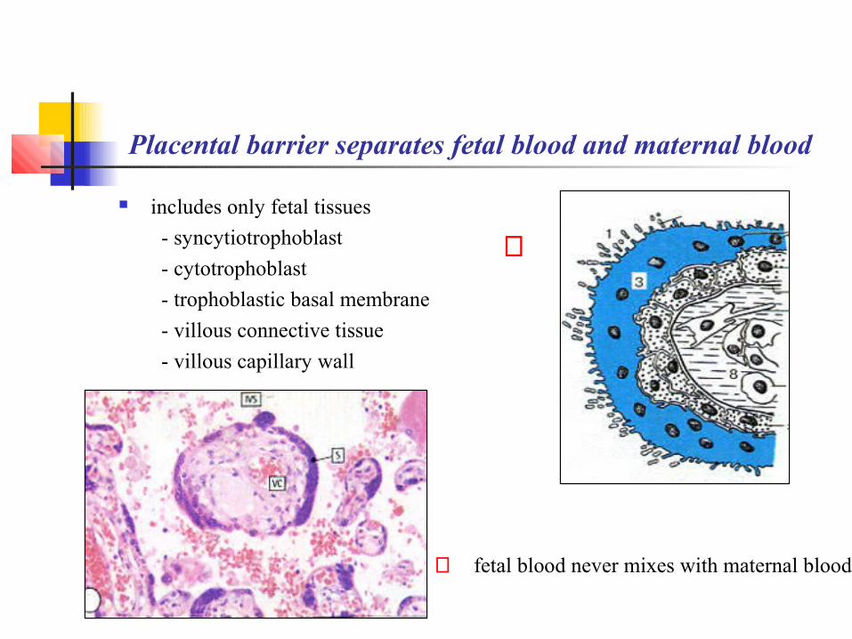
Placental barrier separates fetal blood and maternal blood
includes only fetal tissues
- syncytiotrophoblast
- cytotrophoblast
- trophoblastic basal membrane
- villous connective tissue
- villous capillary wall
⇐ fetal blood never mixes with maternal blood
⇒

Placental barrier ultrastructure Syncytiotrophoblast Cytotrophoblast Trophoblastic basal membrane Endothelium basal membrane Endothelial cells
⇓
⇒ ⇓
⇓⇑
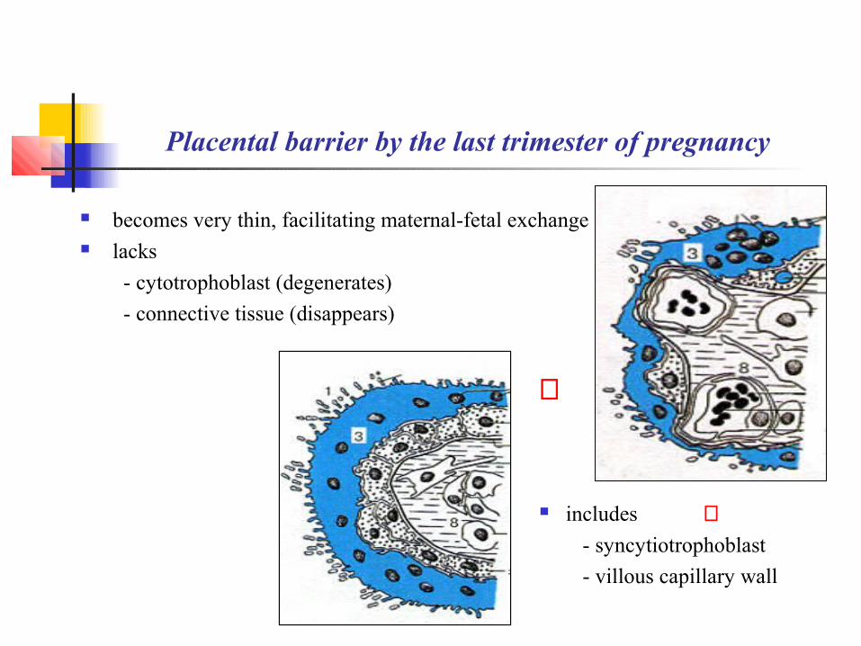
Placental barrier by the last trimester of pregnancy becomes very thin, facilitating maternal-fetal exchange lacks
- cytotrophoblast (degenerates)
- connective tissue (disappears)
includes ⇑- syncytiotrophoblast
- villous capillary wall
⇒

Placenta functions
Selective fetal-maternal blood exchange
- gases, water, electrolytes
- nutrients, hormones, antibodies
- medicine, drugs, infection agents Synthesis of some nutrients
- glycogen, cholesterol, fatty acids Release of enzymes to erode the endometrium Hormone production
- progesterone, estrogens,
- human chorionic gonadotropin (hCG)
- human placental lactogen (hPL)
- relaxin hCG in the villus syncytiotriophoblast
⇑
The End
The End
Thank you for attention!


![Human Fertilisation and Embryology Bill [HL] · ii Human Fertilisation and Embryology Bill [HL] 18 Revocation and variation of licence 19 Procedure for refusal, variation or revocation](https://img.dokumen.tips/doc/110x75/5b9aea6309d3f20b318c74f3/human-fertilisation-and-embryology-bill-hl-ii-human-fertilisation-and-embryology.jpg)