Embed Size (px)
Citation preview
Klinik für Hautkrankheiten
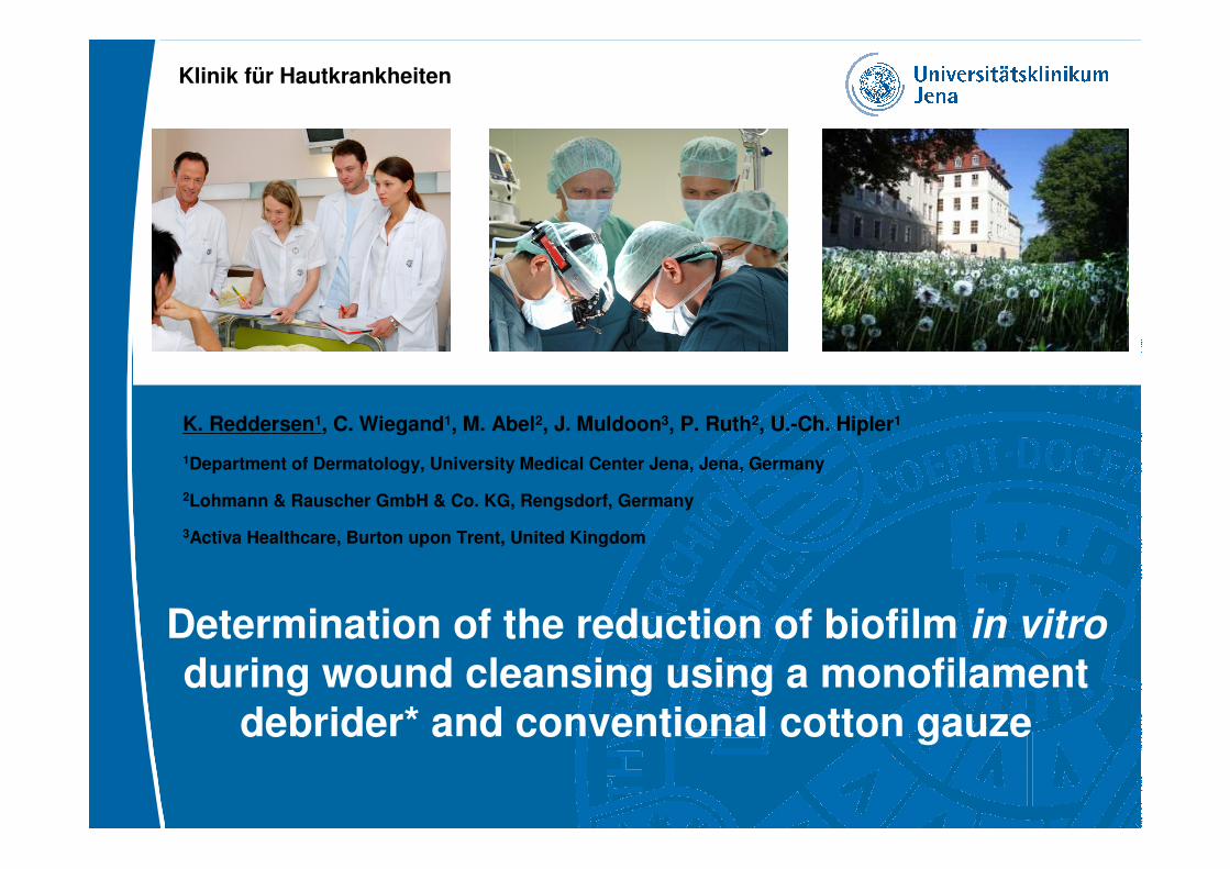
K. Reddersen1, C. Wiegand1, M. Abel2, J. Muldoon3, P. Ruth2, U.-Ch. Hipler1
1Department of Dermatology, University Medical Center Jena, Jena, Germany
2Lohmann & Rauscher GmbH & Co. KG, Rengsdorf, Germany
3Activa Healthcare, Burton upon Trent, United Kingdom
Determination of the reduction of biofilm in vitro
during wound cleansing using a monofilament debrider* and conventional cotton gauze
Klinik für Hautkrankheiten
2
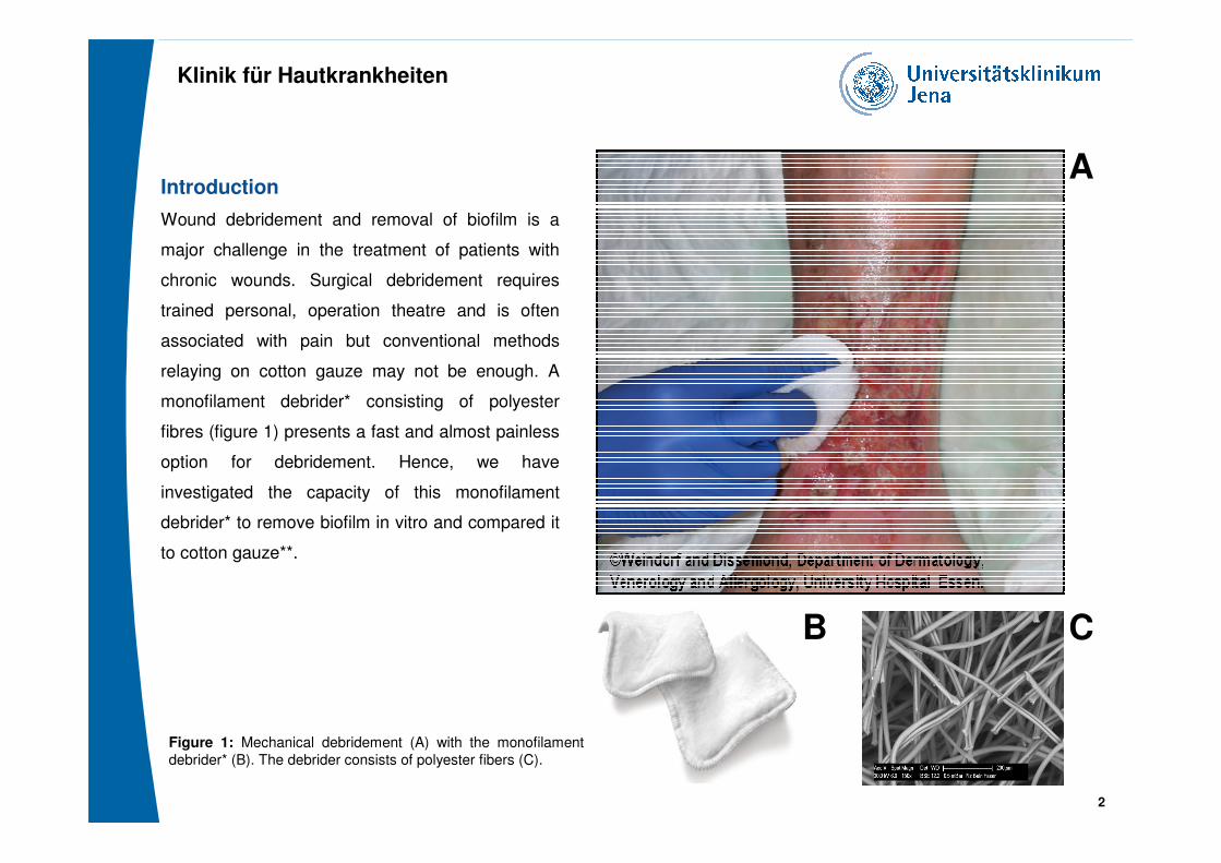
Introduction
Wound debridement and removal of biofilm is a
major challenge in the treatment of patients with
chronic wounds. Surgical debridement requires
trained personal, operation theatre and is often
associated with pain but conventional methods
relaying on cotton gauze may not be enough. A
monofilament debrider* consisting of polyester
fibres (figure 1) presents a fast and almost painless
option for debridement. Hence, we have
investigated the capacity of this monofilament
debrider* to remove biofilm in vitro and compared it
to cotton gauze**.
Figure 1: Mechanical debridement (A) with the monofilament
debrider* (B). The debrider consists of polyester fibers (C).
2
A
B C
Klinik für Hautkrankheiten
3
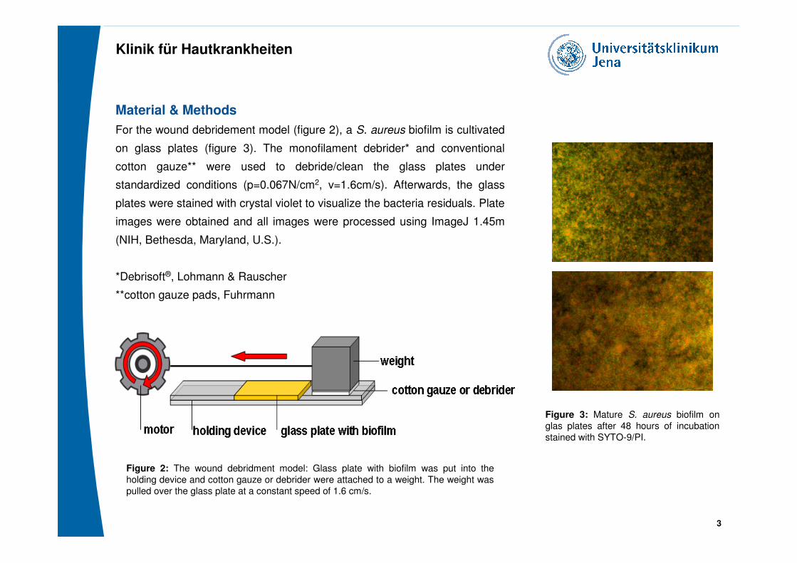
Figure 2: The wound debridment model: Glass plate with biofilm was put into the
holding device and cotton gauze or debrider were attached to a weight. The weight was
pulled over the glass plate at a constant speed of 1.6 cm/s.
Material & Methods
For the wound debridement model (figure 2), a S. aureus biofilm is cultivated
on glass plates (figure 3). The monofilament debrider* and conventional
cotton gauze** were used to debride/clean the glass plates under
standardized conditions (p=0.067N/cm2, v=1.6cm/s). Afterwards, the glass
plates were stained with crystal violet to visualize the bacteria residuals. Plate
images were obtained and all images were processed using ImageJ 1.45m
(NIH, Bethesda, Maryland, U.S.).
*Debrisoft®, Lohmann & Rauscher
**cotton gauze pads, Fuhrmann
3
Figure 3: Mature S. aureus biofilm on
glas plates after 48 hours of incubation
stained with SYTO-9/PI.
Klinik für Hautkrankheiten
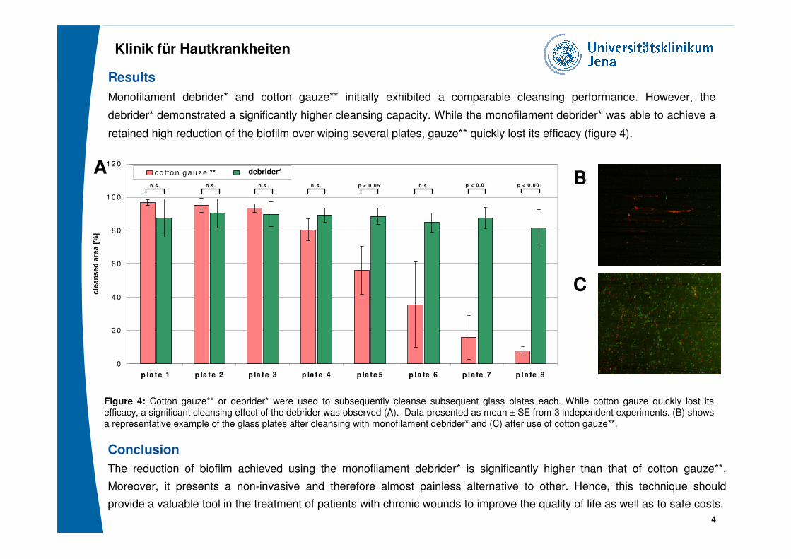
Results
Monofilament debrider* and cotton gauze** initially exhibited a comparable cleansing performance. However, the
debrider* demonstrated a significantly higher cleansing capacity. While the monofilament debrider* was able to achieve a
retained high reduction of the biofilm over wiping several plates, gauze** quickly lost its efficacy (figure 4).
Figure 4: Cotton gauze** or debrider* were used to subsequently cleanse subsequent glass plates each. While cotton gauze quickly lost its
efficacy, a significant cleansing effect of the debrider was observed (A). Data presented as mean ± SE from 3 independent experiments. (B) shows
a representative example of the glass plates after cleansing with monofilament debrider* and (C) after use of cotton gauze**.
4
0
2 0
4 0
6 0
8 0
1 0 0
1 2 0
p la te 1 p la te 2 p la te 3 p la te 4 p la te 5 p la te 6 p la te 7 p la te 8
cle
an
sed
are
a [
%]
c o tto n g a u z e D e b r is o f t
n.s . n .s . n .s . n .s . p < 0 .05 p < 0 .01n.s . p < 0 .0 01
** debrider*AB
C
Conclusion
The reduction of biofilm achieved using the monofilament debrider* is significantly higher than that of cotton gauze**.
Moreover, it presents a non-invasive and therefore almost painless alternative to other. Hence, this technique should
provide a valuable tool in the treatment of patients with chronic wounds to improve the quality of life as well as to safe costs.

Klinik für Hautkrankheiten
Thank you for your attention!
5