Embed Size (px)
Citation preview

Choledochal Cyst PRESENTOR : DR SHIV DNB SURGERY
APOLLO HOSPITAL LUDHIANA

• Cystic dilatation of the common bile duct (CBD) is known as choledochal cyst. It is a fairly uncommon anomaly of the biliary tract.
• It was first described by Vater and Ezler in 1723.
• Douglas published the first complete clinical description of the anomaly in a patient in 1853.
• Population prevalence estimates of choledochal cysts range from approximately 1 case in 13,000 people to 1 case in 2 million people.
• Choledochal cysts can occur in persons of any age.
• 2/3 rd of the cysts are diagnosed before the patient is aged 10 years.
• Approximately 20% of cysts are diagnosed in much older patients.
• In rare cases, choledochal cysts have been detected at prenatal USG as early as 15 weeks' gestation

PATHOPHYSIOLOGY
Congenital cysts may result from an unequal proliferation of embryologic biliary epithelial cells before bile duct cannulation is complete.• Fetal viral ( Reo RNA )infection may also have
a role • cyst formation may be the result of ductal
obstruction or distension during the prenatal or neonatal period.

ACQUIRED
• The exact cause of choledochal cyst remains obscure. Several theories have been postulated
1. Weakness of the wall of the bile duct
2. Obstruction of the distal choledochus
3.Combination of obstruction and weakness
4.Reflux of pancreatic enzymes into the CBD secondary to an anomaly of the pancreaticobiliary junction

–>1 cm common channel (90-100 % cases)
– allows pancreatic secretions to reflux into the common bile duct –Activated pancreatic proenzymes damage
and weak the bile duct wall – (Long common channel/Babbit theory-1969)

Abnormal pancreaticobiliary junction

• In 1984, Todani et al conducted an analysis of ERCP and other cholangiograms and confirmed this long common-channel anomaly.
• All of these theories are applicable to choledochal cyst type I, III, and IV anomalies, but they cannot be used to explain type II and V choledochal cysts in which the CBD is normal

• Perhaps genetic factors play a role. Despite this, the two most accepted theories are still reflux of pancreatic enzymes into the CBD secondary to an anomalous pancreaticobiliary junction and obstruction of the distal CBD.

Type Configuration Biliary Tract Incidence Treatment
Extrahepatic Intra-hepatic
I A Cystic
fusiform
Most or all >50 % to 75 %
Excision of involved portion of extrahepatic tract + Roux-en-Y Hepato-jejunostomy
B Limited
C sacular Most or all
II IsolatedDiverticulumOf CBD
5 % Excision with closure of defect over T-tube or same as above
III IntraduodenalPart ofCBD
5 % < 3 cm = endoscopic sphincterotomy> 3cm = excision via transduodenal approach
CHOLEDOCHOCELE
TODANI(1977) CLASSIFICATION

Type Configuration Biliary Tract Incidence Treatment
Extrahepatic Intra-hepatic
IV A
Multiple Dilations
30 % ExtrahepaticExcision of involved portion + Roux-en-Y Hepato-jejunostomy
IntrahepaticResection of segment or lobeOr transplantation
B
V 1 %
VI Isolated Cyst of Cystic Duct
ExtremelyRare
Cystic Duct ligation near CBD
CAROLI DISEASE (1958)
NOT part of Todani Classification
TODANI(1977) CLASSIFICATION
Jacques CaroliFrench gastroenterologist, 1902-1979

PRESENTATION
1. Classic triad for choledochal cysts is : pain, jaundice, and abdominal mass.
It is found in only a minority of children at the time of presentation.
• Infants commonly presented with elevated conjugated bilirubin (80%), failure to thrive or an abdominal mass (30%)

• older than 2 years of age, abdominal pain is the most common presenting symptom. common presenting symptom.
• Intermittent jaundice , recurrent cholangitis ,and pancreatitis is also a common feature .
• Pancreatitis is more common with type 3 cyst .

Rarely, biliary cysts present with
• intraperitoneal rupture• bleeding due to erosion into adjacent vessels• portal hypertension • secondary biliary cirrhosis due to prolonged biliary
obstruction and recurrent cholangitis. • In addition, type III cysts can case gastric outlet
obstruction due to the obstruction of the duodenal lumen or intussusception.

Investigation
• Transabdominal ultrasound• Computed tomography• CT cholangiogarphy• Endoscopic ultrasound• Intraductal ultrasound• Endoscopic retrograde cholangiopancreatography• Magnetic resonance cholangiopancreatography

Transabdominal ultrasound
• First imaging modality used for the evaluation• Not detect type III and type V cysts. • sensitivity of 71 to 97 %
• Factors that may limit the usefulness of an ultrasound include the patient's body habitus, the presence of bowel gas, and limited visualization due to overlying structures.

Computed tomography
• CT can detect all types of biliary cysts.• Can evaluate for the presence of malignancy.
It is also useful for determining the extent of intrahepatic disease in patients with type IVA or V cysts.

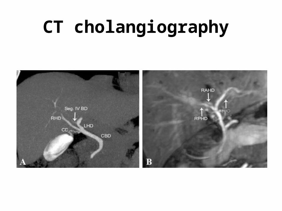
CT cholangiography
has high sensitivities for visualizing the • biliary tree (93%), • biliary cysts (90%),• intraductal stones (93%)
• However, its sensitivity is lower for imaging the pancreatic duct (64 %)
CT cholangiography

Endoscopic ultrasound(EUS)
• EUS can demonstrate extrahepatic biliary cysts and provide detailed images of the cyst wall and pancreaticobiliary junction.
• unlike transabdominal U/S, it is not limited by body habitus, bowel gas, or overlying structures.

Intraductal ultrasound (IDUS)
• has been used for the diagnosis of early malignant changes in a biliary cyst .
• This technique is likely to be more sensitive than direct cholangiography for detecting early malignancy in the cyst wall.

Hepatobiliary scintigraphy
• using radio-labeled dyes : technetium-99m-labeled hepatic iminodiacetic acid (HIDA), which is selectively taken-up by hepatocytes and excreted into the bile.
• HIDA scanning is useful for extrahepatic cysts, with a sensitivity up to 100% for type I cysts. However, it is inadequate at visualizing the intrahepatic bile ducts
• HIDA scanning may also be useful in cases of cyst rupture

HIDA SCAN

Cholangiography
• Direct cholangiography (whether intraoperative, percutaneous, or endoscopic) has a sensitivity of up to 100 percent for diagnosing biliary cysts and previously was a commonly obtained test.
• can identify abnormal pancreatobiliary junction, and filling defects due to stones or malignancy.
• Increase risk of cholangitis and pancreatitis. [ Patients with cystic disease are greater risk for these complications ]

Radiology
Magnetic resonance cholangiopancreatography [MRCP]
• Does not have the risks of cholangitis and pancreatitis as direct cholangiography
• Sensitivity 73 - 100 %.• less sensitive than direct cholangiography for
excluding obstruction. • The data are variable with regard to its ability to
diagnose an abnormal pancreatobiliary junction. [46-75%]

Magnetic Resonant CholangioPancreatography (MRCP)

TREATMENT
• If pt presents with pancreatitis /cholangitis should be treated supportively before definitive operative management.
• In choledochal cyst pb maljunction are high risk for pancreatitis .chance of panceatitis increase with ercp and ampulary stenting.

• The treatment of choice for choledochal cysts is complete excision of the cyst with construction of a biliary-enteric anastomosis to restore continuity with the gastrointestinal tract.
• partial resection of the cyst and internal drainage procedures expose patients to increased risks of cholangitis, pancreatitis, and cholangiocarcinoma.

• Type 1 : KOCHER maneuver to explore distal portion of cyst .
• Type 1(B) : Extend distally to entrance of the cbd into pancrease . Goal is to excise intrapancreatic portion of cyst without injuring pancreatic duct or long common duct .
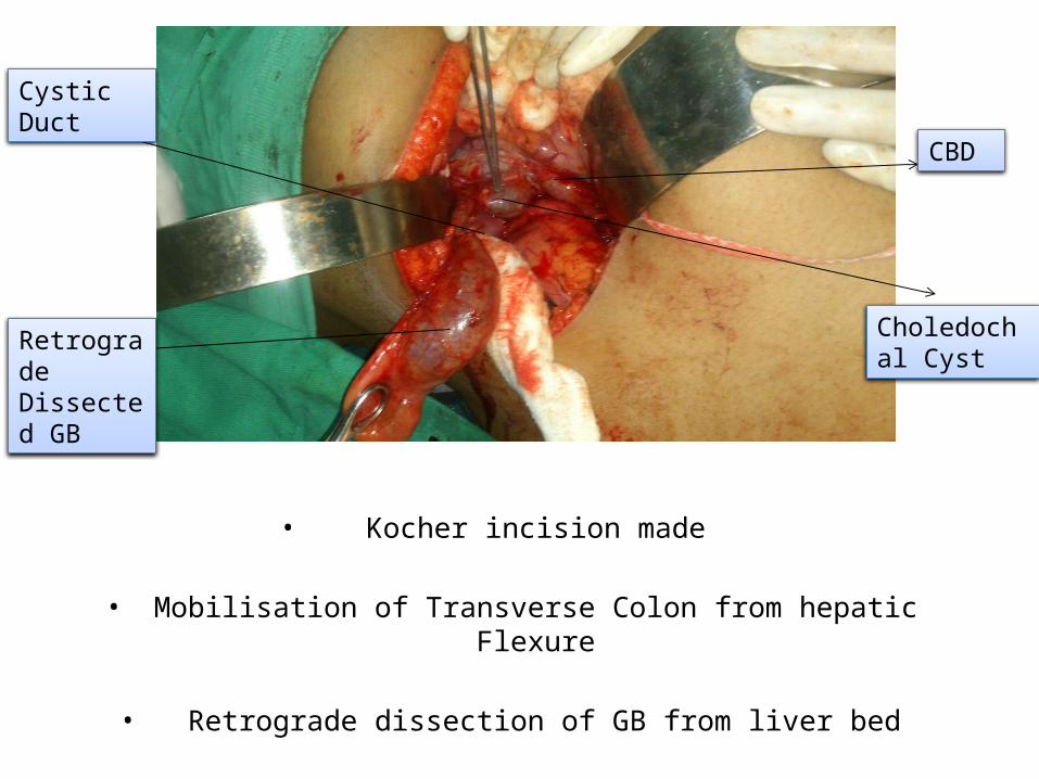
• Kocher incision made
• Mobilisation of Transverse Colon from hepatic Flexure
• Retrograde dissection of GB from liver bed
Retrograde Dissected GB
Cystic Duct
Choledochal Cyst
CBD

• Choledochal Cyst dissected away from portal vein and hepatic artery ( Lilly Technique?)
• Cyst wall opened till common hepatic duct junction

Occasionally, Cyst adheres densely to the portal vein secondary to long-standing inflammatory reaction
complete, full-thickness excision of the cyst may not be possible
serosal surface of the duct is left adhering to the portal vein, while the mucosa of the cyst wall is obliterated by curettage or cautery
Theoretically, this removes the risk of malignant transformation in that segment of the duct

• Hepatic ducts washed with normal saline
• Hepatic ducts patency confirmed with bougies
Left HepaticDuct
Right HepaticDuct

• Roux-en-Y (french: rōō'ěn-wī')
Hepatojejunostomy done(retrocolic isoperistaltic functional side-to-side)
Cesar Roux Swiss Surgeon (1857-1934),
(Performed 1st successful excision of pheochromocytoma in 1926)
(End-to-side)
(End-to-side ORSide-to-side)
• Type 2 : simple cyst excision along with cholecystectomy .close the defect transversely , which reduce the cbd stricture.
• Type 3: endoscopic sphincterotomy is benificial .
• Type 3 : pt may benefit from endoscopy sphincterotomy .
• Surgical resection is via transverse duodenotomy in second or 3 rd part . Duodenotomy allow both biliary and pancreatic duct to be identify individually.
• After resection of cyst both pancreatic and bile duct mucosa are sutured individually to duodenal mucosa .

• Type 4 : 4a and 4b are managed in similarly to type 1 cyst .
• Type 5 : if confined to single lobe resection of involved parenchyma.
• In bilobar absence of cirrhosis or malignancy roux –en- y hepaticojejunostomy with bilateral trans transhepatic silastic stent may be indicated to improve biliary drainage.
• Patient with carolis disease and liver failure may warrant liver transplantation .
• Summary : patient with holedochal cyst require long term surveillance for recurrent cholangitis , intrahepatic stone , pancreatitis ,postoperative biliary strictures and malignancy.