Embed Size (px)
Citation preview

Chest X-ray positioning
outline
• Plain films different views
• Positioning
• Interpretation of the plain film

Different views of Xray chest
• PA
• Lateral
• AP,decubitis,supine,oblique
• Inspiratory-expiratory
• Lordotic,apical

PA view
• Most frequently requested
• Visualization of the lungs excellent
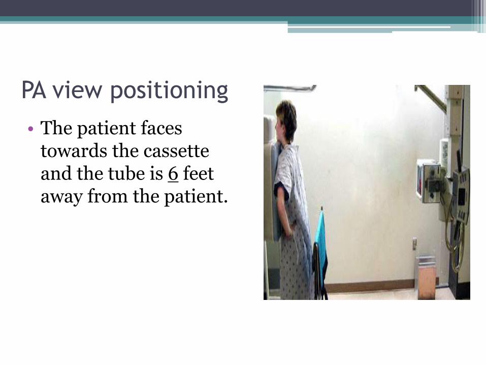
PA view positioning
• The patient faces towards the cassette and the tube is 6 feet away from the patient.

Technical aspect
• Inspiration
• On full inspiration the diaphragm should lie at the level of 8-10th
posterior rib or 5-6th
anterior rib.

Technical aspect
Inspiratory film

Technical aspects
• Penetration

Over penetrated Under penetrated

rotation

Rotated x ray

Interpretation of the PA films
1.request form:
name,age,sex,date and clinical presentation
2.trachea
3.Heart and mediastinum
4.Diaphragm

6.Pleural spaces
7.Lungs
8.Hidden areas
9.Hila
10.Below diaphragm
11.Soft tissues
12.Bones

Trachea
Examined for
• Position
• outline

• Caliber coronal diameter is 25mm for males and 21mm for females
• Para tracheal stripe<5mm
• Azygos vein<10mm
• Carina angle:60-75degree.

Heart
• Size
• Shape
Transverse cardiac diameter:<14.5cm in females and <15.5cm in males. An increase of 1.5 cm is significant
Cardiothoracic ratio<50%

mediastinum
• Right superior mediastinalshadow formed by SVC and innominate vessels.
• Left superior mediastinalshadow formed by the subclavian artery
• Ant junction line
• Post junction line
• Thymus
• Paraspinal lines 10 mm on the left and 3mm on the right

Ant and post junction lines
• Ant junction line
• Parietal and visceral pleurae meeting anteromedially.oblique course
• Post juction line.formed by posteromedial surfaces of the pleurae of the upper lobes post to oesophagus

thymus
• Triangular sail-shaped structure, well defined borders projecting from one or both side of the mediastinum.

Para spinal lines
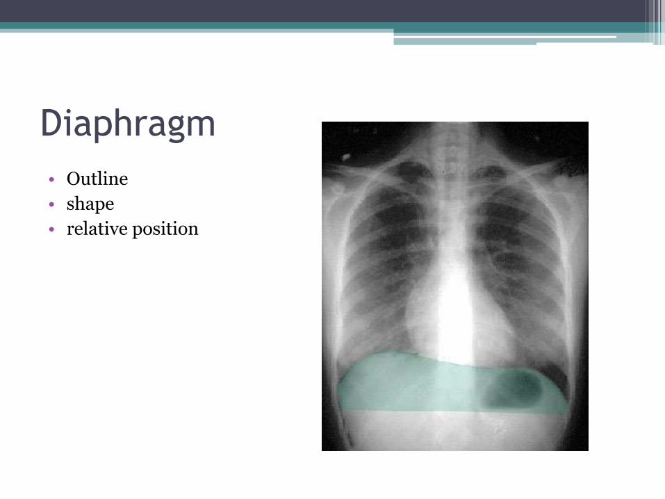
Diaphragm
• Outline
• shape
• relative position
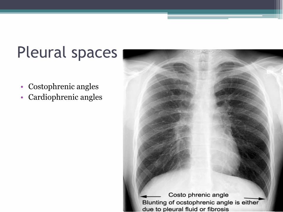
Pleural spaces
• Costophrenic angles
• Cardiophrenic angles

lungs
• Local,generalised abnormality
• Comparison of the translucency
• Vascular markings of the lungs

Zones

Hidden areas
• The apices
• Mediastinum and hila
• Diaphragm
• bones

Hila
• Contain the following structures
• The inferior pulmonary ligament
• The pulmonary vessels
• The bronchial vessels
• the bronchi
• The lymphatic system
• The lymph nodes

Right hilum

Left hilum

Below diaphragms
• Gas shadows
• Calcifications
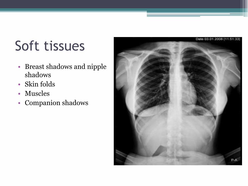
Soft tissues
• Breast shadows and nipple shadows
• Skin folds
• Muscles
• Companion shadows

Nipple markers

Skin fold

Muscles and companion shadows
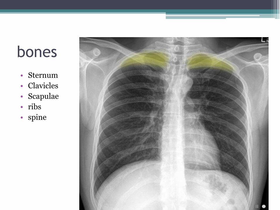
bones
• Sternum
• Clavicles
• Scapulae
• ribs
• spine

Lateral film
• positioning

Interpretation of lateral film
• The clear spaces
• Retrosternal space
• Retrotracheal space
• Vertebral translucency
• Diaphragm outline
• The fissures
• The trachea
• The sternum

Retrosternal space
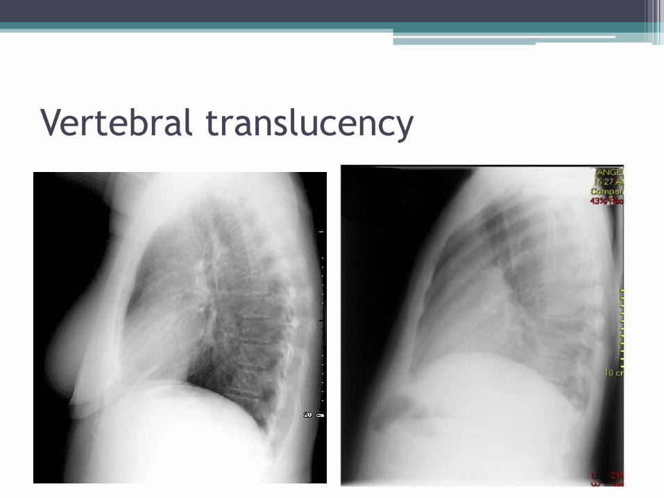
Vertebral translucency

Diaphragm outline
• Right diaphragm continues anteriorly
• Left is silhouetted posteriorly by heart shadow

The fissures

AP view
• the patient back is towards the cassette and tube is 40 inches away from the patient.
• for patients unable to stand


Decubitus position
• The patient faces towards the cassette while lying in decubitus position and tube Is towards the back

Decubitus position
• To asses the volume of pleural fluid.
• Loculated pleural effusion or mobile
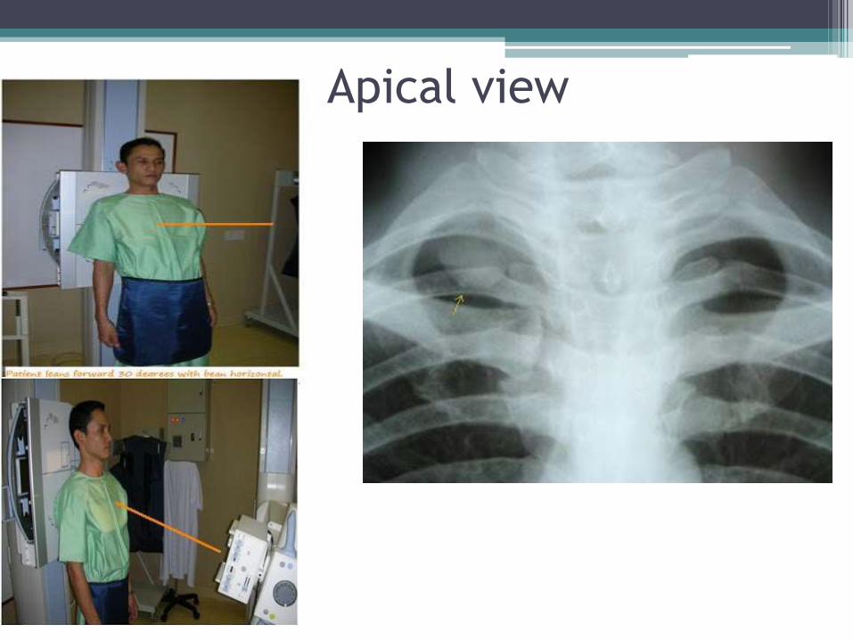
Apical view

Oblique view
• positioning
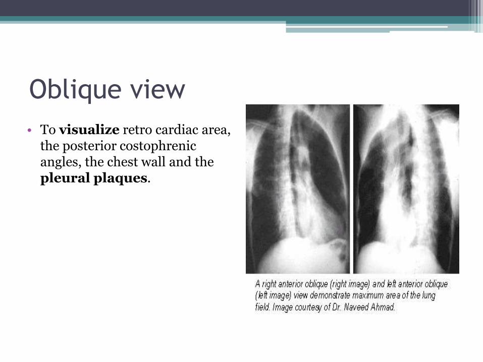
Oblique view
• To visualize retro cardiac area, the posterior costophrenicangles, the chest wall and the pleural plaques.

• Lordotic PA view

Paired inspiratory and expiratory
• Demonstrate air trapping and diaphragm movements.
• Very important in diagnosis of inhaled foreign body in children.
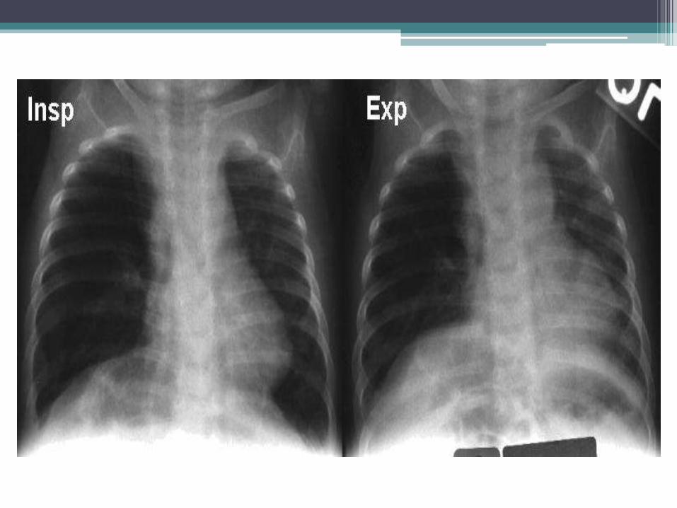
Paired inspiratory and expiratory view

Thank you