Embed Size (px)
Citation preview

Reading Chest X-Ray
Farah Amer
By the Name of Allah,
The Most Gracious, The Most Merciful

Reading Chest X-Ray
-This document contains only some CXR findings that you should be familiar for medicine OSCE .No theoretical material included . This is just a collection of X-rays that I made during studying. -I hope it will help you for your exam. Resources : -Macleod’s clinical examination-12th edition -Davidson’s essentials of medicine -Different websites. Good Luck

Index: • Basic knowledge. • Pleural diseases. • Pneumothorax. • TB. • Pneumonia. • Interstitial pulmonary fibrosis • COPD • Sarcoidosis • Pericardial Effusion.

Chest X-Ray Basic knowledge Normal labeled X-Ray

Normal CXR
Normally lungs are full of air (Black in color) Fluid or
Blood are white in color. Look at the blood vessels.
Bones are white in color and any calcification will appear white too.
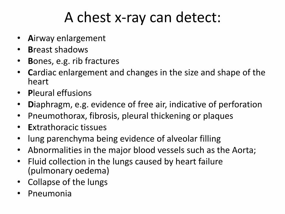
A chest x-ray can detect: • Airway enlargement
• Breast shadows • Bones, e.g. rib fractures • Cardiac enlargement and changes in the size and shape of the
heart • Pleural effusions • Diaphragm, e.g. evidence of free air, indicative of perforation • Pneumothorax, fibrosis, pleural thickening or plaques • Extrathoracic tissues • lung parenchyma being evidence of alveolar filling • Abnormalities in the major blood vessels such as the Aorta; • Fluid collection in the lungs caused by heart failure
(pulmonary oedema) • Collapse of the lungs • Pneumonia

Systemic approach to CXR interpretation
• Pt details & date Note the pt’s name and date of birth as well as the date and time the CXR was
performed . • Technical Quality Orientation : Most CXR are taken using a postero-anterior (PA) view , if patients
are too unwell to stand , then an antero-posterior (AP) X-ray will be done with the X-ray source in front of them & the plate behind them. With AP film the heart appears magnified relative to PA film.
Posture : If the pt is supine , the distribution of air & fluid is changed & it is impossible to exclude a pneumothorax , pleural effusion or subdiaphragmatic air .
Rotation : If the pt is not rotated , the spinous processes of the thoracic vertebrae will be projected midway between the medial borders of the clavicles.
Penetration : The thoracic vertebral bodies should be just visible behind the heart. If they cannot be seen at all , the film is under-exposed and will appear too white. If they can be seen in detail , then the film is over-exposed and will be too dark.
Inspiration : The right hemidiaphragm should be at the level of the anterior end of the 6th rib or the posterior end of the 9th – 10th ribs. If more ribs are seen , hyperinflation is present.
Field of view : All of the lungs should be visible ; make sure that lung apices and especially costophrenic angles have not been missed .

Systemic approach to CXR interpretation • Trachea Should be central .It might be deviated toward the area of loss of volume (e.g.
Lung collapse) or away from an area of increased pressure (e.g. Tension pneumothorax).
• Heart A cardiac shadow of >50% of the total thoracic width on a PA film is abnormal &
occurs with ventricular dilatation or pericardial effusion. The left heart border consist of the left ventricle and left atrium , while the right
heart border is made up of the right atrium . Consolidation in the immediately adjacent lung blurs the heart borders.
• Lung and pleura are discussed in the next slides • Diaphragm The hemidiaphragms should have a well-defined edges , and the costophrenic and
cardiophrenic angles should be sharp. The right hemidiaphragm is usually higher due to the liver below.
• Soft tissues and bones Assess the soft tissues , including breast shadows . Look for surgical emphysema &
free air under the diaphragm . Examine each rib , looking for fractures or metastatic lesions . Then check clavicles and scapula.
• Review areas Rechek areas in which abnormalities are commonly missed : lung apices ,
subdiaphragmatic air , behind the cardiac shadow & behind hemidiaphragms .

Lung Diseases


RML : Right Middle Lobe , RLL: Right Lower Lobe

RLL : Right Lower Lobe, LLL : Left lower Lobe , LUL : Left Upper Lobe , RUL : Right Upper Lobe.


A: Ascending aorta B: Left heart margin C: Left diaphragm D: Aortic knob E: Right heart margin F: Right diaphragm
Silhouette Sign


Pleural Diseases


Pleural Effusion
Right side pleural effusion
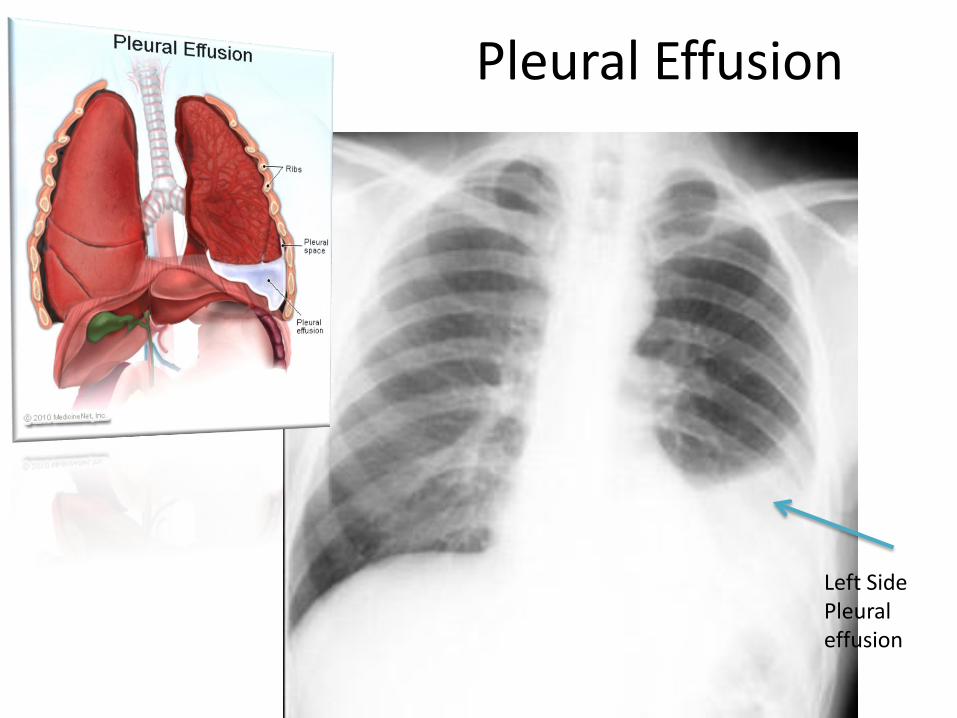
Pleural Effusion
Left Side Pleural effusion

Pleural Effusion
PA film of a patient with bilateral pleural effusions. Note the concave menisci blunting both posterior costophrenic angles.


Tension Pneumothorax
This film shows a right sided tension pneumothorax with right sided lucency and leftward mediastinal shift. This is a medical emergency. Failure to place a right chest tube immediately could allow venous return to diminish and lead to possible death.

Tension Pneumothorax
Right Sided tension pneumothorax Left Sided tension pneumothorax

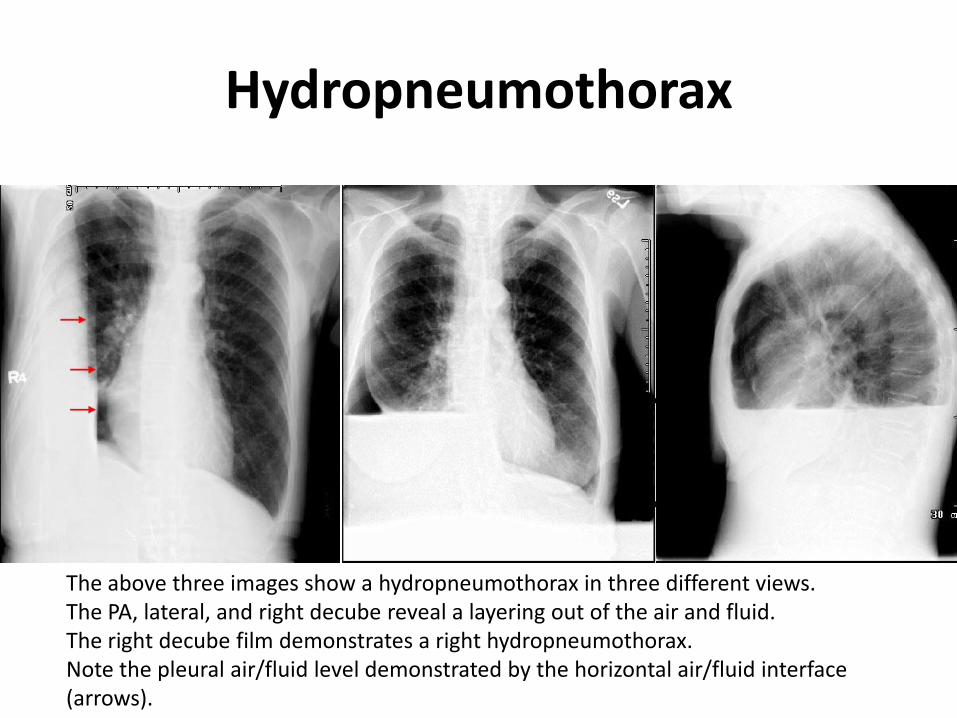
Hydropneumothorax
• Hydropneumothorax : implies presence of both air and fluid in the pleural space ( i.e. between two layers of pleura. An erect chest x-ray will show the air fluid level. The horizontal fluid level is usually well defined and extends across the whole length of hemithorax.
Signs of hydropneumothorax can be remembered by 4 'S'
• Straight line dullness
• Shifting dullness
• Succussion splash
• Sound of coin

Hydropneumothorax
hydropneumothorax with white arrow point to the pulmonary pleura
Hydropneumothorax
The above three images show a hydropneumothorax in three different views. The PA, lateral, and right decube reveal a layering out of the air and fluid. The right decube film demonstrates a right hydropneumothorax. Note the pleural air/fluid level demonstrated by the horizontal air/fluid interface (arrows).


TB • Primary TB
-Calcification in 1ry complex is overall relatively rare.
-Few pts have clinical manifestaions.
• Post-primary TB (TB Reactivation)
-Calcification is usually rarer than in 1ry.
-Limited mainly to the apical & posterior segments of upper lobes & superior segment of lower lobe.
-Bilateral upper lobe disease is very common.
-Cavitation may result ; Cavity is usually thin walled ,smooth or inner margin with no air-fluid level.
-Transbronchial spread might occur from one upper lobe to opposite one.
• Miliary TB
- Hematogenous dissemination of bacilli. Fine (1-2mm) lesions “millet seed” throughout the lung fields.

TB (post-primary)
Bilateral Upper lobe cavitary disease with transbronchial spread to Lingula . (Cavitary looks like a circle )

Infection in both lungs is marked by white arrow-heads, and the formation of a cavity is marked by black arrows.

TB (post-primary)
Ill-defined opacity situated in one of the upper lobes. In this situation Cavitary in the right upper lobe. *As the disease progress consolidation & collapse may develop.
Transbronchial spread might occur from one upper lobe to opposite one. This CXR shows Transbronchial spread to left lower lobe.

TB (post-primary)
Also here Lung Cavitation

TB (post-primary)
Left upper lobe Cavitation

TB (post-primary)
Right upper lobe cavity.

TB (miliary)
Miliary opacities all over the lungs . (Not very important)

Pneumonia
• The type of pneumonia is sometimes characteristic on chest x-ray: • Lobar - classically Pneumococcal pneumonia, entire lobe consolidated and
air bronchograms common
• Lobular - often Staphlococcus, multifocal, patchy, sometimes without air bronchograms
• Interstitial - Viral or Mycoplasma; latter starts perihilar and can become confluent and/or patchy as disease progresses, no air bronchograms Aspiration pneumonia - follows gravitational flow of aspirated contents; impaired consciousness, post anesthesia, common in alcoholics, debilitated, demented pts; anaerobic (Bacteroides and Fusobacterium) Diffuse pulmonary infections - community acquired (Mycoplasma, resolves spontaneoulsy) nosocomial (Pseudomonas, debilitated, mechanical vent pts, high mortality rate, patchy opacities, cavitation, ill-defined nodular) immunocompromised host(bacterial, fungal, PCP).

Pneumonia
These are PA and lateral films of RML pneumonia (arrows). Note the indistinct borders, air bronchograms, and silhouetting of the right heart border.

Pneumonia
PA and Lateral films of RUL pneumonia

Pneumonia
Right upper lobe lobar pneumonia.

Pneumonia
Right upper lobe pneumonia

Interstitial pulmonary fibrosis
• Interstitial pulmonary fibrosis has many causes. The six most common causes of diffuse interstitial pulmonary fibrosis are idiopathic (IPF, >50% of cases), collagen vascular disease, cytotoxic agents and nitrofurantoin, pneumoconioses, radiation, and sarcoidosis.
• Clinically the patient with IPF will present with progressive exertional dyspnea and a nonproductive cough.
• Radiographically : IPF is associated with hazy "ground glass" opacification early and volume loss with linear opacities bilaterally, and honeycomb lung in the late stages.
• IPF carries a poor prognosis with death due to pulmonary failure usually occurring within 3-6 years of the diagnosis unless lung transplant is performed.

Interstitial pulmonary fibrosis

COPD COPD includes chronic bronchitis , chronic bronchiolitis & Emphysema.
Emphysema is commonly seen on CXR as :
• diffuse hyperinflation with flattening of diaphragms
• increased retrosternal space
• bullae (lucent, air-containing spaces that have no vessels that are not perfused)
• enlargement of PA/RV (secondary to chronic hypoxia) an entity also known as cor pulmonale.
• Hyperinflation and bullae are the best radiographic predictors of emphysema. However, the radiographic findings correlate poorly with the patientâs pulmonary function tests.
• CT and HRCT (high resolution CT) has emerged as a technique to evaluate different types, panlobular, intralobular, paraseptal and for guidance prior to volume reduction surgery.
• Occasionally the trachea is very narrow in the mediolateral plane in emphysema.
• In smokers with known emphysema the upper lung zones are commonly more involved than the lower lobes. This situation is reversed in patients with alpha-1 anti-trypsin deficiency, where the lower lobes are affected.


COPD
Chest X-ray demonstrating severe COPD. Note the small heart size in comparison to the lungs

COPD
A lateral chest x-ray of a person with emphysema. Note the barrel chest and flat diaphragm.

COPD
Lung bulla as seen on CXR in a person with severe COPD

Sarcoidosis
Chest X-ray changes are divided into four stages:
• Stage 1: Bihilar lymphadenopathy.(BHL)
• Stage 2: bihilar lymphadenopathy and parenchymal infiltrates.
• Stage 3: parenchymal infiltrates without BHL.
• Stage 4: pulmonary fibrosis ; fibrocystic sarcoidosis typically with upward hilar retraction, cystic and bullous changes.

Sarcoidosis
X-rays show Disease progression

Pericardial Effusion
• Pericardial effusion causes an enlarged heart shadow that is often globular shaped (transverse diameter is disproportionately increased).
• A "fat pad" sign, a soft tissue stripe wider than 2mm between the epicardial fat and the anterior mediastinal fat can be seen anterior to the heart on a lateral view.
• Serial films can be helpful in the diagnosis especially if rapid changes in the size of the heart shadow are observed. Approximately 400-500 ml of fluid must be in the pericardium to lead to a detectable change in the size of the heart shadow on PA CXR.

Pericardial Effusion
PA

Pericardial Effusion

Done
تم بحمد هللا
اللهم إنا نسألك علماً نافعاً و رزقاً طيباً وعمالً متقبالً
Farah Amer