Embed Size (px)
Citation preview
Chapter 12
Oncology: Non–Positron Emission Tomography
Many tumor imaging topics are described elsewhere, including thyroid cancer in Chapter 6, “Endocrinology”; bone tumors in Chapter 7, “Skeletal Scintigraphy”; brain neoplasms in Chapter 15, “Central Nervous System”; and a variety of uses of whole-body and brain fluorine-18 fluo-rodeoxyglucose (F-18 FDG) in Chapter 11, “Oncology: Positron Emission Tomography.” This chapter covers a range of additional topics, including agents that accumu-late in tumors without specific binding and molecules designed to target a characteristic attribute of the tumor cell for imaging or therapy. An overview of some imaging and therapeutic radiopharmaceuticals used in oncology is provided in Box 12-1. Although F-18 FDG positron emis-sion tomography (PET) has replaced many agents clini-cally, the use of non-PET radioisotopes for oncology applications is also showing a surge in clinical growth.
PEPTIDE RECEPTOR IMAGING
Numerous endogenous peptides that modulate tumor cell growth and metabolism have been identified, including several hormones and growth factors that interact with receptors on the tumor cell membrane. Among these are somatostatin, vasoactive intestinal peptide (VIP), tumor necrosis factor, and angiogenesis factor. This section dis-cusses somatostatin derivatives that are available for neu-roendocrine tumor imaging and therapy.
Neuroendocrine Tumors
Neuroendocrine tumors can arise in most organs. Although the histiologic grade is important, they are most often classi-fied by their site of origin, as unique pathologic and clinical features of the tumors are often dependent on where the tumors arise. However, other shared characteristics are seen, including the presence of membrane-bound secretory gran-ules and an ability to produce specific peptides and hor-mones. Examples of these neoplasms include carcinoid, several gastropancreatic tumors (e.g., gastrinoma, insulin-oma), and small and large cell neuroendocrine cancers. Tumors arising from the neural crest, including those deriv-ing from chromaffin cells such as pheochromocytoma and paraganglioma and those of neuroectodermal origin such as neuroblastoma and ganglioneuroblastoma, are often included in these discussions. Neuroendocrine tumors have long been associated with the inheritable multiple endocrine neoplasia (MEN) syndromes and other tumors (Table 12-1).
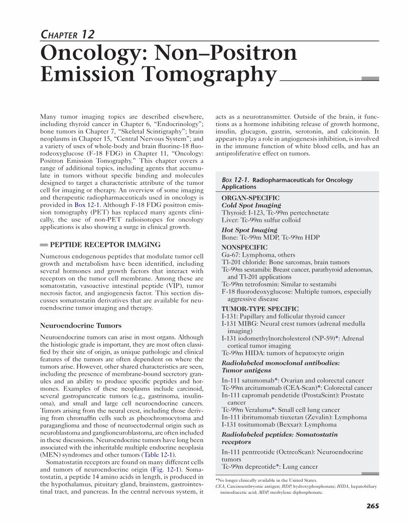
Somatostatin receptors are found on many different cells and tumors of neuroendocrine origin (Fig. 12-1). Soma-tostatin, a peptide 14 amino acids in length, is produced in the hypothalamus, pituitary gland, brainstem, gastrointes-tinal tract, and pancreas. In the central nervous system, it
265
acts as a neurotransmitter. Outside of the brain, it func-tions as a hormone inhibiting release of growth hormone, insulin, glucagon, gastrin, serotonin, and calcitonin. It appears to play a role in angiogenesis inhibition, is involved in the immune function of white blood cells, and has an antiproliferative effect on tumors.
Box 12-1. Radiopharmaceuticals for Oncology Applications
ORGAN-SPECIFICCold Spot ImagingThyroid: I-123, Tc-99m pertechnetateLiver: Tc-99m sulfur colloid
Hot Spot ImagingBone: Tc-99m MDP, Tc-99m HDP
NONSPECIFICGa-67: Lymphoma, othersTl-201 chloride: Bone sarcomas, brain tumorsTc-99m sestamibi: Breast cancer, parathyroid adenomas,
and Tl-201 applicationsTc-99m tetrofosmin: Similar to sestamibiF-18 fluorodeoxyglucose: Multiple tumors, especially
aggressive disease
TUMOR-TYPE SPECIFICI-131: Papillary and follicular thyroid cancerI-131 MIBG: Neural crest tumors (adrenal medulla
imaging)I-131 iodomethylnorcholesterol (NP-59)*: Adrenal
cortical tumor imagingTc-99m HIDA: tumors of hepatocyte origin
Radiolabeled monoclonal antibodies: Tumor antigens
In-111 satumomab*: Ovarian and colorectal cancerTc-99m arcitumomab (CEA-Scan)*: Colorectal cancerIn-111 capromab pendetide (ProstaScint): Prostate
cancerTc-99m Veraluma*: Small cell lung cancerIn-111 ibritumomab tiuxetan (Zevalin): LymphomaI-131 tositumomab (Bexxar): Lymphoma
Radiolabeled peptides: Somatostatin receptors
In-111 pentreotide (OctreoScan): Neuroendocrine tumorsTc-99m depreotide*: Lung cancer
*No longer clinically available in the United States.CEA, Carcinoembryonic antigen; HDP, hydroxyphosphonate; HIDA, hepatobiliary
iminodiacetic acid; MDP, methylene diphosphonate.
266 Nuclear Medicine: The Requisites
Several agents have been developed that readily bind to somatostatin receptors (Fig. 12-2). Octreotide is an 8-amino-acid peptide that maintains the ability to bind to native hormone receptors but is resistant to enzymatic
Table 12-1 Multiple Endocrine Neoplasia (MEN) Syndromes
Lesion MEN-I MEN-IIA MEN-IIB
Pituitary adenoma +
Pancreatic islet cell tumor +
Parathyroid adenoma + +
Pheochromocytoma + +
Medullary thyroid cancer + +
Ganglioneuroma +
Figure 12-1. Somatostatin receptors are found on many tumors, includ-ing those derived from neuroendocrine cells. GH, Growth hormone; TSH, thyroid-stimulating; VIPoma, vasoactive intestinal polypeptide-secreting tumors; Z-E, Zollinger Ellison.
degradation with a 1.5- to 2-hour half-life, as opposed to the 2- to 3-minute half-life of endogenous somatostatin. Nonradiolabeled octreotide (Sandostatin) has been approved by the U.S. Food and Drug Administration (FDA) as a therapeutic agent, suppressing growth in acro-megaly and controlling symptoms in carcinoid syndrome.
Indium-111 Pentetreotide (Indium-111 OctreoScan)
Pharmacokinetics and DosimetryRadiolabeling indium-111 pentetreotide In-111-DTPA-pentetreotide (In-111 pentetreotide [OctreoScan; Mallinck-rodt, Hazelwood, MO]) involves complexing octreotide with diethylenetriaminepentaacetic acid (DTPA) to bind In-111. In-111 pentetreotide is rapidly cleared by the kid-neys, with 50% of the dose excreted by 6 hours and 85% by
Ala-Gly-Cys-Lys-Asn-Phe-
Cys-Ser-Thr-Phe-
Somatostatin Octreotide
I-123 octreotide In-111 pentetreotide
Phe
D-Trp
Lys
Thr
ss
D-Phe-Cys-
123I
Thr-OL-Cys-
Phe
D-Trp
Lys
Thr
ss
D-Phe-Cys-
Thr-OL-Cys-
Phe
D-Trp
Lys
Thr
ss
111In-DTPA-D-Phe-Cys-
Thr-OL-Cys-
Phe
D-Trp
Lys
Thr
ss
Figure 12-2. Comparison of somatostatin analogs octreotide, I-123 pentetreotide, and In-111 pentetreotide.
AntA
Octreoscan 4 hr
Post B Ant
Octreoscan 24 hr
Post
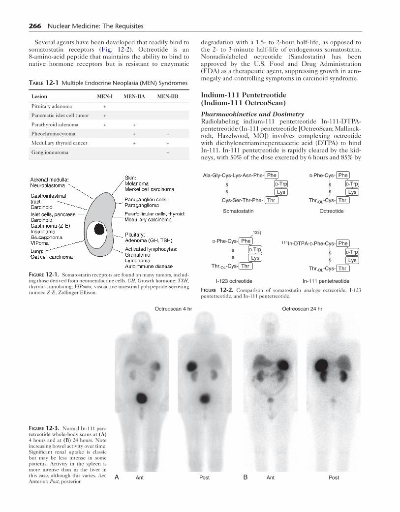
Figure 12-3. Normal In-111 pen-tetreotide whole-body scans at (A) 4 hours and at (B) 24 hours. Note increasing bowel activity over time. Significant renal uptake is classic but may be less intense in some patients. Activity in the spleen is more intense than in the liver in this case, although this varies. Ant, Anterior; Post, posterior.
24 hours after injection. A low level (2%) of hepatobiliary excretion also occurs. At 4 hours after injection, 10% of the dose remains in circulation; at 20 hours, less than 1% is in circulation. Whereas rapid clearance enhances the target-to-background ratio, bowel activity increases over time and can cause problems detecting abdominal lesions (Fig. 12-3). Estimates of radiation dosimetry are provided in Table 12-2.
At least five different subtypes of human somatostatin receptors (SSTR) have been identified (SSTR1 to SSTR5). These receptors are expressed to varying degrees on dif-ferent tumors. The commercially available radiopharma-ceutical In-111 pentetreotide binds with high affinity to the SSTR2 and SSTR5 subtypes, to a lesser extent with SSTR3, and not at all with SSTR1 or SSTR4. Identifying
Table 12-2 Indium-111 Pentetreotide (Indium-111 Octreotide) Radiation Dosimetry
Administered dosemCi (MBq)
Organ receiving highest doserad/mCi (mGy/MBq)
Effective doserem/mCi(mSv/MBq)
6 (222) Spleen 2.1 (0.57) 0.20 (0.054)
Data from SNM Procedure Guideline version 2.0. 2011. Modified from ICRP 106(37).
Box 12-2. Indium-111 Pentetreotide (Octreotide): Protocol Summary
PATIENT PREPARATIONHydrate patient before injection and at least 1 day afterConsider:
Oral laxative (e.g., bisacodyl) for abdominal lesions (when no active diarrhea present)
Discontinuing octreotide therapy 24 hours before injection
RADIOPHARMACEUTICALChildren: 0.14 mCi/kg (5 MBq/kg)Adults: 6 mCi (222 MBq) In-111 octreotide, intrave-
nously
INSTRUMENTATIONGamma camera: Large field of view,Collimator: Medium energyWindows: 20% centered at 173 keV and 247 keV
ACQUISITIONImaging at 24 hours preferable but 4-hour images
may be helpful; 48-hour images may be used if gut activity is initially limiting.
Planar Spot Views10 to 15 minutes per view512 × 512 or 256 × 256 word matrix
Whole-Body ImagesDual-head camera 3 cm/min (approximately 30
minutes head to below hips)1024 × 512 or 1024 × 256 word matrix
SPECT Critical Areas128 × 128 matrix, 3-degree angular sampling,
360-degree rotation, 20 to 30 sec/stopFusion to CT or SPECT/CT preferable
OncOlOgy: nOn–POsiTrOn emissiOn TOmOgraPhy 267
the specific receptor subtypes on tumors is also important as future targeted therapeutic agents.
MethodologyA sample protocol for imaging In-111 pentetreotide is pro-vided in Box 12-2. Although early imaging allows visualiza-tion of the abdomen in the absence of significant bowel activity, 24-hour images are generally most sensitive. Addi-tional images at 48 hours can be done with further bowel cleansing if needed. The addition of single-photon emis-sion computed tomography (SPECT) adds considerably to specificity and may improve sensitivity, as well. Fusing SPECT images to a CT or, ideally, acquisition with SPECT/CT allows the best localization and identification of lesions.
Image Interpretation and ApplicationsNormal activity can be seen not only in the kidneys, blad-der, liver, spleen, and colon but also occasionally in the thyroid and gallbladder. False positive uptake can occur in benign inflammatory conditions such as granulomatous disease, inflammatory bowel disease, chest radiation, bleo-mycin therapy, and sites of recent surgery. Close correla-tion with history is needed to avoid confusion.
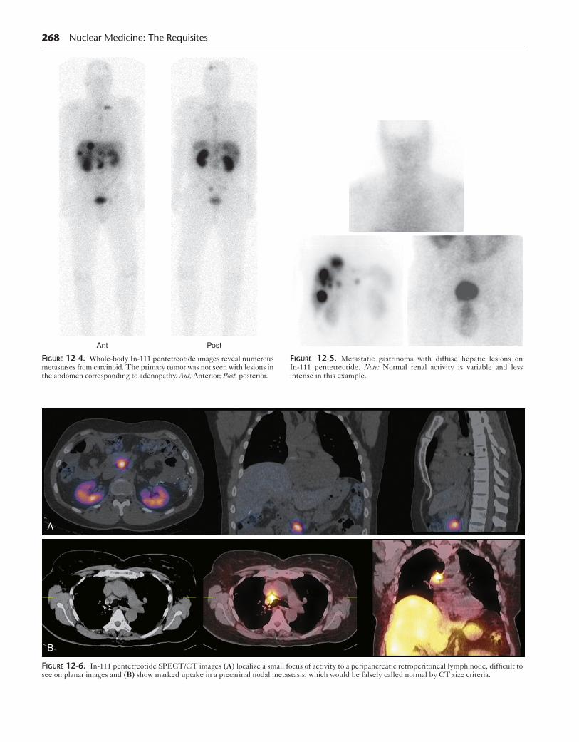
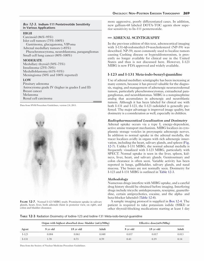
Approximately 80% to 90% of tumors are visible by 4 hours. Because of decreasing background, more lesions are seen by 24 hours. Many tumors can be diagnosed with pla-nar imaging (Figs. 12-4 and 12-5). However, SPECT is essential in the abdomen and can help identify disease in normal-size nodes in any area (Fig. 12-6). Lesions around the kidneys can be difficult to visualize because of the high renal uptake.
Overall, the sensitivity of In-111 pentetreotide is highest for carcinoid at 85% to 95%. This is particularly the case for extrahepatic sites of disease because liver lesions may show only background levels of activity. Neuroendocrine tumors of the pancreas such as gastrinomas, glucaganomas, vasoactive intestinal polypeptide-secreting tumors (VIPo-mas), and nonfunctioning islet cell tumors are also detected approximately 85% of the time (75%-100%). Pheochromo-cytomas, neuroblastomas, and paragangliomas can be eval-uated with iodine-123 meta-iodo-benzyl-guanidine (MIBG), but In-111 pentetreotide may be preferable for the detection of extraadrenal lesions. The use of In-111 pentetreotide is more limited in insulinomas, with low sensitivity. In addition, sensitivity for medullary thyroid cancer is only moderate (Box 12-3).
False negative examination results are seen in tumors that do not express sufficient receptor quantities or the appropriate receptor subtype (e.g., SSTR2). Tumors that do demonstrate In-111 pentetreotide uptake are good can-didates for octreotide drug therapy. Sensitivity appears to be lower in patients on octreotide therapy during imaging; however, many clinicians do not want to stop treatment for imaging examinations.
Experimental protocols with beta-emitters such as iodine-131, yttrium-90, and lutetium-177 labeled to octreotide have been used to treat poorly controlled neuro-endocrine tumors. Some sites have used even higher doses of In-111 with its Auger electron emissions for therapy.
Although the use of F-18 FDG PET has grown in most areas of tumor imaging, the sensitivity is low for many neu-roendocrine tumors. F-18 FDG PETCT may be useful in
268 Nuclear Medicine: The Requisites
Ant Post
Figure 12-4. Whole-body In-111 pentetreotide images reveal numerous metastases from carcinoid. The primary tumor was not seen with lesions in the abdomen corresponding to adenopathy. Ant, Anterior; Post, posterior.
Figure 12-5. Metastatic gastrinoma with diffuse hepatic lesions on In-111 pentetreotide. Note: Normal renal activity is variable and less intense in this example.
A
B
Figure 12-6. In-111 pentetreotide SPECT/CT images (A) localize a small focus of activity to a peripancreatic retroperitoneal lymph node, difficult to see on planar images and (B) show marked uptake in a precarinal nodal metastasis, which would be falsely called normal by CT size criteria.
Box 12-3. Indium-111 Pentetreotide Sensitivity in Various Applications
HIGHCarcinoid (86%-95%)Islet cell tumors (75%-100%)
Gastrinoma, glucaganoma, VIPomaAdrenal medullary tumors (>85%)
Pheochromocytoma, neuroblastoma, paragangliomasSmall cell lung cancer (80%-100%)
MODERATEMedullary thyroid (50%-75%)Insulinoma (25%-70%)Medulloblastoma (61%-93%)Meningioma (50% and 100% reported)
LOWPituitary adenomaAstrocytoma grade IV (higher in grades I and II)Breast cancerMelanomaRenal cell carcinoma
Data from SNM Procedure Guidelines, version 2.0, 2011.
Figure 12-7. Normal I-123 MIBG study. Prominent uptake in salivary glands, heart, liver, both adrenals (faint in posterior view, on right), and colon and bladder clearance.
OncOlOgy: nOn–POsiTrOn emissiOn TOmOgraPhy 269
more aggressive, poorly differentiated cases. In addition, new gallium-68 labeled DOTA-TOC agents show supe-rior sensitivity to In-111 pentetreotide.
ADRENAL SCINTIGRAPHY
In the previous edition of this text, adrenocortical imaging with I-131-6β-iodomethyl-19-norcholesterol (NP-59) was described. NP-59, most commonly used to localize tumors causing Cushing disease or hyperaldosteronism, is pres-ently no longer available for clinical use in the United States and thus is not discussed here. However, I-123 MIBG is now FDA approved and widely available.
I-123 and I-131 Meta-iodo-benzyl-guanidine
Use of adrenal medullary scintigraphy has been increasing at many centers, because it has proved valuable for the diagno-sis, staging, and management of adrenergic neuroectodermal tumors, particularly pheochromocytomas, extraadrenal para-gangliomas, and neuroblastomas. MIBG is a norepinephrine analog that accumulates in adrenergic and neuroblastic tumors. Although it has been labeled for clinical use with both I-131 and I-123, the I-123 radiolabel is generally pre-ferred. The major advantage is improved image quality, but dosimetry is a consideration as well, especially in children.
Radiopharmaceutical Localization and DosimetryAdrenal uptake occurs via a type I, energy-dependent, active amine transport mechanism. MIBG localizes in cyto-plasmic storage vesicles in presynaptic adrenergic nerves. In addition to normal uptake in the adrenal medulla, the tracer localizes avidly in organs with rich adrenergic inner-vation, including the heart, salivary glands, and spleen (Fig. 12-7). Unlike I-131 MIBG, the normal adrenal medulla is frequently visualized with I-123 MIBG, particularly with SPECT. Normal uptake is seen in the liver, spleen, kid-neys, liver, heart, and salivary glands. Genitourinary and colon clearance is often seen. Variable activity has been reported in lungs, gallbladder, salivary glands, and nasal mucosa. The bones are not normally seen. Dosimetry for I-123 and I-131 MIBG is outlined in Table 12-3.
MethodologyNumerous drugs interfere with MIBG uptake, and a careful drug history should be obtained before imaging. Interfering drugs include tricyclic antidepressants, reserpine, guanethi-dine, certain antipsychotics, cocaine, and the alpha- and beta-blocker labetalol (Table 12-4).
A sample imaging protocol is supplied in Box 12-4. The patient is required to take potassium iodide (SSKI) or other thyroid-blocking medications starting at least 1 day
Table 12-3 Radiation Dosimetry of Iodine-123 and Iodine-131 Meta-iodo-benzyl-guanidine
Organ with highest absorbed dose: bladder (mGy/MBq) Effective dose(mSv/MBq)
Agent 5 yr old 15 yr old Adult 5 yr old 15 yr old Adult
I-123 0.084 0.061 0.048 0.037 0.017 0.013
I-131 1.70 0.73 0.59 0.43 0.19 0.14
Data from the Society of Nuclear Medicine Procedure Guidelines.
270 Nuclear Medicine: The Requisites
Table 12-4 Medications Discontinue Before MIBG Imaging
Drug Related drugs Mechanism No. days to stop
Antihypertensive/cardiac agents
Bretylium, guanethidine, reserpineCalcium channel blockers (amlodipine, nifedipine, nicardipine)Labetalol
Deplete granulesDeplete granulesDeplete granlues and inhibit uptake
7-14
Antipsychotics Butyrophenones (droperidol, haloperidol)LoxapinePhenothiazines (chlorpromazine, fluphenazine, promethazine, others)
Inhibit uptakeInhibit uptakeInhibit uptake
21-287-21
21-28
Cocaine/opioids Inhibit uptake 7-14
Sympathicomimetics Amphetamine, dopamine, ephedrine, isoproterenol, phenoterol, phenylephrine, phenylpropanolamine, pseudoephedrine, salbutamol, terbutaline, xylometazoline
Deplete granules 7-14
Tramadol Inhibit uptake 7-14
Tricyclic antidepressants Amitriptyline (and derivatives), amoxapine, doxepine, others Inhibit uptake 7-21
MIBG, Meta-iodobenzylguanidine.
Box 12-4. Iodine-123 and Iodine-131 MIBG: Summary Protocol
PATIENT PREPARATIONHydrationThyroid blockade (Table 12-5) with Lugol solution or
potassium iodide. Begin day before injection and continue 1 to 2 days for I-123 and 3 to 6 days for I-131 MIBG
Discontinue interfering medications (Table 12-4)
RADIOPHARMACEUTICALIV dose injections must be done slowly, over at least
5 minutesI-123 MIBG
Children: 0.14 mCi/kg (5.2 MBq/kg); minimum 1.0 mCi (20 MBq) and maximum 10.8 mCi (400 MBq)*
Adults: 10.8 mCi (400 MBq)I-131 MIBG: Adults 1.2-2.2 mCi (40-80 MBq)
INSTRUMENTATIONGamma camera: Large field of view for planar imagesModern SPECT/CT hybrid systems recommended
for SPECTCollimator:
I-123: Medium energy, parallel holeI-131: High energy, parallel hole
ACQUISITIONI-123: Image at 20 to 24 hours. Delayed images less
than 2 days for equivocal cases.I-131: Image 1 to 2 days after injection. Repeat
images at day 3 if needed.Spot views: 75,000 to 100,000 counts for I-123
preferred, 256 × 256 matrix or 128 × 128 with zoom.Whole body planar images (5 cm/sec) and limited
spot views (500,000 counts or 10 minutes) can be done in adults.
SPECT: 3-degree steps, 25 to 35 sec/step, 120 projections, 128 × 128 matrix.
Uncooperative patients: consider 6-degree steps, or 64 × 64 matrix with shorter time per frame.
*Data from Gelfand MJ, Parisi MT, Treves ST. North American consensus guide-lines for administered radiopharmaceutical activities in children and adolescents. J Nucl Med. 2011;52(2):318-322.
MIBG, Meta-iodo-benzyl-guanidine.
before injection to prevent uptake of free radioiodine by the thyroid and continuing for 3 to 6 days to prevent free radioiodine uptake in thyroid (Table 12-5). Although I-123 can be imaged with a low-energy collimator, a small frac-tion of the photons (<3%) may be high energy (440-625 keV [2.4%] and 625-784 keV [0.15%]), reducing image quality. Therefore a medium-energy collimator may be preferable. Images are acquired 24 hours after injection. For pheochromocytoma, posterior and anterior views of the abdomen are most important. Additional images from the pelvis to the base of the skull are indicated to detect extraadrenal pheochromocytoma and neuroblastomas. Whole-body imaging is indicated for patients with neuro-blastoma. SPECT and or SPECT/CT are routine at many centers, improving sensitivity and accuracy.
Clinical ApplicationsPheochromocytomaPheochromocytoma is an uncommon catecholamine-secret-ing tumor derived from chromaffin cells. When these tumors arise outside of the adrenal gland, they are termed extraadre-nal pheochromocytomas, or paragangliomas. Because of excessive catecholamine secretion, they can precipitate life-threatening hypertension or cardiac arrhythmias. Ten percent of pheochromocytomas are bilateral, 10% are extraadrenal (paragangliomas), and 10% are malignant. Paragangliomas may be found from the bladder up to the base of the skull.
Table 12-5 Daily Doses of Thyroid Blockade Compounds
Drug AdultsChild
(15-50 kg)Child
(5-15 kg)Child
(<5 kg)
CAPSULES*
Potassium iodate 170 80 40 20
Potassium iodide 130 65 32 16
Potassium perchlorate
400 300 200 100
SOLUTION
Lugol solution 1% 1 drop/kg to max 40 (20 drops twice daily)
*Dose in milligram per day. Data from Giammarile F, Chiti A, Lassmann M, et al. EANM procedure guidelines for I-131 MIBG therapy. Eur J Nucl Med Mol Imaging. 2008;35(5):1039-1047.
Paragangliomas are often associated with multiple endo-crine neoplasia (MEN) types IIA and IIB, von Hippel-Lindau disease, neurofibromatosis, tuberous sclerosis, and Carney syndrome. Adrenomedullary hyperplasia develops in patients with MEN type IIA and can be difficult to diag-nose with CT or magnetic resonance imaging (MRI). MIBG scintigraphy is uniquely suited to detect medullary hyperplasia and has been used to assist decision making for timing of surgery.
The diagnosis of pheochromocytoma is suggested by detection of elevated blood or urinary catecholamines, although there are many other causes for these laboratory findings. CT or MRI is often the first imaging modality used. If an adrenal mass is demonstrated in this setting, the diagnosis is inferred and further workup before surgery is often unnecessary. I-131 or I-123 MIBG can sometimes be helpful to confirm the cause of a detected mass on ana-tomical imaging and detect extraadrenal tumors and less commonly, metastatic disease.
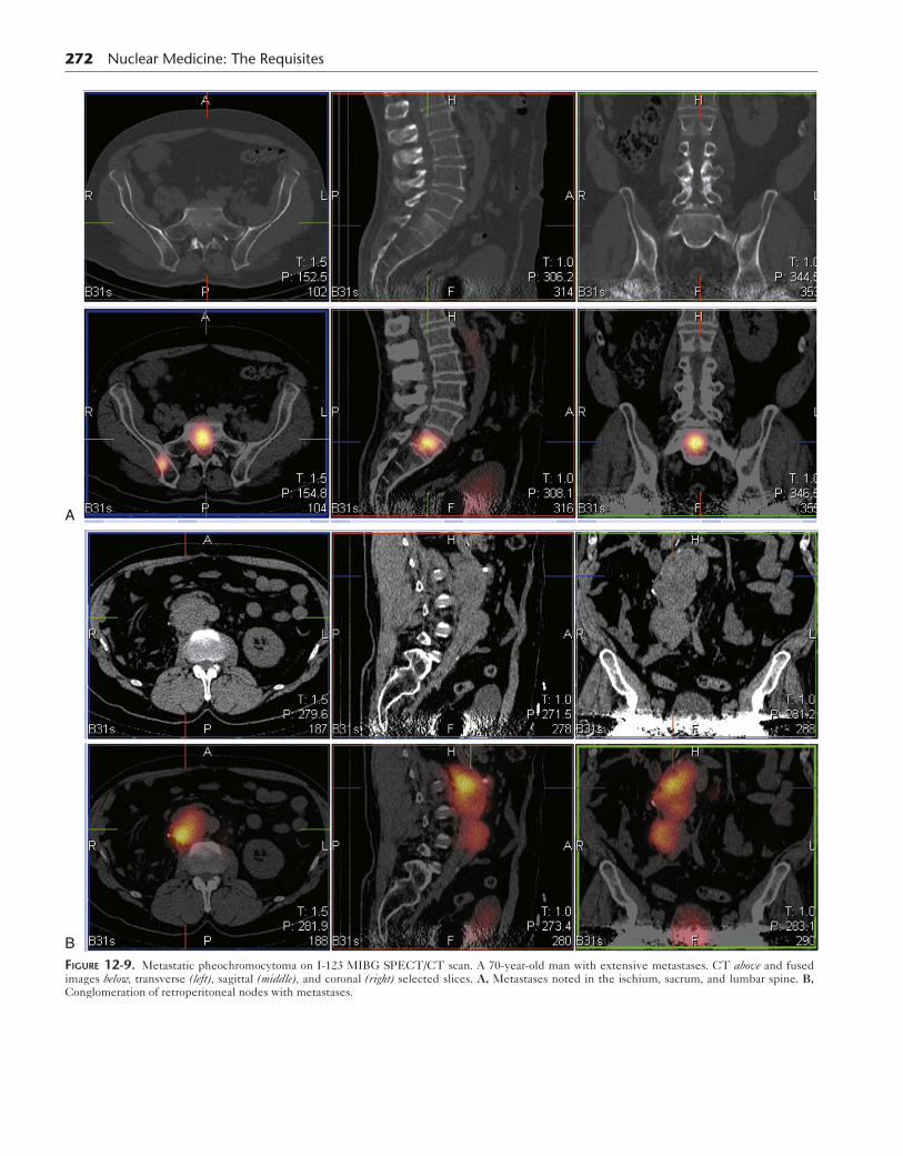
The characteristic scintigraphic appearance of a pheo-chromocytoma or extraadrenal paraganglioma is focal intense MIBG uptake (Figs. 12-8 and 12-9). The tumor-to-background ratio is quite high. The sensitivity for detection is approximately 90% and specificity greater than 95%.
NeuroblastomaNeuroblastoma is an embryonal malignancy of the sympa-thetic nervous system, typically occurring in children younger than 4 years of age. Seventy percent of tumors originate in the retroperitoneal region, from the adrenal or the abdominal sympathetic chain, and 20% occur in the chest, derived from the thoracic sympathetic chain (Fig. 12-10). Patients with localized tumors have good prognosis and outcome; however, those with metastatic disease fare
Figure 12-8. Pheochromocytoma on I-123 MIBG scan. Patient with poorly controlled hypertension and very elevated serum and urinary cat-echolamines. Focal increased uptake is noted in the region of the left adrenal consistent with a pheochromocytoma.
OncOlOgy: nOn–POsiTrOn emissiOn TOmOgraPhy 271
poorly. At the time of diagnosis, more than 50% of patients present with metastatic disease, 25% have localized dis-ease, and 15% have regional extension. Metastatic disease typically involves lymph nodes, liver, bone, and bone mar-row. Most patients present with signs and symptoms related to tumor growth.
Bone scans are used to detect osseous metastases. How-ever, a common location for metastases is in the metaphy-seal areas of long bones, which can be hard to detect due to high normal uptake in growth plates. MIBG has supe-rior sensitivity for detection of metastases compared to the bone scan, partly because these tumors initially involve the bone marrow. The combination of the two studies results in the highest sensitivity for metastatic detection.
MIBG sensitivity for detection of neuroblastoma is greater than 90%, with specificity of 95%. The study is used for staging, detecting metastatic disease, restaging, and determining patient response to therapy. Whole-body scanning is routine for imaging patients with this disease (Fig. 12-11). SPECT and SPECT/CT for specific areas can be quite useful (Fig. 12-12).
Other tumors that take up MIBG include carcinoid and medullary carcinoma of the thyroid. However, the sensi-tivity for tumor detection is considerably lower than for neuroblastoma or pheochromocytoma.
The high uptake of MIBG in neuroectodermal tumors has led to investigations of the use of high-dose I-131 MIBG therapy to treat metastatic neuroblastoma in patients who have failed prior conventional therapies. Investigations are ongoing.
MONOCLONAL ANTIBODY IMAGING AND THERAPY
In recent years, important radiolabeled antibodies have been developed and approved by the FDA for tumor imaging and therapy. Although some are no longer clini-cally available, agents for treating lymphoma and imaging prostate cancer remain valuable problem-solving tools.
Background
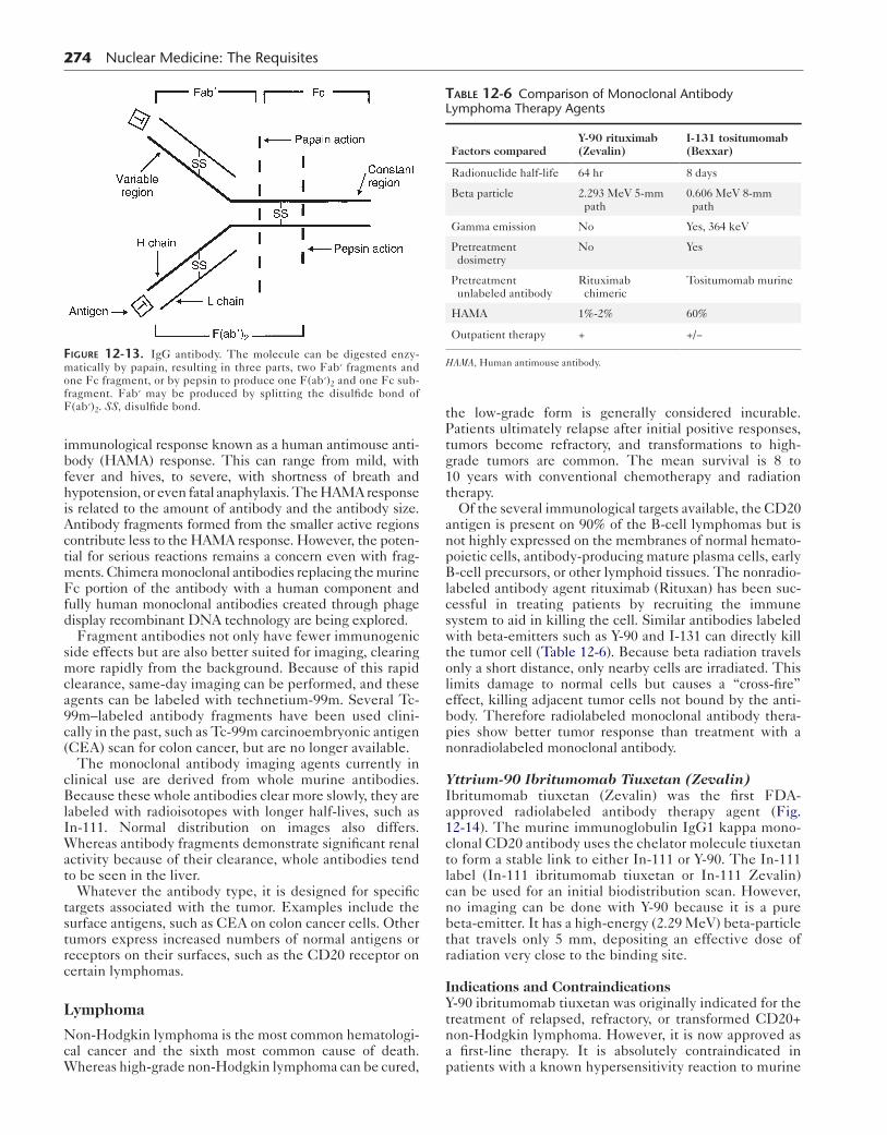
Antibodies are proteins produced by lymphocyte plasma cells in response to exposure to foreign antigens. An IgG antibody consists of two identical heavy (H) and two light (L) chains linked by a disulfide bridge (Fig. 12-13). Each chain is made up of two regions. The variable region (Fab′) is responsible for specifically binding to a cell surface anti-gen. The constant region (Fc) is involved with cell destruc-tion through complement fixation and antibody-dependent cell cytotoxicity.
A desired monoclonal antibody can be produced by fus-ing myeloma cancer cells with lymphocytes from the spleen of a mouse immunized with a particular antigen. These “hybridoma” cells retain both the specific antibody production capacity of the lymphocytes and the immortal-ity of cancer cells. Immunoassay screening identifies the murine (or mouse) monoclonal antibody clone desired. A monoclonal antibody with high affinity and specificity for the antigen of interest is then harvested.
However, the human immune system recognizes these murine monoclonal antibodies as foreign and mounts an
272 Nuclear Medicine: The Requisites
A
BFigure 12-9. Metastatic pheochromocytoma on I-123 MIBG SPECT/CT scan. A 70-year-old man with extensive metastases. CT above and fused images below, transverse (left), sagittal (middle), and coronal (right) selected slices. A, Metastases noted in the ischium, sacrum, and lumbar spine. B, Conglomeration of retroperitoneal nodes with metastases.

Figure 12-10. Imaging findings of neuroblastoma. Coronal CT image (top) reveal a large left abdominal mass (arrows) that had arisen from the retroperitoneum in an adult. I-131 MIBG images (bottom left) show increased uptake in the mass as well as throughout the bones from diffuse metastases. Tc-99m MDP bone scan (bottom right) shows abnormal soft tis-sue uptake in the mass, which is not uncommon. The bone lesions are seen but were harder to detect in some areas.
OncOlOgy: nOn–POsiTrOn emissiOn TOmOgraPhy 273
Figure 12-11. Metastatic neuroblastoma on I-123 MIGB whole-body scan. A 7-year-old boy with stage IV tumor after two bone marrow trans-plants. Extensive metastases are seen throughout the bones.
A B
Figure 12-12. Primary neuroblastoma on I-123 MIBG scan. A 9-year-old girl with posterior mediastinal mass. A, Planar anterior and posterior whole body images. The posterior planar image shows uptake in the chest just above the liver. B, Low-resolution SPECT/CT clearly localizes the paraspinal mass.
274 Nuclear Medicine: The Requisites
immunological response known as a human antimouse anti-body (HAMA) response. This can range from mild, with fever and hives, to severe, with shortness of breath and hypotension, or even fatal anaphylaxis. The HAMA response is related to the amount of antibody and the antibody size. Antibody fragments formed from the smaller active regions contribute less to the HAMA response. However, the poten-tial for serious reactions remains a concern even with frag-ments. Chimera monoclonal antibodies replacing the murine Fc portion of the antibody with a human component and fully human monoclonal antibodies created through phage display recombinant DNA technology are being explored.
Fragment antibodies not only have fewer immunogenic side effects but are also better suited for imaging, clearing more rapidly from the background. Because of this rapid clearance, same-day imaging can be performed, and these agents can be labeled with technetium-99m. Several Tc-99m–labeled antibody fragments have been used clini-cally in the past, such as Tc-99m carcinoembryonic antigen (CEA) scan for colon cancer, but are no longer available.
The monoclonal antibody imaging agents currently in clinical use are derived from whole murine antibodies. Because these whole antibodies clear more slowly, they are labeled with radioisotopes with longer half-lives, such as In-111. Normal distribution on images also differs. Whereas antibody fragments demonstrate significant renal activity because of their clearance, whole antibodies tend to be seen in the liver.
Whatever the antibody type, it is designed for specific targets associated with the tumor. Examples include the surface antigens, such as CEA on colon cancer cells. Other tumors express increased numbers of normal antigens or receptors on their surfaces, such as the CD20 receptor on certain lymphomas.
Lymphoma
Non-Hodgkin lymphoma is the most common hematologi-cal cancer and the sixth most common cause of death. Whereas high-grade non-Hodgkin lymphoma can be cured,
Figure 12-13. IgG antibody. The molecule can be digested enzy-matically by papain, resulting in three parts, two Fab′ fragments and one Fc fragment, or by pepsin to produce one F(ab′)2 and one Fc sub-fragment. Fab′ may be produced by splitting the disulfide bond of F(ab′)2. SS, disulfide bond.
the low-grade form is generally considered incurable.Patients ultimately relapse after initial positive responses, tumors become refractory, and transformations to high-grade tumors are common. The mean survival is 8 to 10 years with conventional chemotherapy and radiation therapy.
Of the several immunological targets available, the CD20 antigen is present on 90% of the B-cell lymphomas but is not highly expressed on the membranes of normal hemato-poietic cells, antibody-producing mature plasma cells, early B-cell precursors, or other lymphoid tissues. The nonradio-labeled antibody agent rituximab (Rituxan) has been suc-cessful in treating patients by recruiting the immune system to aid in killing the cell. Similar antibodies labeled with beta-emitters such as Y-90 and I-131 can directly kill the tumor cell (Table 12-6). Because beta radiation travels only a short distance, only nearby cells are irradiated. This limits damage to normal cells but causes a “cross-fire” effect, killing adjacent tumor cells not bound by the anti-body. Therefore radiolabeled monoclonal antibody thera-pies show better tumor response than treatment with a nonradiolabeled monoclonal antibody.
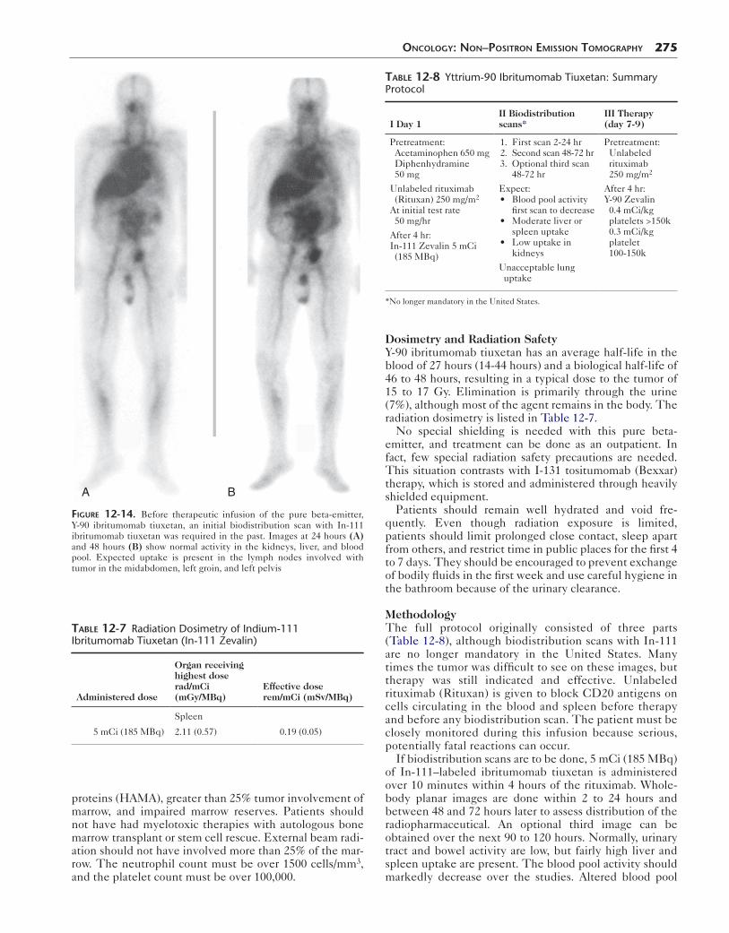
Yttrium-90 Ibritumomab Tiuxetan (Zevalin)Ibritumomab tiuxetan (Zevalin) was the first FDA-approved radiolabeled antibody therapy agent (Fig. 12-14). The murine immunoglobulin IgG1 kappa mono-clonal CD20 antibody uses the chelator molecule tiuxetan to form a stable link to either In-111 or Y-90. The In-111 label (In-111 ibritumomab tiuxetan or In-111 Zevalin) can be used for an initial biodistribution scan. However, no imaging can be done with Y-90 because it is a pure beta-emitter. It has a high-energy (2.29 MeV) beta-particle that travels only 5 mm, depositing an effective dose of radiation very close to the binding site.
Indications and ContraindicationsY-90 ibritumomab tiuxetan was originally indicated for the treatment of relapsed, refractory, or transformed CD20+ non-Hodgkin lymphoma. However, it is now approved as a first-line therapy. It is absolutely contraindicated in patients with a known hypersensitivity reaction to murine
Table 12-6 Comparison of Monoclonal Antibody Lymphoma Therapy Agents
Factors comparedY-90 rituximab (Zevalin)
I-131 tositumomab (Bexxar)
Radionuclide half-life 64 hr 8 days
Beta particle 2.293 MeV 5-mm path
0.606 MeV 8-mm path
Gamma emission No Yes, 364 keV
Pretreatment dosimetry
No Yes
Pretreatment unlabeled antibody
Rituximab chimeric
Tositumomab murine
HAMA 1%-2% 60%
Outpatient therapy + +/−
HAMA, Human antimouse antibody.
proteins (HAMA), greater than 25% tumor involvement of marrow, and impaired marrow reserves. Patients should not have had myelotoxic therapies with autologous bone marrow transplant or stem cell rescue. External beam radi-ation should not have involved more than 25% of the mar-row. The neutrophil count must be over 1500 cells/mm3, and the platelet count must be over 100,000.
A B
Figure 12-14. Before therapeutic infusion of the pure beta-emitter, Y-90 ibritumomab tiuxetan, an initial biodistribution scan with In-111 ibritumomab tiuxetan was required in the past. Images at 24 hours (A) and 48 hours (B) show normal activity in the kidneys, liver, and blood pool. Expected uptake is present in the lymph nodes involved with tumor in the midabdomen, left groin, and left pelvis
Table 12-7 Radiation Dosimetry of Indium-111 Ibritumomab Tiuxetan (In-111 Zevalin)
Administered dose
Organ receiving highest doserad/mCi(mGy/MBq)
Effective doserem/mCi (mSv/MBq)
5 mCi (185 MBq)
Spleen
2.11 (0.57) 0.19 (0.05)
OncOlOgy: nOn–POsiTrOn emissiOn TOmOgraPhy 275
Dosimetry and Radiation SafetyY-90 ibritumomab tiuxetan has an average half-life in the blood of 27 hours (14-44 hours) and a biological half-life of 46 to 48 hours, resulting in a typical dose to the tumor of 15 to 17 Gy. Elimination is primarily through the urine (7%), although most of the agent remains in the body. The radiation dosimetry is listed in Table 12-7.
No special shielding is needed with this pure beta- emitter, and treatment can be done as an outpatient. In fact, few special radiation safety precautions are needed. This situation contrasts with I-131 tositumomab (Bexxar) therapy, which is stored and administered through heavily shielded equipment.
Patients should remain well hydrated and void fre-quently. Even though radiation exposure is limited, patients should limit prolonged close contact, sleep apart from others, and restrict time in public places for the first 4 to 7 days. They should be encouraged to prevent exchange of bodily fluids in the first week and use careful hygiene in the bathroom because of the urinary clearance.
MethodologyThe full protocol originally consisted of three parts (Table 12-8), although biodistribution scans with In-111 are no longer mandatory in the United States. Many times the tumor was difficult to see on these images, but therapy was still indicated and effective. Unlabeled rituximab (Rituxan) is given to block CD20 antigens on cells circulating in the blood and spleen before therapy and before any biodistribution scan. The patient must be closely monitored during this infusion because serious, potentially fatal reactions can occur.
If biodistribution scans are to be done, 5 mCi (185 MBq) of In-111–labeled ibritumomab tiuxetan is administered over 10 minutes within 4 hours of the rituximab. Whole-body planar images are done within 2 to 24 hours and between 48 and 72 hours later to assess distribution of the radiopharmaceutical. An optional third image can be obtained over the next 90 to 120 hours. Normally, urinary tract and bowel activity are low, but fairly high liver and spleen uptake are present. The blood pool activity should markedly decrease over the studies. Altered blood pool
Table 12-8 Yttrium-90 Ibritumomab Tiuxetan: Summary Protocol
I Day 1II Biodistribution scans*
III Therapy (day 7-9)
Pretreatment: Acetaminophen 650 mg Diphenhydramine 50 mg
Unlabeled rituximab (Rituxan) 250 mg/m2
At initial test rate 50 mg/hr
After 4 hr:In-111 Zevalin 5 mCi (185 MBq)
1. First scan 2-24 hr 2. Second scan 48-72 hr 3. Optional third scan
48-72 hr
Expect: • Blood pool activity
first scan to decrease • Moderate liver or
spleen uptake • Low uptake in
kidneys
Unacceptable lung uptake
Pretreatment: Unlabeled rituximab 250 mg/m2
After 4 hr:Y-90 Zevalin 0.4 mCi/kg platelets >150k 0.3 mCi/kg platelet 100-150k
*No longer mandatory in the United States.
276 Nuclear Medicine: The Requisites
Table 12-9 Therapy Protocol for I-131 Tositumomab
Day 0 Day 1 Day 2, 3, or 4 Day 6 or 7 Day 7 up to 14
Thyroid blockade (continue 2 wk)
1. Premedicate acetaminophen 650 mg,tositumomab 450 mg over 60 min2. I-131 tositumomab 5 mCi over 20 min
3. Prevoid whole-body dosimetry scan
Dosimetry scan Dosimetry scanCalculate drug dose:>150k, 75 cGy100-150k, 65 cGy
Therapy administration:1. Pretreat with tositumomab450 mg over 60 min2. Therapy dose I-131 tositumomab over 20 min
distribution would include activity increasing rather than decreasing over time in lung, liver, heart, urinary tract, or bowel uptake. Any of these changes could lead to unac-ceptable radiation to the organ in question, such as the kidneys.
ToxicitySignificant side effects may occur from this therapy. Usu-ally within 7 to 9 weeks, blood counts reach a nadir with a 30% to 70% reduction in platelets and neutrophils (median neutrophil count 800, platelet count 40,000, hemoglobin 10.3). The cytopenia may last from 7 to 35 days. Roughly 7% of neutropenic patients are prone to febrile neutrope-nia and infections. Thrombocytopenia may result in hem-orrhage. Up to one third of patients will subsequently experience disease transformation to a more aggressive lymphoma. It is unclear whether this is a side effect of therapy or the natural course of the disease. A small num-ber (1.4%) of patients will develop myelodysplasia or acute myelogenous leukemia.
Patients treated with Y-90 ibritumomab tiuxetan experi-ence a HAMA response only 1% to 2% of the time. This low level could be due to the protective effects of the pre-dosing administration of rituximab or the immunocompro-mised status of the patient.
ResultsThis is a very effective therapy. Overall, 75% (67%-83%) of patients experience some response, with 15% to 37% of patients showing complete remission. These values are significantly better than the results of nonlabeled rituxan monoclonal antibody therapy alone. The duration of response ranges from 0.5 to 24.9 months.
Iodine-131 Tositumomab (Iodine-131 Bexxar)I-131 tositumomab (I-131 Bexxar) is a murine IgG2a mono-clonal antibody developed to target CD20, which is the same target for Y-90 ibritumomab tiuxetan. Y-90 ibritumomab tiuxetan and I-131 tositumomab have several similarities and differences. I-131 tositumomab is recommended for CD20+ follicular non-Hodgkin lymphoma (with or without transformation) refractory to rituximab but is not considered a first-line therapy.
MethodA protocol outline is provided in Table 12-9. Unlike Y-90 ibritumomab tiuxetan, the I-131 tositumomab therapy dose can be imaged using the 364-keV gamma emissions. The tositumomab regimen uses the same tositumomab monoclonal antibody for pretreatment (in the nonradio-labeled form) as for dosimetry and therapy (in the I-131–labeled form). The I-131 label means that, unlike a pure
beta-emitter, the dose requires shielding. Also, it must be determined before discharging the patient that the expo-sure to others will not be greater than 500 mrem from the patient. I-131 tositumomab protocols require additional scans to determine dosimetry, which adds to inconve-nience compared to Y-90 ibritumomab tiuxetan.
Because the I-131 radiolabel can disassociate from the monoclonal antibody and result in unwanted thyroid expo-sure, the patient must receive thyroid-blocking medica-tion, such as with SSKI beginning at least 1 day before the studies and continuing for 2 weeks.
The patient is first treated with nonlabeled tositumomab to block excess CD20 sites. This helps decrease nonspe-cific antibody targeting. Following this, the dosimetry stud-ies are done with a low dose of I-131 tositumomab. Serial scans allow calculation of how fast activity clears from the body (residence time) before calculating the therapy dose, which is also based on the patient’s platelet levels.
ToxicityAs with Y-90 ibritumomab tiuxetan, similar significant hematological side effects can occur. Up to 15% of patients may require supportive care such as transfusions and colony-stimulating factor. Long-term side effects are possible, such as the myelodysplastic syndrome and secondary leukemia. Hypothyroidism may occur if proper premedication is not given to block the thyroid from taking up I-131.
HAMA titers are commonly elevated initially, although reports of symptomatic reactions are rare. The patients who had extensive previous chemotherapy became sero-positive approximately 10% of the time, whereas patients who received I-131 tositumomab as a first-line therapy had initially elevated titers up to 70% of the time.
ResultsOverall, 63% (54%-71%) of patients refractory to rituximab showed response. Of these, 29% of the patients experi-enced complete response. The median duration of response was 26 months, significantly longer than with Y-90 ibritumomab tiuxetan.
Prostate Cancer
BackgroundProstate cancer staging is based on the combination of physical examination, histopathological Gleason score, and serum prostate-specific antigen (PSA). Therapy with radical prostatectomy is not undertaken when evidence shows nodal involvement or distant spread. Lymph nodes are the most common site of metastatic involvement, usually occurring in a stepwise fashion from periprostatic or obturator nodes to internal or external iliac nodes, and
then to common iliac and periaortic nodes. Frequent sites of distant metastases are the skeleton, liver, and lungs.
If the PSA fails to decline after prostatectomy or begins to rise, residual or recurrent tumor is likely. Identification of patients with isolated disease in the prostate bed is important. If disease is localized to the prostate fossa or pelvis, radiation therapy offers the potential for effective treatment. However, if recurrence involves periaortic lymph nodes or other distant sites outside the therapy field, radiation therapy exposes the patient to significant morbidity with no potential for cure.
Staging is often difficult. Bone scans are indicated with a serum PSA greater than 10 to 20 ng/mL or a high Gleason score. CT and MRI have limited value because of their low sensitivity for detecting nodal involvement. In addi-tion, F-18 FDG PET has low sensitivity for prostate can-cer and plays no role in staging or detecting early recurrence.
Indium-111 Capromab Pendetide (ProstaScint)Attempts to target PSA with radiolabeled antibodies proved insensitive and resulted in significant HAMA
A B
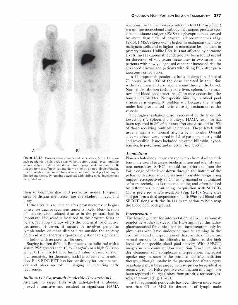
Figure 12-15. Prostate cancer lymph node metastases. A, In-111 capro-mab pendetide whole-body scans 96 hours after dosing reveal multiple abnormal foci in the midabdomen from lymph node metastases. B, Images from a different patient show a slightly altered biodistribution. Even though uptake in the liver is more intense, blood pool activity is limited and the study remains diagnostic with visible nodal involvement in the abdomen.
OncOlOgy: nOn–POsiTrOn emissiOn TOmOgraPhy 277
reactions. In-111 capromab pendetide (In-111 ProstaScint) is a murine monoclonal antibody that targets prostate-spe-cific membrane antigen (PMSA), a glycoprotein expressed by more than 95% of prostate adenocarcinomas (Fig. 12-15). PMSA expression is higher in malignant than non-malignant cells and is higher in metastatic lesions than in primary tumors. Unlike PSA, it is not affected by hormone levels. In-111 capromab pendetide has been found useful for detection of soft tissue metastases in two situations: patients with newly diagnosed cancer at increased risk for advanced disease and patients with rising PSA after pros-tatectomy or radiation.
In-111 capromab pendetide has a biological half-life of 72 hours, with 10% of the dose excreted in the urine within 72 hours and a smaller amount through the bowel. Normal distribution includes the liver, spleen, bone mar-row, and blood pool structures. Clearance occurs into the bowel and bladder. Nonspecific binding in blood pool structures is especially problematic because the lymph nodes being evaluated lie in close approximation to the vessels.
The highest radiation dose is received by the liver, fol-lowed by the spleen and kidneys. HAMA response has been reported in 8% of patients after one dose and in 19% of those receiving multiple injections. These levels will usually return to normal after a few months. Overall adverse effects were noted in 4% of patients, mostly mild and reversible. Issues included elevated bilirubin, hyper-tension, hypotension, and injection site reaction.
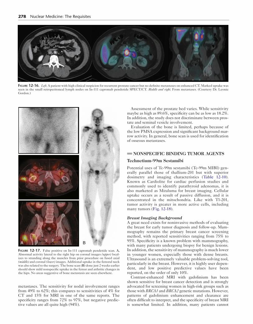
AcquisitionPlanar whole-body images or spot views from skull to mid-femur are useful to assess biodistribution and identify dis-tant metastases. SPECT should be performed from the lower edge of the liver down through the bottom of the pelvis, with attenuation correction if possible. Registering images retrospectively to CT using manual or automated software techniques is time consuming and often limited by differences in positioning. Acquisition with SPECT/CT is preferred where available (Fig. 12-16). Some sites still perform a dual acquisition of a Tc-99m red blood cell SPECT along with the In-111 examination to help map the blood pool background.
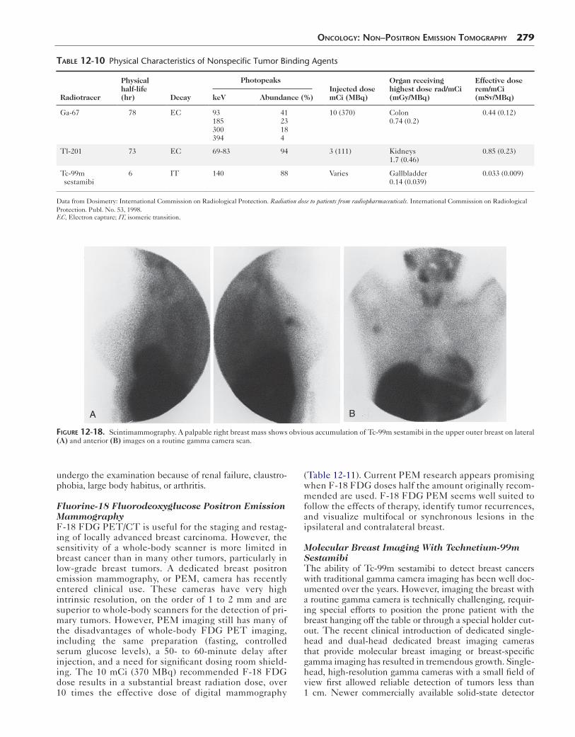
InterpretationThe learning curve for interpretation of In-111 capromab pendetide studies is steep. The FDA approved this radio-pharmaceutical for clinical use and interpretation only by physicians who have undergone specific training in the acquisition and interpretation of these studies. There are several reasons for the difficulty in addition to the high levels of nonspecific blood pool activity. With SPECT, images are low count and low resolution. Bowel and blad-der clearance can complicate interpretation. Increased uptake may be seen in the prostate bed after radiation therapy, although uptake in the prostate bed after surgery or radiation must be regarded with suspicion for residual or recurrent tumor. False positive examination findings have been reported at surgical sites, from arthritis, tortuous ves-sels, and bowel (Fig. 12-17).
In-111 capromab pendetide has been shown more accu-rate than CT or MRI for detection of lymph node
278 Nuclear Medicine: The Requisites
Figure 12-16. Left, A patient with high clinical suspicion for recurrent prostate cancer but no definite metastases on enhanced CT. Marked uptake was seen in the small retroperitoneal lymph nodes on In-111 capromab pendetide SPECT/CT. Middle and right, From metastases. (Courtesy Dr. Leonie
Gordon.)metastases. The sensitivity for nodal involvement ranges from 49% to 62%; this compares to sensitivities of 4% for CT and 15% for MRI in one of the same reports. The specificity ranges from 72% to 97%, but negative predic-tive values are all quite high (94%).
A B
Figure 12-17. False positive on In-111 capromab pendetide scan. A, Abnormal activity lateral to the right hip on coronal images (upper) local-izes to stranding along the muscles from prior procedure on fused axial (middle) and coronal (lower) images. Additional uptake in the femoral neck was also related to the surgery. The bone scan (B) done just 2 weeks earlier should show mild nonspecific uptake in the femur and arthritic changes in the hips. No areas suggestive of bone metastasis are seen elsewhere.
Assessment of the prostate bed varies. While sensitivity maybe as high as 89.6%, specificity can be as low as 18.2%. In addition, the study does not discriminate between pros-tate and seminal vesicle involvement.
Evaluation of the bone is limited, perhaps because of the low PMSA expression and significant background mar-row activity. In general, bone scan is used for identification of osseous metastases.
NONSPECIFIC BINDING TUMOR AGENTS
Technetium-99m Sestamibi
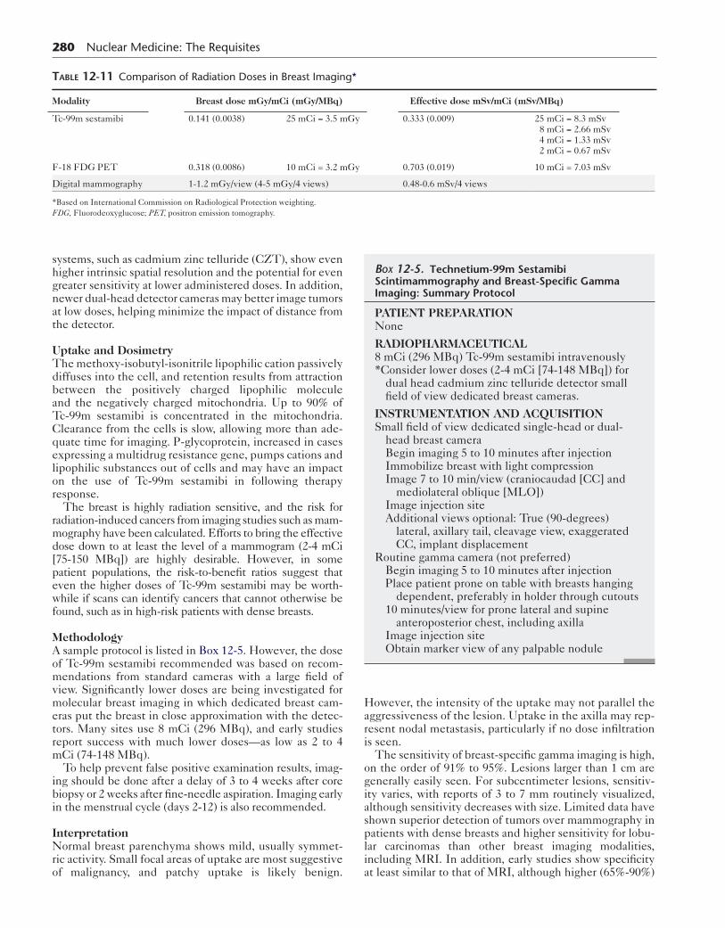
Potential uses of Tc-99m sestamibi (Tc-99m MIBI) gen-erally parallel those of thallium-201 but with superior dosimetry and imaging characteristics (Table 12-10). Known as Cardiolite for cardiac perfusion studies and commonly used to identify parathyroid adenomas, it is also marketed as Miraluma for breast imaging. Cellular uptake occurs as a result of passive diffusion, and it is concentrated in the mitochondria. Like with Tl-201, tumor activity is greater in more active cells, including many tumors (Fig. 12-18).
Breast Imaging BackgroundA great need exists for noninvasive methods of evaluating the breast for early tumor diagnosis and follow-up. Mam-mography remains the primary breast cancer screening method, with reported sensitivities ranging from 75% to 95%. Specificity is a known problem with mammography, with many patients undergoing biopsy for benign lesions. In addition, the sensitivity of mammography is much lower in younger women, especially those with dense breasts. Ultrasound is an extremely valuable problem-solving tool, particularly in the breast. However, it is highly user depen-dent, and low positive predictive values have been reported, on the order of only 10%.
Contrast-enhanced MRI with gadolinium has been shown sensitive for breast cancer detection and is strongly advocated for screening women in high-risk groups such as those with BRCA1 and BRCA2 genetic mutations. However, patterns of gadolinium enhancement and clearance are often difficult to interpret, and the specificity of breast MRI is somewhat limited. In addition, many patients cannot
OncOlOgy: nOn–POsiTrOn emissiOn TOmOgraPhy 279
Table 12-10 Physical Characteristics of Nonspecific Tumor Binding Agents
Radiotracer
Physical half-life (hr) Decay
PhotopeaksInjected dose mCi (MBq)
Organ receiving highest dose rad/mCi(mGy/MBq)
Effective dose rem/mCi(mSv/MBq)keV Abundance (%)
Ga-67 78 EC 93185300394
4123184
10 (370) Colon0.74 (0.2)
0.44 (0.12)
Tl-201 73 EC 69-83 94 3 (111) Kidneys1.7 (0.46)
0.85 (0.23)
Tc-99m sestamibi
6 IT 140 88 Varies Gallbladder0.14 (0.039)
0.033 (0.009)
Data from Dosimetry: International Commission on Radiological Protection. Radiation dose to patients from radiopharmaceuticals. International Commission on Radiological Protection. Publ. No. 53, 1998.EC, Electron capture; IT, isomeric transition.
A B
Figure 12-18. Scintimammography. A palpable right breast mass shows obvious accumulation of Tc-99m sestamibi in the upper outer breast on lateral (A) and anterior (B) images on a routine gamma camera scan.
undergo the examination because of renal failure, claustro-phobia, large body habitus, or arthritis.
Fluorine-18 Fluorodeoxyglucose Positron Emission MammographyF-18 FDG PET/CT is useful for the staging and restag-ing of locally advanced breast carcinoma. However, the sensitivity of a whole-body scanner is more limited in breast cancer than in many other tumors, particularly in low-grade breast tumors. A dedicated breast positron emission mammography, or PEM, camera has recently entered clinical use. These cameras have very high intrinsic resolution, on the order of 1 to 2 mm and are superior to whole-body scanners for the detection of pri-mary tumors. However, PEM imaging still has many of the disadvantages of whole-body FDG PET imaging, including the same preparation (fasting, controlled serum glucose levels), a 50- to 60-minute delay after injection, and a need for significant dosing room shield-ing. The 10 mCi (370 MBq) recommended F-18 FDG dose results in a substantial breast radiation dose, over 10 times the effective dose of digital mammography
(Table 12-11). Current PEM research appears promising when F-18 FDG doses half the amount originally recom-mended are used. F-18 FDG PEM seems well suited to follow the effects of therapy, identify tumor recurrences, and visualize multifocal or synchronous lesions in the ipsilateral and contralateral breast.
Molecular Breast Imaging With Technetium-99m SestamibiThe ability of Tc-99m sestamibi to detect breast cancers with traditional gamma camera imaging has been well doc-umented over the years. However, imaging the breast with a routine gamma camera is technically challenging, requir-ing special efforts to position the prone patient with the breast hanging off the table or through a special holder cut-out. The recent clinical introduction of dedicated single- head and dual-head dedicated breast imaging cameras that provide molecular breast imaging or breast-specific gamma imaging has resulted in tremendous growth. Single-head, high-resolution gamma cameras with a small field of view first allowed reliable detection of tumors less than 1 cm. Newer commercially available solid-state detector
280 Nuclear Medicine: The Requisites
Table 12-11 Comparison of Radiation Doses in Breast Imaging*
Modality Breast dose mGy/mCi (mGy/MBq) Effective dose mSv/mCi (mSv/MBq)
Tc-99m sestamibi 0.141 (0.0038) 25 mCi = 3.5 mGy 0.333 (0.009) 25 mCi = 8.3 mSv8 mCi = 2.66 mSv4 mCi = 1.33 mSv2 mCi = 0.67 mSv
F-18 FDG PET 0.318 (0.0086) 10 mCi = 3.2 mGy 0.703 (0.019) 10 mCi = 7.03 mSv
Digital mammography 1-1.2 mGy/view (4-5 mGy/4 views) 0.48-0.6 mSv/4 views
*Based on International Commission on Radiological Protection weighting.FDG, Fluorodeoxyglucose; PET, positron emission tomography.
systems, such as cadmium zinc telluride (CZT), show even higher intrinsic spatial resolution and the potential for even greater sensitivity at lower administered doses. In addition, newer dual-head detector cameras may better image tumors at low doses, helping minimize the impact of distance from the detector.
Uptake and DosimetryThe methoxy-isobutyl-isonitrile lipophilic cation passively diffuses into the cell, and retention results from attraction between the positively charged lipophilic molecule and the negatively charged mitochondria. Up to 90% of Tc-99m sestamibi is concentrated in the mitochondria. Clearance from the cells is slow, allowing more than ade-quate time for imaging. P-glycoprotein, increased in cases expressing a multidrug resistance gene, pumps cations and lipophilic substances out of cells and may have an impact on the use of Tc-99m sestamibi in following therapy response.
The breast is highly radiation sensitive, and the risk for radiation-induced cancers from imaging studies such as mam-mography have been calculated. Efforts to bring the effective dose down to at least the level of a mammogram (2-4 mCi [75-150 MBq]) are highly desirable. However, in some patient populations, the risk-to-benefit ratios suggest that even the higher doses of Tc-99m sestamibi may be worth-while if scans can identify cancers that cannot otherwise be found, such as in high-risk patients with dense breasts.
MethodologyA sample protocol is listed in Box 12-5. However, the dose of Tc-99m sestamibi recommended was based on recom-mendations from standard cameras with a large field of view. Significantly lower doses are being investigated for molecular breast imaging in which dedicated breast cam-eras put the breast in close approximation with the detec-tors. Many sites use 8 mCi (296 MBq), and early studies report success with much lower doses—as low as 2 to 4 mCi (74-148 MBq).
To help prevent false positive examination results, imag-ing should be done after a delay of 3 to 4 weeks after core biopsy or 2 weeks after fine-needle aspiration. Imaging early in the menstrual cycle (days 2-12) is also recommended.
InterpretationNormal breast parenchyma shows mild, usually symmet-ric activity. Small focal areas of uptake are most suggestive of malignancy, and patchy uptake is likely benign.
However, the intensity of the uptake may not parallel the aggressiveness of the lesion. Uptake in the axilla may rep-resent nodal metastasis, particularly if no dose infiltration is seen.
The sensitivity of breast-specific gamma imaging is high, on the order of 91% to 95%. Lesions larger than 1 cm are generally easily seen. For subcentimeter lesions, sensitiv-ity varies, with reports of 3 to 7 mm routinely visualized, although sensitivity decreases with size. Limited data have shown superior detection of tumors over mammography in patients with dense breasts and higher sensitivity for lobu-lar carcinomas than other breast imaging modalities, including MRI. In addition, early studies show specificity at least similar to that of MRI, although higher (65%-90%)
Box 12-5. Technetium-99m Sestamibi Scintimammography and Breast-Specific Gamma Imaging: Summary Protocol
PATIENT PREPARATIONNone
RADIOPHARMACEUTICAL8 mCi (296 MBq) Tc-99m sestamibi intravenously*Consider lower doses (2-4 mCi [74-148 MBq]) for
dual head cadmium zinc telluride detector small field of view dedicated breast cameras.
INSTRUMENTATION AND ACQUISITIONSmall field of view dedicated single-head or dual-
head breast cameraBegin imaging 5 to 10 minutes after injectionImmobilize breast with light compressionImage 7 to 10 min/view (craniocaudad [CC] and
mediolateral oblique [MLO])Image injection siteAdditional views optional: True (90-degrees)
lateral, axillary tail, cleavage view, exaggerated CC, implant displacement
Routine gamma camera (not preferred)Begin imaging 5 to 10 minutes after injectionPlace patient prone on table with breasts hanging
dependent, preferably in holder through cutouts10 minutes/view for prone lateral and supine
anteroposterior chest, including axillaImage injection siteObtain marker view of any palpable nodule
in some cases. A 2011 multicenter trial, Weigert et al reported sensitivity of 91%, specificity of 77%, positive predictive value of 57%, and negative predictive value of 96%. The high negative predictive value suggests that Tc-99m sestamibi could be used to evaluate questionable lesions on mammogram. However, with a false negative rate of 6% for breast-specific gamma imaging, biopsy should be performed when mammogram indicates a need but scintigraphic imaging is normal.
Thallium-201 Tumor Imaging
Although the potassium analog Tl-201 chloride is com-monly known as a cardiac perfusion radiopharmaceutical, it has long been known to accumulate in many tumors, with some listed in Table 12-12. Multiple factors influence cellular uptake, including blood flow delivery and the membrane sodium-potassium adenosine triphosphatase (ATPase) pumping. Overall, distribution is proportional to blood flow, although activity in tumor relates to cellular activity and viability. Biological clearance is primarily via the kidneys. Given the suboptimal imaging characteristics and poor dosimetry compared to technetium-based agents (Table 12-10), it is not surprising that applications are lim-ited to occasional use, such as in the brain to assess possi-ble recurrent glioma or help differentiate toxoplasmosis from intracranial lymphoma.
Gallium-67 Tumor Imaging
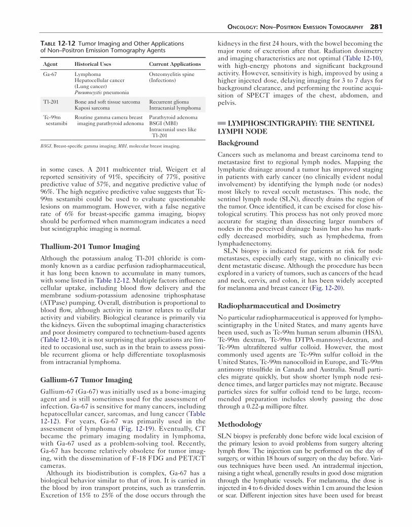
Gallium-67 (Ga-67) was initially used as a bone-imaging agent and is still sometimes used for the assessment of infection. Ga-67 is sensitive for many cancers, including hepatocellular cancer, sarcomas, and lung cancer (Table 12-12). For years, Ga-67 was primarily used in the assessment of lymphoma (Fig. 12-19). Eventually, CT became the primary imaging modality in lymphoma, with Ga-67 used as a problem-solving tool. Recently, Ga-67 has become relatively obsolete for tumor imag-ing, with the dissemination of F-18 FDG and PET/CT cameras.
Although its biodistribution is complex, Ga-67 has a biological behavior similar to that of iron. It is carried in the blood by iron transport proteins, such as transferrin. Excretion of 15% to 25% of the dose occurs through the
Table 12-12 Tumor Imaging and Other Applications of Non–Positron Emission Tomography Agents
Agent Historical Uses Current Applications
Ga-67 LymphomaHepatocellular cancer(Lung cancer)Pneumocystis pneumonia
Osteomyelitis spine(Infections)
Tl-201 Bone and soft tissue sarcomaKaposi sarcoma
Recurrent gliomaIntracranial lymphoma
Tc-99m sestamibi
Routine gamma camera breast imaging parathyroid adenoma
Parathyroid adenomaBSGI (MBI)Intracranial uses like Tl-201
BSGI, Breast-specific gamma imaging; MBI, molecular breast imaging.
OncOlOgy: nOn–POsiTrOn emissiOn TOmOgraPhy 281
kidneys in the first 24 hours, with the bowel becoming the major route of excretion after that. Radiation dosimetry and imaging characteristics are not optimal (Table 12-10), with high-energy photons and significant background activity. However, sensitivity is high, improved by using a higher injected dose, delaying imaging for 3 to 7 days for background clearance, and performing the routine acqui-sition of SPECT images of the chest, abdomen, and pelvis.
LYMPHOSCINTIGRAPHY: THE SENTINEL LYMPH NODE
Background
Cancers such as melanoma and breast carcinoma tend to metastasize first to regional lymph nodes. Mapping the lymphatic drainage around a tumor has improved staging in patients with early cancer (no clinically evident nodal involvement) by identifying the lymph node (or nodes) most likely to reveal occult metastases. This node, the sentinel lymph node (SLN), directly drains the region of the tumor. Once identified, it can be excised for close his-tological scrutiny. This process has not only proved more accurate for staging than dissecting larger numbers of nodes in the perceived drainage basin but also has mark-edly decreased morbidity, such as lymphedema, from lymphadenectomy.
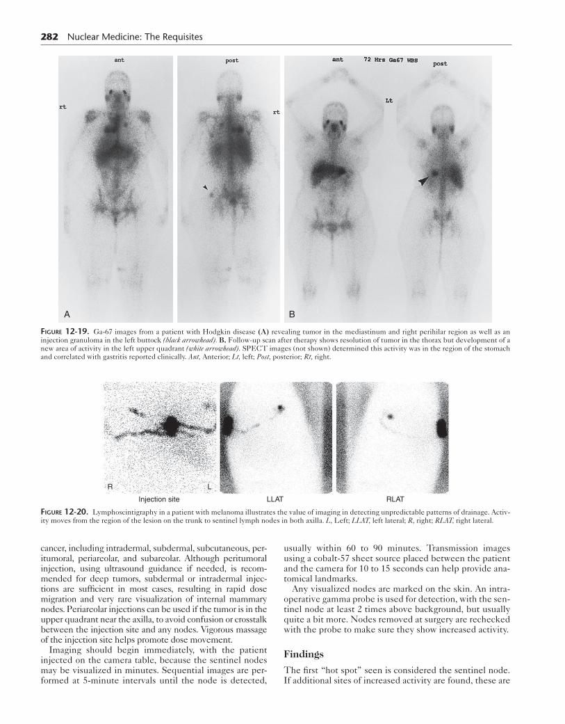
SLN biopsy is indicated for patients at risk for node metastases, especially early stage, with no clinically evi-dent metastatic disease. Although the procedure has been explored in a variety of tumors, such as cancers of the head and neck, cervix, and colon, it has been widely accepted for melanoma and breast cancer (Fig. 12-20).
Radiopharmaceutical and Dosimetry
No particular radiopharmaceutical is approved for lympho-scintigraphy in the United States, and many agents have been used, such as Tc-99m human serum albumin (HSA), Tc-99m dextran, Tc-99m DTPA-mannosyl-dextran, and Tc-99m ultrafiltered sulfur colloid. However, the most commonly used agents are Tc-99m sulfur colloid in the United States, Tc-99m nanocolloid in Europe, and Tc-99m antimony trisulfide in Canada and Australia. Small parti-cles migrate quickly, but show shorter lymph node resi-dence times, and larger particles may not migrate. Because particles sizes for sulfur colloid tend to be large, recom-mended preparation includes slowly passing the dose through a 0.22-μ millipore filter.
Methodology
SLN biopsy is preferably done before wide local excision of the primary lesion to avoid problems from surgery altering lymph flow. The injection can be performed on the day of surgery, or within 18 hours of surgery on the day before. Vari-ous techniques have been used. An intradermal injection, raising a tight wheal, generally results in good dose migration through the lymphatic vessels. For melanoma, the dose is injected in 4 to 6 divided doses within 1 cm around the lesion or scar. Different injection sites have been used for breast
282 Nuclear Medicine: The Requisites
A B
Figure 12-19. Ga-67 images from a patient with Hodgkin disease (A) revealing tumor in the mediastinum and right perihilar region as well as an injection granuloma in the left buttock (black arrowhead). B, Follow-up scan after therapy shows resolution of tumor in the thorax but development of a new area of activity in the left upper quadrant (white arrowhead). SPECT images (not shown) determined this activity was in the region of the stomach and correlated with gastritis reported clinically. Ant, Anterior; Lt, left; Post, posterior; Rt, right.
R L
Injection site LLAT RLAT
Figure 12-20. Lymphoscintigraphy in a patient with melanoma illustrates the value of imaging in detecting unpredictable patterns of drainage. Activ-ity moves from the region of the lesion on the trunk to sentinel lymph nodes in both axilla. L, Left; LLAT, left lateral; R, right; RLAT, right lateral.
cancer, including intradermal, subdermal, subcutaneous, per-itumoral, periareolar, and subareolar. Although peritumoral injection, using ultrasound guidance if needed, is recom-mended for deep tumors, subdermal or intradermal injec-tions are sufficient in most cases, resulting in rapid dose migration and very rare visualization of internal mammary nodes. Periareolar injections can be used if the tumor is in the upper quadrant near the axilla, to avoid confusion or crosstalk between the injection site and any nodes. Vigorous massage of the injection site helps promote dose movement.
Imaging should begin immediately, with the patient injected on the camera table, because the sentinel nodes may be visualized in minutes. Sequential images are per-formed at 5-minute intervals until the node is detected,
usually within 60 to 90 minutes. Transmission images using a cobalt-57 sheet source placed between the patient and the camera for 10 to 15 seconds can help provide ana-tomical landmarks.
Any visualized nodes are marked on the skin. An intra-operative gamma probe is used for detection, with the sen-tinel node at least 2 times above background, but usually quite a bit more. Nodes removed at surgery are rechecked with the probe to make sure they show increased activity.
Findings
The first “hot spot” seen is considered the sentinel node. If additional sites of increased activity are found, these are
also removed. Although intraoperative vital blue dye injec-tion and preoperative radiolabeled colloids have been used independently, together they appear to result in the great-est sensitivity for the SLN. There is no consensus on a procedure if the node cannot be visualized.
MelanomaOf the many factors influencing prognosis in melanoma, the primary lesion’s thickness (depth of tumor invasion measured in millimeters), lesion ulceration, and mitotic rate are the most useful in staging early tumors and pre-dicting occult lymph node involvement. Lymph node involvement is the most important independent predictor of survival.
Currently, the American Joint Committee on Cancer uses lesion thickness cutoffs of 1.0, 2.0, and 4.0 mm, simi-lar to the old Breslow classification, to set the T-stage parameters. Patients with intermediate thickness tumors, greater than 1.0 mm but less than 4.0 mm, may benefit most from SLN biopsy. Of patients with tumors less than 1.0 mm (stage I), the procedure can be considered if there are other high-risk factors such as the presence of a high mitotic rate in the biopsy (≥1 mitosis/mm2) or lesion ulcer-ation (stage IB).
SLN biopsy accuracy is much higher than clinical assess-ment. False negative rates are reported at 5%, with recur-rences of 3% to 6% seen among those who had been found negative for disease initially on SLN assessment. Also, complication rates are much lower than for complete lymph node dissection: 10.1% versus 37.2% in Multicenter Selec-tive Lymphadenectomy Trial I (MSLTI) and 4.6 versus 23.2% in the Sunbelt Melanoma Trial.
Limited data have been reported on survival benefit from SLN biopsy. Preliminary results from the large MSLTI showed no difference in survival between patients who were in an observation group and those who under-went SLN biopsy (86.6% vs. 87.1% p = 0.58). However, 5-year survival among patients in the observation group who did develop lymph node metastases was much lower than in patients in the SLN biopsy group who had positive lymph nodes initially (52.4% vs. 72.4%).
Breast CancerAxillary node status is a major prognostic factor in early-stage breast cancer. Even in small, T1 tumors (≤2 cm), axillary nodes are involved at initial staging 10% to 30% of the time, and this number increases to 45% for T2 lesions (2.1-3.0 cm). Some centers limit SLN biopsy to those with unifocal tumors smaller than 2 to 3 cm; others offer the procedure to patients with large T2 or T3 lesions (>5 cm) and multifocal or multicentric lesions. In breast cancer, use of the sentinel node biopsy has largely replaced axillary lymph node dissection.
Most of the time, the SLN is identified in the axillary region. The presence of internal mammary nodes may be demonstrated with greater frequency with periareolar injections. The significance of internal mammary lymph node visualization is often unclear. Some disagreement exists on surgical management of these nodes, with many not including the findings in their assessment at staging. Clearly, the internal mammary nodes are frequently involved in breast cancer and attention to this region is
OncOlOgy: nOn–POsiTrOn emissiOn TOmOgraPhy 283
warranted, particularly in medially located primary tumors, when restaging, such as with PET/CT.
Lymphoscintigraphy detects the SLN in 90% to 98% of cases. False negative rates of 7% to 8.5% are typical, although some significantly higher levels have been reported. Although this is above the 5% target felt to be acceptable, many surgeons do not perform confirmatory axillary dissection if the SLN is free of tumor. The risk for lymphedema is considerable lower after SLN biopsy than from axillary dissection (5% vs. 13%).
Some recent trials have shown low recurrence rates (0.12%-0.6%) in patients initially free of spread on SLN biopsy. This is well within the expected range. However, a clear impact on survival has not been demonstrated. The preliminary results in the National Surgical Adjuvant Breast and Bowel Project trial, for example, showed an 8-year survival rate of 90.3% in SLN biopsy cases com-pared to 91.8% in the axillary lymph node dissection group.
INTRAARTERIAL RADIOACTIVE MICROSPHERES
Background
Tumors, both primary and metastatic, commonly involve the liver. In the case of hepatocellular cancer, surgical resec-tion and liver transplantation are the only methods for cure, but the majority of patients present with unresectable dis-ease. In the setting of liver metastases, additional palliative or adjuvant therapy is frequently needed in addition to che-motherapy to reduce tumor burden or symptoms.
Many new treatments have grown over the past several years, including direct ablation, hepatic arterial chemo-therapy pumps, chemoembolization, and most recently, drug-eluting and radioactive microspheres. Procedures such as thermal ablation (microwave and radiofrequency), cryoablation, and percutaneous injections have proved effective but are not suitable for patients with large or mul-tiple lesions.
Transarterial chemoembolization (TACE) has been rec-ommended as a front-line therapy for patients with large or multifocal hepatocellular tumors. TACE involves a combi-nation of a chemotherapy agent with an embolic agent (steel coils, microspheres, particles, sponges) that induces ischemic necrosis and locally delivers chemotherapy. Newer drug-eluting microspheres provide an advantageous sustained chemotherapy agent release in comparison with TACE. Radiolabeled microspheres also can be delivered via the hepatic artery, thereby offering the advantage of delivering a large dose of radiation directly to the region of the tumor, so-called selective internal radiation therapy.
These directed intraarterial therapy techniques take advantage of the fact that 80% of the blood supply to hepatic tumors originates from the hepatic artery and three quarters of the blood perfusing normal liver parenchyma is from the portal venous system. Therefore lesions preferentially take up the dose and do not need to be ablated individually.
Two radioembolization microsphere agents are available clinically: Y-90 SIR-Sphere and Y-90 Therasphere. Y-90 SIR-Sphere has been approved for use with adjuvant che-motherapy in hepatic metastases from colon cancer and Y-90 Therasphere for unresectable hepatocellular carcinoma.
284 Nuclear Medicine: The Requisites
Radiopharmaceutical
Physical characteristics of the two Y-90 microsphere agents are outlined in Table 12-13. The beta emissions from the Y-90 label have a mean penetration length of 2.5 mm and energy of 0.94 MeV, resulting in 100 to 150 Gy intratu-moral dose. However, nearby tumor cells are relatively spared. With the physical half-life of Y-90 at 2.68 days, approximately 94% of the dose is delivered by 11 days. Dose calculation is based on many factors, including the tumor burden with in the liver (Table 12-14) and the amount of shunting present from the lungs into the liver. The typical doses given are in the range of 40 to 70 mCi (1.5-2.5 GBq). The microspheres will remain in the liver and do not degrade physically.
Methodology
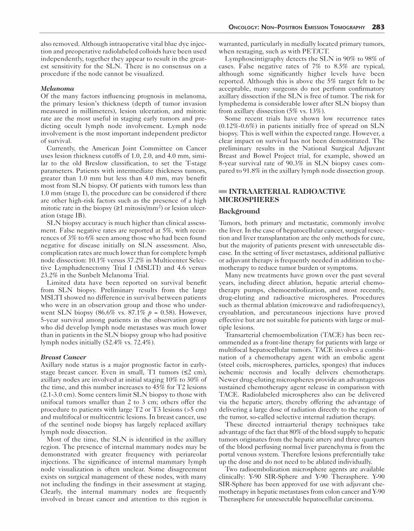
Potential patients must be carefully screened before undergoing radiolabeled microsphere therapy. The func-tional status of the patient, liver function, and estimated tumor burden are all examined. Patency of the portal vein must be established because portal vein thrombosis has been a contraindication, although some studies suggest it need not be absolute. Arteriography of hepatic vasculature is performed, and anomalous vessels that could result in accidental delivery into stomach, bowel, or other struc-tures may be embolized. With the catheter in the expected position for therapy administration, arteriovenous shunt-ing is assessed with a Tc-99m macroaggregated albumin (MAA) scan (Fig. 12-21). The Tc-99m MAA is adminis-tered into the liver. The catheter is removed and the groin stabilized. The patient is then scanned anteriorly and pos-teriorly. The shunt fraction is calculated using a geometric mean calculation based on counts obtained from regions of interest over the lungs and over the liver. For Y-90 SIR-spheres, the dose is adjusted according to Table 12-15 to help prevent radiation pneumonitis. When using Y-90 Theraspheres, the activity is higher on the glass beads, so a lower level of shunting (<10%) is accepted.
Table 12-13 Physical Characteristics of Radiolabeled Microspheres
Agent RadiolabelParticle size(microns)
Particle material
Activity(Bq/particle)
SIR-Sphere Y-90 35
20-60
Resin 50
Therasphere Y-90 25
20-30
Glass 2500
Table 12-14 Yttrium-90 Microsphere Therapy Calculations
Liver involvement by tumor (%)Recommended Y-90 dose (GBq)
>50 3
25-50 2.5
<25 2
After these procedures, the patient returns another day for the therapy itself. The catheter is placed in the same position under fluoroscopic guidance. The dose is admin-istered with a very slow push to prevent refluxing the dose into the systemic circulation. The patient can then be taken to the nuclear medicine department and imaged using the bremsstrahlung radiation emitted from the Y-90 to confirm proper localization (Fig. 12-22). SPECT and SPECT allow the best visualization and comparison to the CT findings.
Patients can be discharged to home after the procedure. Some radiation safety precautions are needed concerning urine because a small amount of activity is excreted through the kidneys. The patient should be instructed to flush the toilet twice and use careful hand hygiene in the bathroom for the first 24 hours after the procedure.
Findings
Y-90 microsphere therapy has been shown to be relatively safe and effective. Side effects potentially include gastric ulcers, radiation hepatitis or cholecystitis, and radiation pneumonitis. Myelosuppression is not expected with cur-rent agents.
After Y-90 microsphere therapy, significant response can be seen in tumor appearance on CT with a decrease in size
Figure 12-21. Intraarterial administration of Tc-99m MAA into the liver allows quantification of any shunt through the liver. Heterogeneous uptake seen above in the liver is not unusual, but the high levels of lung activity indicate a problem, despite all visible accessory vessels having been embolized. The liver-lung shunt was calculated to be 40%, and therapy with the Y-90–labeled microspheres was canceled.
Table 12-15 Yttrium-90 SIR-SPHERE Dose Correction Based on Lung Shunting*
Hepatopulmonary shunting (%) Dose reduction (%)
<10 0
10-15 20
15-20 40
>20 100
*Maximum allowable shunting for Y-90 Therasphere = 10%
and development of necrosis within the lesion. F-18 FDG PET/CT has been done at 1 month and may help monitor response (Fig. 12-23). The majority of patients demon-strate at least partial response. Studies have shown responses in patients who were not responding to chemo-therapy, with hepatocellular cancer in some patients becoming resectable. Limited data suggest some improve-ment in median survival, particularly in those receiving higher doses.
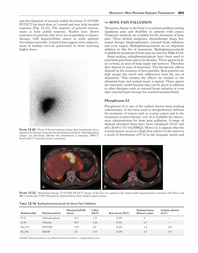
Figure 12-22. Planar Y-90 microspheres image after intrahepatic artery injection for therapy using the bremsstrahlung radiation. Although planar images can determine whether the distribution is adequate, SPECT fused with CT provides better correlation.
OncOlOgy: nOn–POsiTrOn emissiOn TOmOgraPhy 285
BONE PAIN PALLIATION
Metastatic disease to the bone is a common problem causing significant pain and disability in patients with cancer. Numerous methods are available for the treatment of bone pain. These include analgesics, chemotherapy drugs, hor-monal therapy, bisphosphonates, external beam radiation, and even surgery. Radiopharmaceuticals are an important addition to this list of treatments. Radiopharmaceuticals available for treatment of bone pain are listed in Table 12-16.
Bone-seeking radiopharmaceuticals have been used to treat bone pain from cancer for decades. These agents local-ize to bone, in areas of bone repair and turnover. Therefore they deposit in areas of metastasis. The therapeutic effects depend on the emission of beta particles. Beta particles are high energy but travel only millimeters from the site of deposition. This ensures the effects are limited to the abnormal bone and normal tissue is spared. These agents are extremely useful because they can be given in addition to other therapies such as external beam radiation or even after external beam therapy has reached maximal limits.
Phosphorus-32
Phosphorus-32 is one of the earliest known bone-seeking radioisotopes. It has been used in intraperitoneal infusion for treatment of tumors such as ovarian cancer and in the treatment of polycythemia vera. It is available for intrave-nous administration for bone pain palliation. A range of skeletal absorbed doses have been calculated (25-63 rad/mCi [0.68-1.733 cGy/MBq]). However, it appears that the normal marrow receives a high dose relative to the tumor as a result of distribution of P-32 in the inorganic matrix and
A B
Figure 12-23. Monitoring therapy. F-18 FDG PET/CT images of the liver in a patient with unresectable hepatocellular carcinoma (A) before and (B) 1 month after Y-90 Therasphere administration show marked improvement.
Table 12-16 Radiopharmaceuticals for Bone Pain Palliation
Radionuclide PharmaceuticalPhysical half-life (days)
β Max(MeV) Beta mean (MeV)
Maximal tissue distance (mm)
Gamma photon (keV)
P-32 Orthophosphate 14.3 1.71 0.695 8 —
Sr-89 Chloride 50.5 1.46 0.583 6.7 —
Sm-153 EDTMP 1.95 0.8 0.224 3.4 103
Re-186 HEDP 3.8 1.07 0.349 4.7 137
EDTMP, Ethylenediamine tetra; HEDP, hydroxyethylidene-1, 1-diphosphonic acid.

286 Nuclear Medicine: The Requisites
A B
Figure 12-24. Sm-153 palliation of metastatic disease bone pain. A, The whole-body Tc-99m MDP bone scan before therapy confirms the presence of osseous metastasis that will accumulate the therapy agent. B, The whole-body scan done with the Sm-153 therapy dose shows close correlation with the lesions seen on bone scan. Ant, Anterior; Post, posterior.
cellular regions. Also, the lack of a gamma emission means no external imaging can be done to assess distribution.
Strontium-89
Strontium-89 is a pure beta-emitter approved by the FDA for management of metastatic bone pain under the trade name Metastron. It selectively localizes in areas of bony turnover, predominantly in blastic lesions. It is retained lon-ger in regions of metastasis than normal bone. The pathway of excretion is predominantly through urine, with about one third bowel excretion. Dosimetry is outlined in Table 9-18.
A 4-mCi (148 MBq) dose is administered intravenously slowly over 1 to 2 minutes. An alternative dose of 55 μCi/kg (2.04 MBq/kg) may be used. Repeat dosing is possible, but factors such as initial response, hematological status, and current status must be considered in each case. In gen-eral, a repeat administration is not recommended before 90 days have elapsed.
ToxicityBecause toxicity occurs to marrow components, platelets, and white blood cells, the hematological status of each patient must be evaluated before therapy. After obtaining a baseline platelet count, platelets should be measured at least every other week. Typically, platelets will decrease 30% from baseline and reach the nadir 12 to 16 weeks after
therapy. Toxicity is generally mild; however, Sr-89 must be used with caution in those with white blood cell counts less than 2400 and platelets less than 60,000. A small number of patients experience transient worsening of symptoms.
Approximately 20% of patients will become pain free. Around 75% of patients will experience some significant decrease in pain, although some series have reported up to 90% of patients experience some symptom relief. Pain relief begins approximately 7 to 20 days after injection and generally lasts 3 to 6 months.
Samarium-153
Samarium-153 (Quadramet) is a beta-emitting radiophar-maceutical that has the added advantage of a gamma emis-sion that can be detected for external imaging. It has been approved for use in patients with osteoblastic metastases that can be visualized on a nuclear medicine bone scan (Fig. 12-24).
Sm-153 is administered in a 1.0-mCi/kg (37 MBq/kg) dose intravenously over the course of 1 minute. Approxi-mately 50% of the dose is localized to bone. It accumulates in metastatic lesions in a 5:1 ratio compared with normal bone. Patients should be well hydrated and void fre-quently because the primary route of clearance is through the urine. Approximately 35% of the dose is excreted in the first 6 hours.
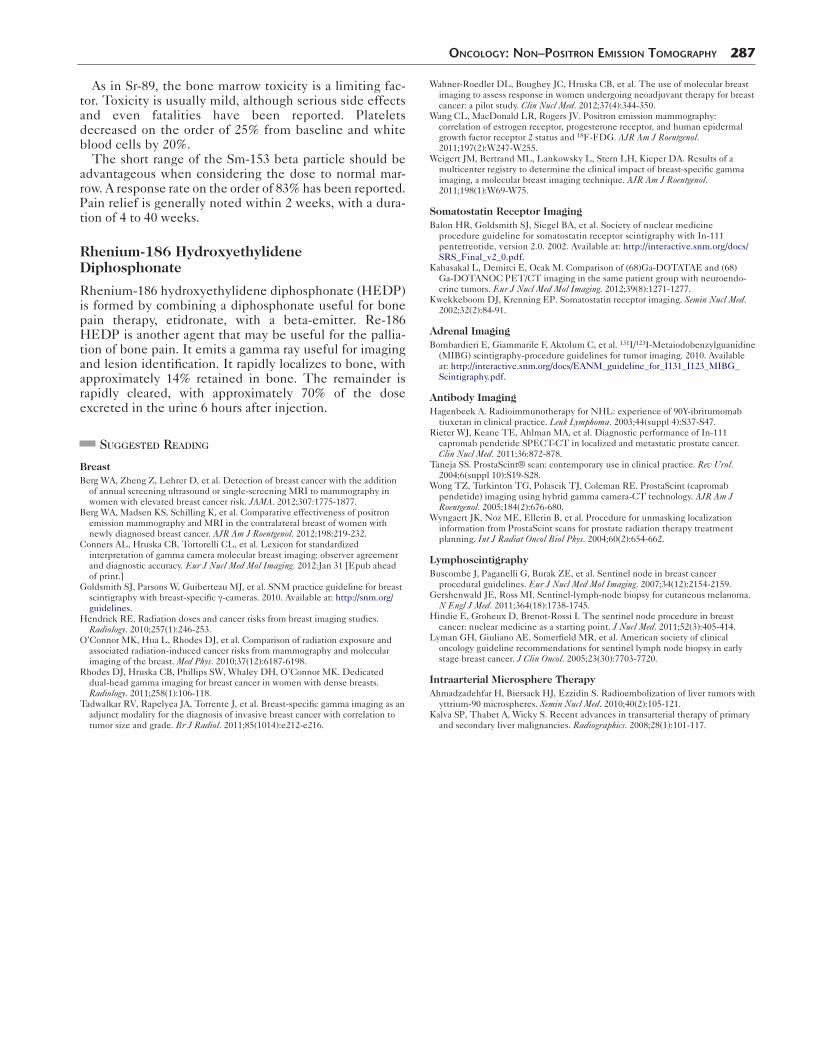
As in Sr-89, the bone marrow toxicity is a limiting fac-tor. Toxicity is usually mild, although serious side effects and even fatalities have been reported. Platelets decreased on the order of 25% from baseline and white blood cells by 20%.
The short range of the Sm-153 beta particle should be advantageous when considering the dose to normal mar-row. A response rate on the order of 83% has been reported. Pain relief is generally noted within 2 weeks, with a dura-tion of 4 to 40 weeks.
Rhenium-186 Hydroxyethylidene Diphosphonate
Rhenium-186 hydroxyethylidene diphosphonate (HEDP) is formed by combining a diphosphonate useful for bone pain therapy, etidronate, with a beta-emitter. Re-186 HEDP is another agent that may be useful for the pallia-tion of bone pain. It emits a gamma ray useful for imaging and lesion identification. It rapidly localizes to bone, with approximately 14% retained in bone. The remainder is rapidly cleared, with approximately 70% of the dose excreted in the urine 6 hours after injection.
SuggeSted Reading
BreastBerg WA, Zheng Z, Lehrer D, et al. Detection of breast cancer with the addition
of annual screening ultrasound or single-screening MRI to mammography in women with elevated breast cancer risk. JAMA. 2012;307:1775-1877.
Berg WA, Madsen KS, Schilling K, et al. Comparative effectiveness of positron emission mammography and MRI in the contralateral breast of women with newly diagnosed breast cancer. AJR Am J Roentgenol. 2012;198:219-232.
Conners AL, Hruska CB, Tortorelli CL, et al. Lexicon for standardized interpretation of gamma camera molecular breast imaging: observer agreement and diagnostic accuracy. Eur J Nucl Med Mol Imaging. 2012:Jan 31 [Epub ahead of print.]
Goldsmith SJ, Parsons W, Guiberteau MJ, et al. SNM practice guideline for breast scintigraphy with breast-specific γ-cameras. 2010. Available at: http://snm.org/guidelines.
Hendrick RE. Radiation doses and cancer risks from breast imaging studies. Radiology. 2010;257(1):246-253.
O’Connor MK, Hua L, Rhodes DJ, et al. Comparison of radiation exposure and associated radiation-induced cancer risks from mammography and molecular imaging of the breast. Med Phys. 2010;37(12):6187-6198.
Rhodes DJ, Hruska CB, Phillips SW, Whaley DH, O’Connor MK. Dedicated dual-head gamma imaging for breast cancer in women with dense breasts. Radiology. 2011;258(1):106-118.
Tadwalkar RV, Rapelyea JA, Torrente J, et al. Breast-specific gamma imaging as an adjunct modality for the diagnosis of invasive breast cancer with correlation to tumor size and grade. Br J Radiol. 2011;85(1014):e212-e216.
OncOlOgy: nOn–POsiTrOn emissiOn TOmOgraPhy 287
Wahner-Roedler DL, Boughey JC, Hruska CB, et al. The use of molecular breast imaging to assess response in women undergoing neoadjuvant therapy for breast cancer: a pilot study. Clin Nucl Med. 2012;37(4):344-350.
Wang CL, MacDonald LR, Rogers JV. Positron emission mammography: correlation of estrogen receptor, progesterone receptor, and human epidermal growth factor receptor 2 status and 18F-FDG. AJR Am J Roentgenol. 2011;197(2):W247-W255.
Weigert JM, Bertrand ML, Lankowsky L, Stern LH, Kieper DA. Results of a multicenter registry to determine the clinical impact of breast-specific gamma imaging, a molecular breast imaging technique. AJR Am J Roentgenol. 2011;198(1):W69-W75.
Somatostatin Receptor ImagingBalon HR, Goldsmith SJ, Siegel BA, et al. Society of nuclear medicine
procedure guideline for somatostatin receptor scintigraphy with In-111 pentetreotide, version 2.0. 2002. Available at: http://interactive.snm.org/docs/SRS_Final_v2_0.pdf.
Kabasakal L, Demirci E, Ocak M. Comparison of (68)Ga-DOTATAE and (68)Ga-DOTANOC PET/CT imaging in the same patient group with neuroendo-crine tumors. Eur J Nucl Med Mol Imaging. 2012;39(8):1271-1277.
Kwekkeboom DJ, Krenning EP. Somatostatin receptor imaging. Semin Nucl Med. 2002;32(2):84-91.
Adrenal ImagingBombardieri E, Giammarile F, Aktolum C, et al. 131I/123I-Metaiodobenzylguanidine
(MIBG) scintigraphy-procedure guidelines for tumor imaging. 2010. Available at: http://interactive.snm.org/docs/EANM_guideline_for_I131_I123_MIBG_Scintigraphy.pdf.
Antibody ImagingHagenbeek A. Radioimmunotherapy for NHL: experience of 90Y-ibritumomab
tiuxetan in clinical practice. Leuk Lymphoma. 2003;44(suppl 4):S37-S47.Rieter WJ, Keane TE, Ahlman MA, et al. Diagnostic performance of In-111
capromab pendetide SPECT-CT in localized and metastatic prostate cancer. Clin Nucl Med. 2011;36:872-878.
Taneja SS. ProstaScint® scan: contemporary use in clinical practice. Rev Urol. 2004;6(suppl 10):S19-S28.
Wong TZ, Turkinton TG, Polascik TJ, Coleman RE. ProstaScint (capromab pendetide) imaging using hybrid gamma camera-CT technology. AJR Am J Roentgenol. 2005;184(2):676-680.
Wyngaert JK, Noz ME, Ellerin B, et al. Procedure for unmasking localization information from ProstaScint scans for prostate radiation therapy treatment planning. Int J Radiat Oncol Biol Phys. 2004;60(2):654-662.
LymphoscintigraphyBuscombe J, Paganelli G, Burak ZE, et al. Sentinel node in breast cancer
procedural guidelines. Eur J Nucl Med Mol Imaging. 2007;34(12):2154-2159.Gershenwald JE, Ross MI. Sentinel-lymph-node biopsy for cutaneous melanoma.
N Engl J Med. 2011;364(18):1738-1745.Hindie E, Groheux D, Brenot-Rossi I. The sentinel node procedure in breast
cancer: nuclear medicine as a starting point. J Nucl Med. 2011;52(3):405-414.Lyman GH, Giuliano AE, Somerfield MR, et al. American society of clinical
oncology guideline recommendations for sentinel lymph node biopsy in early stage breast cancer. J Clin Oncol. 2005;23(30):7703-7720.
Intraarterial Microsphere TherapyAhmadzadehfar H, Biersack HJ, Ezzidin S. Radioembolization of liver tumors with
yttrium-90 microspheres. Semin Nucl Med. 2010;40(2):105-121.Kalva SP, Thabet A, Wicky S. Recent advances in transarterial therapy of primary
and secondary liver malignancies. Radiographics. 2008;28(1):101-117.


![FDG-PET in Large Vessel Vasculitis...FDG-PET in Large Vessel Vasculitis 61 5. [18 F]FDG-PET and [18 F]FDG-PET/CT [18 F]FDG-PET is an operator-independent, non- invasive imaging modality](https://img.dokumen.tips/doc/110x75/5f6c13132f0609183b646bce/fdg-pet-in-large-vessel-vasculitis-fdg-pet-in-large-vessel-vasculitis-61-5.jpg)


![Tecniche di imaging tomografico non distruttivo in ... · Tecniche di imaging tomografico non ... Alcuni Traccianti PET • Metabolismo del glucosio [18F]FDG ... 18FDG-PET 18FDG-PET](https://img.dokumen.tips/doc/110x75/5c6b2b7109d3f262278b69d2/tecniche-di-imaging-tomografico-non-distruttivo-in-tecniche-di-imaging-tomografico.jpg)