
DEVELOPMENT OF QUANTITATIVE AND HIGH-THROUGHPUTASSAYS OF POLYOMAVIRUS AND PAPILLOMAVIRUS DNAREPLICATION
Amélie Fradet-Turcotte1,£, Geneviève Morin1,£, Michaël Lehoux1, Peter A. Bullock2, andJacques Archambault1,*
1Laboratory of Molecular Virology, Institut de Recherches Cliniques de Montréal (IRCM) andDepartment of Biochemistry, Université de Montréal, Montreal, Quebec, Canada2Department of Biochemistry, Tufts University School of Medicine, Boston, Massachusetts
AbstractPolyoma- and papillomaviruses genome replication is initiated by the binding of large T antigen(LT) and of E1 and E2, respectively, at the viral origin (ori). Replication of an ori-containingplasmid occurs in cells transiently expressing these viral proteins and is typically quantified bySouthern blotting or PCR. To facilitate the study of SV40 and HPV31 DNA replication, wedeveloped cellular assays in which transient replication of the ori-plasmid is quantified using afirefly luciferase gene located in cis to the ori. Under optimized conditions, replication of theSV40 and HPV31 ori-plasmids resulted in a 50- and 150-fold increase in firefly luciferase levels,respectively. These results were validated using replication-defective mutants of LT, E1 and E2and with inhibitors of DNA replication and cell-cycle progression. These quantitative and high-throughput assays should greatly facilitate the study of SV40 and HPV31 DNA replication and theidentification of small-molecule inhibitors of this process.
INTRODUCTIONSmall DNA tumor viruses such as polyoma- and papillomaviruses rely widely on the hostcell DNA replication machinery to replicate their double-stranded viral genome. EukaryoticDNA replication is a complex process that is initiated by several factors including the originrecognition complex (ORC), Cdt1, Cdc6 and the mini-chromosome maintenance (MCM)complex, the alleged cellular replicative helicase (Johnson and O’Donnell, 2005; Masai,You, and Arai, 2005; Nishitani and Lygerou, 2002). In contrast, small DNA tumor viruseslike polyoma- and papillomaviruses encode a single initiator protein that performs multiplefunctions during viral genome replication. A well studied example is the large T antigen(LT) of simian virus 40 (SV40). This multifunctional initiator protein can successivelyrecognize the viral origin of replication, assemble into a double hexamer that melts andunwinds the DNA ahead of the replication fork, and interact with the host DNA replicationfactors such as polymerase α-primase, replication protein A (RPA) and topoisomerase I
© 2011 Elsevier Inc. All rights reserved.*To whom correspondence should be addressed: Institut de Recherches Cliniques de Montréal (IRCM), 110 Pine Avenue West,Montreal (Quebec), Canada, H2W 1R7. Tel: (514) 987-5739; Fax: (514) 987-5741; [email protected] .£Both authors contributed equally to this workPublisher's Disclaimer: This is a PDF file of an unedited manuscript that has been accepted for publication. As a service to ourcustomers we are providing this early version of the manuscript. The manuscript will undergo copyediting, typesetting, and review ofthe resulting proof before it is published in its final citable form. Please note that during the production process errors may bediscovered which could affect the content, and all legal disclaimers that apply to the journal pertain.
NIH Public AccessAuthor ManuscriptVirology. Author manuscript; available in PMC 2011 August 10.
Published in final edited form as:Virology. 2010 March 30; 399(1): 65–76. doi:10.1016/j.virol.2009.12.026.
NIH
-PA Author Manuscript
NIH
-PA Author Manuscript
NIH
-PA Author Manuscript

(reviewed in (Borowiec et al., 1990; Bullock, 1997)). The analogous protein frompapillomavirus, E1, has similar activities but also requires the viral protein E2 to initiateviral DNA replication in vivo (reviewed in (Hebner and Laimins, 2006)). Papillomavirus E2is both a replication and transcription factor that binds with high affinity to sites in the viralorigin (Androphy, Lowy, and Schiller, 1987). As a replication factor, E2 interacts directlywith E1 to recruit it to the origin and favor its assembly into a double hexamer (Blitz andLaimins, 1991; Lusky, Hurwitz, and Seo, 1994; Mohr et al., 1990).
LT and E1 are structurally related members of the helicase superfamily III (SF3) (Clertantand Seif, 1984; Hickman and Dyda, 2005; Mansky, Batiza, and Lambert, 1997). The C-terminal domains of LT and E1 have ATPase/helicase activity and are sufficient foroligomerization into hexamers (Li et al., 2003; Titolo et al., 2000; White et al., 2001). Thecentral part of both proteins contains an origin-binding domain (OBD) which recognizesspecific sequences in the origin (McVey, Strauss, and Gluzman, 1989; Simmons, Loeber,and Tegtmeyer, 1990; Titolo et al., 2003a; Titolo et al., 2003b; Wun-Kim et al., 1993). TheOBDs of LT and E1 differ in their primary amino acid sequence but share a common fold.Interestingly, while the LT OBD can bind with high-affinity to its target binding site as amonomer, the E1 OBD needs to dimerize to achieve comparable affinity and specificity(Fradet-Turcotte et al., 2007; Titolo et al., 2003a; Titolo et al., 2003b). Crystal structures ofthe bovine papillomavirus (BPV) and human papillomavirus (HPV) 18 E1 OBDs haverevealed the nature of the dimerization interface and mutations that disrupt this interfacehave been shown to impair viral DNA replication (Auster and Joshua-Tor, 2004; Enemark,Stenlund, and Joshua-Tor, 2002; Schuck and Stenlund, 2005; Titolo et al., 2003a). Both LTand E1 also differ substantially in their N-terminal regions, although in either case thesecontain regulatory elements. The N-terminal domain of LT contains a unique J-domainrequired for replication in vivo (Sullivan and Pipas, 2002), a monopartite nuclearlocalization signal (NLS) (Kalderon et al., 1984a; Kalderon et al., 1984b) and severalphosphorylation sites for different kinases that modulate either the nuclear import of LT(Rihs et al., 1991) or its assembly into a double hexamer at the origin (Cegielska et al., 1994;Moarefi et al., 1993; Mohr, Stillman, and Gluzman, 1987; Scheidtmann et al., 1984;Schneider and Fanning, 1988; Virshup, Kauffman, and Kelly, 1989; Virshup, Russo, andKelly, 1992; Weisshart et al., 1999). As for the N-terminal domain of E1, it contains a bi-partite NLS, a Crm1-dependant nuclear export signal (NES), and a binding site for cyclin A/E-cdk2, which regulates E1 nucleo-cytoplasmic shuttling by phosphorylation (Deng et al.,2004; Ma et al., 1999). In addition, this domain of E1 contains a binding site for the cellularprotein p80, necessary for optimal viral DNA replication (Cote-Martin et al., 2008), and acaspase-3/7 cleavage site required for amplification of the viral genome in differentiatedkeratinocytes (Moody et al., 2007).
Expression of LT and E1/E2 has been shown to be sufficient to support replication of aplasmid encompassing their cognate origin in transiently transfected cells. This type of assayhas been used extensively to define specific DNA sequences required for origin-function invivo. For SV40, the 64-bp core (nt 5193 to 34) was identified as the minimal originhowever, replication was shown to be maximal when the adjacent 21-bp repeat region wasincluded (Bergsma et al., 1982; Lee-Chen and Woodworth-Gutai, 1986). The middle portionof the core contains four binding sites for LT (5′-GAGGC-3′), arranged as two pairs ofinverted repeats. This middle region known as site II is flanked on one side by an AT-richregion and on the other by the early palindrome (EP) (Borowiec et al., 1990; Bullock et al.,1997; Deb et al., 1987; DeLucia et al., 1983; Parsons, Anderson, and Tegtmeyer, 1990;Tegtmeyer et al., 1983). Similarly, the HPV origin (nt 7721 to 100 for HPV31 (Frattini andLaimins, 1994)) was found to contain four binding sites for E1 (5′-ATTGTT-3′), alsoarranged as two pairs of inverted repeats, together with an AT-rich region (Chen andStenlund, 2001; Holt and Wilson, 1995; Lee et al., 1997; Mendoza, Gandhi, and Botchan,
Fradet-Turcotte et al. Page 2
Virology. Author manuscript; available in PMC 2011 August 10.
NIH
-PA Author Manuscript
NIH
-PA Author Manuscript
NIH
-PA Author Manuscript

1995; Sun, Lu, and McCance, 1996; Titolo et al., 2003a; Ustav et al., 1991). In addition andspecific to HPV, the origin of replication contains three binding sites for the E2 protein (5′-ACCN6GGT-3′) (Androphy, Lowy, and Schiller, 1987).
Replication of an origin-containing plasmid (ori-plasmid) by LT, or E1/E2, in transientlytransfected cells is typically detected by Southern blotting or PCR (Del Vecchio et al., 1992;Taylor and Morgan, 2003). In these assays, plasmid replication can be detected from eithertotal or low-molecular weight DNA (i.e. Hirt-extracted DNA) that has been digested withDpnI to restrict any input transfected ori-plasmid that failed to replicate and thus retained amethylation pattern characteristic of bacteria. The numerous steps involved in these transientDNA replication assays have limited their use for high-throughput studies such as thoseinvolving the characterization of large number of mutations or the screening of chemical orsiRNA libraries. To overcome this limitation, we have developed facile and quantitativeassays of SV40 and HPV31 DNA replication that rely on a dual-luciferase readout tomeasure the amount of replicated origin DNA directly from transfected cells.
MATERIALS AND METHODSPlasmid constructions and mutagenesis
The plasmid to express SV40 Large T antigen, pCMV-T-ag (referred herein as pLT forshort), has been described previously (Campbell et al., 1997). Plasmid pFLORI40, whichcontains a firefly luciferase gene under the control of the CMV promoter and the SV40origin of replication, was constructed in two steps. First, the firefly luciferase (Fluc) genefrom pGL3-basic (Promega) was inserted between the XhoI and XbaI restriction sites of pCI(Promega) to create pCI-Fluc. Second, a PCR fragment spanning the SV40 origin ofreplication (nucleotides 5094-161 of the complete genome according to the numberingsystem of Buchman et al. (1981) (Buchman, 1981), NCBI Reference Sequence:NC_001669.1) was inserted into the DraIII site of pCI-Fluc to generate pFLORI40. Primersused for the amplification of the SV40 origin were designed to encode DraIII restrictionsites and were of the following sequence: 5′-CGCATCACGTAGTGCCTCCCCAGCAGGC-3′ and 5′-CGCATCACTACGTGCCACTCCTTTCAAGACC-3′. The plasmids to express HPV31 E1and E2 were obtained by inserting the codon-optimized genes, synthesized commercially(GenScript Corp.), between the BamHI-EcoRI sites (E1) or BamHI-HindIII sites (E2) ofpCMV-3Tag-1a (Stratagene) to create in frame fusions with the triple-Flag (3F) epitope. TheHPV origin-containing plasmid (pFLORI31) was constructed as described above forpFLORI40 with the difference that the PCR fragment spanning the HPV31 origin ofreplication (nucleotides 7721-100 of the complete genome according to numbering schemeof Frattini and Laimins, 1994 (Frattini and Laimins, 1994) NCBI Reference Sequence:J04353.1) was inserted into the NgoMIV site of pCI-Fluc to create pFLORI31. Primers usedfor the amplification of the HPV31 origin were designed to encode NgoMIV restrictionsites, and were of the following sequence: 5′-CGCATGCCGGCAAACTGCTTTTAGGCACATATTTTG-3′ and 5′-CGCATGCCGGCGTAGGTTTGCACAAAATACTATGTG-3′. Plasmid pRL coding forRenilla luciferase (Rluc) was constructed by inserting a PCR fragment encoding Rlucbetween the SalI and NotI sites of pCI. The following primers were used: 5′-CCACCGTCGACGCCACCATGACCAGCAAGGTGTACG-3′ and 5′-CCCGCGGCCGCTTATCTAGATCCGGTGGATCC-3′. Site-directed mutagenesis wasperformed using the QuickChange mutagenesis kit (Stratagene). All DNA constructs wereverified by sequencing. Further details on their construction will be provided upon request.
Fradet-Turcotte et al. Page 3
Virology. Author manuscript; available in PMC 2011 August 10.
NIH
-PA Author Manuscript
NIH
-PA Author Manuscript
NIH
-PA Author Manuscript

Cell culture and transfectionsC33A human cervical carcinoma cells were grown in Dulbecco’s modified Eagle’s medium(DMEM), supplemented with 10% fetal bovine serum, 0.5 IU/ml of penicillin, 50 μg/mlstreptomycin, and 2 mM L-glutamine. Transfections were performed using theLipofectamine 2000 reagent according to the manufacturer’s recommendations (Invitrogen).
Luciferase DNA replication assayC33A cells were plated 20 h before transfection in white flat-bottom 96-well plates(Corning) at a density of 25000 cells/well. The total quantity of plasmid DNA transfectedwas adjusted to 100 ng with the empty vector (pCI) for the SV40 assay, and with(pCMV-3Tag-1a) for the HPV assay, as carrier DNA. For all experiments, 2.5 ng of theorigin-containing plasmid pFLORI40 (Fluc) or pFLORI31 (Fluc) and 0.5ng of pRL (Rluc)were transfected, along with the indicated amount of pLT or pE1 and pE2, respectively. Theculture medium was changed 4 h post-transfection. Fresh media was added and firefly andRenilla luciferase activities were measured using the Dual-Glo Luciferase assay system(Promega) 24 to 120 h post-transfection.
Fluorescence polarization DNA binding assayThe wild-type and G230R HPV31 E1 OBDs were expressed as fusions to GST and purifiedfrom bacteria as described previously (Fradet-Turcotte et al., 2007). The duplex DNAprobes were prepared by annealing a fluorescein-labeled oligonucleotide to itscomplementary oligonucleotide as described previously (Titolo et al., 2003a). 150 μl bindingreactions were assembled in 96- well HTRF plates (Packard) using 10 nM fluorescein-labeled probe and the indicated concentrations of protein in the following buffer: 20 mMTris (pH 7.6), 50 mM NaCl, 0.01% NP-40, and 1 mM DTT. Fluorescence readings wererecorded and KD values calculated as previously described (Fradet-Turcotte et al., 2007;Titolo et al., 2003a).
Quantitative PCR (qPCR) analysisApproximately 1.2 × 10⁶ C33A cells were transfected in 6-well plates with 60 ng ofpFLORI40 and 300 ng of pLT for the SV40 DNA replication assay or with 60 ngpFLORI31, 240 ng of p31E1 and 240 ng of p31E2 for the HPV31 DNA replication assay. Inboth assays, 12 ng of pRL were also transfected to reproduce the conditions used in theluciferase-based assay. The total quantity of plasmid DNA transfected was adjusted to 2400ng with empty vector, either pCI for the SV40 assay or pCMV-3Tag-1a for the HPV31assay, as carrier DNA. Total genomic DNA was extracted 72 h post-transfection using theDNeasy kit from Qiagen. This method of DNA isolation was found to yield morereproducible results than the commonly used Hirt-extraction protocol (Taylor and Morgan,2003) (data not shown). To measure the amount of replicated pFLORI31 or pFLORI40, 25μl of total genomic DNA was digested with 10 units of DpnI (New England Biolabs) for 16hrs in a final volume of 30 μl. The digested DNA samples were then incubated for 30minutes with 100 units of Exonuclease III (New England Biolabs) and the enzyme was heatinactivated at the end of the reaction by incubation at 70°C for 30 minutes. The primers usedto amplify an 80 bp-portion of the firefly luciferase gene present on the pFLORI40 (SV40ori), pFLORI31 (HPV31 ori) or pCI-Fluc (No ori) plasmids as well as the probe used todetect the amplicon were synthesized according to unpublished sequences validated by DrIain Morgan (personal communication). These primers amplify a portion of Fluc thatencompasses two DpnI sites and the probe, which is labeled with FAM and TAMRA at its 5′and 3′ end, respectively, hybridizes to a region that overlaps these two DpnI sites. Real-timeqPCR was performed using 4.5 μl of a 5-fold dilution of digested genomic DNA, 900 nM ofprimers and 125 nM of probe in “PerfeCTa™ qPCR SuperMix, UNG, Low ROX” buffer
Fradet-Turcotte et al. Page 4
Virology. Author manuscript; available in PMC 2011 August 10.
NIH
-PA Author Manuscript
NIH
-PA Author Manuscript
NIH
-PA Author Manuscript

(Quanta Biosciences, this buffer contains: dATP, dCTP, dGTP and dUTP; MgCl2;AccuStart Taq DNA Polymerase; UNG; ROX reference dye and stabilizers). PCR reactionswere performed on a real-time PCR system (Mx3005P, Stratagene) using the following PCRamplification conditions: 95°C for 15 sec and 60°C for 1 min, for 40 cycles. Quantificationwas performed using a 7-point standard curve of pCI-Fluc plasmid ranging from 104 to 10−2
pg (10-fold serial dilutions). Plasmid quantities were reported in pg per mg of total genomicDNA extracted. Each value is the average of at least three replicates.
InhibitorsC33A cells were plated and transfected essentially as described for the luciferase DNAreplication assay. For SV40, a constant amount of 12.5 ng of pLT was used in each wellalong with 2.5 ng of pFLORI40 and 0.5 ng of pRL plasmid. The culture medium wasreplaced 4 h post-transfection with fresh medium. The inhibitors were added 48 h post-transfection by changing the culture medium with fresh medium containing the inhibitor orvehicle alone (0.1 % DMSO). Finally, fresh media was added again 72 h post-transfectionand firefly and Renilla luciferase activities measured using the Dual-Glo Luciferase assaysystem (Promega). For HPV, the assay was essentially the same, but the inhibitors wereadded directly to the assay 4 h post-transfection and the firefly and Renilla luciferaseactivities were measured 24 h post-transfection. For both assays, two-fold serial dilutions ofeach inhibitor were assayed starting at a maximal concentration of 50 nM for gemcitabine, 2mM for hydroxyurea, 500 μM for mimosine, 0.2 μM for geldanamycin, 2 μM for 17-AAGand 10 μM for lactacystin. Lactacystin was purchased from Cayman (Cat: 70980),geldanamycin and 17-AAG from BIOMOL international (Cat: EI-280 and EI-308,respectively) and both hydroxyurea and mimosine from Sigma-Aldrich (Cat: H8627 andM0253). Gemcitabine was a kind gift Dr. William W. Bachovchin, Tufts University.
Western blotting and antibodiesLT proteins were detected using a mouse monoclonal antibody from Santa CruzBiotechnology that recognizes an epitope located within residues 1-82 of the protein (Cat:sc-148). Flag-tagged E1 and E2 proteins were detected using a mouse monoclonal antibodyfrom Sigma-Aldrich (M2-Flag antibody, Cat: F1804). β-tubulin was detected using a mousemonoclonal antibody from Sigma-Aldrich (Cat: T0426). For Western blot analysis, proteinswere transferred onto polyvinylidene difluoride membranes and detected using horseradishperoxidase-conjugated sheep anti-mouse secondary antibody from GE healthcare (Cat:NA931) and an enhanced chemiluminescence detection kit (GE Healthcare).
Confocal fluorescence microscopy8 × 105 C33A cells were transfected with 500 ng of RFP-PCNA expression plasmid(previously described in (Sporbert et al., 2005)) and grown on coverslips. Inhibitors wereadded 4 h post-transfection and cells were fixed 24 h later with 4% formaldehyde. Cellswere mounted using Vectashield mounting medium (Vector Laboratories). Images wereacquired using a LSM510 confocal laser coupled to an Axiovert 100M inverted scanningmicroscope (Zeiss, Toronto, CAN) and analyzed using LSM Image Browser version3.2.0.70 (Zeiss, Toronto, Canada).
RESULTS AND DISCUSSIONDevelopment of a novel SV40 DNA replication assay based on a dual-luciferase readout
To facilitate the study of SV40 DNA replication, we set out to develop a cellular assay inwhich transient replication of an origin-containing plasmid (ori-plasmid) by large T antigen(LT) would result in increased expression of a firefly luciferase (Fluc) reporter gene encoded
Fradet-Turcotte et al. Page 5
Virology. Author manuscript; available in PMC 2011 August 10.
NIH
-PA Author Manuscript
NIH
-PA Author Manuscript
NIH
-PA Author Manuscript

on the same ori-plasmid. A previous study has demonstrated that LT could boost theexpression of several genes, including Fluc, present in cis of the SV40 origin (de Chassevaland de Villartay, 1992), thus making it likely that this strategy could be adapted to quantifySV40 DNA replication. First, we constructed a plasmid containing both the SV40 origin ofreplication (nucleotides 5094-161) (Buchman, 1981) and a firefly luciferase reporter geneexpressed from the CMV promoter (pFLORI40, Fig. 1A). For LT expression, we used thepreviously described plasmid pCMV-T-ag (Campbell et al., 1997) referred herein as pLT forshort, Fig. 1A) in which LT is expressed from the CMV promoter. A third plasmid encodingRenilla luciferase (Rluc) expressed from the CMV promoter (pRL, Fig. 1A) was alsoconstructed as an internal control to correct for differences in transfection efficiencies. Inprinciple, co-transfection of these plasmids into cells should result in LT-catalyzedreplication of the ori-plasmid, which in turn should increase specifically the expression offirefly luciferase but not that of Renilla (Fig. 1B). To verify this assumption, we co-transfected the pFLORI40 (2.5 ng), pLT (12.5 ng) and pRL (0.5 ng) plasmids in 5 × 10⁴
C33A cervical carcinoma cells, in duplicates, and measured the amounts of firefly andRenilla luciferase activities at different time points post-transfection (Fig. 2A). As a negativecontrol, pLT was co-transfected with a firefly luciferase vector lacking the SV40 origin (Ori−). C33A cells were used in this study because they support both SV40 and HPV DNAreplication and allow a direct comparison of both DNA replication assays and inhibitorsthereof. The results from these experiments are presented in Fig. 2A as the ratio of firefly(Fluc) to Renilla (Rluc) luciferase activity measured 24, 48, 72, 96 and 120 h post-transfection. High Fluc/Rluc ratios were obtained only from cells transfected with the origin-containing plasmid (Ori+) and not from those transfected with the negative control (Ori−).The Fluc/Rluc ratios gradually increased at 24 and 48 h and reached a maximum at 72 h.Consistent with this observation, we determined by Western blotting that the levels of LTreached a maximum at 48 h post-transfection and then diminished at later time points (96and 120 h; Fig. 2B). During the development of this assay, we noticed that the maximallevel of SV40 DNA replication was occasionally reached at 96 rather than 72 h post-transfection (see Fig. 2C as an example). However, because the assay always remainedlinear up to 72 h, we chose this time point for all subsequent experiments. Collectively,these results indicate that the high Fluc/Rluc ratios are dependent on the presence of theSV40 origin on the Fluc plasmid and establish a correlation between the increase in Fluc/Rluc values and the expression levels of LT. From this data, we surmised that the high Fluc/Rluc ratios were the result of LT-catalyzed replication of the ori-plasmid.
Dependence of the assay on large T antigen expressionTo more rigorously examine the dependence of the assay on the expression levels of LT, wetransfected cells with a gradient of pLT (12.5, 25, 50 and 100 ng) while keeping the amountof the other two plasmids constant (Fig. 2C). Cells not transfected with pLT were used as anegative control. These experiments were performed in duplicates and luciferase activitiesmeasured at different time points post-transfection. These studies revealed that the Fluc/Rlucratios were proportional to the quantity of transfected LT expression vector for the threelowest amounts (12.5, 25 and 50 ng) but then reached a plateau. Throughout thedevelopment of this assay, we observed that the Fluc levels could be increased by as muchas 50-fold by LT (Fig. 2). As expected, the Rluc levels remained relatively constant when 50ng or less of pLT vector was used (data not shown). Altogether, these results indicate thatthe Fluc/Rluc ratio is proportional to the expression levels of LT and further support thenotion that increases of the Fluc/Rluc ratio occur as a consequence of ori-plasmidreplication.
Fradet-Turcotte et al. Page 6
Virology. Author manuscript; available in PMC 2011 August 10.
NIH
-PA Author Manuscript
NIH
-PA Author Manuscript
NIH
-PA Author Manuscript

Validation of the assay with mutant large T antigensTo validate that the Fluc/Rluc ratio increases as a function of plasmid replication, we testedin the luciferase assay several LT mutant proteins whose molecular defects have beenpreviously reported in the literature. Each mutant was tested in three different amounts (2.5,6.25, and 12.5 ng of pLT), in duplicates and on several days. A representative experiment isshown in Fig. 3A. LT mutants that were previously shown to be defective for doublehexamer assembly (T124A) (McVey et al., 1989; McVey et al., 1993; McVey, Woelker, andTegtmeyer, 1996; Moarefi et al., 1993; Schneider and Fanning, 1988; Weisshart et al., 1999)or DNA binding (N153S) (Simmons et al., 1993; Simmons, Wun-Kim, and Young, 1990)were inactive in our luciferase assay, as anticipated. We next tested the effect of the T199Asubstitution in the OBD, which was previously shown to affect transient DNA replicationwhen combined with other substitutions (P200L and S206F or T182I and H192T) (Welsh etal., 1986) but was never tested on its own. We found that T199A LT retained approximately20% activity relative to the wild-type protein. Fluc/Rluc values were also reduced tobackground levels by amino acid substitutions that abrogate the ATPase activity (K432A) ordisrupt the essential β-hairpin (K512A/H513A and H513A alone) (Kumar et al., 2007) ofLT. As a control, we verified by Western blotting that the different LT mutants wereexpressed at comparable levels (Fig. 3B). Together, these findings indicate that the DNAbinding and helicase activities of LT are required to achieve high Fluc/Rluc ratios.
Development of a luciferase HPV31 DNA replication assayBased on our success with SV40, we set out to establish a similar assay for HPV, which usestwo proteins, E1 and E2, rather than a single one to initiate viral DNA replication. To do so,we constructed a plasmid containing the HPV31 origin of replication (nucleotides 7721-100)(Frattini and Laimins, 1994) and a firefly luciferase reporter gene expressed from the CMVpromoter (pFLORI31, Fig. 4A). We also constructed expression vectors for HPV31 E1 andE2 tagged at their N-terminus with a triple-Flag (3F) epitope (p31E1 and p31E2, Fig. 4A).In these vectors, the coding sequences of E1 and E2 have been codon-optimized forexpression in human cells and are transcribed from the CMV promoter. These threeplasmids (2.5 ng pFLORI31, 10 ng p31E1, 10 ng p31E2), along with the pRL controlplasmid described above (0.5 ng), were co-transfected, in duplicates, in 5 × 10⁴ C33A cellsand the amount of firefly and Renilla luciferase activities measured 24, 48, 72, 96 and 120 hpost-transfection. As negative controls, cells were also transfected without E1, E2 or both.The results from these experiments are presented in Fig. 4B as the ratio of firefly to Renillaluciferase activities measured at each time point. High Fluc/Rluc ratios were obtained onlyfrom cells expressing both E1 and E2 and were readily detectable as early as 24 h post-transfection. Maximum DNA replication was reached at 72 h, consistent with the fact thatE1 and E2 expression was maximal at 24 and 48 h post-transfection and then declined (Fig.4C).
To further ascertain that the Fluc/Rluc ratios were dependent on expression of E1 and E2,we titrated the amount of p31E1 or p31E2 (from 0.01 ng to 25 ng) while keeping theamounts of the other three plasmids constant (Fig. 5). These experiments, performed inoctaplicates, revealed that the Fluc levels increased proportionally to the amount of E1 andE2, in contrast to the Rluc levels which varied by two-fold or less (Fig. 5B and C). At highconcentrations of E1 and E2, replication of the ori-plasmid (i.e. Fluc/Rluc ratio) could beincreased by as much as 150-fold (Fig. 5A). Importantly, we also demonstrated that theassay signal measured at the highest quantity of E1 and E2 (25 ng each) was dependent onthe presence of the origin on the Fluc reporter plasmid (No ori, Fig. 5). Collectively, theseresults indicate that the Fluc/Rluc ratio is a good indicator of the amount of ori-plasmidreplicated by E1 and E2.
Fradet-Turcotte et al. Page 7
Virology. Author manuscript; available in PMC 2011 August 10.
NIH
-PA Author Manuscript
NIH
-PA Author Manuscript
NIH
-PA Author Manuscript

Validation of the luciferase HPV31 DNA replication assay with E1 and E2 mutant proteinsTo validate the luciferase assay, we tested the effect of amino acid substitutions in E1 andE2 that were previously characterized in a conventional (i.e. Southern-based) transient DNAreplication assay.
i) E1 mutants—Each mutant was tested at three different amounts (2.5, 5 and 10 ng ofp31E1 together with 10 ng of p31E2) in duplicates and on several days. A representativeexperiment is shown in Fig. 6A. As can be seen, E1 mutants that were previously shown tobe defective for DNA binding (K265A/R267A) (Titolo et al., 2000; Titolo et al., 2003b) oroligomerization and ATPase activity (F372A, K463A) (Titolo et al., 2000; White et al.,2001) were also defective in the luciferase assay. Furthermore, E1 mutants that are defectivefor interaction with the cellular protein p80 (W17A/F18A, V20A/E21A and V23A/I24A)showed a reduction of approximately 60% in replication activity in the luciferase assay,similarly to what we determined previously in a conventional assay (Cote-Martin et al.,2008). As controls, we verified that the different E1 mutants were expressed to comparablelevels by Western blotting against the 3F epitope (Fig. 6B).
We and others previously reported that the OBD from HPV11, 18 and BPV E1 can dimerizein vitro (Auster and Joshua-Tor, 2004; Enemark, Stenlund, and Joshua-Tor, 2002; Schuckand Stenlund, 2005; Titolo et al., 2003a). Furthermore, we have shown for HPV11 that adimerization-defective E1 mutant was capable of only low levels of replication (Titolo et al.,2003a). To determine if our assay is sensitive enough to detect low levels of replication andto assess the contribution of dimerization to transient DNA replication, we characterized anHPV31 E1 mutant carrying the amino acid substitution G230R that should preventdimerization. G230 forms part of the OBD dimer interface and its change to arginine, amuch bulkier and charged amino acid, is expected to prevent dimerization by sterichindrance and by inducing charge repulsion between adjacent OBDs. We first verified thisassumption by testing the effect of the G230R substitution on the ability of the purifiedHPV31 OBD to bind and dimerize on DNA (Fig. 7A), using an in vitro DNA binding assaybased on fluorescence polarization which we previously described (Fradet-Turcotte et al.,2007; Titolo et al., 2003a). Specifically, we measured the affinities of the wild-type andG230R OBDs for a fluorescein-labeled probe containing two E1 binding sites placed in aninverted orientation and separated by 3 bp (2 E1BS probe). We previously determined thatthe HPV11 and BPV E1 OBDs can bind and dimerize efficiently on this combination ofsites which is found twice in the viral origin (Titolo et al., 2003a). As expected, the G230Rmutant bound more weakly to this probe with a 7-fold lower affinity (KD = 281 ± 12 nM)than the wild-type OBD (KD = 40 ± 3 nM) (Fig. 7B). We then measured the affinities ofboth OBDs for a probe containing 2 E1BS spaced by 5 bp, instead of 3, to precludedimerization (2+2 E1BS probe). Binding to this probe was found to be similar for both thewild-type (KD = 134 ± 8 nM) and mutant OBD (KD = 184 ± 5 nM) (Fig. 7C), thus indicatingthat the G230R substitution only affects dimerization and not the interaction of the OBDwith a single binding site. Finally, we confirmed that binding of the wild-type and mutantOBDs was sequence specific as both proteins bound only weakly to a probe lacking aspecific binding site (No E1BS probe, Fig. 7D). Next, we tested the effect of the G230Rsubstitution on the ability of full length HPV31 E1 to support transient HPV DNAreplication in vivo using the luciferase assay. The G230R E1 mutant was severely defectivein supporting viral DNA replication, showing a 8-fold reduction in activity compared towild-type E1 (Fig. 7E). This defect was not due to a reduced expression of the mutantprotein as it was expressed to comparable levels as wild-type E1, as determined by Westernblotting (Fig. 7F). These results suggested that dimerization of the OBD enhances DNAbinding in vitro and origin-replication in vivo to similar extents, namely by 7- and 8-foldrespectively. These results highlight the critical role played by the OBD dimer interface in
Fradet-Turcotte et al. Page 8
Virology. Author manuscript; available in PMC 2011 August 10.
NIH
-PA Author Manuscript
NIH
-PA Author Manuscript
NIH
-PA Author Manuscript

HPV DNA replication and indicate that the assay can measure accurately even low-levels ofreplication.
ii) E2 mutants—E2 mutants were tested essentially as described above for E1 and using1.0, 2.5 and 5.0 ng of p31E2 together with 25 ng of p31E1. A representative experiment isshown in Fig. 8A. The I73L substitution in the E2 TAD that affects transactivation but notDNA replication had no effect in the luciferase assay (Baxter et al., 2005; Brokaw, Blanco,and McBride, 1996; Cooper, Upmeyer, and Winokur, 1998; Ferguson and Botchan, 1996;Sakai et al., 1996; Stubenrauch, Colbert, and Laimins, 1998). This result is in completeagreement with a previous study on HPV31 E2 that showed little to no effect of the I73Lsubstitution on transient DNA replication measured with a Southern-based assay(Stubenrauch, Colbert, and Laimins, 1998). In contrast, the E39A and E39Q substitutionsthat affect E1-binding significantly reduced replication (Abbate, Berger, and Botchan, 2004;Ferguson and Botchan, 1996; Harris and Botchan, 1999; Sakai et al., 1996; Stubenrauch,Colbert, and Laimins, 1998). Interestingly, low levels of replication were obtained with theE39 mutants suggesting that they are not completely defective for interaction with E1. Asimilar finding was previously reported for codon-optimized HPV11 E2 E39A (Wang,Jansen, and McClements, 2003). As controls, we verified that the different E2 mutants wereexpressed to comparable levels by Western blotting against the 3F epitope (Fig. 8B).Interestingly, we repeatedly observed that the E2 mutant E39A was less stable than the wild-type protein.
Altogether, the results obtained with E1 and E2 mutants described above provide additionalevidence that the Fluc/Rluc ratio reflects replication of the ori-plasmid. Furthermore, theyindicate that the luciferase DNA replication assay behaves similarly as the conventionalassay and is well suited for measuring accurately even low levels of DNA replication.
Luciferase activity increases as a function of ori-plasmid replicationThe experiments presented above functionally validated the use of the luciferase readout tomonitor SV40 and HPV31 DNA replication but did not directly correlate the increase inluciferase activity with replication of the ori-plasmid. To establish this correlation, wemeasured, in the same experiment, the levels of firefly luciferase activity originating fromthe ori-plasmid and the amount of replication of this plasmid by quantitative real-time PCR(qPCR). For the SV40 assay, C33A cells were transfected essentially as described abovewith the pFLORI40 plasmid and the LT expression vector (+LT). Cells transfected with theFluc plasmid lacking the ori (pCI-Fluc; No ori), or with pFLORI40 but without the LTexpression vector (No LT), were used as controls. 72 h post-transfection, both the levels ofFluc activity and the amount of replicated ori-plasmid were measured in parallel. Plasmidreplication was determined by amplification of a portion of the Fluc open-reading framefrom DpnI-digested total genomic DNA and the amount of amplified plasmid quantifiedusing a 7-point standard curve (as described in Materials and Methods). Figure 9A (leftpanel) shows that high levels of replicated ori-plasmid were detected only in cells expressingLT and not in control cells, as expected. Importantly, these results paralleled those obtainedby measuring the levels of Fluc activity by luminescence (Fig. 9A, right panel). Thus, onlycells that contain significant amounts of replicated ori-plasmid express high levels of fireflyluciferase activity. We also performed a similar analysis for the HPV31 assay. Once again,the levels of replicated (DpnI-resistant) HPV31 ori-plasmid were detected in high amountsonly in cells expressing both E1 and E2 (+E1/+E2) (Fig. 9B, left panel). These same cellsalso expressed high levels of firefly luciferase (Fig. 9B, right panel). Collectively, the resultspresented above provide direct evidence that the levels of firefly luciferase activity areincreased as a function of ori-plasmid replication, in both the SV40 and HPV31 assays.
Fradet-Turcotte et al. Page 9
Virology. Author manuscript; available in PMC 2011 August 10.
NIH
-PA Author Manuscript
NIH
-PA Author Manuscript
NIH
-PA Author Manuscript

Inhibition of SV40 and HPV DNA replication by the cytidine analogue gemcitabineTo further validate the SV40 and HPV31 transient DNA replication assays and assess theirpotential for testing small molecule inhibitors, we investigated the effect of a known DNAreplication inhibitor, gemcitabine, a cytidine analogue currently used for the treatment ofseveral cancers (Fig. 10A) (Jiang et al., 2000; Mini et al., 2006). We first determined thatboth assays are tolerant to 0.1% DMSO, which was used as a vehicle in our study (data notshown). Increasing concentrations of gemcitabine (1.56 to 50 nM) were then tested. In orderto minimize any potential cytotoxic effects of the drug, gemcitabine was added totransfected cells 24 h prior to measuring luciferase activities. As can be seen in Fig. 10B,gemcitabine inhibited both SV40 and HPV DNA replication in a dose-dependent mannerwith an IC50 of approximately 50 and ~12.5 nM, respectively. Gemcitabine decreasedexpression of the Fluc signal (Fig. 10C) and had no effect on the levels of Rluc (Fig. 10D),suggesting that it specifically affected viral DNA replication. To confirm this suggestion andrule out any non-specific effects of gemcitabine on expression of the Fluc reporter gene, wemeasured its activity in absence of viral DNA replication (i.e. in absence of viral proteins).Forty-fold higher amounts of the ori-plasmid pFLORI31 were transfected in theseexperiments (without LT, E1 and E2 but together with pRL; No replication) in order tomimic the levels of firefly luciferase activity originating from a replicated ori-plasmid.Indeed, whereas SV40 and HPV DNA replication typically results in a firefly luciferasesignal of 1 and 3 × 10⁶ RLU, respectively, transfection of cells with a 40-fold excess of ori-plasmid (in absence of replication proteins) typically yields 2 × 10⁶ RLU. Under these non-replicating conditions, gemcitabine had no effect on firefly luciferase expression, furtherdemonstrating that it specifically inhibits LT- and E1/E2-dependent DNA replication andnot expression of the Fluc reporter gene (Fig. 10E). As an example of a compound thataffects DNA replication indirectly, we used the DNA intercalating agent Actinomycin Dwhich blocks transcription. As expected, Actinomycin D affected the levels of both fireflyand Renilla luciferase (data not shown). Thus, Renilla luciferase can also be used as acontrol to detect compounds that affect transcription from the CMV promoter. This is aparticularly important control given that the CMV promoter is also used to drive expressionof LT, E1 and E2. More generally, these data indicated that the luciferase assay is suitablefor testing small molecule inhibitors and accurately measure their potency (IC50determination).
Inhibition of SV40 and HPV DNA replication by chemical inhibitors of the cell cycleSV40 and HPV DNA replication occurs during the S-phase of the cell cycle. We thereforeanticipated that our assays would be sensitive to chemical inhibitors that cause cell cyclearrest and/or perturb S-phase. To verify this prediction, we measured the activity of severaldifferent inhibitors in the luciferase SV40 and HPV DNA replication assays (Table I). As acontrol, they were also tested for their effect on CMV-Fluc expression in the absence ofviral replication proteins (Table I, Fluc expression (No replication)), as described above forgemcitabine. First, we examined the effect of hydroxyurea (HU), an inhibitor ofribonucleotide reductase that prevents S-phase progression (reviewed in (Krek andDeCaprio, 1995)). As anticipated, HU inhibited viral DNA replication (IC50 = 1000 and 250μM for SV40 and HPV, respectively) with little effect on expression of CMV-Fluc (IC50 >2000 μM). A similar pattern was found for mimosine, a commonly used inhibitor that blockscells in late G1 near the G1/S-phase transition (IC50 = 500 and 250 μM for SV40 and HPV)(Gilbert et al., 1995; Hughes and Cook, 1996; Krek and DeCaprio, 1995; Krude, 1999;Wang, Miskimins, and Miskimins, 2000; Watson et al., 1991). We also tested the activity oftwo HSP90 inhibitors, geldanamycin and its derivative 17-AAG, which have been reportedto block cell-cycle progression in G1 (Bedin et al., 2004; Munster et al., 2001; Srethapakdiet al., 2000). We found that both compounds could prevent viral DNA replication(Geldanamycin: IC50 = 0.025 and 0.05 μM for SV40 and HPV; 17-AAG: IC50 = 0.5 μM for
Fradet-Turcotte et al. Page 10
Virology. Author manuscript; available in PMC 2011 August 10.
NIH
-PA Author Manuscript
NIH
-PA Author Manuscript
NIH
-PA Author Manuscript

both SV40 and HPV), with less of an effect on expression of CMV-Fluc. Finally, weinvestigated the effect of the proteasome inhibitor lactacystin, which was also reported toblock S-phase entry by arresting cells either in G1 or G2/M phase (Katagiri et al., 1995;Keezer and Gilbert, 2002). Like the two HSP90 inhibitors, lactacystin inhibited viral DNAreplication (IC50 = 1.25 μM for both SV40 and HPV). Because the cell cycle inhibitoryeffect of geldanamycin and lactacystin is not as well documented in the literature as that ofHU and mimosine, we verified that both compounds prevented S-phase entry under ourassay conditions. Specifically, we determined by confocal fluorescence microscopy thatgeldanamycin and lactacystin could prevent the accumulation of PCNA (as a fusion to RFP)into nuclear foci characteristics of cells in S-phase (Fig. S1) (Gorisch et al., 2008;Schermelleh et al., 2007; Sporbert et al., 2005). Collectively, these studies providedevidence that our luciferase assays are indeed measuring SV40 and HPV31 DNA replicationthat occurs during S-phase.
Concluding remarksIn this study, we described the development of novel assays to monitor SV40 and HPV31DNA replication in transiently transfected cells. We established that replication of the ori-plasmid during S-phase results in increased luciferase expression and confirmed thesefindings using known replication-defective mutants of LT, E1 and E2, as well with smallmolecule inhibitors of DNA replication and cell-cycle progression. These assays offerseveral unique advantages over traditional Southern- or PCR-based assays. The major onesare probably that they require very few steps, do not rely on radioactivity and arequantitative, thus making them suitable for high-throughput studies. For example, theseassays would be ideal to screen chemical libraries for new antiviral compounds or siRNAlibraries to systematically identify host factors involved in viral DNA replication. In thisrespect, we have shown that these assays are highly suitable for chemical inhibitor testingand IC50 determination in 96-well plates. An important feature of these assays is the use ofRenilla luciferase as an internal control to correct for variations in transfection efficiencyand cell viability. Furthermore, since Renilla luciferase is expressed from the CMVpromoter, like the firefly luciferase reporter and the viral replication proteins LT, E1 and E2,it can be used to detect inhibitors which indirectly affect viral DNA replication by changingexpression of the viral proteins. Finally, the methods described in this manuscript should bedirectly applicable to other polyoma- and papillomaviruses. It is anticipated that these assayswill facilitate the analysis of how different DNA tumor viruses replicate their genomes andthe roles that replication factors play in this process.
Supplementary MaterialRefer to Web version on PubMed Central for supplementary material.
AcknowledgmentsWe thank Dr. Maria Cardoso (Technische Universitat Darmstad) for the RFP-PCNA expression plasmid and Dr.Iain Morgan (University of Glasgow) for sharing qPCR conditions and reagents. This work was supported by grantsfrom the Canadian Institutes of Health Research (CIHR) to J.A. and from the National Institutes of Health (R01GM055397) to P.A.B. A.F.-T. and M. L. hold studentships from the Fonds de la Recherche en Santé du Québec(FRSQ) and CIHR, respectively.
REFERENCESAbbate EA, Berger JM, Botchan MR. The X-ray structure of the papillomavirus helicase in complex
with its molecular matchmaker E2. Genes Dev. 2004; 18(16):1981–1996. [PubMed: 15289463]Androphy EJ, Lowy DR, Schiller JT. Bovine papillomavirus E2 trans-activating gene product binds to
specific sites in papillomavirus DNA. Nature. 1987; 325(6099):70–3. [PubMed: 3025749]
Fradet-Turcotte et al. Page 11
Virology. Author manuscript; available in PMC 2011 August 10.
NIH
-PA Author Manuscript
NIH
-PA Author Manuscript
NIH
-PA Author Manuscript

Auster AS, Joshua-Tor L. The DNA-binding domain of human papillomavirus type 18 E1. Crystalstructure, dimerization, and DNA binding. J.Biol.Chem. 2004; 279(5):3733–3742. [PubMed:14593106]
Baxter MK, McPhillips MG, Ozato K, McBride AA. The mitotic chromosome binding activity of thepapillomavirus E2 protein correlates with interaction with the cellular chromosomal protein, Brd4. JVirol. 2005; 79(8):4806–18. [PubMed: 15795266]
Bedin M, Gaben AM, Saucier C, Mester J. Geldanamycin, an inhibitor of the chaperone activity ofHSP90, induces MAPK-independent cell cycle arrest. Int J Cancer. 2004; 109(5):643–52. [PubMed:14999769]
Bergsma DJ, Olive DM, Hartzell SW, Subramanian KN. Territorial limits and functional anatomy ofthe simian virus 40 replication origin. Proc Natl Acad Sci U S A. 1982; 79(2):381–5. [PubMed:6281769]
Blitz IL, Laimins LA. The 68-kilodalton E1 protein of bovine papillomavirus is a DNA bindingphosphoprotein which associates with the E2 transcriptional activator in vitro. J.Virol. 1991; 65(2):649–656. [PubMed: 1846189]
Borowiec JA, Dean FB, Bullock PA, Hurwitz J. Binding and unwinding- -how T antigen engages theSV40 origin of DNA replication. Cell. 1990; 60(2):181–4. [PubMed: 2153460]
Brokaw JL, Blanco M, McBride AA. Amino acids critical for the functions of the bovinepapillomavirus type 1 E2 transactivator. J Virol. 1996; 70(1):23–9. [PubMed: 8523530]
Buchman, AR.; Burnett, L.; Berg, P. Appendix A: The SV40 nucleotide sequence. In: Tooze, J., editor.DNA TUMOR VIRUSES - SECOND EDITION REVISED. Cold Spring Harbor Laboratory; ColdSpring Harbor: 1981. p. 799-841.
Bullock PA. The initiation of simian virus 40 DNA replication in vitro. Crit Rev Biochem Mol Biol.1997; 32(6):503–68. [PubMed: 9444478]
Bullock PA, Joo WS, Sreekumar KR, Mello C. Initiation of SV40 DNA replication in vitro: analysis ofthe role played by sequences flanking the core origin on initial synthesis events. Virology. 1997;227(2):460–73. [PubMed: 9018145]
Campbell KS, Mullane KP, Aksoy IA, Stubdal H, Zalvide J, Pipas JM, Silver PA, Roberts TM,Schaffhausen BS, DeCaprio JA. DnaJ/hsp40 chaperone domain of SV40 large T antigen promotesefficient viral DNA replication. Genes Dev. 1997; 11(9):1098–110. [PubMed: 9159391]
Cegielska A, Moarefi I, Fanning E, Virshup DM. T-antigen kinase inhibits simian virus 40 DNAreplication by phosphorylation of intact T antigen on serines 120 and 123. J Virol. 1994; 68(1):269–75. [PubMed: 8254738]
Chen G, Stenlund A. The E1 initiator recognizes multiple overlapping sites in the papillomavirusorigin of DNA replication. J Virol. 2001; 75(1):292–302. [PubMed: 11119599]
Clertant P, Seif I. A common function for polyoma virus large-T and papillomavirus E1 proteins?Nature. 1984; 311(5983):276–9. [PubMed: 6090931]
Cooper CS, Upmeyer SN, Winokur PL. Identification of single amino acids in the humanpapillomavirus 11 E2 protein critical for the transactivation or replication functions. Virology.1998; 241(2):312–22. [PubMed: 9499806]
Cote-Martin A, Moody C, Fradet-Turcotte A, D’Abramo CM, Lehoux M, Joubert S, Poirier GG,Coulombe B, Laimins LA, Archambault J. Human papillomavirus E1 helicase interacts with theWD repeat protein p80 to promote maintenance of the viral genome in keratinocytes. J Virol.2008; 82(3):1271–83. [PubMed: 18032488]
de Chasseval R, de Villartay JP. High level transient gene expression in human lymphoid cells bySV40 large T antigen boost. Nucleic Acids Res. 1992; 20(2):245–50. [PubMed: 1346928]
Deb S, Tsui S, Koff A, DeLucia AL, Parsons R, Tegtmeyer P. The T-antigen-binding domain of thesimian virus 40 core origin of replication. J Virol. 1987; 61(7):2143–9. [PubMed: 3035215]
Del Vecchio AM, Romanczuk H, Howley PM, Baker CC. Transient replication of humanpapillomavirus DNAs. J Virol. 1992; 66(10):5949–58. [PubMed: 1326651]
DeLucia AL, Lewton BA, Tjian R, Tegtmeyer P. Topography of simian virus 40 A protein-DNAcomplexes: arrangement of pentanucleotide interaction sites at the origin of replication. J Virol.1983; 46(1):143–50. [PubMed: 6298451]
Fradet-Turcotte et al. Page 12
Virology. Author manuscript; available in PMC 2011 August 10.
NIH
-PA Author Manuscript
NIH
-PA Author Manuscript
NIH
-PA Author Manuscript

Deng W, Lin BY, Jin G, Wheeler CG, Ma T, Harper JW, Broker TR, Chow LT. Cyclin/CDK regulatesthe nucleocytoplasmic localization of the human papillomavirus E1 DNA helicase. J Virol. 2004;78(24):13954–65. [PubMed: 15564503]
Enemark EJ, Stenlund A, Joshua-Tor L. Crystal structures of two intermediates in the assembly of thepapillomavirus replication initiation complex. Embo J. 2002; 21(6):1487–96. [PubMed: 11889054]
Ferguson MK, Botchan MR. Genetic analysis of the activation domain of bovine papillomavirusprotein E2: its role in transcription and replication. J Virol. 1996; 70(7):4193–9. [PubMed:8676438]
Fradet-Turcotte A, Vincent C, Joubert S, Bullock PA, Archambault J. Quantitative analysis of thebinding of simian virus 40 large T antigen to DNA. J Virol. 2007; 81(17):9162–74. [PubMed:17596312]
Frattini MG, Laimins LA. The role of the E1 and E2 proteins in the replication of humanpapillomavirus type 31b. Virology. 1994; 204(2):799–804. [PubMed: 7941349]
Gilbert DM, Neilson A, Miyazawa H, DePamphilis ML, Burhans WC. Mimosine arrests DNAsynthesis at replication forks by inhibiting deoxyribonucleotide metabolism. J Biol Chem. 1995;270(16):9597–606. [PubMed: 7721891]
Gorisch SM, Sporbert A, Stear JH, Grunewald I, Nowak D, Warbrick E, Leonhardt H, Cardoso MC.Uncoupling the replication machinery: replication fork progression in the absence of processiveDNA synthesis. Cell Cycle. 2008; 7(13):1983–90. [PubMed: 18604168]
Harris SF, Botchan MR. Crystal structure of the human papillomavirus type 18 E2 activation domain.Science. 1999; 284(5420):1673–7. [PubMed: 10356398]
Hebner CM, Laimins LA. Human papillomaviruses: basic mechanisms of pathogenesis andoncogenicity. Rev Med Virol. 2006; 16(2):83–97. [PubMed: 16287204]
Hickman AB, Dyda F. Binding and unwinding: SF3 viral helicases. Curr Opin Struct Biol. 2005;15(1):77–85. [PubMed: 15718137]
Holt SE, Wilson VG. Mutational analysis of the 18-base-pair inverted repeat element at the bovinepapillomavirus origin of replication: identification of critical sequences for E1 binding and in vivoreplication. J Virol. 1995; 69(10):6525–32. [PubMed: 7666554]
Hughes TA, Cook PR. Mimosine arrests the cell cycle after cells enter S-phase. Exp Cell Res. 1996;222(2):275–80. [PubMed: 8598214]
Jiang HY, Hickey RJ, Abdel-Aziz W, Malkas LH. Effects of gemcitabine and araC on in vitro DNAsynthesis mediated by the human breast cell DNA synthesome. Cancer Chemother Pharmacol.2000; 45(4):320–8. [PubMed: 10755321]
Johnson A, O’Donnell M. Cellular DNA replicases: components and dynamics at the replication fork.Annu Rev Biochem. 2005; 74:283–315. [PubMed: 15952889]
Kalderon D, Richardson WD, Markham AF, Smith AE. Sequence requirements for nuclear location ofsimian virus 40 large-T antigen. Nature. 1984a; 311(5981):33–8. [PubMed: 6088992]
Kalderon D, Roberts BL, Richardson WD, Smith AE. A short amino acid sequence able to specifynuclear location. Cell. 1984b; 39(3 Pt 2):499–509. [PubMed: 6096007]
Katagiri M, Hayashi M, Matsuzaki K, Tanaka H, Omura S. The neuritogenesis inducer lactacystinarrests cell cycle at both G0/G1 and G2 phases in neuro 2a cells. J Antibiot (Tokyo). 1995; 48(4):344–6. [PubMed: 7775277]
Keezer SM, Gilbert DM. Sensitivity of the origin decision point to specific inhibitors of cellularsignaling and metabolism. Exp Cell Res. 2002; 273(1):54–64. [PubMed: 11795946]
Krek W, DeCaprio JA. Cell synchronization. Methods Enzymol. 1995; 254:114–24. [PubMed:8531680]
Krude T. Mimosine arrests proliferating human cells before onset of DNA replication in a dose-dependent manner. Exp Cell Res. 1999; 247(1):148–59. [PubMed: 10047457]
Kumar A, Meinke G, Reese DK, Moine S, Phelan PJ, Fradet-Turcotte A, Archambault J, Bohm A,Bullock PA. Model for T-antigen-dependent melting of the simian virus 40 core origin based onstudies of the interaction of the Beta-hairpin with DNA. J Virol. 2007; 81(9):4808–18. [PubMed:17287270]
Lee-Chen GJ, Woodworth-Gutai M. Simian virus 40 DNA replication: functional organization ofregulatory elements. Mol Cell Biol. 1986; 6(9):3086–93. [PubMed: 3023962]
Fradet-Turcotte et al. Page 13
Virology. Author manuscript; available in PMC 2011 August 10.
NIH
-PA Author Manuscript
NIH
-PA Author Manuscript
NIH
-PA Author Manuscript

Lee D, Kim H, Lee Y, Choe J. Identification of sequence requirement for the origin of DNAreplication in human papillomavirus type 18. Virus Res. 1997; 52(1):97–108. [PubMed: 9453148]
Li D, Zhao R, Lilyestrom W, Gai D, Zhang R, DeCaprio JA, Fanning E, Jochimiak A, Szakonyi G,Chen XS. Structure of the replicative helicase of the oncoprotein SV40 large tumour antigen.Nature. 2003; 423(6939):512–8. [PubMed: 12774115]
Lusky M, Hurwitz J, Seo YS. The bovine papillomavirus E2 protein modulates the assembly of but isnot stably maintained in a replication-competent multimeric E1-replication origin complex.Proc.Natl.Acad.Sci.U.S.A. 1994; 91(19):8895–8899. [PubMed: 8090740]
Ma T, Zou N, Lin BY, Chow LT, Harper JW. Interaction between cyclin-dependent kinases andhuman papillomavirus replication-initiation protein E1 is required for efficient viral replication.Proc Natl Acad Sci USA. 1999; 96(2):382–7. [PubMed: 9892642]
Mansky KC, Batiza A, Lambert PF. Bovine papillomavirus type 1 E1 and simian virus 40 large Tantigen share regions of sequence similarity required for multiple functions. J Virol. 1997; 71(10):7600–8. [PubMed: 9311841]
Masai H, You Z, Arai K. Control of DNA replication: regulation and activation of eukaryoticreplicative helicase, MCM. IUBMB Life. 2005; 57(4-5):323–35. [PubMed: 16036617]
McVey D, Brizuela L, Mohr I, Marshak DR, Gluzman Y, Beach D. Phosphorylation of large tumourantigen by cdc2 stimulates SV40 DNA replication. Nature. 1989; 341(6242):503–7. [PubMed:2552322]
McVey D, Ray S, Gluzman Y, Berger L, Wildeman AG, Marshak DR, Tegtmeyer P. cdc2phosphorylation of threonine 124 activates the origin-unwinding functions of simian virus 40 Tantigen. J Virol. 1993; 67(9):5206–15. [PubMed: 8394445]
McVey D, Strauss M, Gluzman Y. Properties of the DNA-binding domain of the simian virus 40 largeT antigen. Mol Cell Biol. 1989; 9(12):5525–36. [PubMed: 2555700]
McVey D, Woelker B, Tegtmeyer P. Mechanisms of simian virus 40 T-antigen activation byphosphorylation of threonine 124. J Virol. 1996; 70(6):3887–93. [PubMed: 8648725]
Mendoza R, Gandhi L, Botchan MR. E1 recognition sequences in the bovine papillomavirus type 1origin of DNA replication: interaction between half sites of the inverted repeats. J Virol. 1995;69(6):3789–98. [PubMed: 7745726]
Mini E, Nobili S, Caciagli B, Landini I, Mazzei T. Cellular pharmacology of gemcitabine. Ann Oncol.2006; 17(Suppl 5):v7–12. [PubMed: 16807468]
Moarefi IF, Small D, Gilbert I, Hopfner M, Randall SK, Schneider C, Russo AA, Ramsperger U,Arthur AK, Stahl H, et al. Mutation of the cyclin-dependent kinase phosphorylation site in simianvirus 40 (SV40) large T antigen specifically blocks SV40 origin DNA unwinding. J Virol. 1993;67(8):4992–5002. [PubMed: 8392624]
Mohr IJ, Clark R, Sun S, Androphy EJ, MacPherson P, Botchan MR. Targeting the E1 replicationprotein to the papillomavirus origin of replication by complex formation with the E2transactivator. Science. 1990; 250(4988):1694–1699. [PubMed: 2176744]
Mohr IJ, Stillman B, Gluzman Y. Regulation of SV40 DNA replication by phosphorylation of Tantigen. Embo J. 1987; 6(1):153–60. [PubMed: 3034573]
Moody CA, Fradet-Turcotte A, Archambault J, Laimins LA. Human papillomaviruses activatecaspases upon epithelial differentiation to induce viral genome amplification. Proc Natl Acad SciU S A. 2007; 104(49):19541–6. [PubMed: 18048335]
Munster PN, Srethapakdi M, Moasser MM, Rosen N. Inhibition of heat shock protein 90 function byansamycins causes the morphological and functional differentiation of breast cancer cells. CancerRes. 2001; 61(7):2945–52. [PubMed: 11306472]
Nishitani H, Lygerou Z. Control of DNA replication licensing in a cell cycle. Genes Cells. 2002; 7(6):523–34. [PubMed: 12059957]
Parsons R, Anderson ME, Tegtmeyer P. Three domains in the simian virus 40 core origin orchestratethe binding, melting, and DNA helicase activities of T antigen. J Virol. 1990; 64(2):509–18.[PubMed: 2153220]
Rihs HP, Jans DA, Fan H, Peters R. The rate of nuclear cytoplasmic protein transport is determined bythe casein kinase II site flanking the nuclear localization sequence of the SV40 T-antigen. Embo J.1991; 10(3):633–9. [PubMed: 1848177]
Fradet-Turcotte et al. Page 14
Virology. Author manuscript; available in PMC 2011 August 10.
NIH
-PA Author Manuscript
NIH
-PA Author Manuscript
NIH
-PA Author Manuscript

Sakai H, Yasugi T, Benson JD, Dowhanick JJ, Howley PM. Targeted mutagenesis of the humanpapillomavirus type 16 E2 transactivation domain reveals separable transcriptional activation andDNA replication functions. J Virol. 1996; 70(3):1602–11. [PubMed: 8627680]
Scheidtmann KH, Hardung M, Echle B, Walter G. DNA-binding activity of simian virus 40 large Tantigen correlates with a distinct phosphorylation state. J Virol. 1984; 50(1):1–12. [PubMed:6321781]
Schermelleh L, Haemmer A, Spada F, Rosing N, Meilinger D, Rothbauer U, Cardoso MC, LeonhardtH. Dynamics of Dnmt1 interaction with the replication machinery and its role in postreplicativemaintenance of DNA methylation. Nucleic Acids Res. 2007; 35(13):4301–12. [PubMed:17576694]
Schneider J, Fanning E. Mutations in the phosphorylation sites of simian virus 40 (SV40) T antigenalter its origin DNA-binding specificity for sites I or II and affect SV40 DNA replication activity.J Virol. 1988; 62(5):1598–605. [PubMed: 3357207]
Schuck S, Stenlund A. Role of papillomavirus E1 initiator dimerization in DNA replication. J Virol.2005; 79(13):8661–4. [PubMed: 15956609]
Simmons DT, Loeber G, Tegtmeyer P. Four major sequence elements of simian virus 40 large Tantigen coordinate its specific and nonspecific DNA binding. J Virol. 1990; 64(5):1973–83.[PubMed: 2157865]
Simmons DT, Upson R, Wun-Kim K, Young W. Biochemical analysis of mutants with changes in theorigin-binding domain of simian virus 40 tumor antigen. J Virol. 1993; 67(7):4227–36. [PubMed:8389924]
Simmons DT, Wun-Kim K, Young W. Identification of simian virus 40 T-antigen residues importantfor specific and nonspecific binding to DNA and for helicase activity. J Virol. 1990; 64(10):4858–65. [PubMed: 2168972]
Sporbert A, Domaing P, Leonhardt H, Cardoso MC. PCNA acts as a stationary loading platform fortransiently interacting Okazaki fragment maturation proteins. Nucleic Acids Res. 2005; 33(11):3521–8. [PubMed: 15972794]
Srethapakdi M, Liu F, Tavorath R, Rosen N. Inhibition of Hsp90 function by ansamycins causesretinoblastoma gene product-dependent G1 arrest. Cancer Res. 2000; 60(14):3940–6. [PubMed:10919672]
Stubenrauch F, Colbert AM, Laimins LA. Transactivation by the E2 protein of oncogenic humanpapillomavirus type 31 is not essential for early and late viral functions. J Virol. 1998; 72(10):8115–23. [PubMed: 9733852]
Sullivan CS, Pipas JM. T antigens of simian virus 40: molecular chaperones for viral replication andtumorigenesis. Microbiol Mol Biol Rev. 2002; 66(2):179–202. [PubMed: 12040123]
Sun YN, Lu JZ, McCance DJ. Mapping of HPV-11 E1 binding site and determination of otherimportant cis elements for replication of the origin. Virology. 1996; 216(1):219–22. [PubMed:8614991]
Taylor ER, Morgan IM. A novel technique with enhanced detection and quantitation of HPV-16 E1-and E2-mediated DNA replication. Virology. 2003; 315(1):103–9. [PubMed: 14592763]
Tegtmeyer P, Lewton BA, DeLucia AL, Wilson VG, Ryder K. Topography of simian virus 40 Aprotein-DNA complexes: arrangement of protein bound to the origin of replication. J Virol. 1983;46(1):151–61. [PubMed: 6298452]
Titolo S, Brault K, Majewski J, White PW, Archambault J. Characterization of the minimal DNAbinding domain of the human papillomavirus e1 helicase: fluorescence anisotropy studies andcharacterization of a dimerization-defective mutant protein. J Virol. 2003a; 77(9):5178–91.[PubMed: 12692220]
Titolo S, Pelletier A, Pulichino AM, Brault K, Wardrop E, White PW, Cordingley MG, Archambault J.Identification of domains of the human papillomavirus type 11 E1 helicase involved inoligomerization and binding to the viral origin. J.Virol. 2000; 74(16):7349–7361. [PubMed:10906188]
Titolo S, Welchner E, White PW, Archambault J. Characterization of the DNA-binding properties ofthe origin-binding domain of simian virus 40 large T antigen by fluorescence anisotropy. J.Virol.2003b; 77(9):5512–5518. [PubMed: 12692254]
Fradet-Turcotte et al. Page 15
Virology. Author manuscript; available in PMC 2011 August 10.
NIH
-PA Author Manuscript
NIH
-PA Author Manuscript
NIH
-PA Author Manuscript

Ustav M, Ustav E, Szymanski P, Stenlund A. Identification of the origin of replication of bovinepapillomavirus and characterization of the viral origin recognition factor E1. Embo J. 1991;10(13):4321–9. [PubMed: 1661672]
Virshup DM, Kauffman MG, Kelly TJ. Activation of SV40 DNA replication in vitro by cellularprotein phosphatase 2A. EMBO J. 1989; 8(12):3891–3898. [PubMed: 2555176]
Virshup DM, Russo AA, Kelly TJ. Mechanism of activation of simian virus 40 DNA replication byprotein phosphatase 2A. Mol Cell Biol. 1992; 12(11):4883–95. [PubMed: 1328866]
Wang G, Miskimins R, Miskimins WK. Mimosine arrests cells in G1 by enhancing the levels ofp27(Kip1). Exp Cell Res. 2000; 254(1):64–71. [PubMed: 10623466]
Wang XM, Jansen KU, McClements WL. DNA replicative functions of highly-expressed, codon-optimized human papillomavirus proteins E1 and E2. J Virol Methods. 2003; 108(1):83–90.[PubMed: 12565157]
Watson PA, Hanauske-Abel HH, Flint A, Lalande M. Mimosine reversibly arrests cell cycleprogression at the G1-S phase border. Cytometry. 1991; 12(3):242–6. [PubMed: 1903691]
Weisshart K, Taneja P, Jenne A, Herbig U, Simmons DT, Fanning E. Two regions of simian virus 40T antigen determine cooperativity of double-hexamer assembly on the viral origin of DNAreplication and promote hexamer interactions during bidirectional origin DNA unwinding. J Virol.1999; 73(3):2201–11. [PubMed: 9971803]
Welsh JD, Swimmer C, Cocke T, Shenk T. A second domain of simian virus 40 T antigen in whichmutations can alter the cellular localization of the antigen. Mol Cell Biol. 1986; 6(6):2207–12.[PubMed: 3023922]
White PW, Pelletier A, Brault K, Titolo S, Welchner E, Thauvette L, Fazekas M, Cordingley MG,Archambault J. Characterization of recombinant HPV6 and 11 E1 helicases: effect of ATP on theinteraction of E1 with E2 and mapping of a minimal helicase domain. J.Biol.Chem. 2001; 276(25):22426–22438. [PubMed: 11304544]
Wun-Kim K, Upson R, Young W, Melendy T, Stillman B, Simmons DT. The DNA-binding domain ofsimian virus 40 tumor antigen has multiple functions. J Virol. 1993; 67(12):7608–11. [PubMed:8230479]
Fradet-Turcotte et al. Page 16
Virology. Author manuscript; available in PMC 2011 August 10.
NIH
-PA Author Manuscript
NIH
-PA Author Manuscript
NIH
-PA Author Manuscript

Fig. 1. Principle of the luciferase SV40 DNA replication assay(A) Schematic representation of the three plasmids used in the assay. The name of eachplasmid is written on the left. The location of the SV40 origin of replication is representedby a black box with the position of the core (grey) and 21 bp-repeat regions (black) enlargedabove. The nucleotide (nt) sequence boundaries of the origin are indicated. The locations ofthe CMV promoter and intron are indicated by dark and light grey boxes, respectively. Thecoding regions of firefly and Renilla luciferase as well as those of LT are indicated by whiteboxes. Amino acid boundaries of each protein are indicated below each box. (B) Schematicrepresentation of the assay. A plasmid expressing SV40 LT (pLT) is co-transfected in cellsalong with a second plasmid containing the SV40 origin of replication (pFLORI40) and afirefly luciferase reporter gene. A third plasmid expressing Renilla luciferase (pRL) is alsotransfected as an internal control to normalize for variations in transfection efficiency. ViralDNA replication is measured using a dual-luciferase assay, at different times post-transfection.
Fradet-Turcotte et al. Page 17
Virology. Author manuscript; available in PMC 2011 August 10.
NIH
-PA Author Manuscript
NIH
-PA Author Manuscript
NIH
-PA Author Manuscript
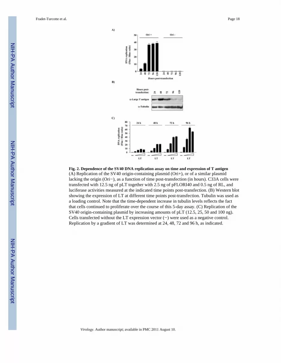
Fig. 2. Dependence of the SV40 DNA replication assay on time and expression of T antigen(A) Replication of the SV40 origin-containing plasmid (Ori+), or of a similar plasmidlacking the origin (Ori−), as a function of time post-transfection (in hours). C33A cells weretransfected with 12.5 ng of pLT together with 2.5 ng of pFLORI40 and 0.5 ng of RL, andluciferase activities measured at the indicated time points post-transfection. (B) Western blotshowing the expression of LT at different time points post-transfection. Tubulin was used asa loading control. Note that the time-dependent increase in tubulin levels reflects the factthat cells continued to proliferate over the course of this 5-day assay. (C) Replication of theSV40 origin-containing plasmid by increasing amounts of pLT (12.5, 25, 50 and 100 ng).Cells transfected without the LT expression vector (−) were used as a negative control.Replication by a gradient of LT was determined at 24, 48, 72 and 96 h, as indicated.
Fradet-Turcotte et al. Page 18
Virology. Author manuscript; available in PMC 2011 August 10.
NIH
-PA Author Manuscript
NIH
-PA Author Manuscript
NIH
-PA Author Manuscript

Fig. 3. Validation of the SV40 DNA replication assay using T antigen mutants(A) Replication activity of mutant T antigens in C33A cells. Each mutant was tested in threedifferent amounts of pLT (2.5, 6.25 and 12.5 ng). Cells transfected without LT expressionvectors (No LT) was used as a negative control. Replication activity is reported as apercentage (%) of the Fluc/Rluc ratio obtained with 12.5 ng of the LT wild-type, which wasset at 100. (B) Western blot showing the expression of the different T antigen mutantproteins. Tubulin was used as a loading control.
Fradet-Turcotte et al. Page 19
Virology. Author manuscript; available in PMC 2011 August 10.
NIH
-PA Author Manuscript
NIH
-PA Author Manuscript
NIH
-PA Author Manuscript

Fig. 4. Principle of the luciferase-based HPV31 DNA replication assay(A) Schematic representation of the three HPV plasmids used in the luciferase assay. Thename of each plasmid is written on the left. The location of the CMV promoter and 3Fepitope are indicated as dark and light grey boxes, respectively. The coding regions offirefly and Renilla luciferase as well as those of codon-optimized (co) E1 and E2 areindicated by white boxes. Amino acid boundaries are indicated below each box. Thelocation (black box) and nucleotide boundaries of the HPV31 origin of replication areindicated with the position of E1 (black) and E2 (grey) binding sites enlarged above. (B)DNA replication activities measured in C33A cells transfected with the indicated amount ofE1 and E2 expression vectors together with 2.5 ng of pFLORI31 and 0.5 ng of pRL. DNAreplication activities are expressed as Fluc/Rluc ratios and were determined at 24, 48, 72, 96and 120 h post-transfection, as indicated. (C) Western blots showing the expression of E1and E2 at different time points post-transfection. Tubulin was used as a loading control.Note that the time-dependent increase in tubulin levels reflects the fact that cells continuedto proliferate over the course of this 5-day assay.
Fradet-Turcotte et al. Page 20
Virology. Author manuscript; available in PMC 2011 August 10.
NIH
-PA Author Manuscript
NIH
-PA Author Manuscript
NIH
-PA Author Manuscript

Fig. 5. Dependence of the HPV31 DNA replication assay on E1 and E2(A) Replication of the HPV31 origin-containing plasmid in C33A cells transfected withincreasing amounts of E1 (white bars) and E2 (black bars) expression vectors (2-foldincrements ranging from 0.01 to 25 ng) as described in the text. Cells transfected without theE1 or E2 expression vector (−) were used as negative controls. Cells transfected without E1and E2 expression vectors (No E1/E2) as well as cells transfected with E1 and E2 but with aplasmid lacking the origin (No ori) were used as controls and are represented as grey bars.(B) and (C) Firefly and Renilla luciferase values (reported as relative light units, RLU) thatwere used to calculate the ratios presented in (A).
Fradet-Turcotte et al. Page 21
Virology. Author manuscript; available in PMC 2011 August 10.
NIH
-PA Author Manuscript
NIH
-PA Author Manuscript
NIH
-PA Author Manuscript
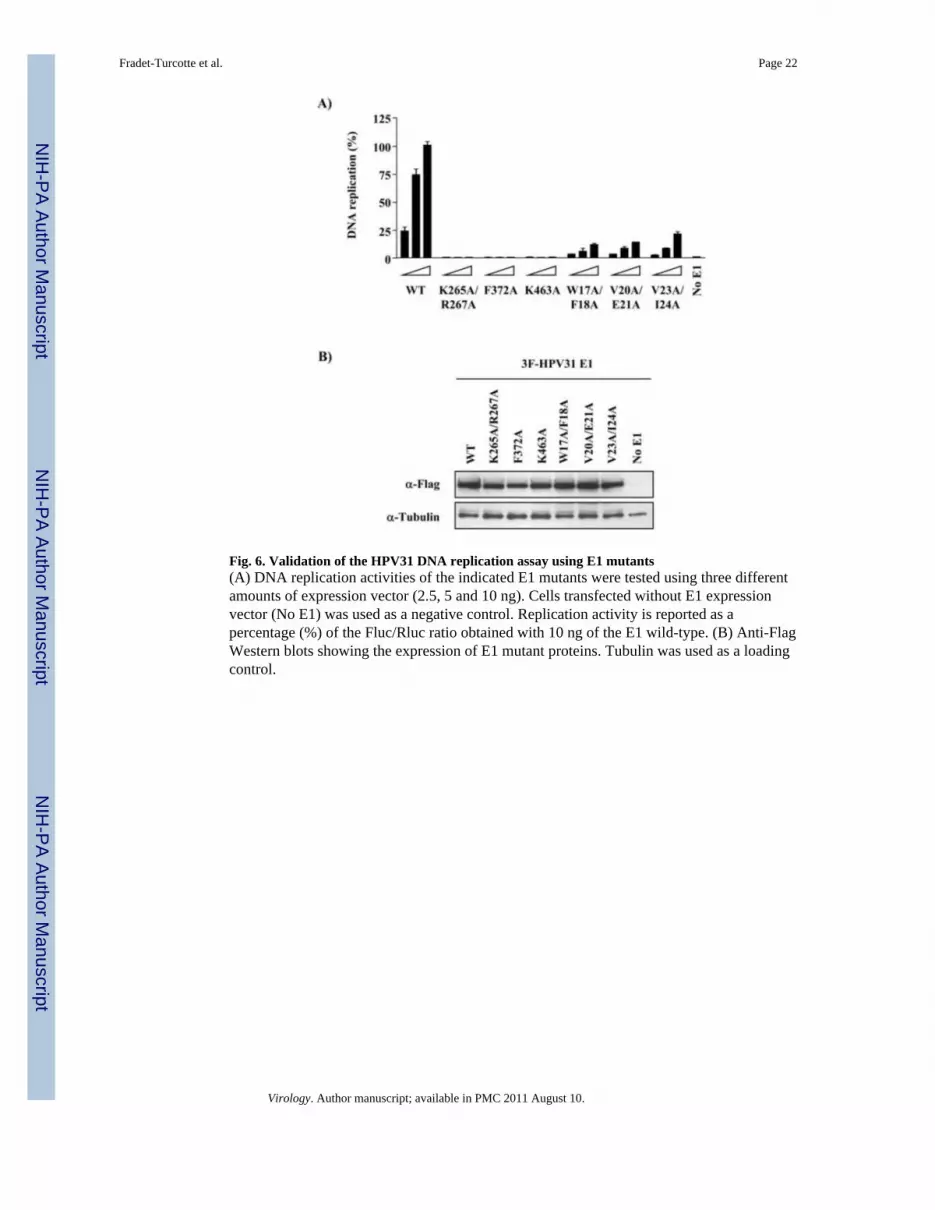
Fig. 6. Validation of the HPV31 DNA replication assay using E1 mutants(A) DNA replication activities of the indicated E1 mutants were tested using three differentamounts of expression vector (2.5, 5 and 10 ng). Cells transfected without E1 expressionvector (No E1) was used as a negative control. Replication activity is reported as apercentage (%) of the Fluc/Rluc ratio obtained with 10 ng of the E1 wild-type. (B) Anti-FlagWestern blots showing the expression of E1 mutant proteins. Tubulin was used as a loadingcontrol.
Fradet-Turcotte et al. Page 22
Virology. Author manuscript; available in PMC 2011 August 10.
NIH
-PA Author Manuscript
NIH
-PA Author Manuscript
NIH
-PA Author Manuscript

Fig. 7. Characterization of a dimerization-defective HPV31 E1 mutant(A) Coomassie-stained SDS-PAGE showing the purified wild-type and G230R E1 OBDs. 3μg of each protein was loaded on the gel. (B-D) Fluorescence polarization DNA bindingassays. Binding isotherms were performed with increasing concentrations of wild-type(filled squares) or G230R (open squares) OBD and 10 nM of fluorescent DNA probe eitherlacking (No E1BS probe (D)) or containing two E1 binding sites spaced by 3 bp (2 E1BSprobe (B)) or 5 bp (2+2 E1BS probe (C)). Only a spacing of 3 bp allows dimerization of theOBD. Each binding isotherm was performed in triplicate. (E) DNA replication activities ofwild-type and G230R E1 were tested using three different amounts of expression vector, asdescribed in the text. (F) Anti-Flag Western blots showing the expression of E1 proteins.Tubulin was used as a loading control.
Fradet-Turcotte et al. Page 23
Virology. Author manuscript; available in PMC 2011 August 10.
NIH
-PA Author Manuscript
NIH
-PA Author Manuscript
NIH
-PA Author Manuscript

Fig. 8. Validation of the HPV31 DNA replication assay using E2 mutants(A) DNA replication activities of the indicated E2 mutants were tested using three differentamounts of expression vector (1, 2.5 and 5 ng). Cells transfected without E2 expressionvector (No E2) was used as a negative control. Replication activity is reported as apercentage (%) of the Fluc/Rluc ratio obtained with 5 ng of E2 wild-type. (B) Anti-FlagWestern blots showing the expression of E2 mutant proteins. Tubulin was used as a loadingcontrol.
Fradet-Turcotte et al. Page 24
Virology. Author manuscript; available in PMC 2011 August 10.
NIH
-PA Author Manuscript
NIH
-PA Author Manuscript
NIH
-PA Author Manuscript

Fig. 9. Correlation between ori-plasmid replication and firefly luciferase expression(A) Levels of SV40 plasmid replication measured by quantitative real-time PCR (qPCR) ordetermined by measuring levels of firefly luciferase activity. C33A cells were transfectedwith the pFLORI40 plasmid and the LT expression vector (+LT). As controls, cells weretransfected without the LT expression vector (No LT) or with a similar plasmid lacking theori (No ori). The amounts of DNA replication measured by qPCR are reported in pg ofDpnI-resistant plasmid per mg of total genomic DNA. Firefly luciferase (Fluc) activity isreported in relative light units (RLU). (B) A similar analysis was performed for HPV31DNA replication with the exception that cells were transfected with the pFLORI31 plasmid,or a similar plasmid lacking the ori (No ori), and with expression vectors for E1 and E2, asindicated.
Fradet-Turcotte et al. Page 25
Virology. Author manuscript; available in PMC 2011 August 10.
NIH
-PA Author Manuscript
NIH
-PA Author Manuscript
NIH
-PA Author Manuscript

Fig. 10. Validation of the SV40 and HPV31 assays using the DNA replication inhibitorgemcitabine(A) Structure of gemcitabine. (B) Replication activity measured in cells treated withincreasing concentrations of gemcitabine (2-fold increments ranging from 1.56 to 50 nM) ortreated with DMSO as a vehicle control (−). Replication activity is reported as a percentage(%) of the Fluc/Rluc ratio obtained in presence of DMSO only. (C) and (D) Firefly andRenilla luciferase values, reported as a percentage of the values obtained withoutgemcitabine (DMSO only), that were used to calculate the replication activity levels shownin (B). (E) Effect of gemcitabine on expression of CMV-Fluc in the absence of plasmidreplication (No replication) is reported as a percentage (%) of the value obtained in absenceof gemcitabine (DMSO only).
Fradet-Turcotte et al. Page 26
Virology. Author manuscript; available in PMC 2011 August 10.
NIH
-PA Author Manuscript
NIH
-PA Author Manuscript
NIH
-PA Author Manuscript

NIH
-PA Author Manuscript
NIH
-PA Author Manuscript
NIH
-PA Author Manuscript
Fradet-Turcotte et al. Page 27
Tabl
e I
Act
ivity
of s
elec
ted
cell-
cycl
e in
hibi
tors
on
vira
l DN
A r
eplic
atio
n
Inhi
bito
rs w
ere
test
ed in
pre
senc
e an
d ab
senc
e of
vira
l DN
A re
plic
atio
n as
des
crib
ed in
the
text
. DN
A p
ol: D
NA
pol
ymer
ases
. RR
: Rib
onuc
leot
ide
redu
ctas
e.
IC50
(μM
)
Inhi
bito
rC
ellu
lar
Tar
get
Cel
l cyc
leph
ase
SV40
HPV
31Fl
ue e
xpre
ssio
n*(N
o re
plic
atio
n)
Gem
cita
bine
DN
A p
ol/R
RS
0.05
0.01
2>
0.05
Hyd
roxy
urea
RR
S10
0025
0>
2000
Mim
osin
eU
nkno
wn
G1/
S50
025
0>
500
Gel
dana
myc
inH
SP90
G1
0.02
50.
05>
0.2
17-A
AG
HSP
90G
10.
50.
5>
2.0
Lact
acys
tinPr
otea
som
eG
1-G
2/M
1.25
1.25
> 10
* Inhi
bito
rs w
ere
test
ed fo
r the
ir ef
fect
on
expr
essi
on o
f the
fire
fly lu
cife
rase
repo
rter g
ene
in a
bsen
ce o
f vira
l DN
A re
plic
atio
n, u
sing
a 4
0-fo
ld e
xces
s of F
luc
ori-p
lasm
id a
s des
crib
ed in
the
text
.
Virology. Author manuscript; available in PMC 2011 August 10.
Recommended


