Embed Size (px)
Citation preview
1
Chapter 17Reproductive
System for Test #3
Copyright © 2016 McGraw-Hill Education. Permission required for reproduction or display.

2
Points to ponder• What are mitosis and meiosis?• How many chromosomes do body cells and sex
cells each have?• Understand the anatomy of both the male and
female.• Know the functions of each structure in the male
and female.• What are the three parts of a sperm?• How do hormones play a role in the male?• Explain the ovarian and uterine cycles.• Be able to discuss the levels of hormones during
the ovarian and uterine cycles. • Where do fertilization and implantation occur?

3
DNA in body and sex cells
Body cells– Each body cell has 46 chromosomes (23 pairs)
within the nucleus. – Cells that have pairs of chromosomes are called
diploid (2n).– 22 pairs of autosomes & a pair of sex
chromosomes – XX or XY
17.1 Human Life Cycle
4
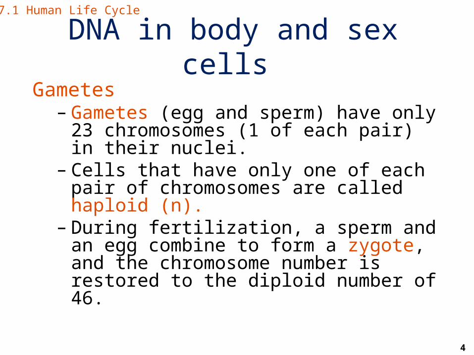
DNA in body and sex cells Gametes
– Gametes (egg and sperm) have only 23 chromosomes (1 of each pair) in their nuclei.
– Cells that have only one of each pair of chromosomes are called haploid (n).
– During fertilization, a sperm and an egg combine to form a zygote, and the chromosome number is restored to the diploid number of 46.
17.1 Human Life Cycle

5
Mitosis and meiosis Review of Mitosis = is
– a type of duplication division in which a cell makes an exact copy of itself.
– a process used for growth and repair of tissues.
– used by body (somatic) cells (cells other than “sex cells” that form gametes in the gonads).
17.1 Human Life Cycle
6
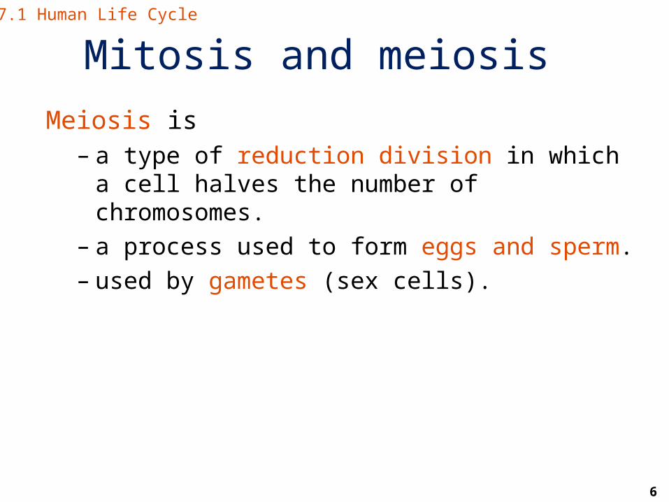
Mitosis and meiosis Meiosis is
– a type of reduction division in which a cell halves the number of chromosomes.
– a process used to form eggs and sperm.– used by gametes (sex cells).
17.1 Human Life Cycle

7
The human life cycle
Figure 17.1 The human life cycle.
17.1 Human Life Cycle
Copyright © The McGraw-Hill Companies, Inc. Permission required for reproduction or display.
2n
zygote
2n = 46diploid (2n)
haploid (n)n = 23
MEIOSIS
n
n
2n
2n
MITOSIS
MITOSIS
2n
eggsperm
FERTILIZATION

8
Male anatomy
1.Scrotum (1)2.Testes (2)3.Epididymides (2)4.Vasa deferentia (2)5.Urethra (1)6.Three glands7.Penis (1)
17.2 Male Reproductive System

9
Male anatomy17.2 Male Reproductive System
Figure 17.2 The male reproductive system.
Copyright © The McGraw-Hill Companies, Inc. Permission required for reproduction or display.
urinary bladder
vas deferens
pubic bone
urethra
penis
glans penis
foreskin
scrotum
seminal vesicleureter (cut)
ejaculatory ductprostate glandbulbourethral gland
anus
epididymis
testis
vas deferens
erectile tissueof penis
urinary bladderureter
prostate gland
seminal vesicle
vas deferens
urethra
bulbourethralgland

10
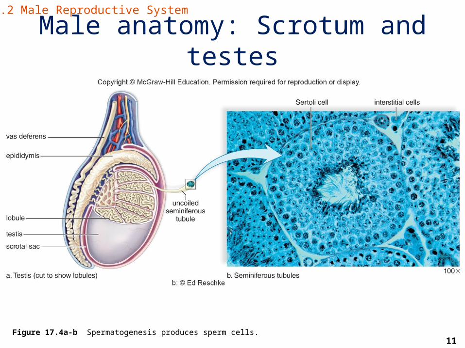
Male anatomy: Scrotum and testes• Scrotum
– Sacs that hold the testes– Help regulate the temperature of the testes
• Testes– Paired organs that produce sperm and male
sex hormones (made by interstitial cells)– Composed of seminiferous tubules where
sperm are being produced
• Epididymis– Sperm mature and are stored here
17.2 Male Reproductive System
11
Male anatomy: Scrotum and testes
Figure 17.4a-b Spermatogenesis produces sperm cells.
17.2 Male Reproductive System

12
Sperm production• Sperm are produced within the seminiferous
tubules of the testes.
• Sertoli cells help nourish sperm and regulate the process of sperm production (spermatogenesis).
• Sperm (spermatozoa) are stored and mature in the epididymis.
17.2 Male Reproductive System
13
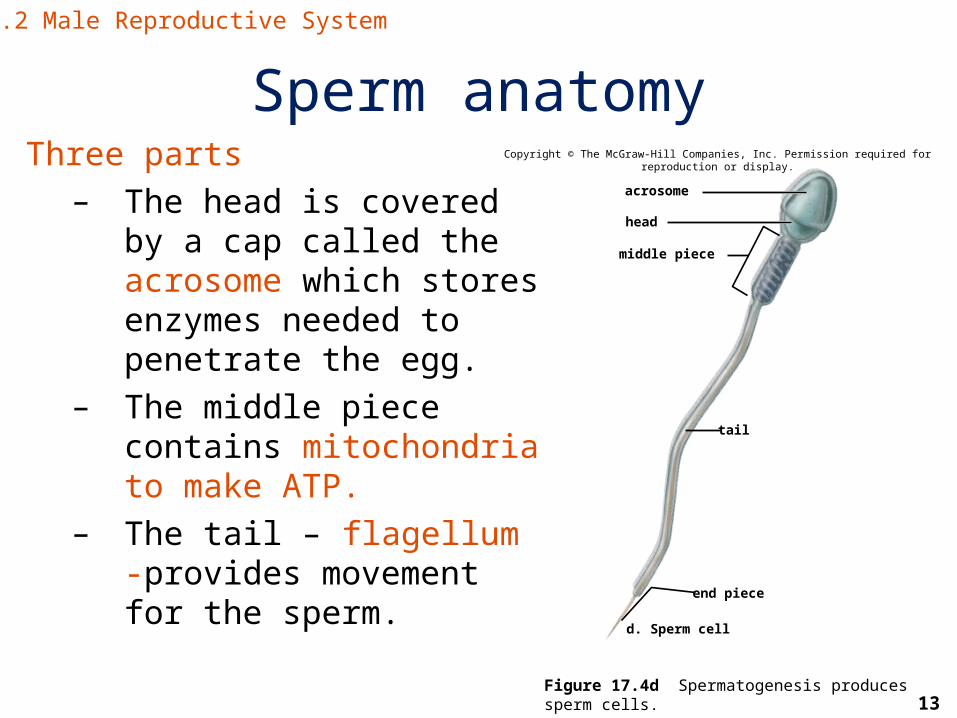
Sperm anatomyThree parts
– The head is covered by a cap called the acrosome which stores enzymes needed to penetrate the egg.
– The middle piece contains mitochondria to make ATP.
– The tail – flagellum -provides movement for the sperm.
Figure 17.4d Spermatogenesis produces sperm cells.
17.2 Male Reproductive System
acrosome
head
middle piece
tail
end piece
d. Sperm cell
Copyright © The McGraw-Hill Companies, Inc. Permission required for reproduction or display.

14
Male anatomy: Vas deferens and urethra
• Vas deferens– Transports sperm to the urethra
• Urethra– Transports sperm out of the body
17.2 Male Reproductive System

15
Male anatomy: 3 glands that contribute to semen
• Seminal vesicles – produce a sugary fluid that provides energy for the sperm
• Prostate gland – produces an alkaline fluid to help buffer the acidic pH of the vagina
• Bulbourethral glands – produce mucus that acts as a lubricant
17.2 Male Reproductive System

16
Male anatomy: Penis• Penis
– This organ is used for sexual intercourse and urination.
Erectile dysfunction (impotency) occurs when the erectile tissue does not expand enough to compress the veins.
• Glans penis
– It is the tip of the penis, usually covered by foreskin, that is intensely sensitive.
– Circumcision is the removal of all or part of the foreskin.
17.2 Male Reproductive System

17
Male anatomy: Penis
Figure 17.3a The structure of the penis.
17.2 Male Reproductive System
erectile tissue foreskinglans penis
a.
skin
connectivelayers
external urethralopening
dorsal veindorsal arterydorsal nerve
Copyright © The McGraw-Hill Companies, Inc. Permission required for reproduction or display.

18
Hormonal regulation in males• Gonadotropin-releasing hormone (GnRH) –
secreted by the thalamus to control release of other hormones
• Follicle-stimulating hormone (FSH) – promotes the production of sperm
• Luteinizing hormone (LH) – controls the production of testosterone
• Testosterone – important for normal development and functioning of the male reproductive organs
17.2 Male Reproductive System

19
Hormonal regulation in malesCopyright © The McGraw-Hill Companies, Inc. Permission required for reproduction or display.
hypothalamus
LHFSH
testis
GnRH
anteriorpituitary
Seminiferoustubulesproducesperm
plus inhibin.
Interstitialcells
producetestosterone.
Figure 17.5 The hormones that control the production of sperm and testosterone by the testes.
17.2 Male Reproductive System

20
Female anatomy
• Genital tract– Ovaries– Oviducts– Uterus– Cervix– Vagina
• External genitals (vulva)– Labia major– Labia minor– Mons pubis– Clitoris
17.3 Female Reproductive System

21
Female anatomy: Genital tract• Ovaries – produce eggs and sex hormones
• Oviducts – move eggs and normal site of fertilization
• Uterus – normal site of implantation and fetal development
• Cervix – opening to the uterus that can dilate during childbirth
• Vagina – birth canal and the copulatory organ of the female
17.3 Female Reproductive System

22
Female anatomy: Genital tract
Figure 17.6 The female reproductive system.
17.3 Female Reproductive System
Copyright © The McGraw-Hill Companies, Inc. Permission required for reproduction or display.
ovary
uterus
urinary bladder
pubic bone
urethra
glans clitoris
labium minora
labium majora
vaginal orifice
fimbriae
cervix
vagina
uterusfimbriae
ovary
rectum
vagina
anus
uterine tube (oviduct)
uterine tube (oviduct)

23
Female anatomy: External anatomy • Labia major – Two large folds of fatty skin
• Labia minor – Two small folds just inside the labia major that contain the openings to the urethra and vagina
• Clitoris – erectile organ and site of intense sexual feeling Essentially what would have become the penis in males
17.3 Female Reproductive System

24
The ovarian cycle: The ovary• An ovary contains many follicles, each containing
an immature egg (oocyte).
• At puberty a female has 300,000-400,000 follicles.
• During the lifetime of a female, only 400 follicles mature .
17.4 The Ovarian Cycle

25
The ovarian cycle: The ovary
• One follicle matures each month from puberty until menopause (end of ovarian and uterine cycles).
• Ovulation is the monthly release of an oocyte from the ovary when a follicle ruptures.– The oocyte remains at the Meiosis II stage until fertilized
by a sperm. It then completes meiosis.
17.4 The Ovarian Cycle

26
Anatomy of the ovary
primaryfollicles1. A primary follicle contains
an oocyte and beginsproducing the sexhormone estrogen.
6. Corpus luteum degenerates.
5. Corpus luteum producesthe sex hormonesprogesterone and someestrogen.
secondaryoocyte
4. Ovulation: The secondary oocyte is released.
3. Vesicular (Graafian) follicle develops.
2. The secondary folliclecontains a secondaryoocyte and produces thesex hormones estrogenand some progesterone.
vesicular (Graafian) folliclesecondaryfollicle
oocyte
corpusluteum
Copyright © The McGraw-Hill Companies, Inc. Permission required for reproduction or display.
© Ed Reschke/Peter Arnold/PhotolibraryFigure 17.8 The ovarian cycle.
17.4 The Ovarian Cycle
27
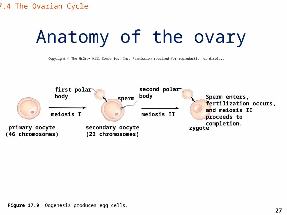
Anatomy of the ovaryCopyright © The McGraw-Hill Companies, Inc. Permission required for reproduction or display.
primary oocyte(46 chromosomes)
secondary oocyte(23 chromosomes)
zygote
Sperm enters,fertilization occurs,and meiosis II proceeds tocompletion.
second polarbodysperm
first polarbody
meiosis I meiosis II
Figure 17.9 Oogenesis produces egg cells.
17.4 The Ovarian Cycle

28
The ovarian cycle
• This is the formation and release of an immature egg.
• It is controlled by GnRH from the hypothalamus.
17.4 The Ovarian Cycle

29
The ovarian cycleTwo phases
- Follicular phase - FSH promotes the development of a follicle
that secretes estrogen.- An estrogen spike leads to a surge in LH and
ovulation around day 14 in the 28-day cycle.- Luteal phase
- LH promotes the development of the corpus luteum that functions to secrete progesterone.
- When successful implantation and pregnancy does not occur, menstruation begins.
17.4 The Ovarian Cycle

30
Hormonal control of the ovariesCopyright © The McGraw-Hill Companies, Inc. Permission required for reproduction or display.
estrogen
hypothalamus
GnRH
FSH
anteriorpituitary
LHfollicle
oocyte
progesterone
corpusluteum
Figure 17.10 The hormones that control the production of estrogen and progesterone by the ovaries.
17.4 The Ovarian Cycle

31
The uterine cycleA 28-day cyclic event in the uterus
• Days 1-5: low levels of estrogen and progesterone cause the inner uterine lining (endometrium) to disintegrate, and menstruation occurs
• Days 6-13 (proliferative phase): increase in estrogen causes the endometrium to thicken
• Day 14: ovulation usually occurs• Days 15-28 (secretory phase): increase in
progesterone causes endometrium to double or triple in thickness in preparation for the developing embryo; if the egg is not fertilized then the corpus luteum regresses and the endometrium breaks down
17.4 The Ovarian Cycle

32
Hormones in the ovarian and uterine phasesCopyright © The McGraw-Hill Companies, Inc. Permission required for reproduction or display.
1 3 5 7 9
1 3 5 7 9
ovulationovarian cycle
developing follicle
LH
mature follicle earlycorpus luteum
regressingcorpus luteum
DaysFollicular Phase Ovulation Luteal Phase
ovulationestrogen
Uterine cycle
progesterone
Secretory PhaseProliferative PhaseMenstruation
menstruation
15 17 19 21 23 25 27 111 13
11 13 15 17 19 21 23 25 27 30Days
FSHEn
dom
etriu
mH
orm
one
Leve
lsO
varia
nEv
ents
Hor
mon
eLe
vels
Figure 17.11 The effects of estrogen and progesterone on the endometrium during the uterine cycle.
17.4 The Ovarian Cycle

33
Fertilization and pregnancy• Fertilization – union of a sperm and egg
nucleus to form a zygote– Normally occurs in an oviduct
• Pregnancy – begins with implantation– Usually six days after fertilization
17.4 The Ovarian Cycle

34
Some common birth control methods• Abstinence – not engaging in sexual intercourseAlso includes the rhythm method
• Hormonal control• Birth control pills block FSH and LH release to
stop follicular development and ovulation.• Contraceptive injections of hormones
(progesterone and/or estrogen) stop ovulation.• Contraceptive implants use synthetic
progesterone to prevent ovulation.
Note: Abstinence and the use of condoms are the only methods that protect against STDs.
17.5 Control of Reproduction
35
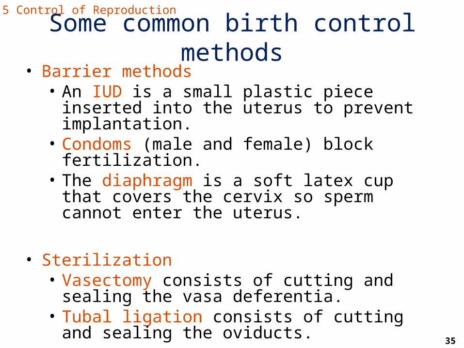
• Barrier methods• An IUD is a small plastic piece inserted into the
uterus to prevent implantation.• Condoms (male and female) block fertilization.• The diaphragm is a soft latex cup that covers
the cervix so sperm cannot enter the uterus.
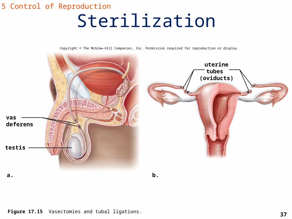
• Sterilization• Vasectomy consists of cutting and sealing the
vasa deferentia.• Tubal ligation consists of cutting and sealing
the oviducts.
17.5 Control of Reproduction
Some common birth control methods

36
Some common birth control methods
Figure 17.14 Placement of birth control devices.
17.5 Control of Reproduction
a. Intrauterine device placement Intrauterine devices
uterus
cervix
femalecondom
Female condomb. Female condom placement
c. Male condom placement Male condom
Copyright © The McGraw-Hill Companies, Inc. Permission required for reproduction or display.
a: © Saturn Stills/Photo Researchers; b: © Keith Brofsky/Getty RF; c: © The McGraw-Hill Companies, Inc. Lars A. Niki, photographer
37
SterilizationCopyright © The McGraw-Hill Companies, Inc. Permission required for reproduction or display.
uterine tubes (oviducts)
testis
a. b.
vasdeferens
Figure 17.15 Vasectomies and tubal ligations.
17.5 Control of Reproduction

38
Sexually transmitted diseases (STDs)
• Viral diseases cannot be treated with antibiotics, but there are some antivirals.– HIV– Genital warts– Genital herpes– Hepatitis
• Bacterial diseases can be treated with antibiotics.– Chlamydia– Gonorrhea– Syphilis
17.6 Sexually Transmitted Diseases

39
STDs: HIV/AIDS• Viral: HIV causes AIDS, the last stage of an HIV
infection, in which the helper T cell count is low and the immune system is compromised leading to a suite of opportunistic infections.
• There is no cure, but there is a treatment called highly active antiretroviral therapy (HAART) that can limit HIV replication.
17.6 Sexually Transmitted Diseases

40
STDs: Genital warts• Caused by human papillomaviruses (HPVs)• Transmitted through skin to skin contact• Can be transmitted without symptoms• Most people do not have symptoms, but for the few
that do, most of them see warts, and flat lesions, most often on the penis or opening to the vagina
• Associated with cervical cancer (up to 90% of cases)• Most often found through regular pap smear
screenings in women• Can be transmitted to a baby during birth• HPV vaccine was developed to help prevent cervical
cancer
17.6 Sexually Transmitted Diseases

41
STDs: Herpes• Caused by herpes simplex virus (HSV)• Two types
– Type 1 is usually found above the waist– Type 2 is usually found below the waist
(genital herpes)• Transmitted through skin to skin contact and
secretions• Can be transmitted when there are no
symptoms
17.6 Sexually Transmitted Diseases

42
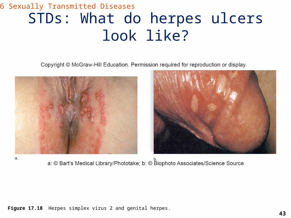
STDs: Herpes• Usually have painful ulcers periodically in the
same place(s) each time• Outbreaks occur when the virus is reactivated
by stress, sunlight, fever, lack of sleep, etc.• No cure, but there are drugs that can keep
outbreaks to a minimum or stop them entirely • Can be transmitted to a baby during birth
17.6 Sexually Transmitted Diseases
43
STDs: What do herpes ulcers look like?
Figure 17.18 Herpes simplex virus 2 and genital herpes.
17.6 Sexually Transmitted Diseases
44
STDs: Hepatitis• An infection of the liver by one of 6 viruses
(Hepatitis A,B,C,D,E,G)
• Hepatitis B: most common sexually transmitted hepatitis
• Transmitted through sexual contact and by contaminated blood
• Hepatitis B can lead to liver failure
• Vaccine available for both Hepatitis A and B
17.6 Sexually Transmitted Diseases

45
STDs: Chlamydia• Very common bacterial infection in men and women
• Some men and most women do not have symptoms (18-21 days after exposure) but if they do:– Male symptoms: burning during urination and a
mucoid discharge– Female symptoms: vaginal discharge and symptoms
of a UTI
17.6 Sexually Transmitted Diseases
46
STDs: Chlamydia• If the infection reaches the uterus, oviducts,
and ovaries, it can lead to pelvic inflammatory disease (PID) and sterility
• Can be transmitted to a baby during birth (causes inflammation of the eyes and/or pneumonia)
17.6 Sexually Transmitted Diseases

47
STDs: Gonorrhea• Also a common bacterial infection in men and women• Male symptoms (3-5 days after contact): pain during
urination and a thick, greenish yellow penile discharge• Female symptoms: uncommon to have vaginal discharge• Can also lead to PID (pelvic inflammatory disease) and
sterility in men and women• Resistance to antibiotics is common in this bacterium• Can be contracted by a baby during birth, leading to an
eye infection and even blindness
17.6 Sexually Transmitted Diseases

48
STDs: Syphilis• A bacterial infection with three stages
– Stage 1 (1-8 weeks after infection): hard chancre at the site of infection
– Stage 2 (3-4 months after infection): nonitchy rash, even found on the soles of feet and the palms of the hands; may also see hair loss and gray patches on mucous membranes
– Stage 3 (5-20 years after infection and continues until death): affects the cardiovascular and/or nervous systems: gummas (large ulcers) can develop on the skin and internal organs
17.6 Sexually Transmitted Diseases

49
STDs: Syphilis• These stages are separated by latent periods
that make the stages hard to link together and the disease hard to diagnose
• Syphilis can cross the placenta and affect the fetus
17.6 Sexually Transmitted Diseases

50
Treponema pallidum, the syphilis bacterium
Figure 17.20 Syphilis.
17.6 Sexually Transmitted Diseases

51
Other common infections of the reproductive tract
• Bacterial vaginosis (BV): – Accounts for 50% of vaginitis in American women– Caused by a disruption of the normal flora in the
vagina leading to an overgrowth of certain bacteria
• Trichomoniasis– Caused by a protozoan (Trichomonas vaginalis)– Can cause a frothy discharge, with a foul smell
and itching– Common cause of vaginitis
17.6 Sexually Transmitted Diseases
52
• Candidiasis – Overgrowth of normal yeast = a fungus
(Candida albicans) in the vagina– Characterized by tissue that is red, inflamed
and itchy; sometimes accompanied by a white, curdy discharge
– Birth control hormones and use of antibiotics make women more prone to this overgrowth
17.6 Sexually Transmitted Diseases
Other common infections of the reproductive tract

53
Preventing transmission of STDs
• Abstain from sexual intercourse.
• Develop long-term monogamous relationships.
• Be aware if your partner is an intravenous drug user because STD prevalence is higher in this group.
• Avoid anal-rectal intercourse.
17.6 Sexually Transmitted Diseases

54
• Use caution with uncircumcised males as they are more likely to be infected.
• Practice safer sex.– Always use a latex condom during intercourse.– Avoid oral sex.– Limit or do not use alcohol and drugs that can
impair your judgment or change your behavior.
17.6 Sexually Transmitted Diseases
Preventing transmission of STDs