Embed Size (px)
Citation preview
HUMAN DIGESTIVE SYSTEM
STRUCTURE AND FUNCTION
Group members
Rameen nadeem (25)
Syed iqra hussain (5)
Hina zamir (4)

OVERVIEW
Major organs Mouth Esophagus Stomach small intestine large intestineAcessory organs: Liver gall bladder Pancreas.

HUMAN DIGESTIVE SYSTEM
Major organs Mouth Esophagus Stomach small intestine large intestine.
Acessory organs: Liver Gall bladder Pancreas.
The process of reducing food into smaller molecules that can be absorbed into the body
Digestive system consists of 2 major parts

MAJOR ORGANSTHE MOUTHpH: 7 The first part of the digestive system the entry point of food.
Structures in the mouth that aids digestion
Teeth – cut, tear, crush and grind food.
Salivary glands – produce and secrete saliva into the oral cavity.
saliva moistens the food contains enzymes (ptyalin or salivary amylase) begins digestion of starch into smaller polysaccharides.
Function: Mechanical digestion. increasing surface area for faster chemical digestion.

The Esophagus a tube connecting the mouth to the stomach running through the Thoracic cavity. Location: lies behind windpipe (Trachea).o The trachea has as an epiglottis o preventing food from entering the windpipe,o moving the food to the esophagus while swallowing.

Food travels down the esophagus, through a series of involuntary rhythmic contractions (wave-like) called peristalsis.
Function:• The lining of the
esophagus secretes mucus
• lubricating• to support the
movement of food.

ESOPHAGEAL SPHINCTER:
• bolus reaches the stomach
• must pass through a muscular ringed valve called the esophageal sphincter (Cardiac Sphincter).
• Function:• prevent stomach
acids from back flowing into the esophagus.

STOMACH J-shaped muscular sac Has inner folds (rugae) Increasing surface area of the stomach.
Function: Stomach performs mechanical digestion HOW
By churning the bolus and mixing it with the gastric juices
secreted by the lining of the stomach. GASTRIC JUICES
HCl, salts, enzymes, water and mucus) HCL helps break down of food and kills bacteria that
came along with the food. The bolus is now called Chyme.
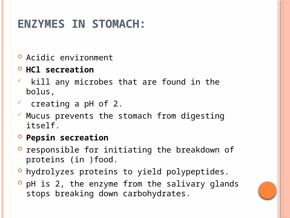
ENZYMES IN STOMACH:
Acidic environment HCl secreation kill any microbes that are found in the bolus, creating a pH of 2. Mucus prevents the stomach from digesting itself. Pepsin secreation responsible for initiating the breakdown of proteins
(in )food. hydrolyzes proteins to yield polypeptides. pH is 2, the enzyme from the salivary glands stops
breaking down carbohydrates.
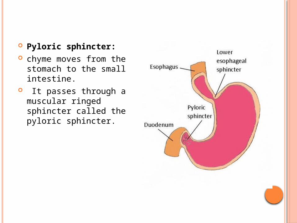
Pyloric sphincter: chyme moves from the
stomach to the small intestine.
It passes through a muscular ringed sphincter called the pyloric sphincter.

STOMACH DOES NOT DIGEST ITSELFWHY ? Protective Mechanism: three protective mechanisms.
1. First the stomach only secretes small amounts of gastric juices until food is present.
2. Second the secretion of mucus coats the lining of the stomach protecting it from the gastric juices.
3. The third mechanism is the digestive enzyme pepsin is secreted in an inactive protein called pepsinogen. Pepsinogen is converted to pepsin in the increased presence of hydrochloric acid (pH 1).

SMALL INTESTINE
responsible for the complete digestion of all macromolecules
and the absorption of their component molecules E.g
glucose Glycerol fatty acids amino acids nucleotides
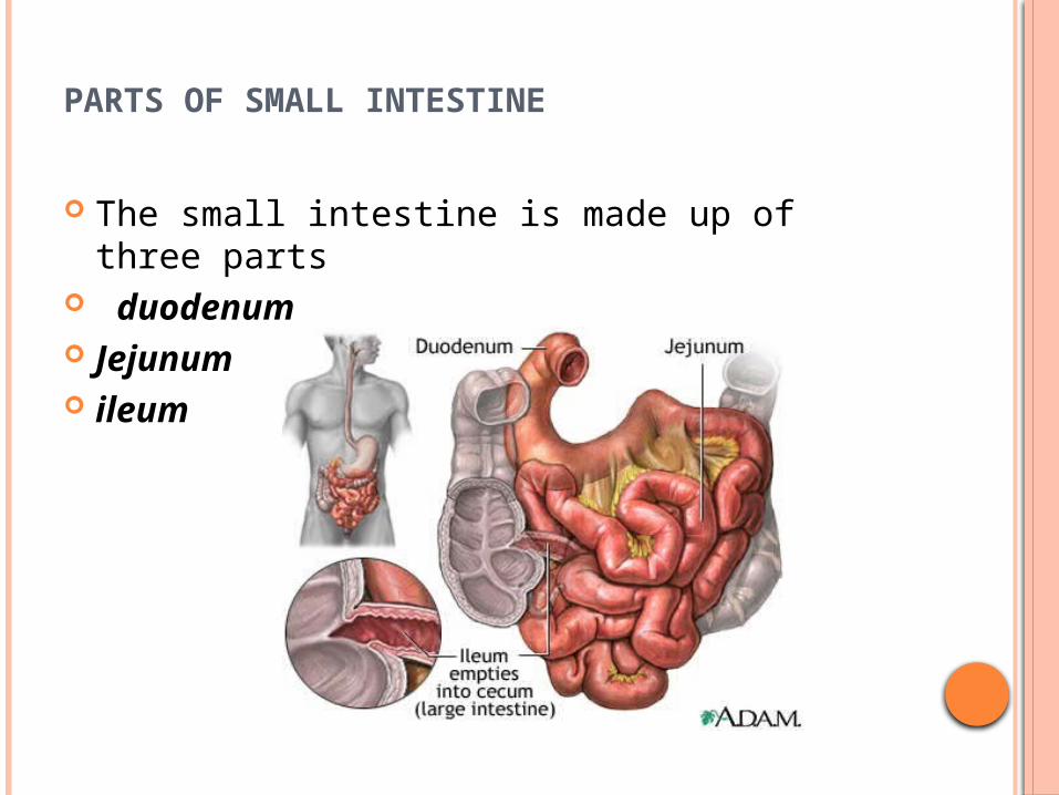
PARTS OF SMALL INTESTINE
The small intestine is made up of three parts duodenum Jejunum ileum

1.DUODENUM
The first part is the duodenum, u-shaped organ.
approximately 30 cm in length. This area completes most of the digestion
processes. Enzymes are secreted into the duodenum
form the pancreas and the gall bladder. The duodenum is lined by folds of tissue called villi.
The villi are covered by fine brush-like microvilli.
These folds increase the surface area of the small intestine increase the rate of absorption.
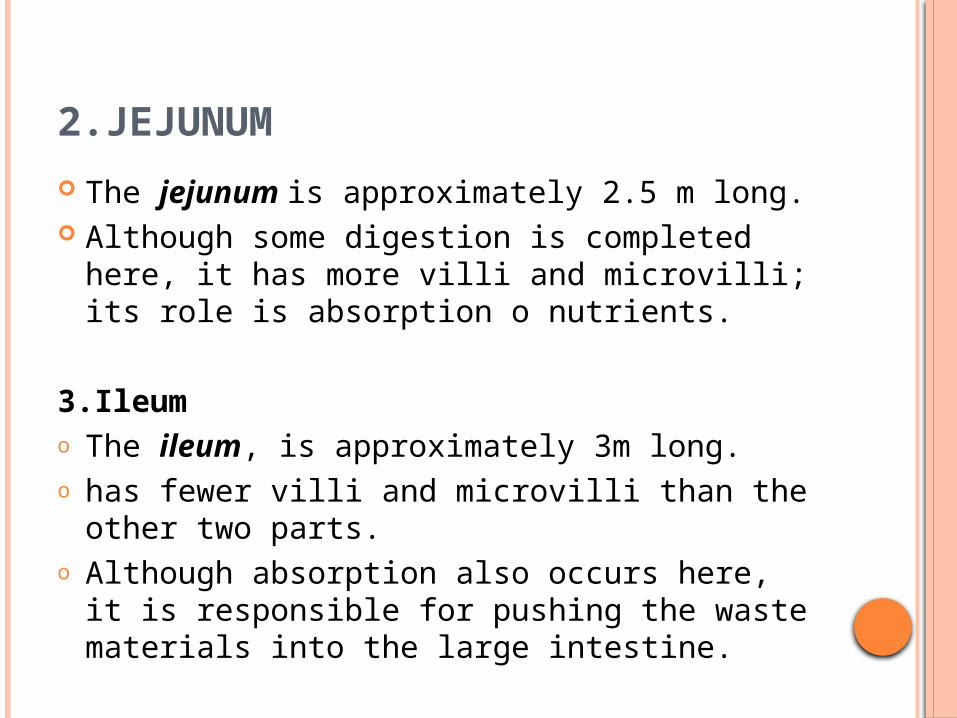
2.JEJUNUM
The jejunum is approximately 2.5 m long. Although some digestion is completed here,
it has more villi and microvilli; its role is absorption o nutrients.
3.Ileumo The ileum, is approximately 3m long.o has fewer villi and microvilli than the other
two parts. o Although absorption also occurs here, it is
responsible for pushing the waste materials into the large intestine.


FUNCTIONS OF THE SMALL INTESTINE
90% of the digestion and absorption of food occurs
other 10% taking place in the stomach and large intestine.
The main function of the small intestine is absorption of nutrients and minerals from food.
Digestion of proteins Proteins, peptides and amino acids are acted
upon by enzymes such as trypsin and chymotrypsin, secreted by the pancreas. This breaks them down to smaller peptides.
DIGESTION OF LIPIDS
Enzymes, like lipases secreted from the pancreas, act on fats and lipids in diet.
lipase can break them into the smaller parts that can enter the intestinal villi for absorption.
Digestion of carbohydrates Carbohydrates are broken down to simple sugars
and monosaccharides like glucose. Pancreatic amylase breaks down some
carbohydrates to oligosaccharides as well. Some carbohydrates and fibers pass undigested to
the large intestine where they may, depending on their type, be broken-down by intestinal bacteria.

ABSORPTION IN THE SMALL INTESTINES
the nutrients are absorbed by the inner walls of the small intestine into the blood stream.
The nutrients are absorbed by processes of simple/passive diffusion, facilitated diffusion, primary active transport, or secondary active transport.
For transport, nutrients commonly rely upon Lipids – undergo passive or simple diffusion Short-chain fatty acids – diffusion Amino acids – primary active transport Glucose – secondary active transport Fructose – facilitated diffusion

ABSORPTION IN THE SMALL INTESTINES
Other absorbed substances in the small intestines include:
1.Water 80% is absorbed by the small intestine 10% by the large intestine remaining 10% excreted in the faeces.2.Electrolytes3.Vitamins and minerals

LARGE INTESTINE (parts & function )

COMPONENTS OF LARGE INTESTINE
The large intestine is composed of several very distinctive parts:
Cecum: Colon:. The colon consists of four parts: Ascending colon Transverse colon Descending colon Sigmoid colon Rectum

CECUM first section of your large intestine looks like a pouch, two inches long. ROLE taking in digested liquid
from the ileum(small intestine) & passes it on to the colon.

COLON :
major section of the large intestine Function: the principal place for water reabsorption, absorbs salts when needed. Components : The colon consists of 4 parts: Ascending colon Transverse colon Descending colon Sigmoid colon

COMPONENTS OF COLON
Ascending colon: 1st portion of the colon pushes any undigested debris up
from the cecum just under the right lower end of the
liver. Transverse colon: 2nd portion of the colon Food traveling from left to right just
under your stomach.

COMPONENTS OF COLON
Descending colon: 3rd portion of colon pushes its contents from
down to the lower left side of your abdomen
Sigmoid colon: final S-shaped length of the
colon, empties into the rectum.

RECTUM
The final section measures from 1 to 1.6 inches (or 2.5 to 4 cm). Leftover waste collects there expanding the rectum emptied through anus

FUNCTION OF LARGE INTESTINE 1. Absorb Water One of the primary functions is to absorb water prepare the waste as a solid stool that will be
expelled from the body. 2. Absorb Vitamin beneficial bacteria role in breaking down undigested sugars and fibers
into fatty acids. produce many vitamins, of which are Vitamin K and
Biotin that are absorbed back into the body.

FUNCTION OF LARGE INTESTINE(LI)
3. Reduce Acidity The fatty acids cause acidic environment. The LI produces alkaline solutions reduce the acidity and balance the pH in the LI
4.Protect from InfectionsThe mucous lining of the large intestine acts as a protective layer prevents harmful bacteria from being reabsorbed into the body.

FUNCTION OF LARGE INTESTINE(LI)
5. Produce Antibodiesalso produces antibodies help to boost immunity. It is believed that the appendix may have been a major producer of antibodies at some point in the evolutionary process

THE ACCESSORY ORGANS:
support the digestive system BUT are not part of the digestive tract
These organs secrete fluids into the digestive tract, and are connect by ducts.
The accessory organs include liver gall bladder pancreas.

1.LIVER
largest of these organs mass of about 1.5 kg. liver produces bile bile
greenish yellow pigment
made up bile pigments and bile salts
it breaksdown old red blood cells.

2.GALL BLADDER
a storage sac. The bile is secreted into it The bile is stored here. HOW IT WORKS food containing fat enters
the digestive tract salts are secreted into
the small intestine to digest fats.
The bile emulsifies fats in partly digested food
thereby assisting their absorption

3.PANCREAS
The pancreas secretes a number of different enzymes into the small intestine]
Role is to digest carbohydrates lipids & proteins completely.
It also secretes bicarbonate ions Role :
neutralize the HCl from the stomach change the pH of the small intestine to apH of 8.
The pancreas will secrete about 1.0 L. of pancreatic fluids per day.

HUMAN DIGESTIVE SYSTEM
STRUCTURE AND FUNCTION
Group members
Rameen nadeem (25)
Syed iqra hussain (5)
Hina zamir (4)