Embed Size (px)
Citation preview

Nanomed. J., 2(4): 231-248, Autumn 2015
231
Functionalization of carbon nanotubes and its application innanomedicine: A review
1, 2*H. Sadegh; 1,2R. Shahryari-ghoshekandi
1Department of Chemistry, Science and Research Branch, Islamic Azad University, Tehran, Iran
2Young Researchers and Elite Club, Science and Research Branch, Islamic Azad University, Tehran, Iran
ABSTRACT:This review focuses on the latest developments in applications of carbon nanotubes (CNTs) in medicine. A brief historyof CNTs and a general introduction to the field are presented.Then, surface modification of CNTs that makes them ideal for use in medical applications is highlighted. Examples ofcommon applications, including cell penetration, drug delivery, gene delivery and imaging, are given. At the same time,there are concerns about their possible adverse effects on human health, since there is evidence that exposure to CNTsinduces toxic effects in experimental models. However, CNTs are not a single substance but a growing family of differentmaterials possibly eliciting different biological responses. As a consequence, the hazards associated with the exposureof humans to the different forms of CNTs may be different. Understanding the structure–toxicity relationships wouldhelp towards the assessment of the risk related to these materials. Finally, toxicity of CNTs, are discussed. This reviewarticle overviews the most recent applications of CNTs in Nanomedicine, covering the period from 1991 to early 2015.
Keywords: Carbon nanotubes, Drug delivery, Functionalization, Nanomedicine, Surface modification
Nanomed. J., 2(4): 231-248, Autumn 2015DOI: 10.7508/nmj. 2015.04.001
*Corresponding Author Email: [email protected]: (+98) 21-65933439
Note. This manuscript was submitted on July 13, 2015;approved on August 21, 2015
Received; 13 July 2015 Accepted; 2 August 2015
INTRODUCTIONSince carbon nanotubes (CNTs) were discovered
by Iijima in 1991(1), they have become the subject ofmany studies because of their unique electrical, optical,thermal, and mechanical properties (2-6). CNTs can bevisualized as a sheet of carbon atoms rolled up into atube with a diameter of around tens of nanometers.There are two main types of CNTs, single-walled(SWCNTs) and multi-walled carbon nanotubes(MWCNTs), the latter being formed by severalconcentric layers of rolled graphite (Fig 1). In particular,SWCNTs are characterized by a high aspect ratio. Inthe last decade, CNTs are intensively explored for invitro and in vivo delivery of therapeutics, which wasinspired by an important finding that CNTs can
penetrate cells by themselves without apparentcytotoxic effect to the cells (7-9). The high aspect ratiomakes CNTs outstanding from other types of roundnanoparticles in that the needle-like CNTs allow loadinglarge quantities of payloads along the longitude oftubes without affecting their cell penetration capability.With the adequate loading capacity, the CNTs can carrymultifunctional therapeutics, including drugs, genesand targeting molecules, into one cell to exert multi-valence effects. In the other side, owing to the ultrahighsurface area along with the strong mechanicalproperties and electrically conductive nature, CNTs areexcellent material for nano-scaffolds and threedimensional nano-composites. In recent year, CNT-based devices have been successfully utilized in tissueengineering and stem cell based therapeuticapplications, including myocardial therapy, boneformation, muscle and neuronal regeneration.Furthermore, owing to the distinct optical properties

Nanomed. J., 2(4): 231-248, Autumn 2015
232
H.R. Sadegh et al.
of CNTs such as high absorption in the near-infrared(NIR) range, photoluminescence, and strong Ramanshift (10), CNTs are excellent agents for biologydetection and imaging. Combined with high surfacearea of CNTs for attaching molecular recognitionmolecules, CNT-based, targeted nano-devices havebeen developed for selective imaging and sensing.There are many areas where CNTs are extremely useful.This review article describes strategies for preparationof CNTs for their use in medicine. Specifically, wefocused and highlighted the important nanomedicineapplications of CNTs in the field of cell penetration,drug delivery, gene delivery and imaging.
Fig. 1. Schematics of a SWCNTs and MWCNTs
CNTs modificationsRaw CNTs, persisting metallic nature, are highly
hydrophobic. Therefore, surfaFce modification ofCNTs, or CNTs functionalization so that they could bedispersed in aqueous solutions becomes a key stepfor their medical applications. The CNTs modificationmethods involved either non-covalent or covalentstrategies. The non-covalent modification utilizes thehydrophobic nature of CNTs, especially, π -πinteractions for coating of amphiphilic molecules. Thecovalent modification generates chemical bonds oncarbon atoms on CNTs surface via chemical reactionsfollowed by further conjugation of hydrophilic organicmolecules or polymers rendering CNTs bettersolubility. These modifications not only offer CNTswater solubility, but also produce functional moietiesthat enable linking of therapeutic agents, such as genes,drugs, and recognition molecules for medicalapplications.
Non-covalent functionalization of CNTsThe methods of functionalization of CNTs based
on non-covalent interaction can be performed without
destroying the intrinsic sp2-hybridized structure of thenanotube sidewall, so that the original electronicstructure and properties of CNTs can be preserved.Different kinds of non-covalent functionalizations havebeen explored. The non-covalent functionalization ofCNTs could be achieved by π–π stacking interactionsbetween conjugated molecules and the graphiticsidewall of CNTs. Compounds with the pyrene moiety,e.g. N-succinimidyl-1-pyrenebutanoate, could beadsorbed irreversibly onto the surface of SWCNTsthrough π–π interaction (11, 12). Furthermore, via thesuccinimidyl ester group on this bifunctional anchor,various molecules with primary or secondary aminessuch as ferritin, streptavidin (SA) or biotin–polyethylene oxide-amine were covalently attached tothe activated pyrene by nucleophilic attack of theamino group (11). Using a similar strategy, some otherbiomolecules (e.g. protein and DNA) (13-15) and goldnanoparticles (16-17) were also coupled to CNTs.Through the π–π stacking interactions, other classesof molecules, including phthalocyanines (18-20),porphyrins (21-31) and their derivatives have beenimmobilized onto the surface of CNTs. It wasdemonstrated that this kind of composite could displayphoto induced charge transfer from the dye moleculesto CNTs (20, 23). The selective interaction between theporphyrin derivative and CNTs was also discovered(34). The use of Raman spectroscopy, near-infrared(NIR) absorption and buck conductivity has shownthat porphyrin derivatives could be selectivelyadsorbed onto semiconducting nanotubes in asolubilized sample. This is a convenient method toseparate the semi-conductive CNTs from metallic CNTs.Polymers with conjugated structures could also becoupled to CNTs by π–π forces (32-54). The stronginteraction between the conjugated polymer and thenanotube remarkably increased CNTs dispersibilty (32,40, 45-47). Some conjugated polymers have been usedto purify CNTs by efficient phase separation from themain impurity, consisting of amorphous graphitic shells(41, 43, 44). In this way, amorphous carbon impuritiestend to sediment out of solution, whereas thenanotubes stay in suspension. In addition, this kind ofcomposite has potential applications in theoptoelectronic field because of energy transfer fromthe excited conjugated polymer to the nanotubes (33-39, 42, 45, 48-54). Another method of non-covalentfunctionalization of CNTs involves association of CNTswith amphiphilic molecules through hydrophobicinteractions in aqueous media. In this system, theamphiphilic molecules interact non-covalently with thearomatic surface of CNTs using their hydrophobic

Nanomed. J., 2(4): 231-248, Autumn 2015
233
parts, while exposing the hydrophilic parts to theaqueous solution to minimize the hydrophobic interfacebetween the nanotubes and their polar environment.Through hydrophobic interactions, water-solublepolymers poly vinyl pyrrolidone (PVP) and poly styrenesulfonate (PSS) wrapped helically around the surfaceof SWCNTs helped to dissolve the nanotubes in water(55). The association between the polymer andSWCNTs, which is reversible by changing the solventsystem, was very robust, independent of the presenceof excess of polymer in solution and uniform along thesurface of the nanotubes. Surfactants were alsoconjugated to CNTs via hydrophobic interactions. Sofar, various anionic, nonionic and cationic surfactantshave been used to suspend CNTs in aqueous medium(56-60). Among them, bile salt surfactants such asdeoxycholic acid (DOC) and taurodeoxycholic acid(TDOC) have been found to be very efficient insolubilizing SWCNTs. This is because the aggregationwas further prevented by the coulomb repulsionbetween surfactant-coated SWCNTs (60). In addition,if aromatic moieties in the hydrophobic part of theamphiphilic molecule were present, particularly stronginteractions between the molecule and the CNTs couldbe established. This kind of aggregates, formsadditional π–π stacking between the aromatic part ofthe molecule and the graphitic sidewalls of CNTs whileexposing the hydrophilic groups to the aqueoussolution. As an example, sodium dodecyl benzenesulfonate (SDBS) presented an increased ability insuspending CNTs in water (59). Some of the surfactantsincluding sodium dodecyl sulfate (SDS), SDBS, DOCor TDOC were found to be very effective in dispersingSWCNTs (58-60). This provided the possibility to studythe properties of individual SWCNTs and furthermanipulate single tubes (61-65). Hydrophobicinteractions were also used to couple biomolecules toCNTs. In particular, an amphiphilic peptide waswrapped around the SWCNT surface. The peptide wasable to adopt α-helical structure in aqueous solution,exposing the hydrophobic face of the helix to thenanotube surface (66, 67). Atomic force microscopy(AFM), transmission electronic microscopy (TEM), andabsorption and Raman spectroscopy proved that theSWCNTs were DE bundled by the peptide. The peptide-coated nanotubes were assembled into macromolecularstructure through charged peptide-peptide interactionsbetween adjacent peptide-wrapped nanotubes. Thesize and morphology of the super-molecular structurecan be regulated by controlling the factors that affectpeptide-peptide interactions. It was also demonstrated
that the ability to disperse individual SWCNTsincreased with increasing the amount of aromaticresidues inside the peptide sequence (68). In a similarmanner, different proteins were adsorbed onto thesurface of SWCNTs and were taken up by themammalian cells (69).Another interesting approach involved the conjugationof single-stranded DNA with SWCNTs resulting inhelical wrapping around the surface of the tubeexposing the hydrophilic sugar-phosphate backbonetowards the exterior (70, 71). Using this strategy,individually dispersed nanotubes in solution wereobtained. CNTs wrapping by single-stranded DNA wasstrongly dependent on the DNA sequence. Moreover,the dependence of the electrostatic properties of theDNA-CNT hybrid on the tube diameter and electronicproperties enable nanotube separation by anionexchange chromatography. In addition to a directinteraction with CNTs, biomolecules have also beenattached to SWCNTs via amphiphilic molecules as abifunctional linker. For instance, phospholipid (PL)molecules with phospholipid–poly(ethylene glycol)(PEG) chains and terminal amine (PL-PEG-NH
2) were
non-covalently conjugated to SWCNTs. PL alkylchains were adsorbed to the surface of SWCNTs viahydrophobic interactions while the PEG chain extendedinto the aqueous phase for SWCNTs dispersion. Afterincorporation of a hetero bifunctional cross-linkedsulfosuccinimidyl 6-(30-[2-pyridyldithio] prop-ionamido) hexanoate (sulfo-LC-SPDP), thefunctionalized SWCNTs (f -SWCNTs) were coupledwith DNA and RNA via disulfide linkage. The complexeswere subsequently delivered to cells (72).
CNTs covalent functionalizationThe CNTs characteristics such as low curvature,
low solubility and bundling tendency render carbonnanomaterials a relatively unreactive to chemicaltreatment. Thus far, a number of ways have beenintroduced for covalent functionalization of CNTs. Withrespect to the location of the functional groups, thestrategies to covalently functionalize CNTs can beclassified into two main types including defect andsidewall functionalizations. The covalentfunctionalization of nanotubes is more robust and isbetter controlled compared to functionalization basedon non-covalent methods.CNTs synthesized by any available method are notfree of defects. The intrinsic defects include five- orseven-membered rings called Stone–Wales defects,sp3- hybridized defects and vacancies in the sp2-

Nanomed. J., 2(4): 231-248, Autumn 2015
234
CNTs in Nanomedicine: A reveiw
hybridized six-membered ring carbon structure of thesidewall (94, 95). A limited number of defects can betolerated by CNTs without losing their macroscopicelectronic and mechanical properties (3, 75-77). Inaddition, the tips of the tubes, forming a hemisphericalfullerene, have stronger reactivity than the sidewallsbecause of their higher curvature (78, 79). When treatedwith strong oxidizing agents such as nitric acid,KMnO
4/H
2SO
4, O
2, K
2Cr
2O
7/H
2SO
4or OsO
4, CNTs could
be opened and cut into short tubes. In the meantime,defects were introduced around the CNT surface (74,79, 80). Moreover, oxygenated functional groups suchas carboxylic acid, ketone, alcohol and ester groupswere generated by the oxidization process to the endsand the defect sites of the nanotubes (81). Thesefunctional groups were very important to furtherfunctionalize CNTs. Sonication could also producedefects on the sidewall of CNTs that were eager forfurther chemical reactions (82-85). Using the nanotube-bound carboxylic acid groups introduced by oxidizationtreatment, further functionalization was performedvia amidation, esterification or the zwitterion(COO-NH
3+) formation. Before covalent modification,
the carboxylic acids are often activated bythionyl chloride, carbodiimide [e.g. N-(3-dimethylaminopropyl)- N’-ethylcarbodiimide (EDC), N,N’-dicyclohexylcarbodiimide (DCC)] or N-hydroxysuccinimide (NHS), to get highly reactiveintermediate groups. Esterification was achieved bythe nucleophilic substitution reaction of nanotubecarboxylate salt with alkyl halides (86). Thefunctionalization of CNTs via nanotube carboxylicacids, located at the ends or pre-existing defects ofCNTs, preserved the essential features of the CNTelectronic structure as well as the bulk properties ofthe material (81, 87). The first shortened solubleSWCNTs were obtained through carboxylic amidationusing octadecylamine (81). Since then, variouslipophilic and hydrophilic Dendrons have beenattached to CNTs via amide or ester linkages, whichsignificantly improved the solubility of CNTs in organicor aqueous solvents (88). These groups could besubsequently removed under basic or acidic hydrolysisconditions (89). In addition, following thisfunctionalization process, CNT bundles were largelyexfoliated into individual nanotubes (75). This enabledfurther manipulation and investigation of thespectroscopic properties of individual SWCNTs. Viaamidation or esterification, polymers like poly-propionyl-ethylenimine-coethylenimine (PPEI-EI), poly-
n-vinylcarbazole (PVK-PS) and PEG were grafted tothe surface of CNTs (90). Another approach called‘‘graft from’’ was developed to attach polymer to CNTs.The polymerization initiators were first covalentlybound to CNTs and then the nanotube-based macro-initiators were exposed to monomers to accomplish thepolymerization. In particular, polymer poly-n-butylmethacry-late (PnBMA), poly-methyl methacrylate(PMMA) and copolymer PMMA-b-polyhydroxyethylmethacrylate (PHEMA) have been coupled to CNTsvia atom transfer radical polymerization (ATRP) (91,92). Defect functionalization was used to link biologicalmolecules to CNTs via stable covalent bonds. Forexample, f -SWCNTs and f -MWCNTs with bovine serumalbumin (BSA) were synthesized via diimide-activatedamidation at ambient condition. It was found that theprotein bound to nanotubes remained active (93). SA(a protein with potential clinical applications inanticancer therapy) was complexed to SWCNTs pre-functionalized with biotin through EDC-activatedamidation (94). Similarly, using the biotin-SAinteraction, a biotinylated DNAzyme was covalentlyattached to MWCNTs pre-functionalized with SA viaamide linkage. The nanotube-bound DNAzymesdisplayed highly active and classical enzymaticbehavior (95). DNA was also bound to CNTs via amidelinkage (96, 97). DNA-CNT conjugates were hybridizedwith their complementary sequences and thishybridization was apparently reversible upon adenaturation process. The derived single-strand DNA-CNTs could be reused in a second-round ofhybridization. In another case, DNA recognition wasachieved by peptide nucleic acid (PNA) f -SWCNTs, inwhich PNA were attached to SWCNTs via NHSactivated SWCNTs (98).The sidewalls of CNTs have an aromatic, six-memberedring structure. So far, various kinds of reactions of thesidewall have been developed such as fluorination,radical addition, nucleophilic addition, electrophilicaddition and cycloaddition (99). There are two principalsources of molecular strain that affect the reactivity innon-planar conjugated molecules: (i) pyramidalizationof the conjugated carbon atom and (ii) π-orbitalmisalignment between adjacent pairs of conjugatecarbon atoms. Unlike fullerene, in which the strain isprimarily from pyramidalization, π-orbital misalignmentis the main source of the strain for the sidewalls ofCNTs. Since π-orbital misalignment, as well aspyramidalization, scales inversely with nanotubediameter, nanotubes with a smaller diameter display a

Nanomed. J., 2(4): 231-248, Autumn 2015
235
higher chemical reactivity (78). Differing from defectreactions, sidewall reactions disrupted the conjugatedstructure of CNTs, as monitored by Raman orabsorption spectroscopy. In the Raman spectrum ofpristine SWCNTs, three main characteristic modes weredisclosed: (i) the radial breathing (ω
r) below 350 cm-1,
(ii) the tangential (ωt) mode at 1550-1600 cm-1 and (iii)
the disorder (sp3) mode at 1250-1450 cm-1. The radialbreathing mode was demonstrated to be in strongrelationship with the diameter of the nanotube (100).The relative intensity of the disorder mode was relatedto the amount of sp3-hybridized carbon atoms in theSWCNTs and thus provided direct information of theextent of sidewall functionalization, in which thecarbons linked to functional groups were convertedfrom sp2-hybridized into sp3-hybridized carbon (101).The absorption spectrum reflected the sidewallcovalent modification of SWCNTs. The disruption ofthe conjugated structure caused by the reaction led tothe loss of optical transitions between van Hovesingularities in the electronic density of state (DOS) ofCNTs, which was detected as the loss of the structureof the absorption spectrum (102).The fluorination of SWCNTs was accomplishedthrough the reaction of SWCNTs with elementalfluorine at temperatures ranging from 150 to 600(103). By controlling the reaction conditions such astemperature and time, the stoichiometry could beregulated. In some cases, a stoichiometry of C
2F was
obtained. The investigation of absorption and Ramanspectra indicated the electronic perturbation offluorinated SWCNTs. The fluorination allowedexfoliation of SWCNTs bundles into individualnanotubes and remarkably improved the solubility ofSWCNTs in tetrahydrofuran (THF), dimethylformamide(DMF) and various alcohols (104). The C-F bonds werevery sensitive to the treatment with hydrazine, whichallowed almost complete recovery of the conductivityand the spectroscopic properties of the pristineSWCNTs (103). It was demonstrated that the Fermienergy level of fluorinated SWCNTs, as well as theconduction band energy, were significantly shifted tolower values compared to those of the pristine SWCNTs.This revealed that fluorinated CNTs were better electronacceptors than pristine nanotubes, and thus displayedhigher chemical reactivity to strong nucleophilicreagents like Grignard, alkyllithium reagents and metalalkoxides. The same result was reached by exploitingthe eclipsing strain effect that weakened the C-F bondsin fluorinated CNTs relative to the C-F bond in alkyl
fluorides (105, 106). It was proposed that thesubstitution of fluorine by nucleophilic reagentsproceeds via a concerted allylic displacement (105).The addition of radicals to SWCNTs was carried outusing different radical sources including perfluorinatedalkyl radicals photo induced from perfluoroalkyl iodide(107), aryl radicals generated by one-electron reductionof the diazonium salt (108-110), radicals produced bydecomposition of benzoyl peroxide in the presence ofalkyl iodides (111), or directly produced bydecomposition of benzoyl or lauroyl peroxide (112).The functionalization of SWCNTs with diazoniumreagents was achieved by three different methods:electrochemical reaction (98), thermochemical reactionin solvent (109) and using a solvent-free system (110).The resultant SWCNTs displayed very high degree offunctionalization. It was also demonstrated that metallicSWCNTs showed higher reactivity in these types ofreactions than semi conductive SWCNTs, which wouldallow CNT separation (113). Due to the high electronaffinity imparted by the curvature of sidewall (114),CNTs were reacted with strong nucleophile reagentssuch as nucleophile carbine (107), sec-butyl lithium(115), polymeric carbanions (116) and Birch reductionreagents (lithium/1, 2-diaminoethane (87) or lithium/ammonia (117)) to form various f-CNTs. In addition,the electrophilic addition was also achieved by thereaction of chloroform with the sidewall of CNTs in thepresence of Lewis acid (118).Various cycloaddition reactions to CNTs have beendeveloped, including [2+1] cycloaddition ofdichlorocarbene (87), nitrenes (107) andbromomalonates under Bingel reaction conditions(119), [4+2]Diels–Alder cycloaddition of o-quinodimethane performed under microwave irradiation(120), [3+2] cycloaddition of azido groups underultraviolet (UV) irradiation (121), and azomethine ylides(122). The 1, 3-dipolar additions of azomethine ylidesis particularly versatile for the functionalization of CNTs(122-124). This strategy is suitable for different typesof CNTs (e.g. pristine and purified SWCNTs orMWCNTs), and involves the in situ generation of anazomethine ylide through the decarboxylation of animmonium salt derived from condensation of α-aminoacid and an aldehyde in the presence of CNTs. Thisafforded functionalization of CNTs with the 2-substituted pyrrolidone. The CNTs modified by thismethod possessed a very high degree offunctionalization and improved solubility in organic oraqueous solvents (122, 125). The disruption of the

Nanomed. J., 2(4): 231-248, Autumn 2015
236
H.R. Sadegh et al.
aromatic structure of the sidewalls of the f-SWCNTswas proven by absorption spectroscopy (122). Thisfunctionalization technique was also used to purify CNTs(126). Protected amino groups were introduced onto thesurface of CNTs via 1, 3-dipolar cycloaddition ofazomethine ylides (122, 127). For instance, f-CNTs withN-protected glycine were achieved using N-protectedamino acid, which were subsequently used to linkbiomolecules (e.g. bioactive peptides) by fragmentcondensation or selective chemical ligation(127,128).The first multi-functionalization of CNTs wasachieved using a vertically aligned MWCNT membrane.One method involved the protection of the sidewalls ofCNTs with polystyrene in the process of plasmaoxidation that produced carboxylate derivatization.Under this process, aligned MWCNT membranesfunctionalized with carboxylic acid groups at the terminalparts were obtained. Thereafter, bifunctional CNTs withthiol groups on one end and carboxylic groups on theother end were accomplished by floating on top of a 2-aminoethanethiol reaction solution one side of theoxidized CNTs membrane (129). Another approach wascarried out by floating directly one side of an alignedMWCNTs membrane on 3’-azido-3’-deoxythymidine(AZT) solution in ethanol under UV irradiation and, afterremoval of absorbed AZT molecules by washing withethanol, laying the unmodified side on the surface of aperfluorooctyl iodide solution in 1,1,2,2-tetrachloroethane (TEC) under UV irradiation. Thus,bifunctionalized MWCNTs with AZT molecules andperfluorooctyl chains were obtained (130). These twomethods of CNT bifunctionalization are quitecomplicated and limited to align MWCNTs. Using adifferent method, Wu and coworkers introduced anorthogonal protecting methodology into the chemicalmodification of nanotube to achieve the multi-functionalization of CNTs (131). In particular,diaminotriethylene glycol chains mono-protected byphthalimide were linked to the carboxylic acids ofoxidized MWCNTs. In a second step, a suitable N-substituted glycine with a diaminotriethylene glycolchain mono-protected by tert-butyloxycarbonyl (Boc)was introduced via 1, 3-dipolar cycloaddition (Fig 2).Since the phthalimide is stable to harsh acidic conditionsand orthogonal to the Boc, the selection and control ofattachment of different molecules was possible (131). Inthis case, CNTs containing both fluorescein andamphotericin B (AmB) were prepared, and bioactivity ofthe conjugates was studied (131). This method of
bifunctionalization is general and suitable for differenttypes of CNTs.In summary, the functionalization and consequentapplications of CNTs have been dramaticallyincreased in the last few years. The chemicalmodification of CNTs cannot only endow thisintriguing nanomaterial with various functions, butalso extend their potential applications to differentfields of research.
Fig. 2. One of the routes to the bifunctionalization of CNTs.(a) Neat (COCl)
2; Pht-N(CH
2CH
2O)
2-CH
2CH
2-NH
2, dry THF,
reflux; (b) Boc-NH-(CH2CH
2O)
2-CH
2CH
2- NHCH
2COOH/
(CH2O)
n, DMF, 125 ; (c) hydrated NH
2-NH
2, EtOH, reflux;
(d) FITC, DMF; (e) HCl 4 M in dioxane; (f) Fmoc-AmB,HOBt/EDC × HCl/DIPEA, DMF; 25% piperidine in DMF.
Boc = tertbutyloxycarbonyl; DIPEA =diisopropylethylamine; FMOC = fluorenyl-
methyloxycarbonyl; FITC = fluorescein isothiocyanate;HOBt = 1-hydroxybenzo-triazole; Pht = phthalimide
Potential Nanomedicine applications of CNTsMany applications of CNTs in medicine and related
fields are envisaged and many research groups areinvolved in these efforts today. One of the majordriving forces for biomedical exploration is the CNTcapability to penetrate into cells. It is necessary tounderline that apparently contradictory results canbe reported, mainly because the structural differencesamong the studied materials are enormous. Forexample, some macroscopic differences, such as theuse of SWCNTs or MWCNTs, type of surfacemodification, and degree of bundling, need to be
Nanomed. J., 2(4): 231-248, Autumn 2015
237
Cell penetrationIt has already been accepted that CNTs can easily
translocate into a wide range of cells (132). Thepossible mechanisms of this process are phagocytosis,endocytosis, or membrane piercing, and the differencesbetween the used CNT materials are probably due tothe different surface coatings on the CNT backbone.Endocytosis has been reported for DNA-CNTcomplexes by Kamet al. (133, 134) and Heller et al. (135),while the membrane piercing (132) has been suggestedfor block copolymer-coated non-covalentlyfunctionalized MWCNTs (136) and oxidized, water-soluble CNTs (137).Liu et al. investigated the possibilityfor carbon nanotubes to penetrate the cell wall and cellmembrane of intact plant cells (BY-2 cells), usingoxidized SWCNTs non-covalently labeled withfluorescein isothiocyanate (Ox-SWCNT/FITC) orpristine SWCNT complexes with FITC-labeled ssDNA(SWCNT/ssDNA). In both cases, the results were reallyencouraging; in fact the CNTs enter cells withouttoxicity. Cells preserved their morphology, cytoplasmicfluidity, and proliferation rate, and, interestingly thessDNA delivered alone was not founded in the BY-2cells. The distribution in the cells is different: the Ox-SWCNT/FITC concentrated in the vacuoles while theSWCNT/ssDNA was seen in the cytoplasmic strands.The internalization was time- and temperaturedependent, suggesting the involvement of endocytosisthat in plant cells is blocked by decrease in thetemperature (138).
Drug deliveryAs already mentioned, the possibility to fill the
internal cavity of CNTs represents a new approach tocreate drug reservoirs. Hampel et al. explored this fieldusing carboplatin as a filling drug (139). They usedoxidized MWCNTs with different diameters (rangingfrom 10 to 30 nm) and lengths (10-30 mm). The oxidationprocess was performed for opening the tubes. Theincorporation seems to be dependent on thetemperature. With different techniques (such as X-rayphotoelectron spectroscopy) the authors confirmed thedrug retention into CNTs, although also traces of
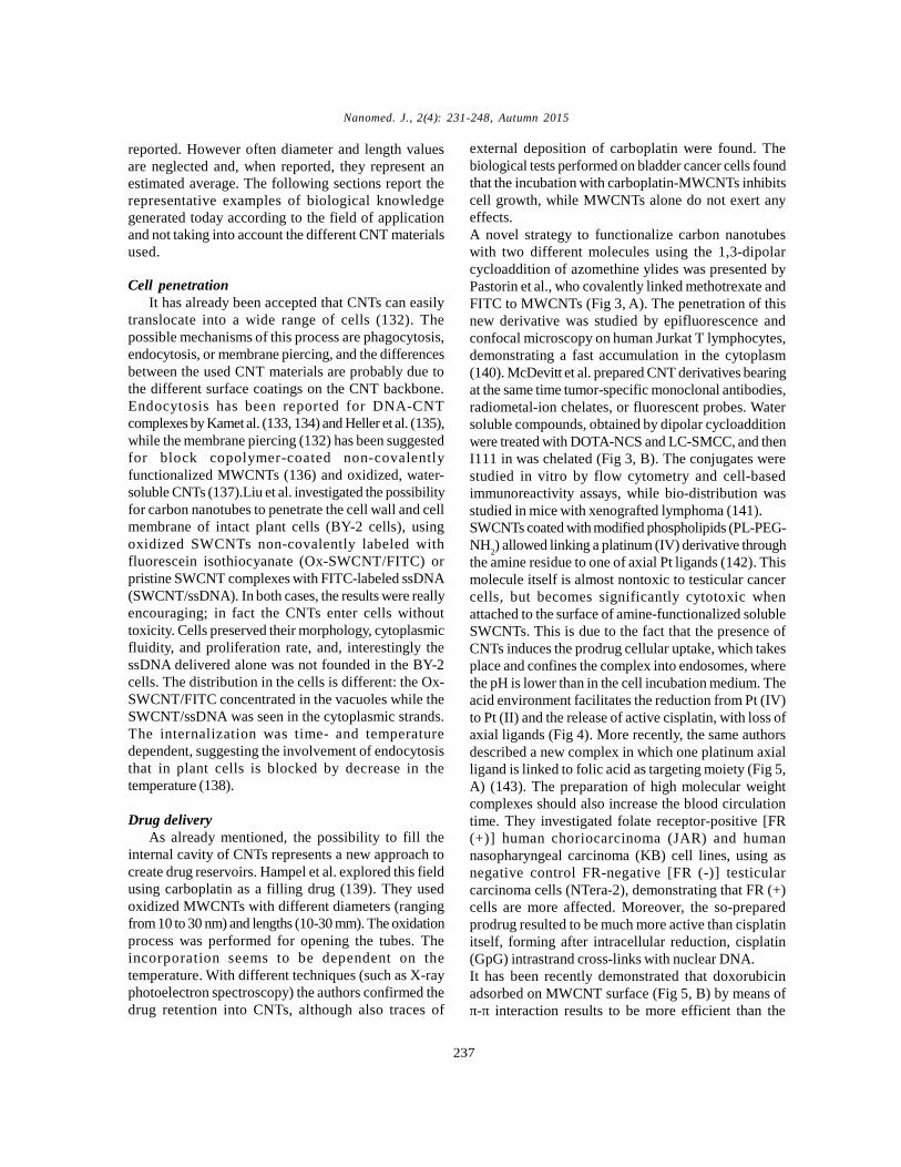
external deposition of carboplatin were found. Thebiological tests performed on bladder cancer cells foundthat the incubation with carboplatin-MWCNTs inhibitscell growth, while MWCNTs alone do not exert anyeffects.A novel strategy to functionalize carbon nanotubeswith two different molecules using the 1,3-dipolarcycloaddition of azomethine ylides was presented byPastorin et al., who covalently linked methotrexate andFITC to MWCNTs (Fig 3, A). The penetration of thisnew derivative was studied by epifluorescence andconfocal microscopy on human Jurkat T lymphocytes,demonstrating a fast accumulation in the cytoplasm(140). McDevitt et al. prepared CNT derivatives bearingat the same time tumor-specific monoclonal antibodies,radiometal-ion chelates, or fluorescent probes. Watersoluble compounds, obtained by dipolar cycloadditionwere treated with DOTA-NCS and LC-SMCC, and thenI111 in was chelated (Fig 3, B). The conjugates werestudied in vitro by flow cytometry and cell-basedimmunoreactivity assays, while bio-distribution wasstudied in mice with xenografted lymphoma (141).SWCNTs coated with modified phospholipids (PL-PEG-NH
2) allowed linking a platinum (IV) derivative through
the amine residue to one of axial Pt ligands (142). Thismolecule itself is almost nontoxic to testicular cancercells, but becomes significantly cytotoxic whenattached to the surface of amine-functionalized solubleSWCNTs. This is due to the fact that the presence ofCNTs induces the prodrug cellular uptake, which takesplace and confines the complex into endosomes, wherethe pH is lower than in the cell incubation medium. Theacid environment facilitates the reduction from Pt (IV)to Pt (II) and the release of active cisplatin, with loss ofaxial ligands (Fig 4). More recently, the same authorsdescribed a new complex in which one platinum axialligand is linked to folic acid as targeting moiety (Fig 5,A) (143). The preparation of high molecular weightcomplexes should also increase the blood circulationtime. They investigated folate receptor-positive [FR(+)] human choriocarcinoma (JAR) and humannasopharyngeal carcinoma (KB) cell lines, using asnegative control FR-negative [FR (-)] testicularcarcinoma cells (NTera-2), demonstrating that FR (+)cells are more affected. Moreover, the so-preparedprodrug resulted to be much more active than cisplatinitself, forming after intracellular reduction, cisplatin(GpG) intrastrand cross-links with nuclear DNA.It has been recently demonstrated that doxorubicinadsorbed on MWCNT surface (Fig 5, B) by means ofπ-π interaction results to be more efficient than the
reported. However often diameter and length valuesare neglected and, when reported, they represent anestimated average. The following sections report therepresentative examples of biological knowledgegenerated today according to the field of applicationand not taking into account the different CNT materialsused.

Nanomed. J., 2(4): 231-248, Autumn 2015
238
H.R. Sadegh et al.
Fig. 3. Molecular structures of bifunctional CNTs derivatives
Fig. 4. Cisplatin release mechanism
Fig. 5. Non-covalent attachments of anti-tumoral drugs toCNTs
Gene DeliveryOne of the first approaches toward genetic material
delivery was performed by Lu et al. who complexedRNA polymer poly (rU) to SWCNTs by nonspecificinteractions (145). The translocation across MCF7breast cancer cells took place and the genetic materialwas found across the cellular and the nuclearmembranes.Covalent derivative SWCNTs, obtained by amidationof the nanotube carboxylic groups with a chainpresenting a terminal ammonium group were studiedas telomerase reverse transcriptase small interferingRNA (TERT-siRNA) delivery system into cells (146).The effect was assessed taking into account theproliferation and the growth of tumor cells (Lewis lungcancer cells and HeLa cells), both in vitro and in vivo.So the silencing of the targeted gene is the evidence ofa good intracellular delivery of siRNA, which is locallyreleased from nanotubes and can induce its effect. Daiand coworkers delivered siRNA to human T cells andperipheral blood mononuclear cells. In this case,SWCNTs were again coated with PL-PEG-NH
2.
Differences in efficacy were found to depend onfunctionalization and degree of hydrophilicity, relatedto the length of the introduced PEG chain in the PL-PEG construct (147). Short SWCNTs, non-covalentlyfunctionalized with PL-PEG, were used. In this case, acleavable bond was introduced, linking by disulfidebond the PLPEG unit and the delivering siRNA (148).The siRNA functionality obtained in this modelresulted to be more potent than lipofectamine used ascontrol transfection agent.Oxidized SWCNTs, bearing positive charges able tointeract with fluorescein-labeled dsDNA (dsDNA-FAM), were also coated with folic acid-modifiedphospholipids (PL-PEG-NH
2), by wrapping the alkyl
chains. The multifunctional system was selectivelyuptaken by tumor cells, in which folic receptors areoverexpressed, driving the oligonucleotide into cells,as demonstrated by fluorescence imaging (149).SWCNTs were utilized as carriers for ssDNA probeinto cells, demonstrating an increased resistance ofthe oligonucleotides toward nuclease digestion. In factthe ssDNA is protected from enzymatic cleavage andinterference from nucleic acid binding proteins, stillexerting its function of targeting mRNA (150). Thisincreases the potentiality of CNTs as oligonucleotidevectors, as already hypothesized by Kateb et al. (136).They studied the possibility of using CNTs as DNA or
free one, probably because of ameliorate delivery intocells (144).

Nanomed. J., 2(4): 231-248, Autumn 2015
239
RNA delivery systems in brain tumors, thusdemonstrating the ability of microglia to efficientlyinternalize MWCNTs coated with Pluronic F108 ascompared to glioma cells.
Anticancer ApproachesThe potential of CNTs as a genetic material delivery
system is great, with application in gene therapy,considering the general biocompatibility of CNTsthemselves. As mentioned already, nanotubes easilybind macromolecules, such as nucleic acids. Li et al.found that oxidized SWCNTs can inhibit DNA duplexassociation (151, 152). Moreover, it has beendemonstrated that they selectively induce humantelomeric i-motif DNA formation, binding to the 5’-endmajor groove and directly stabilizing the chargedCC+ base pairs. These findings can be utilized indesigning new compounds for cancer therapy, wherethe modulation of human telomeric DNA can play afundamental role, although the biological effects of i-motif structure induction is not yet clear.A polyadenylic acid [poly (rA)] tail is present ineukaryotic cells and human poly (rA) polymerase (PAP)and can be considered a tumor-specific target, andcompounds interfering with this structure haveinteresting potential in the therapy. Zhao et al. studiedoxidized SWCNTs and reported that CNTs inducesingle-stranded poly (rA) to self-structure into duplexbut the mechanism is not understood and shouldinvolve the characteristics of the oxidized CNTsconsidering that amino-derivative SWCNTs,surprisingly, do not induce self-structuration (153).Antitumor immunotherapy is actually not veryeffective. In the past, Pantarotto et al. (154) have foundthat viral peptides conjugated to CNTs can improvethe anti-peptide immune response. Meng et al. pavedthe way to improve immunotherapy. Tumor lysateprotein, readily obtainable from most solid tumors, hasbeen linked to MWCNTs and administered as a possibletumor-cell vaccine in a mouse model bearing the H22liver cancer, with positive effects on the cure rate andcellular antitumor immune reaction (155).Phospholipid–polyethylene glycol chains linked to afolic acid residue were linked by means of hydrophobicinteractions to CNTs. The presence of folate shoulddrive toward a selective uptake of the complex intocells overexpressing folate receptors, as it happens insome cancer cells. After internalization, cell death wasinduced irradiating with near-infrared (NIR) light. This
action proved to be selective because the presence ofCNTs in cells increases the temperature, therebyinducing thermal ablation when irradiated (21).Analogously, CNTs bearing two specific monoclonalantibodies (insulin-like growth factor 1 receptor, IGF1Rand human endothelial receptor 2, HER2), prepared bysupra-molecular approach with properly functionalizedpyrene units, have been used to kill breast cancer cellswith NIR irradiation (156). The main problem of nearinfrared irradiation application is due to its poor tissuepenetration capacity, which allows the treatment of onlysuperficial cancer lesions. Radiofrequency waves, onthe contrary, penetrate more into tissues, and theirinteraction with internalized CNTs produces anincrease in temperature of cells, thereby inducing celldeath. Gannon et al. proposed the use of these waves,but in the reported case the limitation is due to thedirect injection into tumor of SWCNT coated with polyphenylene-ethynylene polymer (157).Another atypical approach to cancer treatment isrelated to angiogenesis. In fact, its inhibition byinterfering in the growth factor balance can obstaclethe insurgence of diseases, among which tumors canbe mentioned. A good model to study angiogenesis isthe chick chorio allantoic membrane (CAM). A studyperformed by Murugesan et al. employed carbonmaterials (such as CNTs, fullerene, and graphite) totest their capability to inhibit FGF2- or VEGF-inducedvessel formation in the CAM model. All the materialsshowed significant effects in the inducedangiogenesis, acting more efficiently in comparison toVEGF, while they did not exert any effect on the basalprocess in absence of the already mentioned growthfactors. The mechanism involved in this process is notyet clarified but it is clear that this anti-antigenic actioncould be exploited in cancer treatment (158).
CNTs and Neuron InteractionsFew years ago, Lovat et al. reported the possibility
of improving the neural signal transfer by growingneuronal circuits on CNT substrates, relating thisactivity to the high electrical conductivity of CNTs(159). Later on, the same authors developed anintegrated SWCNT-neuron system to test whetherelectrical stimulation delivered via SWCNTs can induceneuronal signaling. The patch clamped hippocampalcells, cultured on SWCNT substrates, have shown thatthey can be stimulated via the SWCNT layers. Anyresistive coupling between bio membranes and

Nanomed. J., 2(4): 231-248, Autumn 2015
240
CNTs in Nanomedicine: A reveiw
SWCNTs was qualitatively distinguishable from acoupling between SWCNTs and the patch pipettethrough the patch seal path to ground (160). Usingsingle-cell electrophysiology techniques, electronmicroscopy analysis, and theoretical modeling, thesame authors demonstrated that CNTs enhance theresponsiveness of neurons, due to the formation oftight contacts between CNTs and the cell membranes.These could favor electrical shortcuts between theproximal and distal compartments of the neuron (161).While in the already mentioned works, the CNTs weredeposed on glass coverslips for growing cells, adifferent approach has been used by Keefer et al. usingCNTs to electrochemically coat tungsten and stainlesssteel wire electrodes. This treatment led to an increasein recording and electrical stimulation of neuronalcultures (162). Kotov and coworkers also prepared alayer-by-layer composite with SWCNTs and laminin,an essential glycoprotein of human extracellular matrix.The use of this surface for cellular culture inducedneural stem cells (NSC) differentiation and the creationof functional neural network. Also in this case, theapplication of current through CNTs stimulated theaction potentials (163). Carbon nanotubes have alsobeen arranged in vertical alignment to the gate insulatorof an ion-sensitive field-effect transistor to act aselectrical interfaces to neurons, to study theinteractions with them, and an enhance efficiency hasbeen recorded in neuronal electrical activity (164).Water-soluble SWCNTs grafted to polyethylene glycolhave been used in the culturing medium for neuralgrowth. These derivatives demonstrated their abilityto block the stimulated membrane endocytosis inneurons and this could justify the evidenced extendedneurite length (165), while MWCNTs coated withpluronic F127 (PF127) injected in mouse cerebral cortexdo not induce degeneration of the neurons close tothe injection site, while PF127 itself can induceapoptosis of mouse primary cortical neurons, implyingthat the presence of MWCNTs inhibits PF127-inducedapoptosis (166).All these successful works performed on neuronalgrowth and stimulation led to the hope of using CNTsas material for the reconstruction of neural injuredtissues in the near future, paving the way for anapplication in spinal cord disease resolution andneurodegeneration restoration.
CNTs and antioxidantCNTs, when instilled in lungs, induced inflammatory
and fibrotic response, which was supposed to be due
to oxidative stress derived from free radicals, althoughno real evidence of ROS generation from CNTs wasobserved. Moreover Fenoglio et al. reported thatMWCNTs do not generate oxygen or carbon-basedfree radicals in the presence of H
2O
2 or formatted,
respectively, but, on the contrary, when radicals wereinduced, CNTs act as radical scavengers (167). Veryrecently the antioxidant properties of CNTs have beenreported by Tour and coworkers. SWCNTs and ultra-short SWCNTs (US-SWCNTs) were derivatized withbutyrate hydroxyl toluene (BHT) using two differentapproaches as covalent attachment of triazene to thesidewalls of pluronic-wrapped SWCNT or amidationof carboxylic residues in the case of US-SWCNTderivatives. The oxygen radical scavenging ability ofthe different compounds has been evaluated leadingto really interesting results, although the differentcompounds react differently. In the first case, thepluronic-coated SWCNTs were more efficient than thecorresponding BHT derivatives, while in the case ofoxidized US-SWCNTs, higher the loading of BHTresidues, better was the antioxidant activity. This newfinding paves the way for SWCNT application as novelmedical therapeutics in the antioxidant field (168).However, it is necessary to remember that a recentreport presents contradictory results indicatingMWCNT toxicity in A549 cells, which can be reducedby pretreatment with antioxidants to decrease ROSproduction and interleukin-8 gene expression (169).
CNTs and imagingSWCNTs are endowed of fluorescent properties
that can be exploited in in vitro and in vivo imaging,and that have been used to determine the uptake ofnanotubes in macrophages (170), the CNT eliminationin rabbits (171), and their toxicity in fruit fly larvae (172).This surprising use of CNTs for imaging purposes isonly possible when some properties of the nanotubesare preserved, that is, at least 100nm of the tubes mustbe unmodified and the CNTs should not be in bundles,conditions in which the fluorescence is quenched.More recently, Welsher et al. used semiconductingSWCNTs wrapped with polyethylene glycol as near-infrared fluorescent tags. The polyethylene glycol wasconjugated to antibodies for the selective recognitionfor CD20 cell surface receptor on B cells or for HER2/neu positive breast cancer cells. The intrinsic NIRphotoluminescence allowed the detection of thebinding to the cells, in spite of the presence of lowauto fluorescence in different cells (173). MRI is anotherimportant imaging technique used in medicine. Afterthe discovery of the great performances of Gd@C
60,

Nanomed. J., 2(4): 231-248, Autumn 2015
241
Gd@C82
and their derivatives as contrast agents (174-176), the evolution toward the analogous use of CNThas been done by Wilson and coworkers (177). Thetubes, shortened by fluorination down to 20-80 nm,were loaded with GdCl
3. The obtained results
demonstrated that these CNTs have a relaxivity (r1) 40times greater than any current Gd3+ ion-based clinicalagents. The aggregation, that in the case of fullereneendohedrals can vary the relaxivity, does not affectthis parameter in the case of CNTs. However, moresoluble compounds have been prepared by Ashcroftet al. (178). The same authors demonstrated the stabilityof gadonanotubes in buffers, serum, at variation of pHand temperature. Moreover they discovered a greatdependence of r1 on the pH, with ultra-sensitivity in arange of pH 7.0-7.4. This suggests the possibility ofusing the gadonanotubes for the early detection ofcancer cells in which the pH can dramatically decrease(179). A novel study on the bio-distribution and theeffect of SWCNTs (raw and super-purified, raw-SWCNT and SP-SWCNT, respectively) after in vivoexposure has been recently reported. The combinationof 3He and 1Hmagnetic resonance imaging (MRI) wasused in a rat model. Hyperpolarized gases such as 3Heacting a contrast agent diffuses rapidly in lungs,permitting the determination of ventilated air ways andalveolar spaces. The presence of metal impurity in theraw- SWCNTs was sufficient to induce a drop inmagnetic field homogeneity detected in 3He MRI, whileno significant variation was observed after SP-SWCNTexposition (180). Proton MRI was used to follow raw-SWCNTs after intravenous injection, finding them inspleen and kidneys. The absence of metal nanoparticlesin the SP-SWCNT excluded signal modifications. Whenthis technique was used to determine the fate of CNTafter pulmonary instillation, no signal changes in liver,spleen, and kidney were detected, implying the absenceof systemic circulation of CNTs after inhalation. Thisresult was also confirmed by histological analysis,establishing the possibility of using noninvasivemethodology to detect CNT presence, if associatedwith a proper iron impurity concentration.A different approach has been reported by Richard etal. The authors, instead of filling the CNTs with the Gdderivative, used an amphiphilic gadolinium (III) chelate,absorbed on MWCNT external surface, in differentconcentrations.The obtained suspension resulted to be stable and r1was measured in different conditions, finding adependence on the Gd-chelate concentration. On thecontrary, the transversal water proton relaxivity (T2)
was independent from Gd concentration and frequency(181).
Various applications of CNTsCNTs are incredible sorbent materials for different
compounds due to their large surface areas (BET valueis about e”600m2/g, depending on the CNT types takeninto account). They have been already used ascontaminant remover for water pollution in laboratoryexperiments (182, 183) and also for dioxin (184). Chenet al. studied the sorption of americium and thorium onCNT surface, their kinetics and pH dependence (185,186) but different efficiency probably due to differentpretreatment of CNTs, found by other authors (187),and the possibility to use this new form of carbon innuclear waste management cannot be excluded. CNTs,both single- and multiwalled, were also used to capturebacteria, as Streptococcus mutans. The simple mixtureof CNT bundles with bacteria leads to the formation ofa precipitate, which shows bacterial adhesion tonanotubes, that depends on the CNT diameters (188).
Toxicity of CNTsA big debate and many concerns about CNTs
toxicity arouse some years ago and up to now theinformation are fragmentary and apparentlycontradictory. Actually it is rather difficult to stigmatizedefinitive assumptions, considering that the performedstudies cover a wide spectrum of derivatives presentingdifferent characteristics. In general it is possible toaffirm that toxicity decreases proportionally with theincrease in dispersion and/or solubility degree, but itis necessary to go much further in this field (189). Asystematic study on the effect of structural defects ofthe MWCNT carbon cages on lung toxicity has beenperformed by Fenoglio et al., who examined CNTs (i)with introduced structural defects, (ii) with reducedoxygenated carbon functionalities and metallic oxides,(iii) in a purified annealed sample, and (iv)in metal-deprived carbon framework with introduced structuraldefects (190). The hydroxyl radical scavengingactivities of modified CNTs were studied by EPRdemonstrating that the original ground materialexhibited an activity toward hydroxyl radicals, whichdisappeared in the purified annealed sample, but wasdetected again in metal-deprived carbon framework withintroduced structural defects. In vitro experiments onrat lung epithelial cells were performed to assess thegenotoxic potential of the different CNT preparations.Acute pulmonary toxicity and the genotoxicity of CNTswere reduced upon heating but restored upon grinding,

Nanomed. J., 2(4): 231-248, Autumn 2015
242
indicating that the intrinsic toxicity of CNTs is mainlymediated by the presence of defective sites in theircarbon framework (191). The scavenging activity isrelated to the presence of defects and seems to gopaired with the genotoxic and inflammatory potentialof CNT. Tabet et al. used dispersions of MWCNT indipalmitoyl lecithin, ethanol, and phosphate-bufferedsaline (PBS), to study the CNT toxicity on humanepithelial cell line A549. The presence of PBS inducesagglomerate formation on top of the cells, but in allcases MWCNTs decrease the cellular metabolismwithout permeabilization of the cell membrane orapoptosis, while asbestos fibers penetrate into the cellsand increase apoptosis (192). Wick et al. investigatedCNTs with different agglomeration degrees to determinetheir cytotoxicity on human MSTO-211H cells. Well-dispersed CNTs (obtained by using polyoxyethylenesorbitan monooleate) were less toxic than asbestos andrope-like agglomerates induced higher cytotoxic effectsthan asbestos fibers (193). Recently, Guo et al. reportedthe modification of MWCNTs with glucosamine anddecylamine by c-ray irradiation (g-MWCNT and d-MWCNT), and they used these derivatives to performcytotoxicity test on Tetrahymena pyriformis. Thedecylamine derivative exhibits a dose-dependent growninhibition, attributed to the amine toxicity, driven intocells by the nanotubes. The comparison betweenpurified MWCNTs and g-MWCNTs demonstratedgrowth stimulation by the latter. It seems that thehydrophilic compound is able to complex peptone,present in the medium, and to transfer it into cells. Thismeans that any nonspecific interaction between CNTsand components of the culture medium must beconsidered in evaluating the cytotoxicity of CNTs (194).Considering that almost nothing is known about CNTimpact on natural ecosystems, the ciliated protozoanTetrahymena thermophile has been also studiedbecause of its role in the regulation of microbialpopulations through the ingestion and digestion ofbacteria. It is, in fact, an important organism inwastewater treatment and, moreover an indicator ofsewage effluent quality. SWCNTs have been found inthese microorganisms, inducing protozoa aggregationswith a consequent inability to interact with bacteria(195). Salmonella typhimurium and Escherichia colistrains were used to perform mutagenicity test withMWCNTs. The mutagenic activity did not appear, evenin presence of the metabolic activators (196).Different tests devoted to establish the genotoxicpotential of MWCNTs has been performed both in vivoin rats and in vitro on rat lung epithelial cells or human
epithelial cells. Three days after carbon nanotubeintratracheal administration, type II pneumocytespresent micronuclei with a dose-dependent increase.The same behavior was reported for the in vitroexperiments, proving the possibility of MWCNTs toinduce clastogenic and aneugenic events (respectivelyincrease the rate of genetic mutation due to DNA breaksand loss or gain of whole chromosomes) (197). Oftenthe clastogenic is related to ROS generation and thepossible presence of metallic nanoparticles (Fe or Co)would justify this action, but the MWCNT antioxidantbehavior suggests different pathways and indicatesthe necessity to explore these systems more in details.
CONCLUSIONSCNTs have exhibited diverse physical, chemical and
mechanical properties suitable for a variety ofapplications. In last decade, medical applications ofCNTs have undergone rapid progress. Their uniqueproperties such as ultrahigh surface area, high aspectratio and distinct optical properties have been appliedto develop innovative, multi-functional CNT-basednanodevices for broad applications. This review hasdescribed the chemical and physical methods toprepare CNTs for used in medicine. With these methods,targeting molecules are attached onto CNTs fortargeted drug delivery, selective imaging, and othertherapies. As a new type of nanomaterial, the toxicityof CNTs has been extensively investigated. To date,tremendous toxicity studies on CNTs have beenpublished. However, the published data areinconsistent. The reason is that CNTs used in thesestudies vary in dispersion status, size and length oftubes, metal impurities and functionalization methodsetc. Moreover, different analysis methods used in theevaluation CNTs toxicity studies also cause disparities.Despite these disparities, there is a broad agreementthat well-dispersed CNTs have little or no toxicity bothin vitro and in vivo, and therefore are useful for medicalapplications. Finally, an urgent need has been proposedfor long-term studies on the absorption, deposition,metabolism and excretion (ADME) of CNTs. Only afterthe uncertainty on CNTs toxicity is resolved, the CNT-based therapeutics can be possible applied clinically.
ACKNOWLEDGMENTSAuthors would like to thank Islamic Azad University
Science and Research Branch for all supports.
REFERENCES1. Iijima S. Helical Microtubules of Graphitic Carbon. Nature.
1991; 354(6348): 56-58.

Nanomed. J., 2(4): 231-248, Autumn 2015
243
2. Ouyang M, Huang J L, Lieber CM. One-dimensional energydispersion of single-walled carbon nanotubes by resonantelectron scattering. Phys Rev Lett. 2002; 88(6): 066804.
3. Zare K, Najafi F, Sadegh H. Studies of ab initio and MonteCarlo simulation on interaction of fluorouracil anticancerdrug with carbon nanotube. J Nanostruc Chem. 2013; 3(1):1-8.
4. Thostenson ET, Ren Z F, Chou TW. Advances in the scienceand technology of carbon nanotubes and their composites: areview. Compos Sci Technol. 2001; 61(13): 1899-1912.
5. Troiani HE, Miki-Yoshida M, Camacho-Bragado GA, MarquesMAL, Rubio A, Ascencio JA, Jose-Yacaman M. Directobservation of the mechanical properties of single-walledcarbon nanotubes and their junctions at the atomic level.Nano Lett. 2003; 3(6): 751-755.
6. Wan XG, Dong JM, Xing DY. Optical properties of carbonnanotubes. Phys Rev B. 1998; 58(11): 6756-6759.
7. Kostarelos K, Lacerda L, Pastorin G, Wu W, Wieckowski S,Luangsivilay J, Bianco A. Cellular uptake of functionalizedcarbon nanotubes is independent of functional group and celltype. Nat Nanotechnol. 2007; 2(2): 108-113.
8. Sadegh H, Shahryari-ghoshekandi R, Kazemi M. Study insynthesis and characterization of carbon nanotubes decoratedby magnetic iron oxide nanoparticles. Int Nano Lett. 2014;4(4): 129-135.
9. Sadegh H, Shahryari-ghoshekandi R, Agarwal S, Tyagi I, AsifM, Gupta VK. Microwave-assisted removal of malachite greenby carboxylate functionalized multi-walled carbon nanotubes:Kinetics and equilibrium study. J Mol Liq. 2015; 206: 151-158.
10. Ando Y. Carbon nanotube: the inside story. J NanosciNanotechnol. 2010; 10(6): 3726-3738.
11. Chen RJ, Zhang Y, Wang D, Dai H. Noncovalent sidewallfunctionalization of single-walled carbon nanotubes forprotein immobilization, J Am Chem Soc. 2001; 123: 3838–3839.
12. Zare K, Gupta VK, Moradi O, Makhlouf ASH, Sillanpää M,Nadagouda MN, Sadegh H, Shahryari-ghoshekandi R, Pal A,Wang Z, Tyagi I, Kazemi M. A comparative study on thebasis of adsorption capacity between CNTs and activatedcarbon as adsorbents for removal of noxious synthetic dyes:a review. J Nanostruc Chem. 2015; 5(2): 227-236.
13. Besteman K, Lee JO, Wiertz FGM, Heering HA, Dekker C.Enzyme-coated carbon nanotubes as single-moleculebiosensors, Nano Lett. 2003; 3: 727–730.
14. Xin H, Woolley AT. DNAtemplated nanotube localization.J Am Chem Soc. 2003; 125: 8710-8711.
15. Taft BJ, Lazareck AD, Withey GD, Yin A, Xu JM, KelleySO. Site-specific assembly of DNA and appended cargo onarrayed carbon nanotubes. J Am Chem Soc. 2004; 126: 12750-12751.
16. Gupta VK, Tyagi I, Agarwal S, Moradi O, Sadegh H,Shahryari-ghoshekandi R, Makhlouf ASH, Goodarzi M,Garshasbi A. Study on the removal of heavy metal ions fromindustry waste by carbon nanotubes: effect of the surfacemodification-A review. Crit Rev Env Sci Technol (Acceptedfor publish) DOI:10.1080/10643389.2015.1061874 (2015).
17. Liu L, Wang T, Li J, Guo Z, Dai L, Zhang D, Zhu D. Self-assembly of gold nanoparticles to carbon nanotubes using athiol-terminated pyrene as interlinker. Chem Phys Lett.2003; 367: 747-752.
18. Cao L, Chen H, Wang M, Sun J, Zhang X, Kong F.Photoconductivity study of modified carbon nanotube/oxotitanium phthalocyanine composites. J Phys Chem B.2002; 106: 8971-8975.
19. Wang X, Liu Y, Qiu W, Zhu D. Immobilization of tetra-tertbutylphthalocyanines on carbon nanotubes: a first steptowards the development of new nanomaterials. J MaterChem. 2002; 12: 1636-1639.
20. Cao L, Chen HZ, Zhou HB, Zhu L, Sun JZ, Zhang XB, XuJM, Wang M. Carbon nanotube templated assembly of rare-earth phthalocyanine nanowires. Adv Mater. 2003; 15: 909-913.
21. Guldi DM, Rahman GNA, Ramey J, Marcaccio M, PaolucciD, Paolucci F, Qin S, Ford WT, Balbinot D, Jux N,Tagmatarchis N, Prato M. Donor–acceptor nanoensemblesof soluble carbon nanotubes. Chem Commun. 2004; 2034-2035.
22. Guldi DM, Rahman GMA, Prato M, Jux N, Qin S, Ford W.Single-wall carbon nanotubes as integrative building blocksfor solarenergy conversion. Angew Chem Int Ed. 2005; 44:2015-2018.
23. Murakami H, Nomura T, Nakashima N. Noncovalentporphyrin-functionalized single-walled carbon nanotubes insolution and the formation of porphyrin–nanotubenanocomposites. Chem Phys Lett. 2003; 378: 481-485.
24. Li H, Zhou B, Lin Y, Gu L, Wang W, Fernando KAS, KumarS, Allard LF, Sun YP. Selective interactions of porphyrinswith semiconducting single-walled carbon nanotubes. J AmChem Soc. 2004; 126: 1014-1015.
25. Chen J, Collier CP. Noncovalent functionalization of single-walled carbon nanotubes with water-soluble porphyrins. JPhys Chem B. 2005; 109: 7605-7609.
26. Satake A, Miyajima Y, Kobuke Y. Porphyrin–carbonnanotube composites formed by noncovalent polymerwrapping. Chem Mater. 2005; 17: 716-724.
27. Guldi DM, Rahman GMA, Jux N, Tagmatarchis N, Prato M.Integrating single-wall carbon nanotubes into donor–acceptornanohybrids. Angew Chem Int Ed. 2004; 43: 5526-5530.
28. Guldi DM, Rahman GMA, Jux N, Balbinot D, TagmatarchisN, Prato M. Multiwalled carbon nanotubes in donor–acceptornanohybrids – towards long-lived electron transfer products.Chem Commun. 2005; 2038-2040.
29. Guldi DM, Taieb H, Rahman GMA, Tagmatarchis N, PratoM. Novel photoactive single-walled carbon nanotube–porphyrin polymer wraps: efficient and long-livedintracomplex charge separation. Adv Mater. 2005; 17: 871-875.
30. Guldi DM, Rahman GMA, Jux N, Balbinot D, Hartnagel U,Tagmatarchis N, Prato M. Functional single-wall carbonnanotube nanohybrids-associating SWCNTs with water-soluble enzyme model systems J Am Chem Soc. 2005; 127:9830-9838.
31. Chichak KS, Star A, Altoe MVP, Stoddart JF. Single-walledcarbon nanotubes under the influence of dynamiccoordination and supramolecular chemistry. Small. 2005; 1:452-461.
32. Tang BZ, Xu H. Preparation, alignment, and opticalproperties of soluble poly(phenylacetylene)-wrapped carbonnanotubes. Macromol. 1999; 32: 2569-2576.
33. Romero DB, Carrard M, Heer W, Zuppiroli L. A carbonnanotube/organic semiconducting polymer heterojunction.Adv Mater. 1996; 8: 899-902.
34. Ago H, Petritsch K, Shaffer MSP, Windle AH, Friend RH.Composites of carbon nanotubes and conjugated polymersfor photovoltaic devices. Adv Mater.1999; 11: 1281-1285.
35. Moradi O, Sadegh H, Shahryari-Ghoshekandi R, Norouzi M.Application of carbon nanotubes in nanomedicine: newmedical approach for tomorrow. In: Soni, S., Salhotra, A.,

Nanomed. J., 2(4): 231-248, Autumn 2015
244
Suar, M. (eds.) Handbook of Research on Diverse Applicationsof Nanotechnology in Biomedicine, Chemistry, andEngineering, pp. 90–128. (2015). doi:10.4018/978-1-4666-6363-3.ch006. Accessed 16 Jan 2015
36. Ago H, Shaffer MSP, Ginger DS, Windle AH, Friend RH.Electronic interaction between photoexcited poly( p-phenylene vinylene) and carbon nanotubes. Phys Rev B.2000; 61: 2286-2290.
37. Wery J, Aarab H, Lefrant S, Faulques E, Mulazzi E, PeregoR. Photoexcitations in composites of poly(paraphenylenevinylene) and single-walled carbon nanotubes. Phys Rev B.2003; 67(11): 115202.
38. Fournet P, Coleman JN, Lahr B, Drury A, Blau WJ, O’BrienDF, Horhold HH. Enhanced brightness in organic light-emitting diodes using a carbon nanotube composite as anelectron-transport layer. J Appl Phys. 2001; 90: 969-975.
39. Fournet P, O’Brien DF, Coleman JN, Horhold HH, Blau WJ.A carbon nanotube composite as an electron transport layerfor M3EH-PPV based lightemitting diodes. Synth. Met. 2001;121: 1683-1684.
40. Moradi O, Gupta VK, Agarwal S, Tyagi I, Asif M, MakhloufASH, Sadegh H, Shahryari-ghoshekandi R. Characteristicsand electrical conductivity of graphene and graphene oxidefor adsorption of cationic dyes from liquids: Kinetic andthermodynamic study. J Ind Eng Chem. 2015; 28: 294-301.
41. Coleman JN, Dalton AB, Curran S, Rubio A, Davey AP,Drury A, McCarthy B, Lahr B, Ajayan PM, Roth S, BarklieRC, Blau WJ. Phase separation of carbon nanotubes andturbostratic graphite using a functional organic polymer. AdvMater. 2000; 12: 213-216.
42. Star A, Lu Y, Bradley K, Gruner G. Nanotube optoelectronicmemory devices. Nano Lett. 2004; 4: 1587-1591.
43. Murphy R, Coleman JN, Cadek M, McCarthy B, Bent M,Drury A, Barklie RC, Blau WJ. Highyield, nondestructivepurification and quantification method for multiwalled carbonnanotubes. J Phys Chem B. 2002; 106: 3087-3091.
44. Coleman JN, O’Brien DF, Dalton AB, McCarthy B, Lahr B,Barklie RC, Blau WJ. Electron paramagnetic resonance as aquantitative tool for the study of multiwalled carbonnanotubes. J Chem Phys. 2000; 113: 9788-9793.
45. Star A, Stoddart JF, Steuerman D, Diehl M, Boukai A, WongEW, Yang X, Chung SW, Choi H, Heath JR. Preparation andproperties of polymer-wrapped singlewalled carbonnanotubes. Angew Chem Int Ed. 2001; 40: 1721-1725.
46. Sadegh H, Shahryari-ghoshekandi R, Tyagi I, Agarwal S,Gupta VK. Kinetic and thermodynamic studies for alizarinremoval from liquid phase using poly-2-hydroxyethylmethacrylate (PHEMA). J Mol Liq. 2015; 207: 21-27.
47. Star A, Stoddart JF. Dispersion and solubilization of single-walled carbon nanotubes with a hyperbranched polymer.Macromol. 2002; 35: 7516-7520.
48. Musa I, Baxendale M, Amaratunga GAJ, Eccleston W.Properties of regioregular poly(3-octylthiophene)/multi-wallcarbon nanotube composites. Synth Met. 1999; 102: 1250.
49. Alexandrou I, Kymakis E, Amaratunga GAJ. Polymer-nanotube composites: burying nanotubes improves theirfield emission properties. Appl Phys Lett. 2002; 80: 1435-1437.
50. Valentini L, Armentano I, Biagiotti J, Frulloni E, KennyJM, Santucci S. Frequency dependent electrical transportbetween conjugated polymer and single-walled carbonnanotubes. Diam Rel Mater. 2003; 12: 1601-1609.
51. Kymakis E, Amaratunga GAJ. Single-wall carbon nanotube/conjugated polymer photovoltaic devices. Appl Phys Lett.2002; 80: 112-114.
52. Kymakis E, Alexandrou I, Amaratunga GAJ. Highopencircuit voltage photovoltaic devices from carbon-nanotube-polymer composites. J Appl Phys. 2003; 93:1764-1768.
53. Bhattacharyya S, Kymakis E, Amaratunga GAJ.Photovoltaic properties of dye functionalized single-wallcarbon nanotube/ conjugated polymer devices. Chem Mater.2004; 16: 4819-4823.
54. Landi BJ, Raffaelle RP, Castro SL, Bailey SG. Single-wallcarbon nanotube-polymer solar cells. Prog Photovolt ResAppl. 2005; 13: 165-172.
55. O’Connell MJ, Boul P, Ericson, LM, Huffman C, Wang Y,Haroz E, Kuper C, Tour JM, Ausman KD, Smalley RE.Reversible water-solubilization of single-walled carbonnanotubes by polymer wrapping. Chem Phys Lett. 2001;342: 265-271.
56. Islam MF, Rojas E, Bergey DM, Johnson AT, Yodh AG. Highweight fraction surfactant solubilization of single-wall carbonnanotubes in water. Nano Lett. 2003; 3: 269-273.
57. Richard C, Balavoine F, Schultz P, Ebbesen TW, MioskowskiC. Supramolecular self-assembly of lipid derivatives on carbonnanotubes. Science. 2003; 300(5620): 775-778.
58. O’Connell MJ, Bachilo SM, Huffman CB, Moore VC, StranoMS, Haroz EH, Rialon KL, Boul PJ, Noon WH, Kittrell C,Ma J, Hauge RH, Weisman RB, Smalley RE. Band gapfluorescence from individual single-walled carbon nanotubes.Science. 2002; 297: 593-596.
59. Moore VC, Strano MS, Haroz EH, Hauge RH, Smalley RE.Individually suspended single-walled carbon nanotubes invarious surfactants. Nano Lett. 2003; 3: 1379-1382.
60. Wenseleers W, Vlasov II, Goovaerts E, Obraztsova ED,Lobach AS, Bouwen A. Efficient isolation and solubilizationof pristine single-walled nanotubes in bile salt micelles. AdvFunct Mater. 2004; 14: 1105-1112.
61. Bachilo SM, Strano MS, Kittrell C, Hauge RH, Smalley RE,Weisman RB. Structureassigned optical spectra of singlewalledcarbon nanotubes. Science. 2002; 298(5602): 2361–2366.
62. Hagen A, Hertel T. Quantitative analysis of optical spectrafrom individual single-wall carbon nanotubes. Nano Lett.2003; 3: 383-388.
63. Strano MS, Doorn SK, Haroz EH, Kittrell C, Hauge RH,Smalley RE. Assignment of (n, m) Raman and optical featuresof metallic single-walled carbon nanotubes. Nano Lett. 2003;3: 1091-1096.
64. Weisman RB, Bachilo SM. Dependence of optical transitionenergies on structure for singlewalled carbon nanotubes inaqueous suspension: an empirical Kataura plot. Nano Lett.2003; 3: 1235-1238.
65. Dyke CA, Tour JM. Unbundled and highly functionalizedcarbon nanotubes from aqueous reactions. Nano Lett. 2003;3: 1215-1218.
66. Dieckmann GR, Dalton AB, Johnson PA, Razal J, Chen J,Giordano GM, Muňoz E, Musselman IH, Baughman RH,Draper RK. Controlled assembly of carbon nanotubes bydesigned amphiphilic peptide helices. J Am Chem Soc. 2003;125: 1770-1777.
67. Zorbas V, Ortiz-Acevedo A, Dalton AB, Yoshida MM,Dieckmann GR, Draper RK, Baughman RH, Jose-YacamanM, Musselman IH. Preparation and characterization of

Nanomed. J., 2(4): 231-248, Autumn 2015
245
individual peptidewrapped single-walled carbon nanotubes. JAm Chem Soc. 2004; 126: 7222-7227.
68. Zorbas V, Smith AL, Xie H, Ortiz-Acevedo A, Dalton AB,Dieckmann GR, Draper RK, Baughman RH, Musselman IH.Importance of aromatic content for peptide/single-walledcarbon nanotube interactions. J Am Chem Soc. 2005; 127:12323-12328.
69. Kam NWS, Dai H. Carbon nanotubes as intracellular proteintransporters: generality and biological functionality. J AmChem Soc. 2005; 127: 6021-6026.
70. Zheng M, Jagota A, Semke ED, Diner BA, Mclean RS,Lustig SR, Richardson RE, Tassi NG. DNA-assisted dispersionand separation of carbon nanotubes. Nat. Mater. 2003; 2:338-342.
71. Zheng M, Jagota A, Strano MS, Santos AP, Barone P, ChouSG, Diner BA, Dresselhaus MS, Mclean RS, Onoa GB,Samsonidze GG, Semke ED, Usrey M, Walls DJ. Structurebasedcarbon nanotube sorting by sequence-dependent DNAassembly. Science. 2003; 302(5650): 1545-1548.
72. Kam NWS, Liu Z, Dai H. Functionalization of carbonnanotubes via cleavable disulfide bonds for efficientintracellular delivery of siRNA and potent gene silencing. JAm Chem Soc. 2005; 127: 12492-12493.
73. Hirsch A. Functionalization of single-walled carbonnanotubes. Angew Chem Int Ed. 2002; 41: 1853-1859.
74. Banerjee S, Hemraj-Benny T, Wong SS. Covalent surfacechemistry of single-walled carbon nanotubes. Adv Mater.2005; 17: 17-29.
75. Hamon MA, Chen J, Hu H, Chen Y, Itkis ME, Rao AM,Eklund PC, Haddon RC. Dissolution of single-walled carbonnanotubes. Adv Mater. 1999; 11: 834-840.
76. Kukovecz A, Kramberger C, Holzinger M, Kuzmany H,Schalko J, Mannsberger M, Hirsch A. On the stacking behaviorof functionalized single-wall carbon nanotubes. J Phys ChemB. 2002; 106: 6374-6380.
77. Chen J, Rao AM, Lyuksyutov S, Itkis ME, Hamon MA, HuH, Cohn RW, Eklund PC, Colbert DT, Smalley RE, HaddonRC. Dissolution of fulllength single-walled carbon nanotubesJ Phys Chem B. 2001; 105: 2525-2528.
78. Niyogi S, Hamon MA, Hu H, Zhao B, Bhowmik P, Sen R,Itkis ME, Haddon RC. Chemistry of single-walled carbonnanotubes. Acc Chem Res. 2002; 35: 1105-1113.
79. Hiura H, Ebbesen TW, Tanigaki K. Opening and purificationof carbon nanotubes in high yields. Adv Mater. 1995; 7: 275-276.
80. Ajayan PM, Ebbesen TW, Ichihashi T, Iijima S, Tanigaki K,Hiura H. Capillarity-induced filling of carbon nanotubes.Nature. 1993; 361: 333-334.
81. Chen J, Hamon MA, Hu H, ChenY, Rao AM, Eklund PC,Haddon RC. Solution properties of single-walled carbonnanotubes. Science. 1998; 282(5386): 95-98.
82. Lago RM, Tsang SC, Lu KL, Chen YK, Green MLH. Fillingcarbon nanotubes with small palladium metal crystallites:the effect of surface acid groups. Chem Commun. 1995;1355-1356.
83. Gupta VK, Sadegh H, Yari M, Shahryari Ghoshekandi R,Maazinejad B, Chahardori M. Removal of ammonium ionsfrom wastewater: A short review in development of efficientmethods. Global J Environ Sci Manag. 2015; 1(2): 149-158.
84. Monthioux M, Smith BW, Burteaux B, Claye A, FischerJE, Luzzi DE. Sensitivity of single wall carbon nanotubes tochemical processing: an electron microscopy investigation.Carbon. 2001; 39: 1251-1272.
85. Koshio A, Yudasaka M, Zhang M, Iijima SA. simple way tochemically react single-wall carbon nanotubes with organicmaterials using ultrasonication. Nano Lett. 2001; 1: 361-363.
86. Qin Y, Shi J, Wu W, Li X, Guo Z, Zhu D. Concise route tofunctionalized carbon nanotubes. J Phys Chem B. 2003;107: 12899-12901.
87. Chen Y, Haddon RC, Fang S, Rao AM, Eklund PC, Lee WH,Dickey EC, Grulke EA, Pendergrass JC, Chavan A, HaleyBE, Smalley RE. Chemical attachment of organic functionalgroups to single-walled carbon nanotube material. J MaterRes. 1998; 13: 2423-2431.
88. Sun YP, Huang W, Lin Y, Fu K, Kitaygorodskiy A, RiddleLA, Yu YJ, Carroll DL. Soluble dendron-functionalized carbonnanotubes: preparation, characterization, and properties.Chem Mater. 2001; 13: 2864-2869.
89. Fu K, Huang W, Lin Y, Riddle LA, Carroll DL, Sun YP.Defunctionalization of functionalized carbon nanotubes. NanoLett. 2001; 1: 439-441.
90. Sun YP, Fu K, Lin Y, Huang W. Functionalized carbonnanotubes: properties and applications. Acc Chem Res. 2002;35: 1096-1104.
91. Kong H, Gao C, Yan D. Controlled functionalization ofmultiwalled carbon nanotubes by in situ atom transfer radicalpolymerization. J Am Chem Soc. 2004; 126: 412-413.
92. Gupta VK, Tyagi I, Agarwal S, Sadegh H, Shahryari-ghoshekandi R, Yari M, Yousefi-nejat O. Experimental studyof surfaces of hydrogel polymers HEMA, HEMA–EEMA–MA, and PVA as adsorbent for removal of azo dyes fromliquid phase. J Mol Liq. 2015; 206: 129-136.
93. Huang W, Taylor S, Fu K, Lin Y, Zhang D, Hanks TW, RaoAM, Sun YP. Attaching proteins to carbon nanotubes viadiimideactivated amidation. Nano Lett. 2002; 2: 311-314.
94. Kam NWS, Jessop TC, Wender PA, Dai H. Nanotubemolecular transporters: internalization of carbon nanotube–protein conjugates into mammalian cells. J Am Chem Soc.2004; 126: 6850-6851.
95. Yim T, Liu J, Lu Y, Kane RS, Dordick JS. Highly active andstable DNAzyme-carbon nanotube hybrids. J Am Chem Soc.2005; 127: 12200-12201.
96. Baker SE, Cai W, Lasseter TL, Weidkamp KP, Hamers RJ.Covalently bonded adducts of deoxyribonucleic acid (DNA)oligonucleotides with single-wall carbon nanotubes: synthesisand hybridization. Nano Lett. 2002; 2: 1413-1417.
97. Hazani M, Naaman R, Hennrich F, Kappes MM. Confocalfluorescence imaging of DNA-functionalized carbonnanotubes. Nano Lett. 2003; 3: 153-155.
98. Williams KA, Veenhuizen PTM, de la Torre BG, Eritja R,Dekker C. Nanotechnology: carbon nanotubes with DNArecognition. Nature. 2002; 420: 761.
99. Tasis D, Tagmatarchis N, Bianco A, Prato M. Chemistry ofcarbon nanotubes. Chem Rev. 2006; 106: 1105-1136.
100. Jorio A, Dresselhaus G, Dresselhaus MS, Souza M, DantasMSS, Pimenta MA, Rao AM, Saito R, Liu C, Cheng HM.Polarized Raman study of single-wall semiconducting carbonnanotubes. Phys Rev Lett. 2000; 85: 2617-2620.
101. Holden JM, Zhou P, Bi X, Eklund PC, Bandow S, Jishi RA,Chowdhury KD, Dresselhaus G, Dresselhaus MS. Ramanscattering from nanoscale carbons generated in a cobalt-catalyzed carbon plasma. Chem Phys Lett. 1994; 220: 186-191.
102. Bahr JL, Tour JM. Covalent chemistry of single-wall carbonnanotubes. J Mater Chem. 2002; 12; 1952-1958.

Nanomed. J., 2(4): 231-248, Autumn 2015
246
103. Mickelson ET, Huffman CB, Rinzler AG, Smalley RE, HaugeRH, Margrave JL. Fluorination of single-wall carbonnanotubes. Chem Phys Lett. 1998; 296: 188-194.
104. Mickelson ET, Chiang IW, Zimmerman JL, Boul PJ, LozanoJ, Liu J, Smalley RE, Hauge RH, Margrave JL. Solvation offluorinated single-wall carbon nanotubes in alcohol solvents.J Phys Chem B. 1999; 103: 4318-4322.
105. Boul PJ, Liu J, Mickelson ET, Huffman CB, Ericson LM,Chiang IW, Smith KA, Colbert DT, Hauge RH, Margrave JL,Smalley RE. Reversible sidewall functionalization ofbuckytubes. Chem Phys Lett. 1999; 310: 367-372.
106. Khabashesku VN, Billups WE, Margrave JL. Fluorinationof single-wall carbon nanotubes and subsequent derivatizationreactions. Acc. Chem. Res. 2002; 35: 1087-1095.
107. Holzinger M, Vostrowsky O, Hirsch A, Hennrich F, KappesM, Weiss R, Jellen F. Sidewall functionalization of carbonnanotubes. Angew Chem Int Ed Engl. 2001; 40: 4002-4005.
108. Bahr JL, Yang J, Kosynkin DV, Broniskowski MJ, SmalleyRE, Tour JM. Functionalization of carbon nanotubes byelectrochemical reduction of aryl diazonium salts: a buckypaper electrode. J Am Chem Soc. 2001; 123: 6536-6542.
109. Bahr JL, Tour JM. Highly functionalized carbon nanotubesusing in situ generated diazonium compounds. Chem Mater.2001; 13: 3823-3824.
110. Dyke CA, Tour JM. Solvent-free functionalization of carbonnanotubes. J Am Chem Soc. 2003; 125: 1156-1157.
111. Ying Y, Saini RK, Liang F, Sadana AK, Billups WE.Functionalization of carbon nanotubes by free radicals. OrgLett. 2003; 5: 1471-1473.
112. Peng H, Reverdy P, Khabashesku VN, Margrave JL. Sidewallfunctionalization of single-walled carbon nanotubes withorganic peroxides. Chem Comm. 2003; 362-363.
113. Strano MS, Dyke CA, Usrey ML, Barone PW, Allen MJ,Shan H, Kittrell C, Hauge RH, Tour JM. Smalley, R. E.Electronic structure control of single-walled carbon nanotubefunctionalization. Science. 2003; 301(5639): 1519-1522.
114. Hamon MA, Itkis ME, Niyogi S, Alvaraez T, Kuper C,Menon M, Haddon RC. Effect of rehybridization on theelectronic structure of single-walled carbon nanotubes. J AmChem Soc. 2001; 123: 11292-11293.
115. Viswanathan G, Chakrapani N, Yang H, Wei B, Chung H,Cho K, Ryu CY, Ajayan PM. Singlestep in situ synthesis ofpolymergrafted single-wall nanotube composites. J Am ChemSoc. 2003; 125: 9258-9259.
116. Wu W, Zhang S, Li Y, Li J, Lu L, Qin Y, Guo Z, Dai L, YeC, Zhu D. PVK-modified single-walled carbon nanotubes witheffective photoinduced electron transfer. Macromol. 2003;36: 6286-6288.
117. Pekker S, Salvetat JP, Jakab E, Bonard JM, Forro L.Hydrogenation of carbon nanotubes and graphite in liquidammonia. J Phys Chem B. 2001; 105: 7938-7943.
118. Tagmatarchis N, Georgakilas V, Prato M, Shinohara H.Sidewall functionalization of single-walled carbon nanotubesthrough electrophilic addition. Chem Commun. 2002; 2010-2011.
119. Coleman KS, Bailey SR, Fogden S, Green MLH.Functionalization of single-walled carbon nanotubes via theBingel reaction. J Am Chem Soc. 2003; 125: 8722-8723.
120. Worsley KA, Moonoosawmy KR, Kruse P. Long-rangeperiodicity in carbon nanotube sidewall functionalization.Nano Lett. 2004; 4: 1541-1546.
121. Moghaddam MJ, Taylor S, Gao M, Huang S, Dai L, McCallMJ. Highly efficient binding of DNA on the sidewalls and
tips of carbon nanotubes using photochemistry. Nano Lett.2004; 4: 89-93.
122. Georgakilas V, Kordatos K, Prato M, Guldi DM, HolzingerM, Hirsch A. Organic functionalization of carbon nanotubes.J Am Chem Soc. 2002; 124: 760-761.
123. Lu X, Tian F, Xu X, Wang N, Zhang Q. Theoreticalexploration of the 1,3-dipolar cycloadditions onto thesidewalls of (n, n) armchair single-wall carbon nanotubes. JAm Chem Soc. 2003; 125: 10459-10464.
124. Yao Z, Braidy N, Botton GA, Adronov A. Polymerizationfrom the surface of single-walled carbon nanotubes-preparation and characterization of nanocomposites. J AmChem Soc. 2003; 125: 16015-16024.
125. Georgakilas V, Tagmatarchis N, Pantarotto D, Bianco A,Briand JP, Prato M. Amino acid functionalization of watersoluble carbon nanotubes. Chem Commun. 2002; 3050-3051.
126. Georgakilas V, Voulgaris D, Vazquez E, Prato M, Guldi DM,Kukovecz A, Kuzmany H. Purification of HiPCO carbonnanotubes via organic functionalization. J Am Chem Soc.2002; 124: 14318-14319.
127. Pantorotto D, Partidos CD, Graff R, Hoebeke J, Briand JP,Prato M, Bianco A. Synthesis, structural characterization,and immunological properties of carbon nanotubesfunctionalized with peptides. J Am Chem Soc. 2003; 125:6160-6164.
128. Bianco A, Prato M. Can carbon nanotubes be considereduseful tools for biological applications?. Adv Mater. 2003;15: 1765-1768.
129. Chopra N, Majumder M, Hinds BJ. Bifunctional carbonnanotubes by sidewall protection. Adv Funct Mater. 2005;15: 858-864.
130. Lee KM, Li L, Dai L. Asymmetric end-functionalizationof multi-walled carbon nanotubes. J Am Chem Soc. 2005;127: 4122-4123.
131. Wu W, Wieckowski S, Pastorin G, Benincasa M, KlumppC, Briand JP, Gennaro R, Prato M, Bianco A. Targeted deliveryof amphotericin B to cells using functionalized carbonnanotubes. Angew Chem Int Ed. 2005; 44: 6358-6362.
132. Kostarelos K, Lacerda L, Pastorin G, Wu W, WieckowskiS, Luangsivilay J, Godefroy S, Pantarotto D, Briand JP, MullerS, Prato M, Bianco A. Cellular uptake of functionalized carbonnanotubes is independent of functional group and cell type.Nat Nanotechnol. 2007; 2: 108-113.
133. Kam NWS, O.Connell M, Wisdom JA, Dai HJ. Carbonnanotubes as multifunctional biological transporters and near-infrared agents for selective cancer cell destruction. ProcNatl Acad Sci USA. 2005; 102: 11600-11605.
134. Kam NWS, Liu ZA, Dai HJ. Carbon nanotubes as intracellulartransporters for proteins and DNA: an investigation of theuptake mechanism and pathway. Angew Chem Int Ed. 2006;45: 577-581.
135. Heller DA, Jeng ES, Yeung TK, Martinez BM, Moll AE,Gastala JB, Strano MS. Optical detection of DNAconformational polymorphism on single-walled carbonnanotubes. Science. 2006; 311: 508-511.
136. Kateb B, Van Handel M, Zhang LY, Bronikowski MJ,Manohara H, Badie B. Internalization of MWCNTs bymicroglia: possible application in immunotherapy of braintumors. NeuroImage. 2007; 37: S9-S17.
137. Rojas-Chapana J, Troszczynska J, Firkowska I, MorsczeckC, Giersig M. Multi-walled carbon nanotubes for plasmiddelivery intoEscherichia coli cells. Lab Chip. 2005; 5: 536-539.

Nanomed. J., 2(4): 231-248, Autumn 2015
247
138. Liu Q, Chen B,Wang Q, Shi X, Xiao Z, Lin J, Fang X.Carbon nanotubes as molecular transporters for walled plantcells. Nano Lett. 2009; 9: 1007-1010.
139. Hampel S, Kunze D, Haase D, Krämer K, Rauschenbach M,Ritschel M, Leonhardt A, Thomas J, Oswald S, Hoffmann V,B€uchner B. Carbon nanotubes filled with a chemotherapeuticagent: a nanocarrier mediates inhibition of tumor cell growth.Nanomedicine. 2008; 3: 175-182.
140. Pastorin G, Wu W, Wieckowski S, Briand JP, Kostarelos K,Prato M, Bianco A. Double functionalisation of carbonnanotubes for multimodal drug delivery. Chem Commun.2006; 1182-1184.
141. McDevitt MR, Chattopadhyay D, Kappel BJ, Jaggi JS,Schiffman SR, Antczak C, Njardarson JT, Brentjens R,Scheinberg DA. Tumor targeting with antibody-functionalized, radiolabeled carbon nanotubes. J Nucl Med.2007; 48: 1180-1189.
142. Feazell RP, Nakayama-Ratchford N, Dai H, Lippard SJ.Soluble single-walled carbon nanotubes as longboat deliverysystems for platinum (IV) anticancer drug design. J Am ChemSoc. 2007; 129: 8438-8439.
143. Dhar S, Liu Z, Thomale J, Dai H, Lippard SJ. Targetedsingle-wall carbon nanotube-mediated Pt(IV) prodrug deliveryusing folate as a homing device. J Am Chem Soc. 2008; 130:11467-11476.
144. Ali-Boucetta H, Al-Jamal KH, McCarthy D, Prato M, BiancoA, Kostarelos K. Multiwalled carbon nanotube–doxorubicinsupramolecular complexes for cancer therapeutics. ChemCommun. 2008; 459-461.
145. Lu Q, Moore JM, Huang G, Mount AS, Rao AM, LarcomLL, Chun Ke P. RNA polymer translocation with single-walled carbon nanotubes. Nano Lett. 2004; 4: 2473-2477.
146. Zhang Z, Yang X, Zhang Y, Zeng B, Wang S, Zhu T, RodenRBS, Chen Y, Yang R. Delivery of telomerase reversetranscriptase small interfering RNA in complex withpositively charged single-walled carbon nanotubes suppressestumor growth. Clin Cancer Res. 2006; 12: 4933-4939.
147. Liu Z, Winters M, Holodniy M, Dai H. siRNA Deliveryinto human T cells and primary cells with carbon-nanotubetransporters. Angew Chem Int Ed. 2007; 46: 2023-2027.
148. Shi Kam NW, Liu Z, Dai H. Functionalization of carbonnanotubes via cleavable disulfide bonds for efficientintracellular delivery of siRNA and potent gene silencing. JAm Chem Soc. 2005; 127: 12492-12493.
149. Yang X, Zhang Z, Liu Z, Ma Y, Yang R, Chen Y.Multifunctionalized single-walled carbon nanotubes as tumorcell targeting biological transporters. J Nanopart Res. 2008;10: 815-822.
150. Wu Y, Phillips JA, Liu H, Yang R, Tan W. Carbon nanotubesprotect DNA strands during cellular delivery. ACS Nano.2008; 2: 2023-2028.
151. Cheung W, Pontoriero F, Taratula O, Chen AM, He H.DNA and carbon nanotubes as medicine. Adv Drug DeliveryRev. 2010; 62(6): 633-649.
152. Li X, Peng Y, Ren J, Qu X. Carboxyl-modified single-walled carbon nanotubes selectively induce human telomerici-motif formation. Proc Nat Acad Sci USA. 2006; 103:19658-19663.
153. Zhao C, Peng Y, Song Y, Ren J, Qu X. Self-assembly ofsinglestranded RNA on carbon nanotube: polyadenylic acidto form a duplex structure. Small. 2008; 4: 656-661.
154. Pantarotto D, Partidos CD, Hoebeke J, Brown F, KramerE, Briand JP, Muller S, Prato M, Bianco A. Immunizationwith peptidefunctionalized carbon nanotubes enhances virus-
specific neutralizing antibody responses. Chem Biol. 2003;10: 961.
155. Meng J, Meng J, Duan J, Kong H, Li L,Wang C, Xie S, ChenS, Gu N, Xu H, Yang XD. Carbon nanotubes conjugated totumor lysate protein enhance the efficacy of an antitumorimmunotherapy. Small. 2008; 4: 1364-1370.
156. Shao N, Lu S, Wickstrom E, Panchapakesan B. Integratedmolecular targeting of IGF1R and HER2 surface receptorsand destruction of breast cancer cells using single wall carbonnanotubes. Nanotech. 2007; 18: 315101-315110.
157. Gannon CJ, Cherukuri P, Yakobson BI, Cognet L, KanziusJS, Kittrell C, Weisman RB, Pasquali M, Schmidt HK, SmalleyRE, Curley SA. Carbon nanotube-enhanced thermaldestruction of cancer cells in a noninvasive radiofrequencyfield. Cancer. 2007; 111: 2654-2665.
158. Murugesan S, Shaker A, Mousa SA, O’Connor LJ, LincolnDW, Linhardt RJ. Carbon inhibits vascular endothelial growthfactor- and fibroblast growth factor-promoted angiogenesis.FEBS Lett. 2007; 581: 1157-1160.
159. Lovat V, Pantarotto D, Lagostena L, Cacciari B, GrandolfoM, Righi M, Spalluto G, Prato M, Ballerini L. Carbonnanotube substrates boost neuronal electrical signaling. NanoLett. 2005; 5: 1107-1110.
160. Mazzatenta A, Giugliano M, Campidelli S, Gambazzi L,Businaro L, Markram H, Prato M, Ballerini L. Interfacingneurons with carbon nanotubes: electrical signal transfer andsynaptic stimulation in cultured brain circuits. J Neurosci.2007; 27: 6931-6936.
161. Cellot G, Cilia E, Cipollone S, Rancic V, Sucapane A, GiordaniS, Gambazzi L, Markram H, Grandolfo M, Scaini D, Gelain F,Casalis L, Prato M, Giugliano M, Ballerini L. Carbonnanotubes might improve neuronal performance by favouringelectrical shortcuts. Nat. Nanotechnol. 2009; 4: 126-133.
162. Keefer EW, Botterman BR, Romero MI, Rossi AF, GrossGW. Carbon nanotube coating improves neuronal recordings.Nat. Nanotechnol. 2008; 3: 434-439.
163. Kam NWS, Jan E, Kotov NA. Electrical stimulation ofneural stem cells mediated by humanized carbon nanotubecomposite made with extracellular matrix protein. NanoLett. 2009; 9: 273-278.
164. Massobrio G, Massobrio P, Martinoia S. Modeling theneuroncarbon nanotube-ISFET junction to investigate theelectrophysiological neuronalactivity. Nano Lett. 2008; 8:4433-4440.
165. Malarkey EB, Reyes RC, Zhao B, Haddon RC, Parpura V.Water soluble single-walled carbon nanotubes inhibitstimulated endocytosis in neurone. Nano Lett. 2008; 8: 3538-3542.
166. Bardi G, Tognini P, Ciofani G, Raffa V, Costa M, PizzorussoT. Pluronic-coated carbon nanotubes do not inducedegeneration of cortical neurons in vivo and in vitro. NNMB.2009; 5: 96-104.
167. Fenoglio I, Tomatis M, Lison D, Muller J, Fonseca A, NagyJB, Fubini B. Reactivity of carbon nanotubes: free radicalgeneration or scavenging activity?. Free Radic Biol Med.2006; 40: 1227-1233.
168. Lucente-Schultz RM, Moore VC, Leonard AD, Price BK,Kosynkin DV, Lu M, Partha R, Conyers JL, Tour JM.Antioxidant singlewalled carbon nanotubes. J Am Chem Soc.2009; 131: 3934-3941.
169. Ye SF,Wu YH, Hou ZQ, Zhang QQ. ROS and NF-kappaBare involved in upregulation of IL-8 in A549 cells exposedto multi-walled carbon nanotubes. Biochem Biophys ResCommun. 2009; 379: 643-648.

Nanomed. J., 2(4): 231-248, Autumn 2015
248
170. Cherukuri P, Bachino SM, Litovsky SH, Weisman RB.Nearinfrared fluorescence microscopy of single-walled carbonnanotubes in phagocytic cells. J Am Chem Soc. 2004; 126:15638-15639.
171. Cherukuri P, Gannon CJ, Leeuw TK, Schmidt HK, SmalleyRE, Curley SA, Weisman, RB. Mammalian pharmacokineticsof carbon nanotubes using intrinsic near-infraredfluorescence. Proc Natl Acad Sci USA. 2006; 103: 18882-18886.
172. Leeuw TK, Reith RM, Simonette RA, Harden ME, CherukuriP, Tsyboulski DA, Beckingham KM, Weisman RB. Single-walled carbon nanotubes in the intact organism: near-IRimaging and biocompatibility studies in Drosophila. NanoLett. 2007; 7: 2650-2654.
173. Welsher K, Liu Z, Daranciang D, Dai H. Selective probingand imaging of cells with single-walled carbon nanotubes asnear-infrared fluorescent molecules. Nano Lett. 2008; 8:586-590.
174. Zhang S, Sun D, Li X, Pei F, Liu S. Synthesis and solventenhanced relaxation property of water-soluble endohedralmetallofullerenes. Fullerene Sci Tech. 1997; 5: 1635-1643.
175. Bolskar RD, Alford JM. Chemical oxidation of endohedralmetallofullerenes: identification and separation of distinctclasses. Chem Commun. 2003; 11: 1292-1293.
176. Bolskar RD, Benedetto AF, Husebo LO, Price RE, JacksonEF, Wallace S, Wilson LJ, Alford JM. First soluble M@C
60
derivatives provide enhanced access to metallofullerenes andpermit in vivo evaluation of Gd@C
60[C(COOH)2]10 as a
MRI contrast agent. J Am Chem Soc. 2003; 125: 5471-5478.
177. Sitharaman B, Kissell KR, Hartman KB, Tran LA, BaikalovA, Rusakova I, Sun Y, Khant HA, Ludtke SJ, Chiu W, Laus S,Tóth E, Helm L, Merbach AE, Wilson LJ. Superparamagneticgadonanotubes are high-performance MRI contrast agents.Chem Commun. 2005; 31: 3915-3917.
178. Ashcroft JM, Hartman KB, Kissell KR, Mackeyev Y,Pheasant S, Young S, Van der Heide PAW, Mikos AG, Wilson,LJ. Single-molecule I
2@US-tube nanocapsules: A new X-ray
contrastagent design. Adv Mater. 2007; 19: 573-576.179. Hartman KB, Laus S, Bolskar RD, Muthupillai R, Helm L,
Tóth E, Merbach AE, Wilson LJ. Gadonanotubes asultrasensitive pHsmart probes for magnetic resonanceimaging. Nano Lett. 2008; 8: 415-419.
180. Faraj AA, Cieslar K, Lacroix G, Gaillard S, Canet-Soulas E,Cremillieux Y. In vivo imaging of carbon nanotubebiodistribution using magnetic resonance imaging. Nano Lett.2009; 9: 1023-1027.
181. Richard C, Doan BT, Beloeil JC, Bessodes M, Tóth E,Scherman D. Noncovalent functionalization of carbonnanotubes with amphiphilic Gd3+ chelates: Toward powerfulT1 and T2 MRI contrast agents. Nano Lett. 2008; 8: 232-236.
182. Stafiej A Pyrzynska K. Adsorption of heavy metal ionswith carbon nanotubes. Sep Purif Technol. 2007; 58: 49-52.
183. Li YH, Wang SG, Wei JQ, Zhang XF, Xu CL, Luan ZK, WuDH, Wei BQ. Lead adsorption on carbon nanotubes. ChemPhys Lett. 2002; 357: 263-266.
184. Long RQ Yang RT. Carbon nanotubes as superior sorbentfor dioxin removal. J Am Chem Soc. 2001; 123: 2058-2059.
How to cite this article:Sadegh H.R, Shahryari-ghoshekandi R. Functionalization of carbon nanotubes and its application in Nanomedicine: A review. Nanomed. J.,2015; 2(4): 231-248.
185. Wang X, Chen C, Hu W, Ding A, Xu D, Zhou X. Sorptionof Am(III) to multiwall carbon nanotubes. Environ SciTechnol. 2005; 39: 2856-2860.
186. Shahryari-ghoshekandi R, Sadegh H. Kinetic study of theadsorption of synthetic dyes on graphene surfaces. Jordan JChem. 2014; 9(4): 267-278.
187. Belloni F, Kutahyali C, Rondinella VV, Carbol P, Wiss T,Mangione A. Can carbon nanotubes play a role in the field ofnuclear waste management?. Environ Sci Technol. 2009;43: 1250-1255.
188. Akasaka T, Watari F. Capture of bacteria by flexible carbonnanotubes. Acta Biomater. 2009; 5: 607-612.
189. Wick P, Manser P, Limbach LK, Dettlaff-WeglikowskabU, Krumeich F, Roth S, Stark WJ, Bruinink A. The degreeand kind of agglomeration affect carbon nanotubecytotoxicity. Toxicol Lett. 2007; 168: 121-131.
190. Fenoglio I, Greco G, Tomatis M, Muller J, Raymundo-Piñero E, Beguin F, Fonseca A, Nagy JB, Lison D, Fubini B.Structural defects play a major role in the acute lung toxicityof multiwall carbon nanotubes: Physicochemical aspects.Chem Res Toxicol. 2008; 21: 1690-1697.
191. Muller J, Huaux F, Fonseca A, Nagy JB, Moreau N, DelosM, Raymundo- Piñero E, Beguin F, Kirsch-Volders M,Fenoglio I, Fubini B, Lison D. Structural defects play a majorrole in the acute lung toxicity of multiwall carbon nanotubes:toxicological aspects. Chem Res Toxicol. 2008; 21: 1698-1705.
192. Tabet L, Bussy C, Amara N, Setyan A, Grodet A, Rossi MJ,Pairon JC, Boczkowski J, Lanone S. Adverse effects ofindustrial multiwalled carbon nanotubes on human pulmonarycells. J Toxicol Environ Health A. 2009; 72: 60-73.
193. Wick P, Manser P, Limbach LK, Dettlaff-WeglikowskabU, Krumeich F, Roth S, Stark WJ, Bruinink A. The degreeand kind of agglomeration affect carbon nanotubecytotoxicity. Toxicol Lett. 2007; 168: 121-131.
194. Guo J, Zhang X, Zhang S, ZhuY, Li W. The different bio-effects of functionalized multi-walled carbon nanotubes onTetrahymena pyriformis. Curr Nanosci. 2008; 4: 240-245.
195. Ghafari P, St-Denis CH, Power ME, Jin X, Tsou V, MandalHS, Bols NC, Tang XS. Impact of carbon nanotubes on theingestion and digestion of bacteria by ciliated protozoa. NatNanotechnol. 2008; 3: 347-351.
196. Di Sotto A, Chiaretti M, Carru GA, Bellucci S, Mazzanti G.Multi-walled carbon nanotubes: lack of mutagenic activityin the bacterial reverse mutation assay. Toxicol Lett. 2009;184: 192-197.
197. Muller J, Decordier I, Hoet PH, Lombaert N, ThomassenL, Huaux F, Lison D, Kirsch-Volders M. Clastogenic andaneugenic effects of multi-wall carbon nanotubes in epithelialcells. Carcinogenesis. 2008; 29: 427-433.
198. Iijima S, Yudasaka M, Yamada R, Bandow S, Suenaga K,Kokai F, Takahashi K. Nano-aggregates of single-walledgraphitic carbon nanohorns. Chem Phys Lett. 1999; 309:165-170.
199. Yudasaka M, Fan J, Miyawaki J, Iijima S. Studies on theadsorption of organic materials inside thick carbon nanotubes.J Phys Chem B. 2005; 109: 8909-8913.