Embed Size (px)
Citation preview

35 Benign Bone Tumors and Tumor-like Processes on Computed Tomography

CLINICAL IMAGAGINGAN ATLAS OF DIFFERENTIAL DAIGNOSIS
EISENBERG
DR. Muhammad Bin Zulfiqar PGR-FCPS III SIMS/SHL

• Fig B 35-1 Osteoid osteoma. Low-attenuation nidus within an area of thickened cortical bone in the lesser trochanter of the right femur.45

• Fig B 35-2 Osteoblastoma. In this aggressive form, focal areas of bone formation within the lesion and invasion of the cortex simulate a malignant process. (Courtesy of Ibrahim F. Abdelwahab, MD, New York, NY.)45

• Fig B 35-3 Osteochondroma. There is continuity of the cortex, which extends without interruption from the osteochondroma into the tibia. Note also that the medullary portion of the lesion and the tibia communicate.45

Fig B 35-4 Enchondroma. Characteristic calcifications within the lesion. The central area of lucency, devoid of calcifications, was worrisome for malignancy, but a biopsy demonstrated the benign nature of the lesion. (Courtesy of Jim Wu, MD, Boston)

Fig B 35-5 Chondroblastoma. Well-defined lesion in the distal tibial epiphysis with calcifications.46

• Fig B 35-6 Chondromyxoid fibroma. The lytic lesion has well-defined sclerotic margins with no matrix calcifications.46

• Fig B 35-7 Nonossifying fibroma. Well-defined eccentric metaphyseal lesion with scalloped sclerotic margins. (Courtesy of Jim Wu, MD, Boston)

• Fig B 35-8 Aneursymal bone cyst. Coronal image of the anterior portion of the talus demonstrates internal ridges within a well-defined expansile, eccentric lesion of fluid attenuation.45

Fig B 35-9 Fibrous dysplasia. Image through the shoulder joint shows high-attenuation areas of sclerosis in the humeral head and scapula (arrows).45
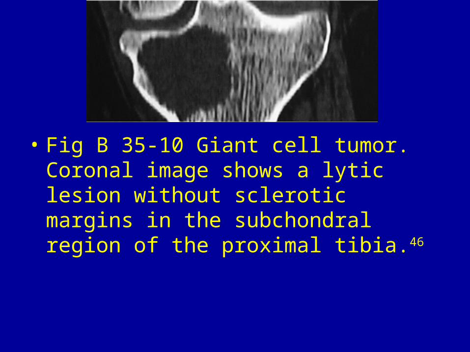
• Fig B 35-10 Giant cell tumor. Coronal image shows a lytic lesion without sclerotic margins in the subchondral region of the proximal tibia.46

• Fig B 35-11 Langerhans cell histiocytosis. Well-defined lucent lesion with medullary expansion in the acetabulum. (Courtesy of Jim Wu, MD, Boston)

Fig B 35-12 Bone infarct. Distal femur lesion with central coarse calcification but no endosteal scalloping of the cortex.45

• Fig B 35-13 Intraosseous ganglion. Eccentric oval lesion in the proximal tibia that has low attenuation and ramifications. Note the rim of reactive sclerosis.45

• Fig B 35-14 Brodie's abscess. Area of low attenuation that contains a central sequestrum (arrowhead) and has a sinus tract extending through the thickening cortex.46