Embed Size (px)
Citation preview

ENTEROBACTERIACEAE
KIMAIGA H.O
MBChB (University of Nairobi)

Taxonomical Classification
Two important classifications
• Based on taxonomy
•Kingdom- Bacteria
• Phylum- Proteobacteria
•Class- Gammaproteobacteria
•Order- Enterobacteriales
• Family – Enterobacteriacaea
• Based on lactose fermentation

TRIBE GENUS
I Escherichieae Escherichia, shigella
II Edwardsielleae Edwardsiella
II KlebsielleaeKlebsiella, Enterobacter, Edwardsiella, Hafnia, Serratia
III Salmonelleae Salmonella, Arizona
IV Citrobactereae Citrobacter
IV Protease Proteus, Providencia, Morganella
V Yersineae, Yersinia, Pasteurella
Erwinieae Erwinia, Pectobacterium
TAXONOMY (Ewing)• Tribe concept proposed by Ewing group in genera
that share similar biochemical reactions and diagnostic importance (7 Tribes)

Classification based on lactose fermentation
Lactose fermenters (LF) • Citrobacter• Enterobacter• Escherichia coli• Klebsiella
Non lactose fermenters (NLF)• Salmonella• Shigella• Yersinia• Proteus• SerratiaLate lactose fermenters• Shigella sonnei• Edwardsiella,, Citrobacter,
Arizona,Providencia, Erwinia


General characteristics• Primarily normal flora in intestinal tract of humans and animals and
others found in water and soil. Also called coli forms or enteric bacteria• Gram negative rods• Ferment carbohydrates/glucose with acid production• Most are oxidase –ve (lack cytochrome oxidase)• Catalase +ve• Facultative anaerobes• Non-capsulated except Klebsiella• Non-spore forming• Some motile by peritrichous flagella few genera non motile e.g
Klebsiella• Contain endotoxin in cell wall• Most reduce nitrate to nitrite via nitrate reductase
• Grow in media with bile salts (MacConkey)• Combinations of chromosomal & plasmid-mediated drug resistance
hence importance of in-vitro susceptibility testing

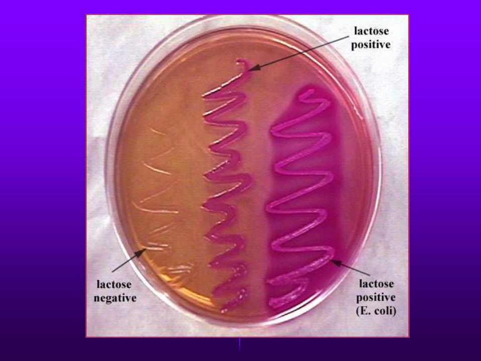
• Media used to distinguish Lactose fermenters and Non-Lactose Fementers
•MacConkey agar - LF-colored/pink, NLF-colorless colonies
• Eosin Methylene Blue (EMB) agar
• Salmonella Shigella (SS) agar
• Triple Sugar Iron (TSI)agar - Slant, butt, gas, H2S


CITROBACTER
• Normal intestinal flora may cause opportunistic infections.
• NLF, Associated with nosocomical UTI and respiratory infections, Endocarditis, neonatal meningitis and brain abscesses
• Treatment
•Multidrug resistance
•Aminoglycosides/ carbapenems/ quinolones/ antipseodomonal penicillins

Enterobacter species
• Previously known as aerobacter spp
• Compromised of 12 species; E. cloacae and E. aerogenes are most common
Characteristics
• Cononies resemble Klebsiella
• Motile peritrichous flagella
• IMViC--++

Clinical manifestations
• Commensals in the human gut
• Most strains cause infections in immunocompromised individual- hospital impatients
• Cause wound infections, burn infection, pneumonia, UTI, Intra-abdominal infections, neonatal
Resistance
• 1st generation cephalosporins
• -lactamase production – resistance to many cephalosporins
Treatment
• Carbapenams,4th generation cephalosporins
• Ampicillin and gentamicin- E.sakazakii

KLEBSIELLASPECIES
• Klebsiella pneumonia – Lobar pneuomina , Most common
• K. oxytoca.- Lobar pneumonia
• K. ozaenae – atrophic rhinitis (ozena)
• K. rhnosceleromatis- rhinoscleroma (Granuloma of the nose and pharynx)
• K. granulomatis- Granuloma inguinale/ donovanosis-painless, non-purulent genital ulcer
• Klebsiella, Citrobacter, Hafnia, Serratia
• Habitat – large intestines, soil, H2O

Pathogenesis
Virulence factors
• Large polysaccharide capsule. Capsular K antigen- antiphagocytic capsule
• Synthesis of siderophores, that are capable of competitively taking up iron bound to proteins
• Pili – adherence to respiratory and urinary epithelia
• Endotoxin – The carry plasmids that code for heat labile and heat stable toxins. Gram negative septic shock

CLINICAL PRESENTATION
• Pulmonary infections- pneumonia lobar;• High fatality• Predisposing factors : elderly, chronic respiratory dx,
diabetes, alcoholism• Extensive necrosis and hemorrhage resulting in thick, mucoid,
brink red sputum currant jelly like.
• Extrapulmonary infections• Meningitis and enteritis in infants• UTI• Septicemia/Bacteremia• Cholecystis, cholangitis, otitis media, sinusitis, peritonitis,
wound/ burn infections,• AN important cause of nosocomial infections
• K.rhinoscleromaticus – inflammatory granulomatous lesions of nose, pharynx, etc

LAB DIAGNOSIS
• Specimens - urine, pus, blood, sputum, CSF• Gram –ve rods• Culture:- MAC , CLED, or BA:—
• Growth conditions, 350C ,18-24 hrs• Mucoid colonies, pink(LF) in MAC and CLED• LF thus yellow in CLED and pink in McC• Non motile
• Biochemical tests• IMViC(--++)• Urease positive• TSI- acid slant, acid butt, gas, no H2S• KSM positive• K pneumoniae can liquefy gelatin.
• Short and plump- Capsule swelling (Quellung reaction)– Typing based on about 90 capsular (K) antigens there are three types –K2,K3 and K21. Rapid identification.

Gram –ve Klebsiella rods

Klebsiella on MAC Klebsiella Spp BA

Klebsiella on NA
Klebsiella on CLED


Klebsiella_Citrate test Indole test

Klebsiella ferments inositol but E. coli does not

TREATMENT
• Hospital- acquired infections – resistance to multiple antibiotics
• Based on antibiotics susceptibility tests
• Beta lactam + beta lactamase inhibitiorcombination
• 3rd generation of cephalosporins e.g cefataxime, ceftriaxone + aminoglycoside