1
Copyright © 2004 Pearson Education, Inc., publishing as Benjamin Cummings
PowerPoint® Lecture Slide Presentation prepared by Christine L. Case
MicrobiologyB.E Pruitt & Jane J. Stein
AN INTRODUCTIONEIGHTH EDITION
TORTORA • FUNKE • CASE
Chapter 11The Prokaryotes:
Domains Bacteria and Archaea
Copyright © 2004 Pearson Education, Inc., publishing as Benjamin Cummings
The Prokaryotes: Domains Bacteria and Archaea• One circular chromosome, not in a membrane• No histones• No organelles• Peptidoglycan cell walls• Binary fission
Learning objective:
Make a dichotomous key to distinguish among the a-proteobacteria described in this chapter.
Copyright © 2004 Pearson Education, Inc., publishing as Benjamin Cummings
Bergey’s Manual
• Categorizes bacteria into taxa based upon rRNA sequences
• Lists identifying characteristics like: • Gram stain reaction• cellular morphology• oxygen requirements• nutritional properties
• Prokaryotes classified into two domains:• Bacteria• Archaea
Copyright © 2004 Pearson Education, Inc., publishing as Benjamin Cummings
• Bacteria essential to life on earth
• Proteobacteria• Mythical
Greek god, Proteus, who could assume many shapes
• Gram-negative
Domain Bacteria
Copyright © 2004 Pearson Education, Inc., publishing as Benjamin Cummings Copyright © 2004 Pearson Education, Inc., publishing as Benjamin Cummings
• Human pathogens:• Bartonella• B. hensela Cat-scratch disease• Brucella Brucellosis
The α (alpha) Proteobacteria

2
Copyright © 2004 Pearson Education, Inc., publishing as Benjamin Cummings
• Includes nitrogen-fixing bacteria, chemoautotrophs, and chemoheterotrophs
• Obligate intracellular parasites:• Ehrlichia. Tick-borne, ehrlichiosis• Rickettsia. Arthropod-borne, spotted fevers
• R. prowazekii Epidemic typhus• R. typhi Endemic murine typhus• R. rickettsii Rocky Mountain Spotted Fever
The α (alpha) Proteobacteria
Copyright © 2004 Pearson Education, Inc., publishing as Benjamin Cummings
The α (alpha) Proteobacteria
Figure 11.1
Copyright © 2004 Pearson Education, Inc., publishing as Benjamin Cummings
• Wolbachia. Live in insects and other animals
• In an infected pair, only female hosts can reproduce
• “Popcorn” strain causes host cells to lyse
• Possible biological control of insects
The α (alpha) Proteobacteria
Copyright © 2004 Pearson Education, Inc., publishing as Benjamin Cummings
• Have prosthecae:• Caulobacter. Stalked
bacteria found in lakes
• Hyphomicrobium. Budding bacteria found in lakes
The α (alpha) Proteobacteria
Figure 11.2 & 3
Copyright © 2004 Pearson Education, Inc., publishing as Benjamin Cummings
• Plant pathogen:• Agrobacterium.
Insert a plasmid into plant cells, inducing a tumor
The α (alpha) Proteobacteria
Figure 9.17 Copyright © 2004 Pearson Education, Inc., publishing as Benjamin Cummings
• Chemoautotrophic:• Oxidize nitrogen for energy• Fix CO2
• Nitrobacter. NH3+ → NO2
–
• Nitrosomonas. NO2– → NO3
–
The α (alpha) Proteobacteria

3
Copyright © 2004 Pearson Education, Inc., publishing as Benjamin Cummings
• Nitrogen-fixing bacteria:• Azospirillum
• Grow in soil, using nutrients excreted by plants
• Fix nitrogen• Rhizobium
• Fix nitrogen in the roots of plants
The α (alpha) Proteobacteria
Figure 27.5 Copyright © 2004 Pearson Education, Inc., publishing as Benjamin Cummings
• Produce acetic acid from ethyl alcohol:• Acetobacter• Gluconobacter
The α (alpha) Proteobacteria
Copyright © 2004 Pearson Education, Inc., publishing as Benjamin Cummings Copyright © 2004 Pearson Education, Inc., publishing as Benjamin Cummings
The β (beta) Proteobacteria
Learning objective:
Make a dichotomous key to distinguish among the β-proteobacteria described in this chapter.
Copyright © 2004 Pearson Education, Inc., publishing as Benjamin Cummings
• Thiobacillus• Chemoautotrophic, oxidize sulfur: H2S → SO4
2–
• Sphaerotilus• Chemoheterotophic, form sheaths
The β (beta) Proteobacteria
Figure 11.5
Sphaerotilus natans:
•Sheathed bacteria found in dilute sewage and aquatic environs
Copyright © 2004 Pearson Education, Inc., publishing as Benjamin Cummings
• Spirillum volutans:• Chemoheterotrophic,
helical• Note polar flagella
• Neisseria• Chemoheterotrophic,
cocci• N. meningitidis• N. gonorrhoeae (diplo-
cocci) – fimbriae attach to mucous membranes for greater pathogenicity
The β (beta) Proteobacteria
Figure 11.4 & 6
4
Copyright © 2004 Pearson Education, Inc., publishing as Benjamin Cummings
• Bordetella• Chemoheterotrophic, rods• B. pertussis (pertussis or whooping cough)
• Burkholderia. Nosocomial infections (hospital infection)• Extraordinary nutritional spectrum, able to degrade
> 100 different organic molecules, can grow in disinfectant!
• Zoogloea. Slimy masses in aerobic sewage-treatment processes – essential to sewage treatment
The β (beta) Proteobacteria
Copyright © 2004 Pearson Education, Inc., publishing as Benjamin Cummings
Copyright © 2004 Pearson Education, Inc., publishing as Benjamin Cummings
The γ (gamma) Proteobacteria
Learning objective:
Make a dichotomous key to distinguish among the γ -proteobacteria described in this chapter.
Copyright © 2004 Pearson Education, Inc., publishing as Benjamin Cummings
• Pseudomonadales:• Pseudomonas
• Gram -• Opportunistic
pathogens• Metabolically
diverse• Polar flagella (characteristic, as in picture)
• Azotobacter and Azomonas. Nitrogen fixing• Moraxella. Conjunctivitis
The γ (gamma) Proteobacteria
Figure 11.7
Copyright © 2004 Pearson Education, Inc., publishing as Benjamin Cummings
• Legionellales:• Legionella
• Found in streams, warm-water pipes, cooling towers of air-conditioning
• L. pneumophilia (Legionnaire's)
• Coxiella• Q fever
transmitted via aerosols or milk
The γ (gamma) Proteobacteria
Figure 24.15b
Coxiella burnetii
Copyright © 2004 Pearson Education, Inc., publishing as Benjamin Cummings
• Vibrionales:• Found in coastal water
• Vibrio cholerae causes cholera
• Slight curvature of rods
• V. parahaemolyticuscauses gastroenteritis (raw/undercooked shellfish)
The γ (gamma) Proteobacteria
Figure 11.8

5
Copyright © 2004 Pearson Education, Inc., publishing as Benjamin Cummings
• The γ (gamma) Proteobacteria• Enterobacteriales (enterics – intestinal tracts):
• Peritrichous flagella, facultatively anaerobic• Enterobacter• Erwinia• Escherichia• Klebsiella• Proteus• Salmonella• Serratia• Shigella• Yersinia
The γ (gamma) Proteobacteria
Copyright © 2004 Pearson Education, Inc., publishing as Benjamin Cummings
The γ (gamma) Proteobacteria
Figure 11.9a, b
Proteus mirabilis – swarmer due to multiple flagella
Copyright © 2004 Pearson Education, Inc., publishing as Benjamin Cummings
• Pasteurellales: • Non-motile• Human and animal pathogens• Pasteurella
• Cause pneumonia and septicemia• Haemophilus
• Require X factor (heme) and V factor (NAD+, NADP+) factors from blood hemoglobin
• H. influenzae – several important diseases (meningitis, earaches, epiglotitis, bronchitis, etc.)
The γ (gamma) Proteobacteria
Copyright © 2004 Pearson Education, Inc., publishing as Benjamin Cummings
• Beggiatoa• Chemoautotrophic, oxidize H2S to S0 for energy• Interface between aerobic and anaerobic layers in
aquatic sediments• Factor in discovery of of autotrophic metabolism
• Francisella• Chemoheterotrophic, tularemia
The γ (gamma) Proteobacteria
Copyright © 2004 Pearson Education, Inc., publishing as Benjamin Cummings Copyright © 2004 Pearson Education, Inc., publishing as Benjamin Cummings
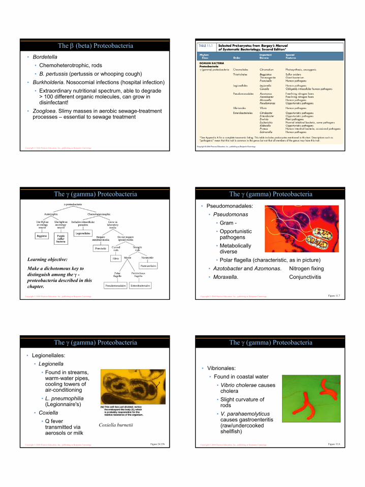
The δ (delta) Proteobacteria
Learning objective:
Make a dichotomous key to distinguish among the δ -proteobacteria described in this chapter.
6
Copyright © 2004 Pearson Education, Inc., publishing as Benjamin Cummings
• Bdellovibrio. Prey on other bacteria• Desulfovibrionales. Use S instead of O2 as final
electron acceptor (sulfur reducing)• Releases tons of H2S into atmosphere, key part in
sulfur cycle• Myxococcales. Gliding. Cells aggregate to form
myxospores (stalked fruiting body – 2nd slide next)• Leave behind a slime trail (next slide)• Nutrition from bacteria they encounter
The δ (delta) Proteobacteria
Copyright © 2004 Pearson Education, Inc., publishing as Benjamin Cummings
The δ (delta) Proteobacteria
Figure 11.10a
Copyright © 2004 Pearson Education, Inc., publishing as Benjamin Cummings
The δ (delta) Proteobacteria
Figure 11.1b Copyright © 2004 Pearson Education, Inc., publishing as Benjamin Cummings
The ε (epsilon) Proteobacteria
Learning objective:
Make a dichotomous key to distinguish among the ε -proteobacteria described in this chapter.
Copyright © 2004 Pearson Education, Inc., publishing as Benjamin Cummings
The ε (epsilon) Proteobacteria
Figure 11.1a
• Helicobacter• Multiple flagella • Peptic ulcers• Stomach cancer
• Campylobacter• One polar flagellum• Gastroenteritis
Copyright © 2004 Pearson Education, Inc., publishing as Benjamin Cummings
Helicobacter pylori:
Example of a helical bacterium that doesn’t make a complete twist (different from spirochetes)
7
Copyright © 2004 Pearson Education, Inc., publishing as Benjamin Cummings Copyright © 2004 Pearson Education, Inc., publishing as Benjamin Cummings
Copyright © 2004 Pearson Education, Inc., publishing as Benjamin Cummings
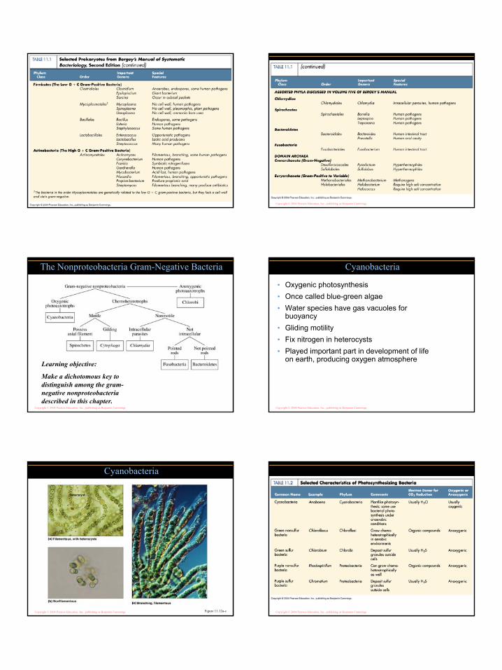
The Nonproteobacteria Gram-Negative Bacteria
Learning objective:
Make a dichotomous key to distinguish among the gram-negative nonproteobacteria described in this chapter.
Copyright © 2004 Pearson Education, Inc., publishing as Benjamin Cummings
• Oxygenic photosynthesis • Once called blue-green algae• Water species have gas vacuoles for
buoyancy• Gliding motility• Fix nitrogen in heterocysts• Played important part in development of life
on earth, producing oxygen atmosphere
Cyanobacteria
Copyright © 2004 Pearson Education, Inc., publishing as Benjamin Cummings
Cyanobacteria
Figure 11.12a-c Copyright © 2004 Pearson Education, Inc., publishing as Benjamin Cummings

8
Copyright © 2004 Pearson Education, Inc., publishing as Benjamin Cummings
• Anoxygenic photosynthesis• Purple and green sulfur bacteria (bottom formula)
Purple and Green Photosynthetic Bacteria
2H2O + CO2light
(CH2O) + H2O + O2
2H2S + CO2light
(CH2O) + H2O + 2S0
Learning objective:
Compare and contrast purple and green photosynthetic bacteria with cyanobacteria
Copyright © 2004 Pearson Education, Inc., publishing as Benjamin Cummings
Purple sulfur bacteria: intracellular sulfur granules (multicolored refractile objects (anoxygenic photoautotrophs)
Copyright © 2004 Pearson Education, Inc., publishing as Benjamin Cummings
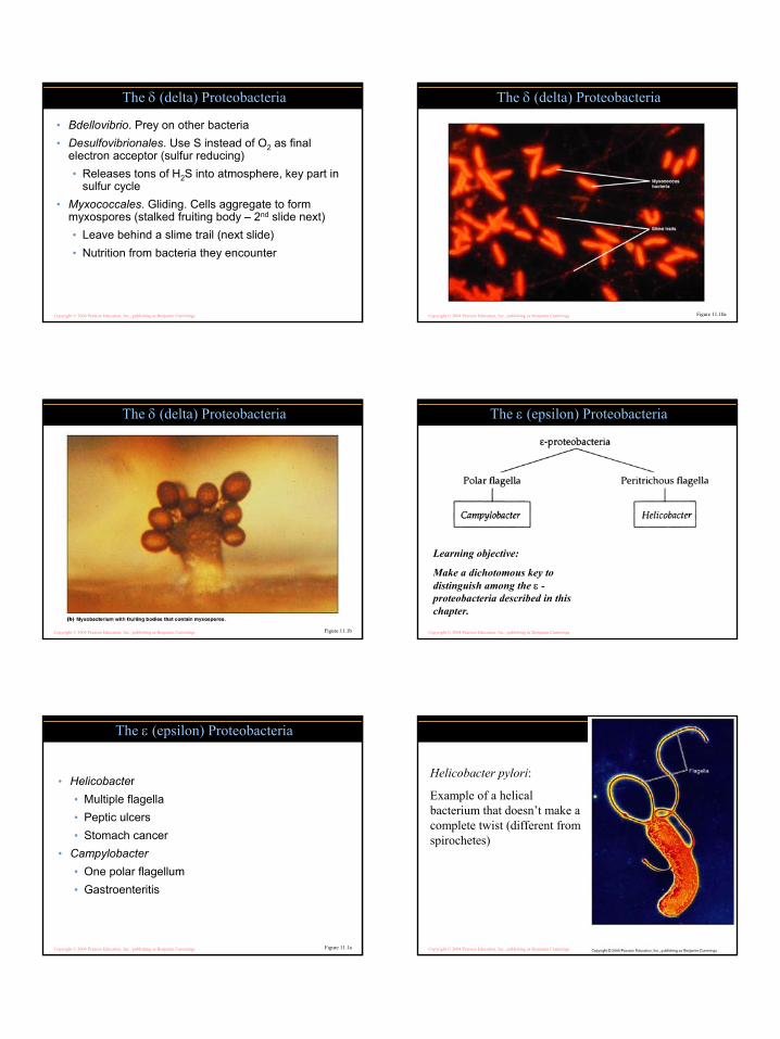
• Low G + C• Gram-positive
FirmicutesLearning objective:
Make a dichotomous key to distinguish among the low G + C gram-positivedescribed in this chapter.
Copyright © 2004 Pearson Education, Inc., publishing as Benjamin Cummings
• Clostridium• Endospore-
producing• Obligate anaerobes• Endospores usually
distend the cell wall• Epulopiscium
• Very large, shown on the head of a pin
• rRNA determined placement with prokaryotes
Clostridiales
Figure 11.14 & 15
Copyright © 2004 Pearson Education, Inc., publishing as Benjamin Cummings
• Bacillus• Endospore-producing
rods• B. anthracis - anthrax
Bacillales
Figure 11.16b Copyright © 2004 Pearson Education, Inc., publishing as Benjamin Cummings
• Staphylococcus aureus• Cocci in grapelike clusters• Gram-positive, produces enterotoxin• Grow fairly well under high osmotic pressure
and low moisture (nasal secretions, skin, ham and other cured meats)
Bacillales
Figure 1.17

9
Copyright © 2004 Pearson Education, Inc., publishing as Benjamin Cummings
• Generally aerotolerant anaerobes, lack an electron-transport chain• Lactobacillus – lactic-
acid producing• Streptococcus –
more illnesses and diseases than any other bacteria group
• Enterococcus –intestinal tract, oral cavity
• Listeria –contaminates dairy
Lactobacillales
Figure 11.18
Streptococcus – many of spherical cells are dividing and somewhat oval
Copyright © 2004 Pearson Education, Inc., publishing as Benjamin Cummings
• Mycoplasma pneumoniae• No cell walls• Pleomorphic (irregular
cells)• Arrows indicate terminal
structures that likely aid attachment to eukaryotic cells
• 0.1 - 0.24 µm
• Filamentous growth of M. pneumoniae• Reproduces by
fragmentation of the filaments
Mycoplasmatales
Figure 11.19a, b
Copyright © 2004 Pearson Education, Inc., publishing as Benjamin Cummings
• High G + C• Gram-positive
ActinobacteriaLearning objective:
Make a dichotomous key to distinguish among the high G + C gram-positivedescribed in this chapter.
Copyright © 2004 Pearson Education, Inc., publishing as Benjamin Cummings
• Actinomyces• Corynebacterium• Frankia• Gardnerella• Mycobacterium• Nocardia• Propionibacterium• Streptomyces
Actinobacteria
Figure 11.20b
Copyright © 2004 Pearson Education, Inc., publishing as Benjamin Cummings
• Streptomyces –• Filamentous
branching growth with asexual reproductive conidiospores at tips
• Make up much of soil bacteria
Copyright © 2004 Pearson Education, Inc., publishing as Benjamin CummingsActinomyces – notice branched filamentous morphology

10
Copyright © 2004 Pearson Education, Inc., publishing as Benjamin Cummings
• C. trachomatis• Trachoma• STD, urethritis
• C. pneumoniae• C. psittaci
• Causes psittacosis
Chlamydiae
Copyright © 2004 Pearson Education, Inc., publishing as Benjamin Cummings
Generalized life cycle of Chlamydia (48 hours)
Figure 11.22a
Copyright © 2004 Pearson Education, Inc., publishing as Benjamin Cummings
Generalized life cycle of Chlamydia
Figure 11.22b
Elementary bodies – infectious stage
Reticulate bodies – reproduce in host cell
Intermediate bodies – stage in between Copyright © 2004 Pearson Education, Inc., publishing as Benjamin Cummings
• Borrelia• Leptospira• Treponema
Spirochaetes
Figure 11.23
Spirochetes –
•Helical, axial filaments under outer sheath
•Move by corkscrewlike rotation
Copyright © 2004 Pearson Education, Inc., publishing as Benjamin Cummings
• Anaerobic• Bacteroides. In mouth and large intestine• Cytophaga. Cellulose-degrading in soil
Phyla Bacteroidetes & Fusobacteria
• Fusobacterium• Found in mouth• May be involved in
dental diseases
Copyright © 2004 Pearson Education, Inc., publishing as Benjamin Cummings
• Hyperthermophiles (heat)• Pyrodictium• Sulfolobus
• Methanogens (methane)• Methanobacterium
• Extreme halophiles (salt)• Halobacterium
Domain Archaea
Figure 11.25
Archaea – Pyrodictium abyssi:
•Deep ocean, 110 degrees C
•Cells disk-shaped with network of tubules (cannulae)
11
Copyright © 2004 Pearson Education, Inc., publishing as Benjamin Cummings
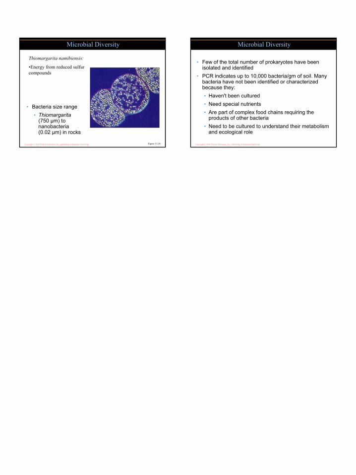
• Bacteria size range• Thiomargarita
(750 µm) to nanobacteria (0.02 µm) in rocks
Microbial Diversity
Figure 11.26
Thiomargarita namibiensis:
•Energy from reduced sulfur compounds
Copyright © 2004 Pearson Education, Inc., publishing as Benjamin Cummings
• Few of the total number of prokaryotes have been isolated and identified
• PCR indicates up to 10,000 bacteria/gm of soil. Many bacteria have not been identified or characterized because they:• Haven't been cultured• Need special nutrients• Are part of complex food chains requiring the
products of other bacteria• Need to be cultured to understand their metabolism
and ecological role
Microbial Diversity
Recommended