Molecules 2013, 18, 8535-8549; doi:10.3390/molecules18078535
molecules ISSN 1420-3049
www.mdpi.com/journal/molecules
Article
Synthesis and Biological Evaluation of a New Acyclic Pyrimidine Derivative as a Probe for Imaging Herpes Simplex Virus Type 1 Thymidine Kinase Gene Expression
Andrijana Meščić 1, Thomas Betzel 2, Adrienne Müller 2, Roger Slavik 2, Stjepko Čermak 2,
Silvana Raić-Malić 1,* and Simon M. Ametamey 2,*
1 Department of Organic Chemistry, Faculty of Chemical Engineering and Technology,
University of Zagreb, Marulićev trg 20, P.O. Box 177, HR-10 000 Zagreb, Croatia;
E-Mails: [email protected]
2 Center for Radiopharmaceutical Sciences, ETH Zurich (Swiss Federal Institute of Technology),
Wolfgang-Pauli-Strasse 10, CH-8093 Zurich, Switzerland;
E-Mails: [email protected] (T.B.); [email protected] (A.M.);
[email protected] (R.S.); [email protected] (S.Č.)
* Authors to whom correspondence should be addressed; E-Mails: [email protected] (S.R.-M.);
[email protected] (S.M.A.); Tel.: + 385-1-4597-213 (S.R.-M.);
Fax: +385-1-4597-224 (S.R.-M.); Tel.: +41-44-633-7463 (S.M.A.); Fax: +41-44-633-1367 (S.M.A.).
Received: 27 May 2013; in revised form: 12 July 2013 / Accepted: 16 July 2013 /
Published: 19 July 2013
Abstract: With the idea of finding a more selective radiotracer for imaging herpes simplex
virus type 1 thymidine kinase (HSV1-tk) gene expression by means of positron emission
tomography (PET), a novel [18F]fluorine radiolabeled pyrimidine with 4-hydroxy-3-
(hydroxymethyl)butyl side chain at N-1 (HHB-5-[18F]FEP) was prepared and evaluated as
a potential PET probe. Unlabeled reference compound, HHB-5-FEP, was synthesized via a
five-step reaction sequence starting from 5-(2-acetoxyethyl)-4-methoxypyrimidin-2-one.
The radiosynthesis of HHB-[18F]-FEP was accomplished by nucleophilic radiofluorination
of a tosylate precursor using [18F]fluoride-cryptate complex in 45% ± 4 (n = 4)
radiochemical yields and high purity (>99%). The biological evaluation indicated the
feasibility of using HHB-5-[18F]FEP as a PET radiotracer for monitoring HSV1-tk
expression in vivo.
OPEN ACCESS
Molecules 2013, 18 8536
Keywords: acyclic pyrimidine nucleoside analogues; PCV-like side chain; fluorination;
radiosynthesis; positron emission tomography (PET); HSV1-TK
1. Introduction
Positron emission tomography (PET) is a noninvasive imaging modality for the in vivo visualization
of various metabolic processes such as cellular proliferation, HSV1-tk reporter gene expression [1–3],
determination of receptor concentrations and the assessment of treatment response to therapy [4–6]. A
paradigm for the non-invasive imaging of transgene expression involves the appropriate combination
of a reporter gene and a reporter substrate or probe [7]. In essence, the reporter gene product
selectively converts a reporter probe into a negatively charged metabolite that is trapped and
accumulates within the transduced cells as it is unable to cross the cell membrane [3]. The
accumulation of radioactivity within the transfected cell can be imaged by PET. The most studied
reporter gene for the visualization of gene expression in animals and humans is herpes virus type 1
thymidine kinase (HSV1-tk) which is visualized by its enzyme product HSV1-TK. A number of 18F-labeled pyrimidine ([18F]-FIAU, [18F]-FMAU [8,9], [18F]-FEAU [10]) and purine ([18F]FHBG
[11], [18F]FHPG [12]) based nucleosides have shown promise as reporter probes for non-invasive
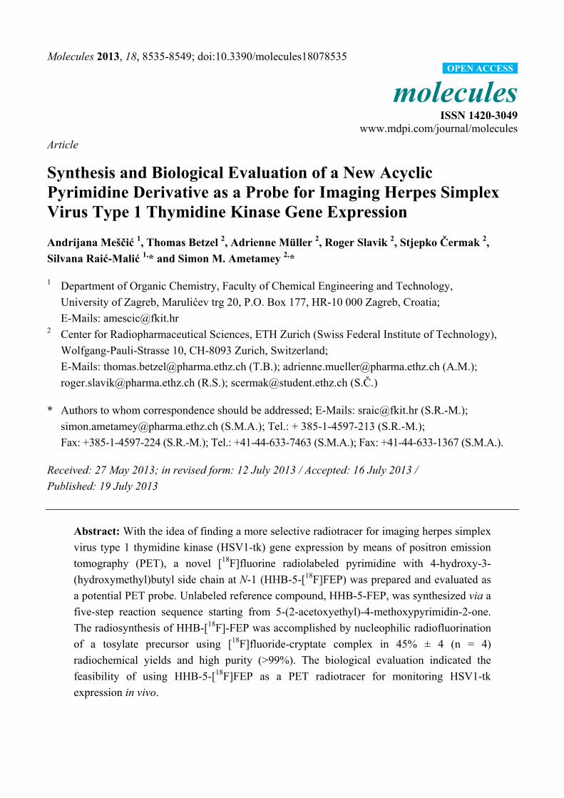
imaging of HSV1-tk gene expression (Figure 1).
Figure 1. Structures of some pyrimidine and purine nucleoside analogues and HHB-5-[18F]FEP.
In recent years, [18F]FEAU has emerged as a tracer with improved sensitivity and selectivity for
imaging HSV1-tk expressing cells [13,14]. Whereas purine [15] and pyrimidine [16] nucleoside
analogues have found application as imaging agents for imaging HSV1-tk expression, cellular
proliferation is mainly imaged with thymidine analogues [17]. The purine analogue, [18F]FHBG,
considered as the gold standard in clinical studies for HSV1-tk reporter gene imaging with PET [18,19]
exhibits high abdominal activity due to hepatobiliary elimination [20,21]. When compared with
pyrimidines, [18F]FHBG shows less sensitivity towards the native HSV1-TK.
We have previously reported on the synthesis, radiosynthesis and the in vivo evaluation of pyrimidine
nucleoside analogues in which acyclic 6-(1,3-dihydroxyisobutyl) and 6-(1,3-dihydroxyisobutenyl) side
chains have been attached at the C-6 position rather than at the N-1 position [22–24]. More recently, we
have also published results on N-Me-[18F]FHBT, a N-methylated thymine derivative bearing a 6-(1,3-
dihydroxyisobutyl) side chain. Compared to [18F]FHBG, N-Me-[18F]FHBT showed a higher
background radioactivity in most tissues [25]. Taking into account the aforementioned potential of
HN
N
HHOH H
H F18OHO
R
O
O
R = I
R = CH3
R = CH3CH2
18F FIAU
18F FMAU
18F FEAU
HN
N
O
H2N N
N
XOH
F1818F FHBG18F FHPG
HN
N
O
O
OH
OH
F18
HHB-5-18F FEP, ( 18F 7 ), this work
X = CH2
X = O
Molecules 2013, 18 8537
pyrimidine based nucleosides for the development of novel fraudulent substrates of HSV-1 TK with
improved pharmacodynamic and pharmacokinetic profile, we prepared a series of novel C-5 and N-1-
substituted pyrimidine derivatives [26]. Among these series, a pyrimidine acyclonucleoside bearing a
penciclovir (PCV)-like side chain, which was shown to be a substrate of HSV-1 TK, was selected as a
lead compound for development as a PET imaging agent for measuring HSV-1 TK expression.
Here we report on the synthesis of unlabeled HHB-5-FEP and the radiosynthesis of its fluorine-18
labeled counterpart, HHB-5-[18F]FEP (Figure 1). We further present results of the in vitro cellular
uptake and the in vivo evaluation of HHB-5-[18F]FEP. A direct comparison of the small animal PET
imaging and biodistribution for HHB-5-[18F]FEP and [18F]FHBG, as the most commonly used
HSV1-TK imaging agent, is also presented.
2. Results and Discussion
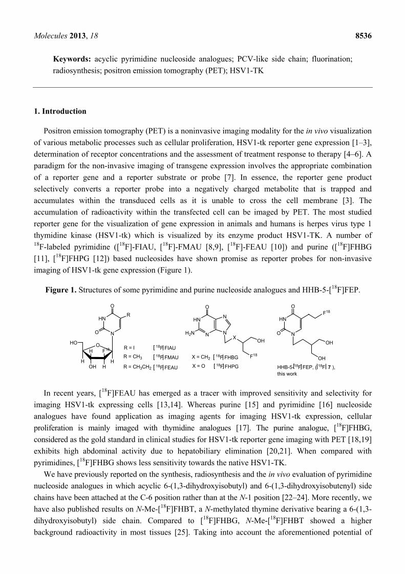
2.1. Synthesis of the Reference HHB-5-FEP (7) and Precursor 8
The target compound, N-1-[4-hydroxy-3-(hydroxymethyl)butyl]-5-(2-fluoroethyl)pyrimidin-2,4-
dione (HHB-5-FEP, 7) was prepared as outlined in Scheme 1.
Scheme 1. Synthesis of reference compound HHB-5-FEP (7) and precursor 8.
Reagents and conditions: (i) K2CO3, DMF, 20 min; (ii) 0.1 M NaOMe/MeOH in MeOH, 1 h; (iii) p-TsOH ×
H2O, 2,2-dimethoxypropane, DMF, 2 h; (iv) DAST, CH2Cl2, −40 °C, 3 h; (v) Method A. NaI, TMSCl,
MeCN, reflux, 10 min; Method B. HCl, reflux, 10 min; (vi) TsCl, pyridine, 2 h.
N
NH
O
OCH3
OAc
I
OAc
OAcN
NO
OCH3
OAcN
NO
OCH3
OH
N
NO
OCH3
OH
O
O
N
NO
OCH3
F
O
O
i ii
iii
vi
ivv
1 2
3 4
5
8
6HHB-5-FEP (7)
HN
NO
O
F
OH
OH
N
NO
OCH3
OTs
O
O
OAC
OAc
OH
OH
Molecules 2013, 18 8538
The introduction of penciclovir-like chain at N-1 position of pyrimidine scaffold was performed by
reaction of 5-(2-acetoxyethyl)-4-methoxypyrimidin-2-one (1) with 4-acetoxy-(3-acetoxymethyl)butyl
iodide (2) to give acyclic C-5-substituted pyrimidine derivative 3. Acetyl groups in both N-1 and C-5
side chains were then removed under basic conditions to give the triol 4 [27]. Protection of the
1,3-diols in penciclovir-like side chain was carried out using p-toluenesulfonic acid monohydrate
(p-TsOH×H2O) and 2,2-dimethoxypropane to afford acetonide 5 in 59% chemical yield. Transformation
of compound 5 to the fluorinated derivative 6 was achieved in a one-step reaction in 34% yield using
diethylaminosulfur trifluoride (DAST) as fluorinating reagent. Initial attempts to prepare compound 7
from 6 using NaI, TMSCl in MeCN (Method A) afforded 7 in a somewhat lower yield (24%). Besides
the formation of several by-products, purification of 7 also proved tedious. Method B which involves
the use of concentrated acid provided target compound 7 in an optimal 29% yield.
In order to synthesize tosylate precursor 8 for the radiosynthesis of HHB-5-[18F]FEP,
5-(2-hydroxyethyl)pyrimidine derivative 5 was treated with p-toluenesulfonyl chloride in pyridine to
give compound 8 in 68% yield (Scheme 1). The identities of all the synthesized compounds were
confirmed by MS and NMR-spectroscopy.
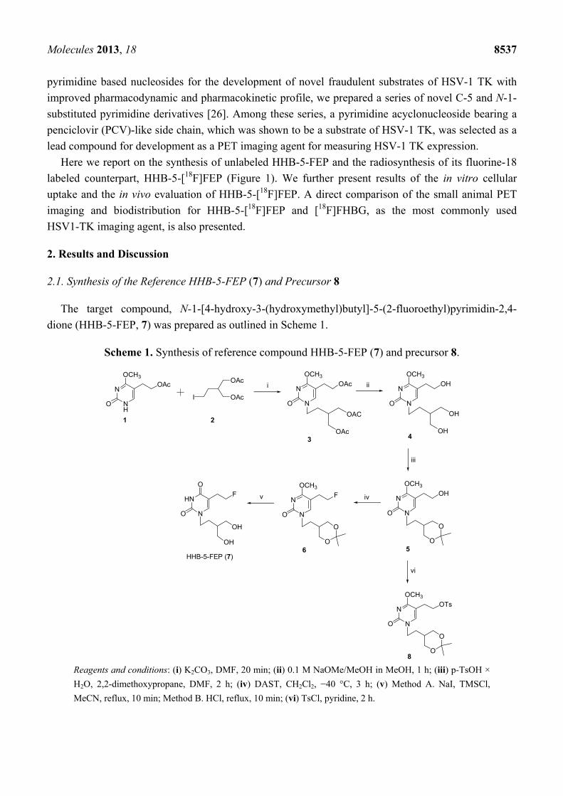
2.2. Radiosynthesis of HHB-5-[18F]FEP ([18F]7)
The radiosynthesis of [18F]7 was carried out using a two-step procedure and consisted of
[18F]fluorination and cleavage of the protecting groups (Scheme 2). Nucleophilic substitution of the
tosyl group by [18F]fluoride was performed at 100 °C using MeCN as solvent. A maximal
incorporation yield of 60% was accomplished for [18F]6 after a reaction time of 8 min. Cleavage of the
protecting groups under acidic conditions at 100 °C afforded HHB-5-[18F]FEP in quantitative
radiochemical yield.
Scheme 2. Radiosynthesis of HHB-5-[18F]FEP
Reagents and conditions: (i) [18F]KF-K2.2.2, MeCN, 100 °C, 8 min; (ii) 37% HCl, 100 °C, 10 min.
HHB-5-[18F]FEP was purified by a semipreparative radio-HPLC within 25 min (tR = 21.08 min)
and formulated for in vitro and in vivo studies. The radiochemical yield was 45% ± 4, (n = 4) decay
corrected. The total amount of radioactivity at the end of the synthesis (EOS) was up to 7.36 GBq in a
radiochemical purity of > 99%. The specific activity ranged between 50 and 135 GBq/µmol after a
total synthesis time of approx. 90 min. The chemical identity of HHB-5-[18F]FEP was confirmed by
coinjection with the non-radiolabeled reference compound HHB-5-FEP (7).
N
NO
OCH3
OTs
O
O
i
8
N
NO
OCH318F
O
O
[18F]6
HN
NO
O18F
OH
OH
HHB-5-[18F]FEP ([18F]7)
ii
Molecules 2013, 18 8539
2.3. Cell Uptake Studies
The in vitro uptake of HHB-5-[18F]FEP was performed on HEK293TK+ cells and compared to wild
type HEK293 cells. The uptake of HHB-5-[18F]FEP was at all time points (60, 120, 240 min) higher in
HEK293TK+ cells than in control cells. For the investigated time points, the ratio of radioactivity
uptake in HEK293TK+ and HEK293 cells was 35–41-fold higher than in wild type cells (Figure 2).
This favorable in vitro properties encouraged the further in vivo testing of HHB-5-[18F]FEP.
Figure 2. Uptake ratios of HHB-5-[18F]FEP in HEK293TK+ and wild type cells.
2.4. Small Animal PET Imaging with HHB-5-[18F]FEP and [18F]FHBG
Static whole body (two beds, 60–90 min) PET images of xenograft-bearing mice after i.v. injection
of [18F]FHBG and HHB-5-[18F]FEP are shown in Figure 3.
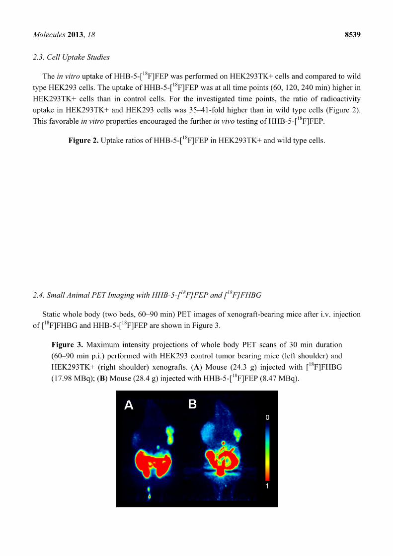
Figure 3. Maximum intensity projections of whole body PET scans of 30 min duration
(60–90 min p.i.) performed with HEK293 control tumor bearing mice (left shoulder) and
HEK293TK+ (right shoulder) xenografts. (A) Mouse (24.3 g) injected with [18F]FHBG
(17.98 MBq); (B) Mouse (28.4 g) injected with HHB-5-[18F]FEP (8.47 MBq).
Molecules 2013, 18 8540
2.5. Biodistribution
Similar to the in vitro cell uptake studies, a higher uptake of HHB-5-[18F]FEP in TK-positive
xenograft was observed compared to HEK293 control xenograft. The SUVPET for the TK-positive
xenograft was 0.32. Uptake in the control xenograft was slightly higher (SUVPET = 0.17) compared to
background activity (SUVPET = 0.08). Compared to [18F]FHBG, HHB-5-[18F]FEP revealed a higher
background activity but a lower abdominal radioactivity. [18F]FHBG showed similar imaging
characteristics as previously shown [28]. The SUVPET for HHB-5-[18F]FEP was higher than the
SUVPET for [18F]FHBG, however, due to the higher background activity of HHB-5-[18F]FEP, a lower
TK+/control ratio was obtained (Table 1).
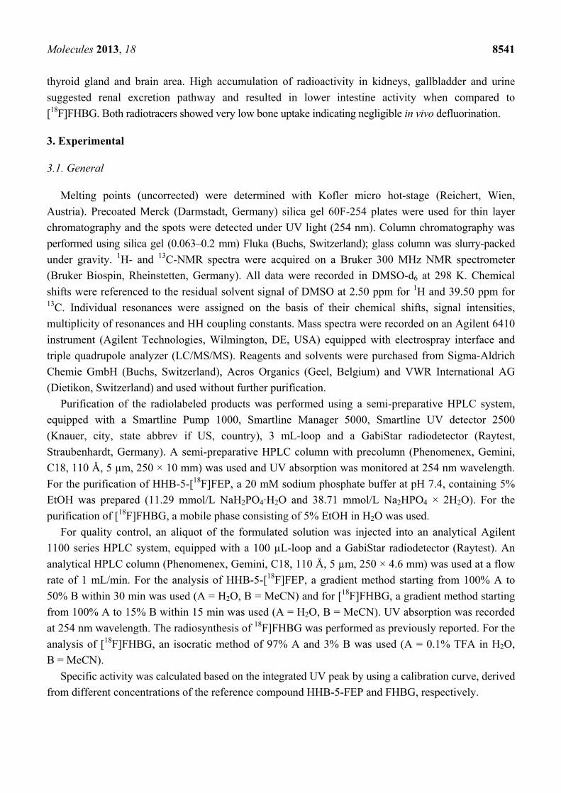
Table 1. Biodistribution (SUVbiodis) and PET data (SUVPET) of [18F]FHBG and
HHB-5-[18F]FEP in xenograft-bearing mice at 95 min after radiotracer injection.
Tissue
[18F]FHBG HHB-5-[18F]FEP
SUVbiodis SUVPET Ratio
SUVbiodis SUVbiodis SUVPET
Ratio
SUVbiodis
Xenograft TK+ 0.24 ± 0.07 0.22 ± 0.02 TK+/control
5.7 ± 1.9 0.37 ± 0.15 0.32 ± 0.06
TK+/control
3.4 ± 1.1
Xenograft control 0.04 ± 0.00 0.09 ± 0.004 0.11 ± 0.01 0.17 ± 0.02
Blood 0.03 ± 0.02 0.07 ± 0.04
Spleen 0.08 ± 0.08 0.05 ± 0.00
Liver 0.16 ± 0.19 0.06 ± 0.00
Kidney 0.12 ± 0.06 0.25 ± 0.09
Lung 0.03 ± 0.01 0.05 ± 0.00
Bone 0.04 ± 0.03 0.05 ± 0.01
Heart 0.04 ± 0.05 0.04 ± 0.01
Brain 0.002 ± 0.00 0.01 ± 0.00
Stomach w. cont. 0.02 ± 0.01 0.05 ± 0.01
Intestine w. cont. 1.78 ± 0.04 0.78 ± 0.05
Pancreas 0.03 ± 0.01 0.06 ± 0.02
Muscle 0.02 ± 0.00 0.04 ± 0.01 0.08 ± 0.02 0.08 ± 0.002
Thyroid 0.02 ± 0.01 0.12 ± 0.11
Gallbladder 0.01 to 3.6 1.5 to 3.5
Urine 15 to 29 13 to 182
The region with the highest uptake radioactivity was the abdomen followed by the gallbladder and
the TK-positive xenograft. Table 1 summarizes the biodistribution data of [18F]FHBG and HHB-5-
[18F]FEP in xenograft-bearing mice at 95 min after radiotracer injection. SUVbiodis data are the mean ±
standard deviation of three animals for [18F]FHBG and HHB-5-[18F]FEP. Radioactivity accumulation
for [18F]FHBG and HHB-5-[18F]FEP in TK-positive xenografts was significantly higher than in the
control xenografts (p = 0.004 and 0.033, respectively, Student’s t-Test). The uptake ratio of TK+ to
control xenograft was 5.7 ± 1.9 for [18F]FHBG (n = 3) and 3.4 ± 1.1 for HHB-5-[18F]FEP (n = 3). The
distribution patterns of the two tracers were similar, however, HHB-5-[18F]FEP exhibited lower
radioactivity levels in the intestine, liver and spleen and higher activity in the muscles, blood, kidneys,
Molecules 2013, 18 8541
thyroid gland and brain area. High accumulation of radioactivity in kidneys, gallbladder and urine
suggested renal excretion pathway and resulted in lower intestine activity when compared to
[18F]FHBG. Both radiotracers showed very low bone uptake indicating negligible in vivo defluorination.
3. Experimental
3.1. General
Melting points (uncorrected) were determined with Kofler micro hot-stage (Reichert, Wien,
Austria). Precoated Merck (Darmstadt, Germany) silica gel 60F-254 plates were used for thin layer
chromatography and the spots were detected under UV light (254 nm). Column chromatography was
performed using silica gel (0.063–0.2 mm) Fluka (Buchs, Switzerland); glass column was slurry-packed
under gravity. 1H- and 13C-NMR spectra were acquired on a Bruker 300 MHz NMR spectrometer
(Bruker Biospin, Rheinstetten, Germany). All data were recorded in DMSO-d6 at 298 K. Chemical
shifts were referenced to the residual solvent signal of DMSO at 2.50 ppm for 1H and 39.50 ppm for 13C. Individual resonances were assigned on the basis of their chemical shifts, signal intensities,
multiplicity of resonances and HH coupling constants. Mass spectra were recorded on an Agilent 6410
instrument (Agilent Technologies, Wilmington, DE, USA) equipped with electrospray interface and
triple quadrupole analyzer (LC/MS/MS). Reagents and solvents were purchased from Sigma-Aldrich
Chemie GmbH (Buchs, Switzerland), Acros Organics (Geel, Belgium) and VWR International AG
(Dietikon, Switzerland) and used without further purification.
Purification of the radiolabeled products was performed using a semi-preparative HPLC system,
equipped with a Smartline Pump 1000, Smartline Manager 5000, Smartline UV detector 2500
(Knauer, city, state abbrev if US, country), 3 mL-loop and a GabiStar radiodetector (Raytest,
Straubenhardt, Germany). A semi-preparative HPLC column with precolumn (Phenomenex, Gemini,
C18, 110 Å, 5 µm, 250 × 10 mm) was used and UV absorption was monitored at 254 nm wavelength.
For the purification of HHB-5-[18F]FEP, a 20 mM sodium phosphate buffer at pH 7.4, containing 5%
EtOH was prepared (11.29 mmol/L NaH2PO4·H2O and 38.71 mmol/L Na2HPO4 × 2H2O). For the
purification of [18F]FHBG, a mobile phase consisting of 5% EtOH in H2O was used.
For quality control, an aliquot of the formulated solution was injected into an analytical Agilent
1100 series HPLC system, equipped with a 100 µL-loop and a GabiStar radiodetector (Raytest). An
analytical HPLC column (Phenomenex, Gemini, C18, 110 Å, 5 µm, 250 × 4.6 mm) was used at a flow
rate of 1 mL/min. For the analysis of HHB-5-[18F]FEP, a gradient method starting from 100% A to
50% B within 30 min was used (A = H2O, B = MeCN) and for [18F]FHBG, a gradient method starting
from 100% A to 15% B within 15 min was used (A = H2O, B = MeCN). UV absorption was recorded
at 254 nm wavelength. The radiosynthesis of 18F]FHBG was performed as previously reported. For the
analysis of [18F]FHBG, an isocratic method of 97% A and 3% B was used (A = 0.1% TFA in H2O,
B = MeCN).
Specific activity was calculated based on the integrated UV peak by using a calibration curve, derived
from different concentrations of the reference compound HHB-5-FEP and FHBG, respectively.
Molecules 2013, 18 8542
3.2. Cell Lines
3.2.1. Cell Culture
Cell uptake and internalization experiments were performed as previously described [25]. In brief,
HEK293 human embryonic kidney cells and HEK293 stable transfected with nonmutant HSV1-tk
(HEK293TK+ cells) were cultured in high glucose DMEM media supplemented with 10% FBS and
1% Penicillin/Streptomycin. Cells were grown in humidified atmosphere with 5% CO2 at 37 °C.
Thymidine kinase expression in the HEK293TK+ cells was maintained with 0.3 mg/mL G418 in the
culture medium and was routinely verified by visualization of the cotransfected RFP with a
fluorescence microscope.
3.2.2. Cell Uptake
HEK293TK+ and HEK293 control cells were seeded in 12-well culture plates (8 × 10⁵ cells per
well) in DMEM supplemented with 10% FBS. After 24 h, when cultures reached 80% confluence,
cells were incubated for 30, 60, and 240 min with medium (1 mL) containing 130 kBq HHB-5-
[18F]FEP per well. At the end of the incubation, cells were washed twice with PBS and detached with
0.25% trypsin (0.3 mL). The cells were resuspended in culture medium (0.7 mL), sedimented and
lysed in lysis buffer (0.5 mL, 0.0625 M Tris, 2% sodium dodecyl sulfate, 7% glycerol, pH 6.8). The
radioactivity of the cell lysate and the combined incubation medium were measured in a gamma
counter (Wizard; PerkinElmer). Radioactivity of the cell lysates was normalized to total protein
determined in cell lysate (50 μL) with the DC™ Protein Assay Kit I (BioRad, Hercules, CA). Data
were expressed as percent accumulated activity/μg protein ((dpm cells × 100%/(dpm cells + dpm
medium))/μg protein) and uptake ratios (dpm HEK293TK+/dpm HEK293 control), respectively [29].
3.3. Animals
All animal experiments were approved by the local veterinarian department and complied with
Swiss and local laws on animal protection. Six week-old female NMRI nude mice were purchased
from Charles River Laboratories (Sulzfeld, Germany). Under 2-3% isoflurane anesthesia, animals were
injected subcutaneously with 5 × 10⁶ cells in Matrigel (100 µL). Transfected HEK293TK+ cells were
injected on the right side of the shoulder region, control HEK293 cells on the left side. Xenograft
growth and body weight were monitored regularly. Experiments were conducted when the xenografts
reached a volume of 1–2 cm3, which was approx. four weeks after inoculation.
3.3.1. In Vivo PET Scan
For PET imaging, eXplore VISTA PET/CT tomograph (Sedecal/GE Healthcare, Madrid, Spain)
was used. Nude mice, bearing HEK293TK+ xenografts on the right shoulder and HEK293 xenografts
(control) on the left shoulder were injected with HHB-5-[18F]FEP or [18F]FHBG formulations (100 μL
per injection) via lateral tail vein injection (t = 0). After injection of the radiotracer, mice were
anesthetized by inhalation of isoflurane in an air/oxygen mixture approximately 5 min prior to PET
data acquisition and scanned as described previously [30]. In dynamic PET mode, one animal was
Molecules 2013, 18 8543
scanned from 0–90 min. For the second animal, data were acquired from 60–150 min p.i. For static
PET scans, mice were scanned from 60–90 min. as it revealed to be the optimal time frame. After
acquisition, PET data were reconstructed and fused datasets of PET and CT were analyzed with
PMOD software (version 3.4).
3.3.2. Ex Vivo Biodistribution Studies of [18F]FHBG and of HHB-5-[18F]FEP
To perform ex vivo biodistribution studies of HHB-5-[18F]FEP and [18F]FHBG, HEK293 control
and HEK293TK+ xenograft bearing nude mice were euthanized immediately after the static PET
experiments, which was 95 min post tracer application (n = 3). Organs and tissue samples were
weighed and the radioactivity determined in a gamma counter. Decay corrected radioactivity was
expressed in analogy to the SUV (standardized uptake values) as the ratio of the detected activity per
gram tissue and the injected dose per gram body weight (SUVbiodis).
3.4. Procedures for the Preparation of Compounds
N-1-[4-Acetoxy-3-(acetoxymethyl)butyl]-5-(2-acetoxyethyl)-4-methoxypyrimidin-2-one (3) and
N-1-[4-hydroxy-3-(hydroxymethyl)butyl]-5-(2-hydroxyethyl)-4-methoxypyrimidin-2-one (4) were
prepared according to a previously published procedure [27].
N-1-[2-(2,2-Dimethyl-1,3-dioxane-5-yl)ethyl]-5-(2-hydroxyethyl)-4-methoxypyrimidin-2-one (5). To a
stirred solution of compound 4 (726 mg, 2.67 mmol) in dry DMF (9 mL), 2,2,-dimethoxypropane
(0.61 mL, 3.89 mmol) and p-toluenesulfonic acid monohydrate (15.17 mg, 0.08 mmol) were added.
The stirring was continued for 2 h at room temperature. The reaction mixture was then neutralized by
triethylamine. The solvent was removed under reduced pressure and the residue was purified by
column chromatography on silica gel (CH2Cl2-CH3OH = 10:1) to give compound 5 as white crystals
(494.7 mg, 59%, mp: 120–121 °C). 1H-NMR: (δ) 7.78 (1H, s, H-6), 4.60 (1H, t, OH, J = 4.5 Hz), 3.83
(3H, s, OCH3), 3.72–3.82 (4H, m, H-4'', H-2'), 3.44–3.57 (4H, m, H-4'', H-1''), 2.41 (2H, t, H-1',
J = 6.7 Hz), 1.58–1.70 (1H, m, H-3''), 1.53 (2H, q, H-2'', J = 7.1 Hz), 1.33 (3H, s, CH3), 1.27 (3H, s,
CH3) ppm. 13C-NMR: (δ) 170.0 (C-4), 155.7 (C-2), 147.2 (C-6), 104.6 (C-5), 97.5 (C-5''), 63.9 (C-4''),
61.9 (C-2'), 60.0 (C-4''), 54.3 (OCH3), 47.8 (C-1''), 41.3 (C-3''), 30.0 (C-1'), 28.3 (C-2''), 27.1 (CH3),
21.6 (CH3) ppm. MS (ESI): m/z = 313.2 ([M+H]+).
N-1-[2-(2,2-Dimethyl-1,3-dioxane-5-yl)ethyl]-5-(2-fluoroethyl)-4-methoxypyrimidin-2-one (6). A
solution of compound 5 (296.7 mg, 0.95 mmol) in anhydrous CH2Cl2 (7 mL) was cooled to −68 °C
and diethylaminosulfur trifluoride (DAST) (0.27 mL, 2.04 mmol) was added under argon atmosphere.
The reaction mixture was stirred at −40 °C for 3 h and then allowed to warm up to room temperature.
The solvent was evaporated and the residue chromatographed (CH2Cl2-CH3OH = 15:1). Compound 6
was isolated as a colourless oil (100.2 mg, 34%). 1H-NMR: (δ) 7.89 (1H, s, H-6), 4.52 (2H, dt, H-2',
JH-F = 47.1 Hz, JH-H = 6.3 Hz), 3.84 (3H, s, OCH3), 3.74–3.80 (2H, m, H-4''), 3.50–3.55 (4H, m, H-4'',
H-1''), 2.64–2.72 (2H, dt, H-1', JH-F = 23.4 Hz, JH-H = 6.3 Hz), 1.61–1.67 (1H, m, H-3''), 1.51–1.55
(2H, m, H-2''), 1.32 (3H, s, CH3), 1.27 (3H, s, CH3) ppm. MS (ESI): m/z 315.1 ([M+H]+).
Molecules 2013, 18 8544
N-1-[4-Hydroxy-3-(hydroxymethyl)butyl]-5-(2-fluoroethyl)pyrimidin-2,4-dione (HHB-5-FEP, 7).
Method A. To a solution of compound 6 (40 mg, 0.13 mmol) and MeCN (2 mL), trimethylsilyl
chloride (TMSCl) (0.05 mL, 0.39 mmol) and NaI (39.5 mg, 0.26 mmol) were added under argon
atmosphere. The mixture was stirred at reflux for 10 min, the solvent was evaporated under reduced
pressure and the residue chromatographed (EtOAc-CH3OH = 10:1). Compound 7 was obtained as
yellow oil (8.0 mg, 24%).
Method B. Compound 6 (37.3 mg, 0.12 mmol) was dissolved in concentrated 37% HCl (1.6 mL) and
the mixture was stirred at reflux for 10 min. Then, it was neutralized by the addition of 6 M NaOH.
The salt was filtered of, the solvent was evaporated under reduced pressure and the residue
chromatographed (CH2Cl2-CH3OH = 10:1). Compound 7 was obtained as colourless oil (9.0 mg,
29%). 1H-NMR: (δ) 11.24 (1H, s, NH), 7.58 (1H, s, H-6), 4.50 (2H, dt, H-2', JH-F = 47.3 Hz,
JH-H = 6.3 Hz), 4.37 (2H, bs, OH), 3.70 (2H, t, H-1'', J = 7.6 Hz), 3.39–3.42 (2H, m, H-4''), 3.33–3.35
(2H, m, H-4''), 2.58 (2H, t, H-1', JH-F = 23.1 Hz, JH-H = 6.3 Hz), 1.55 (2H, q, H-2'', J = 7.2 Hz),
1.45–1.49 (1H, m, H-3'') ppm. 13C-NMR: (δ)163.8 (C-4), 150.6 (C-2), 143.1 (C-6), 115.3 (C-5), 82.2,
81.1 (C-2’, J = 164.7 Hz), 61.4 (C-4''), 45.9 (C-1''), 40.7 (C-3''), 27.8 (C-2''), 27.5, 27.3 (C-1',
J = 21.1 Hz) ppm. MS (ESI): m/z = 261.2 ([M+H]+).
N-1-[2-(2,2-Dimethyl-1,3-dioxane-5-yl)ethyl]-5-[2-((p-toluenesulfonyl)oxy)ethyl]-4-methoxy-pyrimidin-
2-one (8). p-Toluenesulfonyl chloride (TsCl, 357 mg, 1.87 mmol) dissolved in pyridine
(3 mL) was added to a cooled solution (0 °C) of compound 5 (100 mg, 0.32 mmol) in anhydrous
pyridine (5 mL). The reaction mixture was stirred at 0 °C for 1 h then allowed to warm to room
temperature and additionally stirred for 1 h. Ethyl acetate (50 mL) was added to the reaction mixture
and the organic layer was extracted three times with water (2 × 30 mL). The aqueous washings were
extracted again with ethyl acetate (2 × 30 mL) and the combined organic layer was dried over
anhydrous MgSO4. The drying agent was filtered off, the solvent was removed by rotary evaporation
and the residue chromatographed on silica gel (CH2Cl2-CH3OH = 15:1). Compound 8 was isolated as a
colourless oil (102.2 mg, 68%). 1H-NMR: (δ) 7.77 (1H, s, H-6), 7.65–7.69 (m, 2H, Ph), 7.39–7.43 (2H,
m, Ph), 4.14 (2H, t, H-2', J = 6.3 Hz), 3.76–3.82 (2H, m, H-4''), 3.69–3.73 (within OCH3 signal, H-1''),
3.71 (3H, s, OCH3), 3.51–3.56 (2H, m, H-4''), 2.57 (2H, t, H-1', J = 6.3 Hz), 2.41 (3H, s, CH3),
1.60–1.67 (1H, m, H-3''), 1.52 (2H, q, H-2'' J = 7.3 Hz), 1.34 (3H, s, CH3), 1.28 (3H, s, CH3) ppm. 13C-NMR: (δ )168.9 (C-4), 155.0 (C-2), 147.4 (C-6), 145.0 (Ph-4), 132.1 (Ph-1), 130.0 (Ph-3), 127.3
(Ph-2), 101.4 (C-5), 97.2 (C-5''), 68.6 (C-4''), 63.3 (C-2'), 61.5 (C-4''), 53.7 (OCH3), 46.3 (C-1''), 40.1
(C-3''), 31.2 (CH3), 27.7 (C-1'), 26.4 (CH3), 25.9 (C-2''), 21.4 (CH3) ppm. MS (ESI): m/z = 467.3
([M+H]+).
Production of Dried [18F]fluoride
No-carrier-added [18F]fluoride was obtained by irradiation of a liquid target filled with isotopically
enriched [18O]H2O (1900 µL, 97%, Cambridge Isotope Laboratories, Burgdorf, Switzerland), by an
18 MeV proton beam on a 18/9 cyclotron (IBA, Ottignies-Louvain-la-Neuve, Belgium). After the end
of bombardment radioactivity (31–54 GBq) was transferred to a synthesis hot cell by a continuous
Molecules 2013, 18 8545
stream of helium. The aqueous [18F]fluoride solution was trapped on a Sep-Pak Light Accell Plus carb
QMA cartridge (Waters, Baden, Switzerland) without preconditioning. Elution was performed by
using a solution of K2CO3 (1.8 mg, 13 μmol) and Kryptofix K2.2.2 (10 mg, 26.6 μmol) in a mixture of
H2O (0.6 mL) and MeCN (1.4 mL). The eluate was collected in a sealed Wheaton reactor (5 mL) and the
solvent was evaporated at 90 °C under reduced pressure and a gentle stream of nitrogen for 10 min.
Subsequently, MeCN (1 mL) was added three times and evaporated to dryness within 3 min at 90 °C.
Finally, full vacuum without nitrogen stream was applied for 5 min at 90 °C.
Radiosynthesis of N-1-[4-hydroxy-3-(hydroxymethyl)butyl]-5-(2[18F]fluoroethyl)pyrimidine-2,4-dione
(HHB-5-[18F]FEP, [18F]7).
For the preparation of [18F]7, the precursor (8) (4 mg, 8.6 µmol) was dissolved in anhydrous MeCN
(300 µL) and added to the azeotropically dried [18F]fluoride-cryptate complex (25–42 GBq) (Scheme 2).
The solution was heated at 100 °C for 8 min and was allowed to cool down for 5 min. MeCN (1 mL)
was added and the solution was passed through a Sep-Pak light silica cartridge (Waters, preconditioned
with 5 mL Et2O) to remove unreacted [18F]fluoride and kryptofix/carbonate salts from [18F]6. The
reactor was additionally rinsed with MeCN (1 mL) and the organic phase was also passed through the
Sep-Pak light silica cartridge. The combined MeCN phase was evaporated to dryness under reduced
pressure and a gentle stream of nitrogen at 90 °C. For deprotection, concentrated hydrochloric acid
(0.5 mL) was added to the reaction vessel and the solution was heated for 10 min at 100 °C to give
[18F]7. After cooling for 5 min, 5 M NaOH (1 mL) was added to neutralize the acidic solution and 0.6 M
PBS (1.0 mL) and water (0.5 mL) were added for dilution to a total volume of 3 mL. The final
radiolabeled product was purified by using a semi-preparative radio-HPLC. The product fraction was
collected and passed through a sterile filter into a sterile pyrogen-free vial ready to use for further
experiments. The specific activity ranged between 50 and 135 GB q/µmol. A chromatogram of the
HPLC analysis of the formulated radiotracer is available in the Supplementary Materials.
Radiosynthesis of [18F]FHBG ([18F]11).
For preparation of [18F]FHBG, the tosyl precursor (3 mg, 3.15 µmol) was dissolved in anhydrous
MeCN (400 µL) and added to the azeotropically dried [18F]fluoride-cryptate complex (30–38 GBq).
The solution was heated at 115 °C for 20 min and was allowed to cool down for 10 min. A mixture of
15% MeOH in CH2Cl2 (1 mL) was added and the solution was passed through a Sep-Pak light silica
(preconditioned with 5 mL Et2O). The reactor was additionally rinsed with 15% MeOH in CH2Cl2
(3 mL) and the solution was also passed through the Sep-Pak light silica cartridge. The organic phase
was evaporated to dryness under reduced pressure and a gentle stream of nitrogen at 90 °C. For
deprotection, 1 M hydrochloric acid (0.6 mL) was added to the reaction vessel and the solution was
heated for 10 min at 115 °C to give [18F]FHBG. After cooling for 5 min, 1 M NaOH (0.6 mL) was
added to neutralize the acidic solution. Then, 0.6 M PBS (1 mL) and water (0.8 mL) were added for
dilution to a total volume of 3 mL. The radioproduct was purified by using a semi-preparative radio-HPLC.
The product fraction was collected and passed through a sterile filter into a sterile pyrogen-free vial.
Molecules 2013, 18 8546
4. Conclusions
The novel N-acyclic 5-(2-fluoroethyl)pyrimidine nucleoside analogue, HHB-5-FEP, was
synthesized in a five-step reaction sequence starting from 5-(2-acetoxyethyl)-4-methoxypyrimidin-2-
one. Synthesis of its 18F labeled structural analogue, HHB-5-[18F]FEP ([18F]7), was accomplished in
two steps by nucleophilic substitution on a tosyl leaving using [18F]fluoride-cryptate complex and
subsequent removal of the 4-methoxy and isopropylidene protecting groups under acidic conditions.
The overall maximal radiochemical yield for HHB-5-[18F]FEP was significantly higher (45%) than
that for the routinely used [18F]FHBG (16%).
Cell uptake studies of HHB-5-[18F]FEP showed 35–41-fold higher accumulation of radioactivity in
TK+ cells than in control cells. HHB-5-[18F]FEP in tumor bearing mice clearly visualized HSV1-tk
expressing tumors but the contrast between transduced and non-transduced xenografts was higher for
[18F]FHBG due to the low background radioactivity. A clear advantage of HHB-5-[18F]FEP is the
lower abdominal radioactivity when compared to [18F]FHBG. HHB-5-[18F]FEP may thus allow the
monitoring of HSV1-tk expression in areas close to the abdominal region which otherwise would not
be possible with [18F]FHBG. Although pyrimidine acyclonucleoside HHB-5-[18F]FEP was not
superior to [18F]FHBG, we have successfully demonstrated its potential for the in situ monitoring of
HSV1-tk expression.
Supplementary Materials
Supplementary Materials containing 1H- and 13C-NMR spectra of compounds 4, 5, 7 and 8 and
HPLC chromatogram of [18F]7 coinjected with the nonradioactive reference compound 7 can be
accessed at: http://www.mdpi.com/1420-3049/18/7/8535/s1.
Acknowledgments
This study was performed in the framework of the SCOPES 2009–2012 (Swiss National Science
Foundation). Support of this study by the Ministry of Science, Education and Sports of the Republic of
Croatia (project #125-0982464-2925) is also gratefully acknowledged.
Conflict of Interest
The authors declare no conflict of interest.
References
1. Rosé, C.; Dose, J.; Avril, N. Positron emission tomography for the diagnosis of breast cancer.
Nucl. Med. Commun. 2002, 23, 613–618.
2. Vasselle, H.; Grierson, J.; Muzi, M.; Pugsley, J.M.; Schmidt, R.A.; Rabinowitz, P.; Peterson,
L.M.; Vallie`res, E.; Wood, D.E. In vivo validation of 3'-deoxy-3'-[18F]fluorothymidine
([18F]FLT) as a proliferation imaging tracer in humans: co-relation of 18F-FLT uptake by positron
emission tomography with Ki-67 immunohistochemistry and flow cytometry in human lung
tumors. Clin. Cancer Res. 2002, 8, 3315–3323.
Molecules 2013, 18 8547
3. Soghomonyan, S.; Hajitou, A.; Rangel, R.; Trepel, M.; Pasqualini, R.; Arap, W.; Gelovani, J.G.;
Alauddin, M.M. Molecular PET imaging of HSV1-tk reporter gene expression using [18F]FEAU.
Nat. Protoc. 2007, 2, 416–423.
4. Yeh, H.H.; Ogawa, K.; Balatoni, J.; Mukhapadhyay, U.; Pal, A.; Gonzales-Lepera, C.; Shavrina,
A.; Soghomonyana, S.; Flores II, L.; Younga, D.; et al. Molecular imaging of active mutant
L858R EGFR kinase expressing non small cell lung carcinomas using PET/CT with [18F]F-PEG6-
IPQA. PNAS 2002, 108, 1603–1608.
5. Kostakoglu, L.; Goldsmith, S.J. [18F]-FDG PET evaluation of the response to therapy for
lymphoma and for breast, lung, and colorectal carcinoma. J. Nucl. Med. 2003, 44, 224–239.
6. Paolillo, V.; Yeh, H.H.; Mukhopadhyay, U.; Gelovani, J.G.; Alauddin, M.M. Improved detection
and measurement of low levels of [18F]fluoride metabolized from [18F]-labeled pyrimidine
nucleoside analogues in biological samples. Nucl. Med. Biol. 2011, 38, 1129–1134.
7. Brader, P., Wong, R.J.; Horowitz, G.; Gil, Z. Combination of PET imaging with viral vectors for
identification of cancer metastases Adv. Drug Deliv. Rev. 2012, 64, 749–755.
8. Alauddin, M.M.; Conti, P.S.; Fissekis, J.D. Synthesis of [18F]-labeled 2'-deoxy-2'-fluoro-5-
methyl-1-b-D-arabinofuranosyluracil) [18F]-FMAU. J. Labelled Compd. Radiopharm. 2002, 45,
583–590.
9. Mangner, T.J.; Klecker, R.W.; Anderson, L.; Shields, A.F. Synthesis of 2'-deoxy-2'-[18F]fluoro-
beta-D-arabinofuranosyl nucleosides, [18F]FAU, [18F]FMAU, [18F]FBAU and[18F]FIAU, as
potential PET agents for imaging cellular proliferation. Synthesis of [18F]labelled FAU, FMAU,
FBAU, FIAU. Nucl. Med. Biol. 2003, 30, 215–224.
10. Buursma, A.R.; Rutgers, V.; Hospers, G.A.; Mulder, N.H.; Vaalburg, W.; de Vries, E.F.J. 18FFEAU as a radiotracer for herpes simplex virus thymidine kinase gene expression: in vitro
comparison with other PET tracers. Nucl. Med. Commun. 2006, 27, 25–30.
11. Yaghoubi, S.S.; Couto, M.A.; Chen, C.C.; Polavaram, L.; Cui, G.; Sen, L.; Gambhir S.S.
Preclinical safety evaluation of 18F-FHBG: a PET reporter probe for imaging herpes simplex virus
type 1 thymidine kinase (HSV1-tk) or mutant HSV1-sr39tk’s expression. J. Nucl. Med. 2006, 47,
706–715.
12. Brust, P.; Haubner, R.; Friedrich, A.; Scheunemann, M.; Anton, M.; Koufaki, O.N.; Hauses, M.;
Noll, S.; Noll, B.; Haberkorn, U.; et al. Comparison of [18F]FHPG and [124/125I]FIAU for imaging
herpes simplex virus type1 thymidine kinase gene expression. Eur. J. Nucl. Med. 2001, 28, 721–729.
13. Alauddin, M.M.; Shahinian, A.; Park, R.; Tohme, M.; Fissekis, J.D.; Conti, P.S. In vivo
evaluation of 2'-deoxy-2'-[(18)F]fluoro-5-iodo-1-beta-D-arabinofuranosyluracil ([18F]FIAU) and
2'-deoxy-2'-[18F]fluoro-5-ethyl-1-beta-D-arabinofuranosyluracil ([18F]FEAU) as markers for
suicide gene expression. Eur. J. Nucl. Med. Mol. Imaging 2007, 34, 822–829.
14. Miyagawa, T.; Gogiberidze, G.; Serganova, I.; Cai, S.; Balatoni, J.A.; Thaler, H.T.; Ageyeva, L.;
Pillarsetty, N.; Finn, R.D.; Blasberg, R.G. Imaging of HSV-tk Reporter gene expression:
comparison between[18F]FEAU, [18F]FFEAU, and other imaging probes. J. Nucl. Med. 2008, 49,
637–648.
15. Gambhir, S.S.; Herschman, H.R.; Cherry, S.R.; Barrio, J.R.; Satyamurthy, N.; Toyokuni, T.;
Phelps, M.E.; Larson, S.M.; Balatoni, J.; Finn, R.; et al. Imaging transgene expression with
radionuclide imaging technologies. Neoplasia 2000, 2, 118–138.
Molecules 2013, 18 8548
16. De Clercq, E. Antivirals and antiviral strategies. Nat. Rev. Microbiol. 2004, 2, 704–720.
17. Huang, H.-L; Chiang, L.-W.; Chen, J.-R.; Yang, W.K.; Jeng, K.-C.; Chen, J.-T.; Duh, T.-S.; Lin,
W.-J.; Farn, S.-S.; Chiang, C.-S.; et al. Study of [18F]FLT and [123I]IaraU for cellular imaging in
HSV1 tk-transfected murine fibrosarcoma cells: evaluation of the tracer uptake using 5-fluoro,
5-iodo and 5-iodovinyl arabinosyl uridines as competitive probes. Nucl. Med. Biol. 2012, 39,
371–376.
18. Yaghoubi, S.S.; Jensen, M.C.; Satyamurthy, N.; Budhiraja, S.; Paik, D.; Czernin, J.; Gambhir,
S.S. Noninvasive detection of therapeutic cytolytic T cells with 18F-FHBG PET in a patient with
glioma. Nat. Clin. Pract. Oncol. 2009, 6, 53–58.
19. Yaghoubi, S.S.; Gambhir, S.S. PET imaging of herpes simplex virus type 1 thymidine kinase
(HSV1-tk) or mutant HSV1-sr39tk reporter gene expression in mice and humans using
[18F]FHBG. Nat. Protoc. 2006, 1, 3069–3074.
20. Tjuvajev, J.G.; Doubrovin, M.; Akhurst, T.; Cai, S.; Balatoni, J.; Alauddin, M.M.; Finn, R.;
Bornmann, W.; Thaler, H.; Conti, P.S.; et al. Comparison of radiolabeled nucleoside probes
(FIAU, FHBG, and FHPG) for PET imaging of HSV1-tk gene expression. J. Nucl. Med. 2002, 43,
1072–1083.
21. Yaghoubi, S.; Barrio, J.R.; Dahlbom, M.; Iyer, M.; Namavari, M.; Satyamurthy, N.; Goldman, R.;
Herschman, H.R.; Phelps, M.E.; Gambhir, S.S. Human pharmacokinetic and dosimetry studies of
[18F]FHBG: a reporter probe for imaging herpes simplex virus type-1 thymidine kinase reporter
gene expression. J. Nucl. Med. 2001, 42, 1225–1234.
22. Raić-Malić, S.; Johayem, A.; Ametamey, S.M.; Batinac, S.; De Clercq, E.; Folkers, G.; Scapozza,
L. Synthesis, 18F-radiolabelling and biological evaluations of C-6 alkylated pyrimidine
nucleoside analogues. Nucleosides Nucleotides Nucleic Acids 2004, 23, 1707–1721.
23. Johayem, A.; Raić-Malić, S.; Lazzati, K.; Schubiger, P.A.; Scapozza, L.; Ametamey, S.M.
Synthesis and characterization of a C(6) nucleoside analogue for the in vivo imaging of the gene
expression of herpes simplex virus type-1 thymidine kinase (HSV1 TK). Chem. Biodiv. 2006, 3,
274–283.
24. Krištafor, S.; Novaković, I.; Gazivoda Kraljević, T.; Kraljević Pavelić, S.; Lučin, P.; Westermaier,
Y.; Pernot, L.; Scapozza, L.; Ametamey, S.M.; Raić-Malić, S. Synthetic Approach to New N-
methyl Thymine Derivative Comprising Dihydroxyisobutenyl Unit as Ligand for Thymidine
Kinase of Herpes Simplex Virus Type 1 (HSV1-TK). Bioorg. Med. Chem. Lett. 2011, 21, 6161–6165.
25. Müller, U.; Martić, M.; Gazivoda Kraljević, T.; Krištafor, S.; Ross, T.L.; Ranadheera, C.; Müller,
A.; Born, M.; Krämer, S.D.; Raić-Malić, S.; et al. Synthesis and evaluation of a C-6 alkylated
pyrimidine derivative for the in vivo imaging of HSV1-TK gene expression. Nucl. Med. Biol.
2012, 39, 235–246.
26. Meščić, A.; Krištafor, S.; Novaković, I.; Osmanović, A.; Müller, U.; Završnik, D.; Ametamey,
S.M.; Scapozza, L.; Raić-Malić, S. C-5 Hydroxyethyl and Hydroxypropyl Acyclonucleosides as
Substrates for Thymidine Kinase of Herpes Simplex Virus Type 1 (HSV-1 TK): Syntheses and
Biological Evaluation. Molecules 2013, 18, 5104–5124.
27. Meščić, A.; Glavač, D.; Osmanović, A.; Završnik, D.; Cetina, M.; Makuc, D.; Plavec, J.;
Ametamey, S.M.; Raić-Malić, S. N -alkylated and O -alkylated regioisomers of
5-(hydroxyalkyl)pyrimidines: Synthesis and structural study. J. Mol. Struct. 2013, 1039, 160–166.
Molecules 2013, 18 8549
28. Müller, U.; Ross, T.L; Ranadheera, C.; Slavik, R.; Müller, A.; Born, M.; Trauffer, E.; Miličević
Sephton, S.; Scapozza, L.; Krämer, S.D.; Ametamey, S.M. Synthesis and preclinical evaluation of
a new C-6 alkylated pyrimidine derivative as a PET imaging agent for HSV1-tk gene expression.
Am. J. Nucl. Med. Mol. Imaging 2013, 3, 71–84.
29. Yaghoubi, S.S; Gambhir, S.S. Measuring herpes simplex virus thymidine kinase reporter gene
expression in vitro. Nat. Protoc. 2006, 1, 2137–2142.
30. Honer, M.; Brühlmeier, M.; Missimer, J.; Schubiger, A.P.; Ametamey, S.M. Dynamic imaging of
striatal D2 receptors in mice using quad-HIDAC PET. J. Nucl. Med. 2004, 45, 464–470.
Sample Availability: Samples of the compounds 1–8 are available from the authors.
© 2013 by the authors; licensee MDPI, Basel, Switzerland. This article is an open access article
distributed under the terms and conditions of the Creative Commons Attribution license
(http://creativecommons.org/licenses/by/3.0/).
Recommended
![Synthesis, Characterization and Biological Evaluation of ... · pynthesis, Characterization and Biological evaluation of some novel myrazolo IR-a]Pyrimidine derivatives kilesh M](https://img.dokumen.tips/doc/110x75/60f4066a1c78f1609b715fe2/synthesis-characterization-and-biological-evaluation-of-pynthesis-characterization.jpg)


![12 Chapter 3 Synthesis and biological evaluationshodhganga.inflibnet.ac.in/bitstream/10603/13456/12... · Fahmy synthesized a sequence of novel fluorinated thiazole [4,5-d] pyrimidine](https://img.dokumen.tips/doc/110x75/5f1ba72817c90c51377a6a7d/12-chapter-3-synthesis-and-biological-fahmy-synthesized-a-sequence-of-novel-fluorinated.jpg)