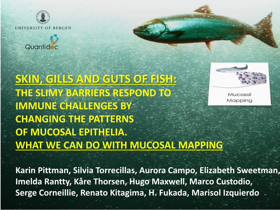
SKIN, GILLS AND GUTS OF FISH:THE SLIMY BARRIERS RESPOND TOIMMUNE CHALLENGES BY CHANGING THE PATTERNS OF MUCOSAL EPITHELIA. WHAT WE CAN DO WITH MUCOSAL MAPPING
Karin Pittman, Silvia Torrecillas, Aurora Campo, Elizabeth Sweetman, Imelda Rantty, Kåre Thorsen, Hugo Maxwell, Marco Custodio, Serge Corneillie, Renato Kitagima, H. Fukada, Marisol Izquierdo
Co-conspirators• Bjarne Ravnøy, Produs AS• Johan Johansen, Gildeskål forskningsstasjon• John Sweetman, Alltech Aqua• Sulefisk A/S• Ingrid Uglenes, IMR• Tine Oen, BIO UiB/Fiskeridirektoratet• Amanda Pittman, UiB/U Copenhagen• Kirsten Redmond, BIO UiB/UiStavanger• Stine Karlson, BIO, UiB/IMR• Aurora Campo, Quantidoc, Bergen• Stanko Skugor, Nofima Averøy• Elizabeth Sweetman, Ecomarine• Marco Custodio, U Algarve, Portugal• Imelda Rantti, BIO UiB, • Hugo Maxwell , BIO UiB• Kåre Thorsen, BIO UiB• Silvia Torrecillas, Univ of Gran Canaria, Spain• Marisol Izquierdo, Las Palmas de Gran Canaria University• Phillippe Sourde, Vet’eau/Aquativ, France• Katerina Koutsoulakis, Nofima, Norway• Arne Skorping, BIO UiB• Mathias Ugelvik, BIO UiB• Mark Powell, NIVA/BIO UiB• Salmon Group• Salmobreed• ILAB, Bergen• Alltech Japan• Cooke Aquaculture, Canada
Funding sources:IndustryRegional Research FundInnovation NorwayNorwegian Research Council
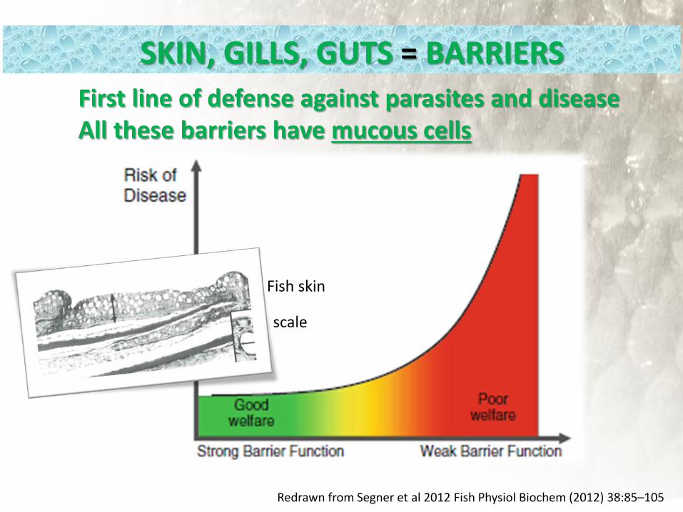
Redrawn from Segner et al 2012 Fish Physiol Biochem (2012) 38:85–105
SKIN, GILLS, GUTS = BARRIERS
Fish skin
scale
First line of defense against parasites and diseaseAll these barriers have mucous cells
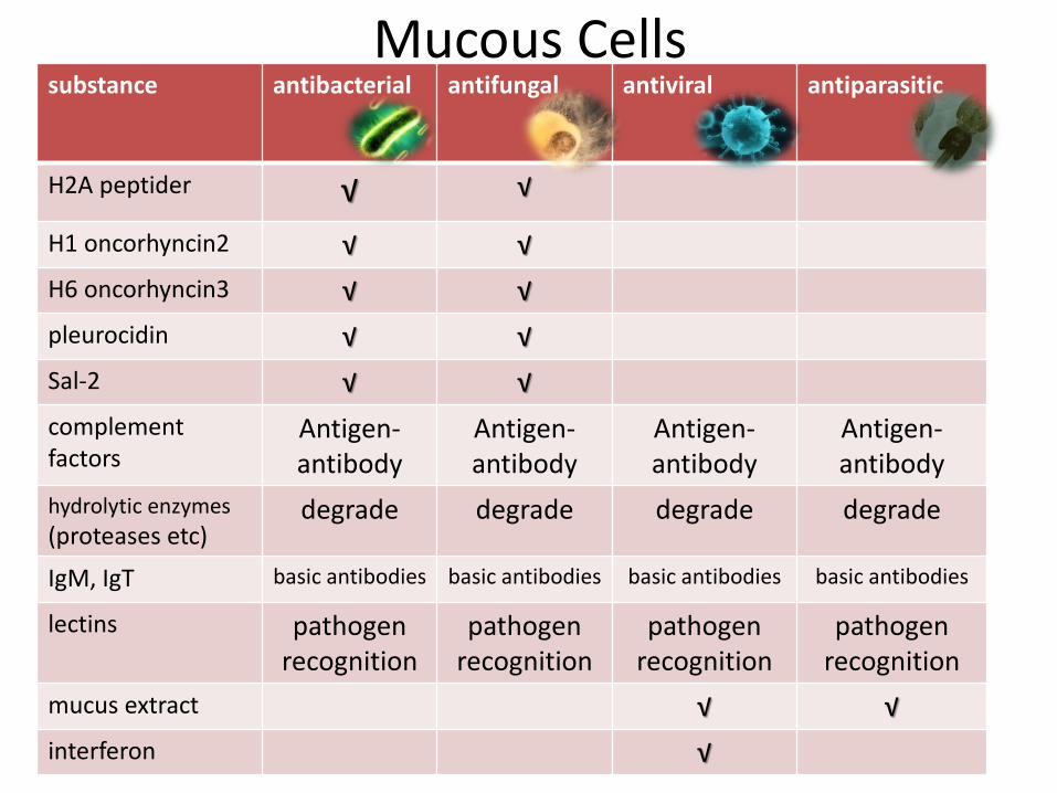
substance antibacterial antifungal antiviral antiparasitic
H2A peptider √ √
H1 oncorhyncin2 √ √H6 oncorhyncin3 √ √pleurocidin √ √Sal-2 √ √complementfactors
Antigen-antibody
Antigen-antibody
Antigen-antibody
Antigen-antibody
hydrolytic enzymes(proteases etc)
degrade degrade degrade degrade
IgM, IgT basic antibodies basic antibodies basic antibodies basic antibodies
lectins pathogenrecognition
pathogenrecognition
pathogenrecognition
pathogenrecognition
mucus extract √ √interferon √
Mucous Cells
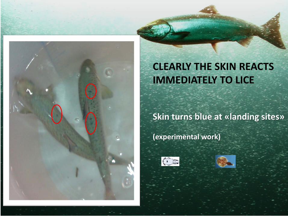
Skin turns blue at «landing sites»
(experimental work)
CLEARLY THE SKIN REACTS IMMEDIATELY TO LICE
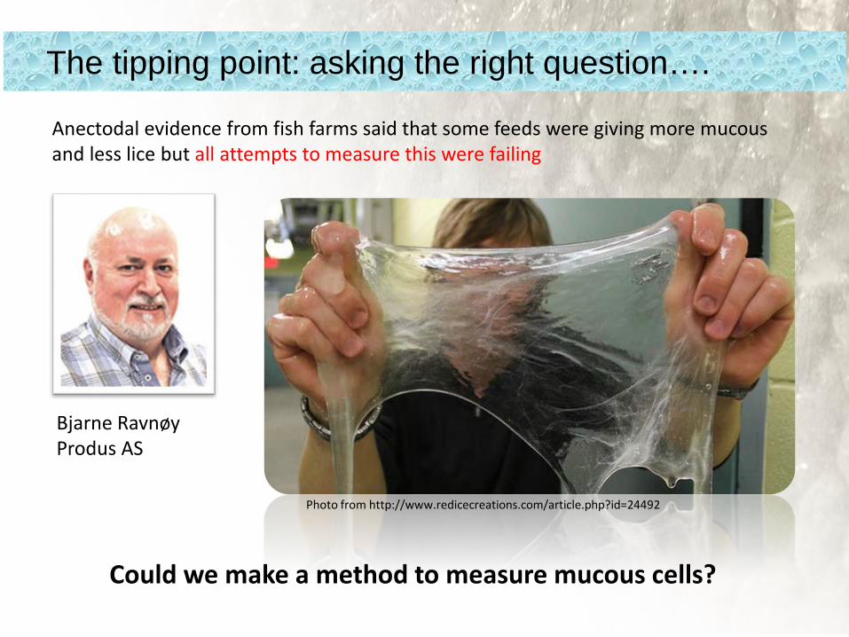
Photo from http://www.redicecreations.com/article.php?id=24492
Bjarne RavnøyProdus AS
The tipping point: asking the right question….
Anectodal evidence from fish farms said that some feeds were giving more mucousand less lice but all attempts to measure this were failing
Could we make a method to measure mucous cells?

A: Design-based stereology
YES: Mucosal mapping
Q: how do you take one (1) slice of the sky andfind out how big the balloons areand how tightly packed they are?
Mucosal mapping is complementary to all other existing technologies

challenges• Salmon lice • Salmon welfare
Weekly lice counts Sampling for skin response

From Gomez et al., 2013
Similar structures between mammals and fish, except fish have live cells at the surfaceGoblet cell = mucous cellNice pictures but not good statistics….
Brief anatomy of barriers
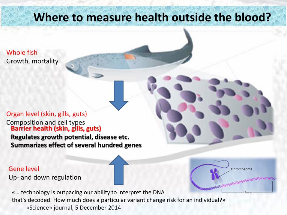
Where to measure health outside the blood?
Whole fishGrowth, mortality
Gene levelUp- and down regulation
Organ level (skin, gills, guts) Composition and cell types
Summarizes effect of several hundred genes
«… technology is outpacing our ability to interpret the DNA that's decoded. How much does a particular variant change risk for an individual?»
«Science» journal, 5 December 2014
Barrier health (skin, gills, guts)Regulates growth potential, disease etc.
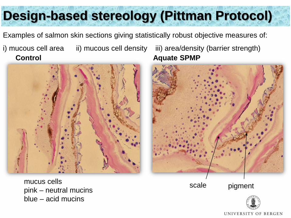
Control Aquate SPMP
mucus cells pink – neutral mucinsblue – acid mucins
pigmentscale
Design-based stereology (Pittman Protocol)Examples of salmon skin sections giving statistically robust objective measures of:
i) mucous cell area ii) mucous cell density iii) area/density (barrier strength)
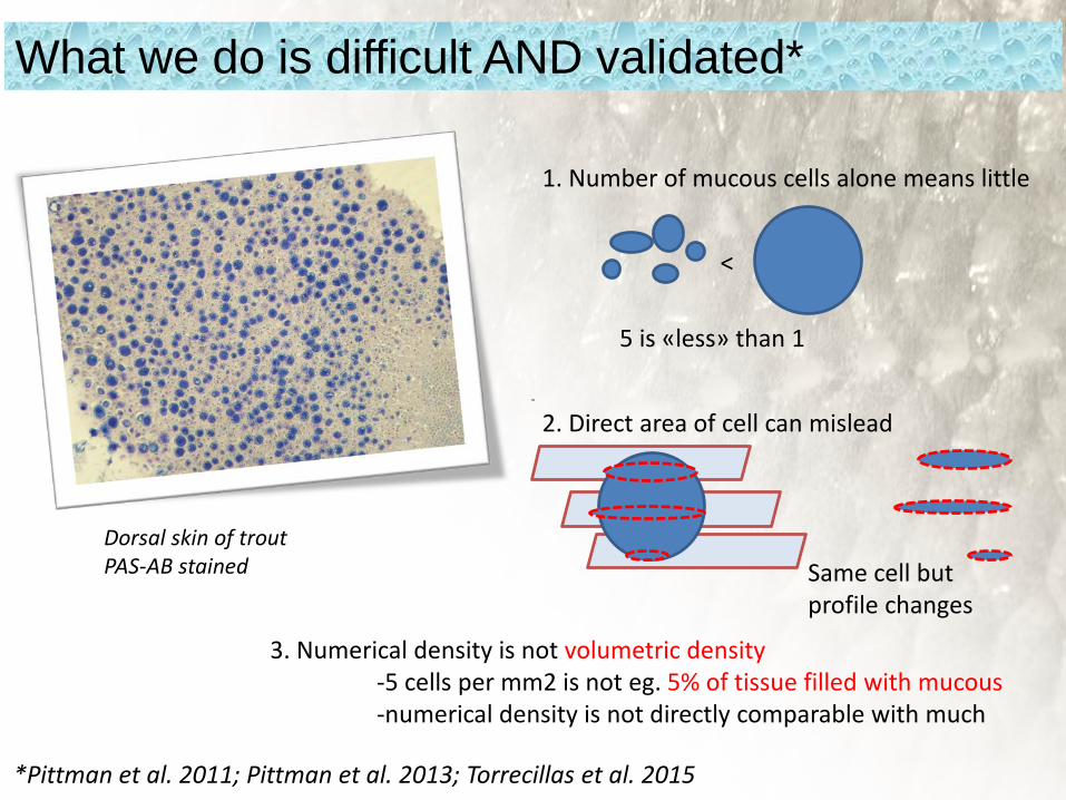
What we do is difficult AND validated*
Dorsal skin of troutPAS-AB stained
1. Number of mucous cells alone means little
5 is «less» than 1
<
2. Direct area of cell can mislead
Same cell butprofile changes
3. Numerical density is not volumetric density-5 cells per mm2 is not eg. 5% of tissue filled with mucous-numerical density is not directly comparable with much
*Pittman et al. 2011; Pittman et al. 2013; Torrecillas et al. 2015

The Repeatable Basics: Significantly larger mucous cellson dorsal (p<0.01)
H S V D
BODY SITE
100
110
120
130
140
150
160
170
Muc
ous c
ell a
rea
(μm
2 )
Mean Mean±0,95 Conf. Interval
n=4 fisk52 cm SLPittman et al. 2012
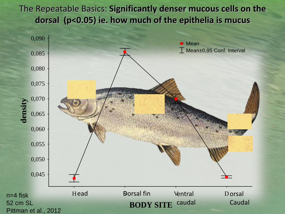
The Repeatable Basics: Significantly denser mucous cells on thedorsal (p<0.05) ie. how much of the epithelia is mucus
H S V D
BODY SITE
0,045
0,050
0,055
0,060
0,065
0,070
0,075
0,080
0,085
0,090
dens
ity
Mean Mean±0,95 Conf. Interval
n=4 fisk52 cm SLPittman et al., 2012
ead Dorsal fin entralcaudal
orsalCaudal
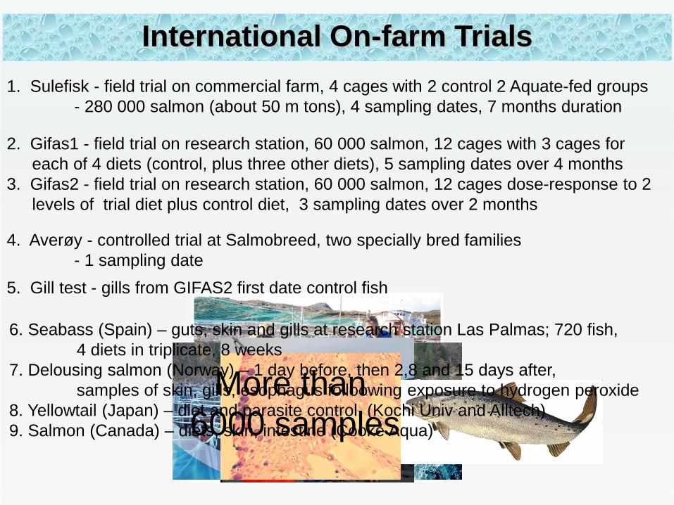
1. Sulefisk - field trial on commercial farm, 4 cages with 2 control 2 Aquate-fed groups- 280 000 salmon (about 50 m tons), 4 sampling dates, 7 months duration
International On-farm Trials
2. Gifas1 - field trial on research station, 60 000 salmon, 12 cages with 3 cages for each of 4 diets (control, plus three other diets), 5 sampling dates over 4 months
3. Gifas2 - field trial on research station, 60 000 salmon, 12 cages dose-response to 2 levels of trial diet plus control diet, 3 sampling dates over 2 months
4. Averøy - controlled trial at Salmobreed, two specially bred families - 1 sampling date
5. Gill test - gills from GIFAS2 first date control fish
More than6000 samples
6. Seabass (Spain) – guts, skin and gills at research station Las Palmas; 720 fish, 4 diets in triplicate, 8 weeks
7. Delousing salmon (Norway) – 1 day before, then 2,8 and 15 days after, samples of skin, gills, esophagus folloowing exposure to hydrogen peroxide
8. Yellowtail (Japan) – diet and parasite control, (Kochi Univ and Alltech)9. Salmon (Canada) – diets, skin, intestine (Cooke Aqua)
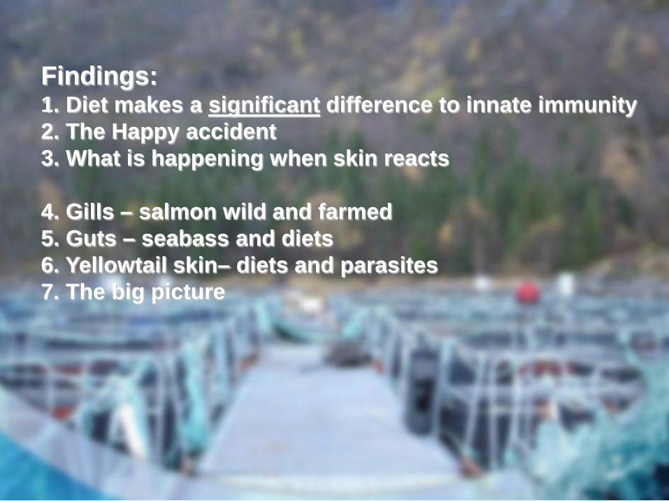
Findings:1. Diet makes a significant difference to innate immunity2. The Happy accident3. What is happening when skin reacts
4. Gills – salmon wild and farmed5. Guts – seabass and diets6. Yellowtail skin– diets and parasites7. The big picture

4 diets, 4 months, 60000 salmon-Diet really does make a difference
The Happy Accident
Aquate SG
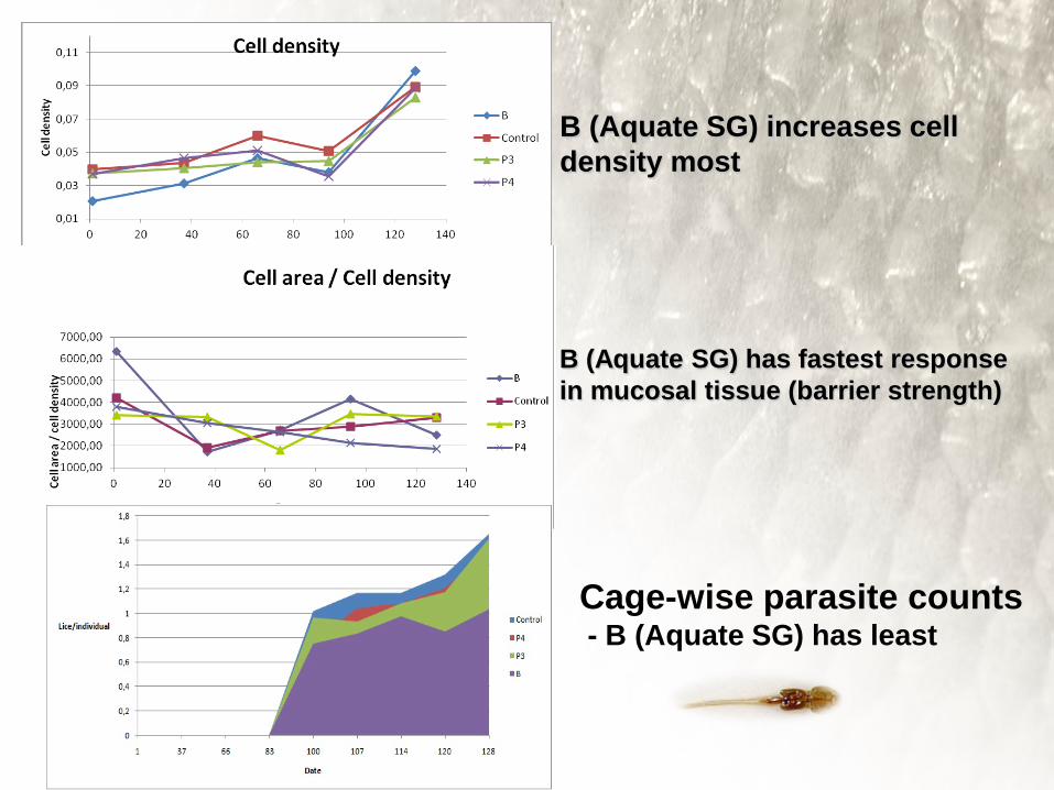
B (Aquate SG) increases celldensity most
Cage-wise parasite counts- B (Aquate SG) has least
B (Aquate SG) has fastest responsein mucosal tissue (barrier strength)

MUCOSAL MAPPING IN SALMONIDS
Parasites go where mucosal cells are small and few
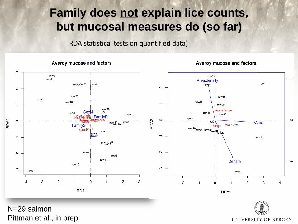
Family does not explain lice counts, but mucosal measures do (so far)
N=29 salmonPittman et al., in prep
RDA statistical tests on quantified data)
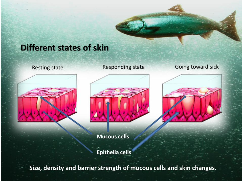
Resting state Responding state Going toward sick
Different states of skin
Epithelia cells
Mucous cells
Size, density and barrier strength of mucous cells and skin changes.
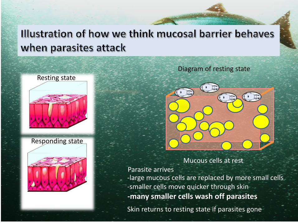
Responding state
Mucous cells at rest
-large mucous cells are replaced by more small cells-smaller cells move quicker through skin-many smaller cells wash off parasites
Parasite arrives
Skin returns to resting state if parasites gone
Resting stateDiagram of resting state
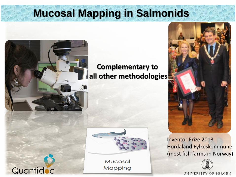
Mucosal Mapping in Salmonids
Complementary to all other methodologies
Inventor Prize 2013Hordaland Fylkeskommune(most fish farms in Norway)
******
***
***
****
*
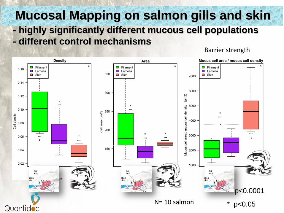
*** p<0.0001
* p<0.05
- highly significantly different mucous cell populations- different control mechanisms
Mucosal Mapping on salmon gills and skin
N= 10 salmon
Barrier strength
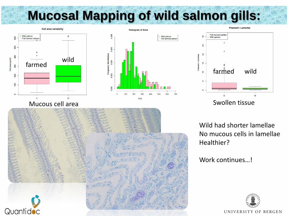
Mucosal Mapping of wild salmon gills:
Wild had shorter lamellaeNo mucous cells in lamellaeHealthier?
Work continues…!
Mucous cell area
wildfarmed
Swollen tissue
farmed wild
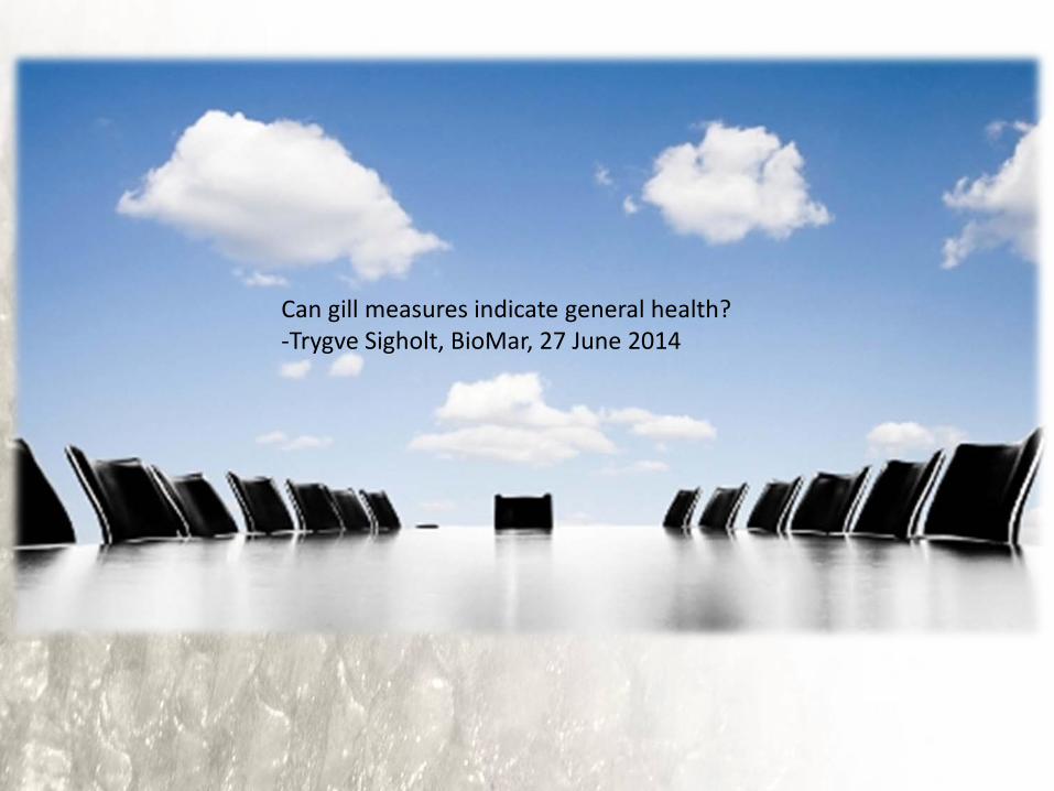
Can gill measures indicate general health?-Trygve Sigholt, BioMar, 27 June 2014
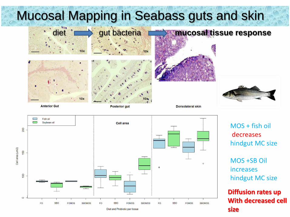
Mucosal Mapping in Seabass guts and skindiet gut bacteria mucosal tissue response
MOS + fish oildecreases
hindgut MC size
MOS +SB Oilincreaseshindgut MC size
Diffusion rates upWith decreased cellsize
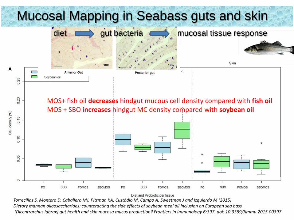
Mucosal Mapping in Seabass guts and skindiet gut bacteria mucosal tissue response
MOS+ fish oil decreases hindgut mucous cell density compared with fish oilMOS + SBO increases hindgut MC density compared with soybean oil
Torrecillas S, Montero D, Caballero MJ, Pittman KA, Custódio M, Campo A, Sweetman J and Izquierdo M (2015) Dietary mannan oligosaccharides: counteracting the side effects of soybean meal oil inclusion on European sea bass(Dicentrarchus labrax) gut health and skin mucosa mucus production? Frontiers in Immunology 6:397. doi: 10.3389/fimmu.2015.00397

foregut hindgut
Fish oil
Fish oil + MOS
Soybean oil
Soybean Oil + MOS
hindgut
Seabass gut Mucous cellsize and density(to scale)
From: Torrecillas et al., 2015
Mucosal Mapping in Seabass guts and skindiet gut bacteria mucosal tissue response
Good epithelial turnover combined with differentiation of cells
Stronger immune reactivity slightly increased ability for immune substances to diffuse
Lowest cell migration & diffusion rates, epithelial hyperplasia,more cell differentiation and turnover, and more anti-inflammatories
Downregulated IL-6, IL-10, and TGFβ (functioning of mucous barrier and immune homeostasis)
Dietary evaluation
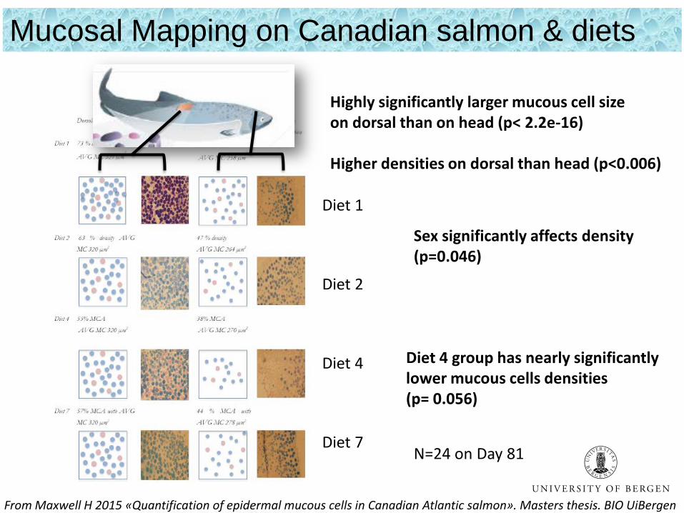
From Maxwell H 2015 «Quantification of epidermal mucous cells in Canadian Atlantic salmon». Masters thesis. BIO UiBergen
Diet 1
Diet 2
Diet 4
Diet 7
Diet 4 group has nearly significantlylower mucous cells densities(p= 0.056)
Highly significantly larger mucous cell sizeon dorsal than on head (p< 2.2e-16)
Higher densities on dorsal than head (p<0.006)
N=24 on Day 81
Sex significantly affects density(p=0.046)
Mucosal Mapping on Canadian salmon & diets
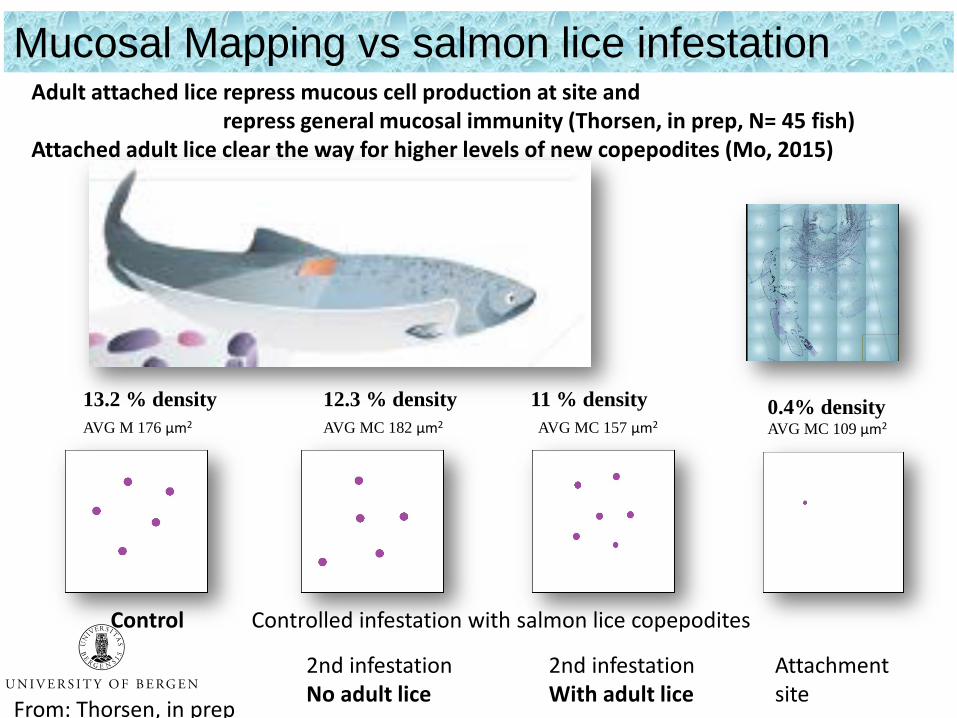
13.2 % density 12.3 % density 11 % densityAVG M 176 µm2 AVG MC 182 µm2 AVG MC 157 µm2
Control Controlled infestation with salmon lice copepodites
2nd infestationNo adult lice
2nd infestationWith adult lice
Adult attached lice repress mucous cell production at site and repress general mucosal immunity (Thorsen, in prep, N= 45 fish)
Attached adult lice clear the way for higher levels of new copepodites (Mo, 2015)
Mucosal Mapping vs salmon lice infestation
0.4% densityAVG MC 109 µm2
Attachmentsite
From: Thorsen, in prep
Skin response to salmon lice is stage specific
• proper animation first draft by Egil Paulsen
Mucosal Mapping on salmon and lice problem
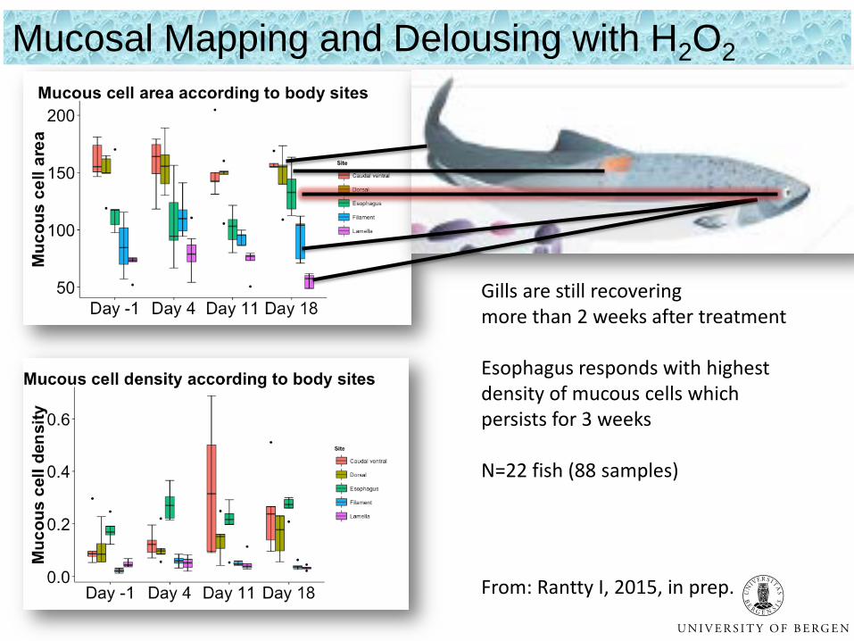
Delousing with hydrogen peroxide
Gills are still recoveringmore than 2 weeks after treatment
Esophagus responds with highestdensity of mucous cells whichpersists for 3 weeks
N=22 fish (88 samples)
From: Rantty I, 2015, in prep.
Mucosal Mapping and Delousing with H2O2
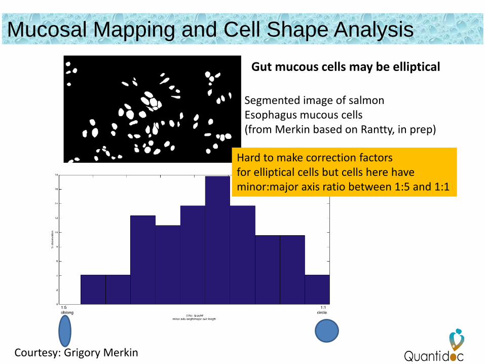
Mucosal Mapping and Cell Shape Analysis
Courtesy: Grigory Merkin
Segmented image of salmonEsophagus mucous cells(from Merkin based on Rantty, in prep)
Hard to make correction factorsfor elliptical cells but cells here have minor:major axis ratio between 1:5 and 1:1
Gut mucous cells may be elliptical
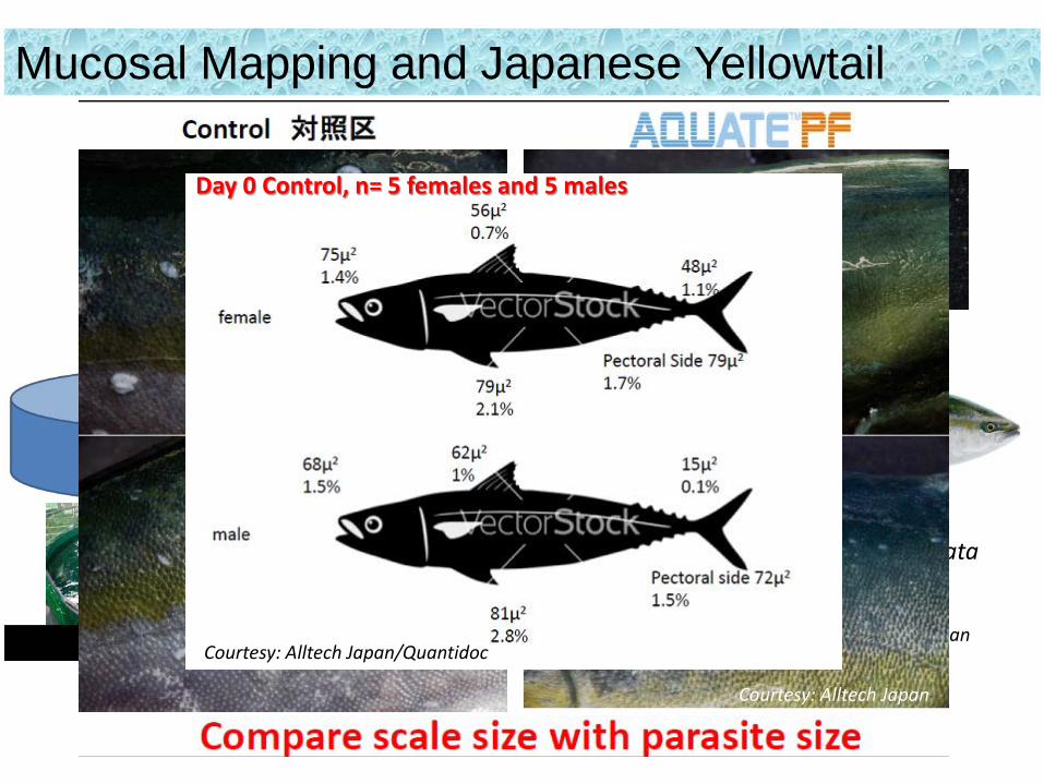
Courtesy: Alltech Japan
Mucosal Mapping and Japanese YellowtailBenedenia seriolae(skin fluke)
YellowtailSeriola quinqueradiata
Day 0 Control, n= 5 females and 5 males
Courtesy: Alltech Japan/Quantidoc
Courtesy: Alltech Japan
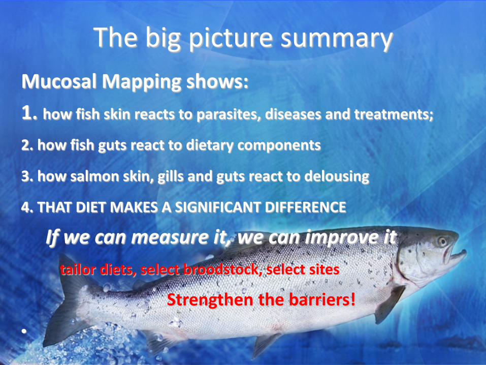
The big picture summaryMucosal Mapping shows:1. how fish skin reacts to parasites, diseases and treatments;
2. how fish guts react to dietary components
3. how salmon skin, gills and guts react to delousing
4. THAT DIET MAKES A SIGNIFICANT DIFFERENCE
If we can measure it, we can improve ittailor diets, select broodstock, select sites
Strengthen the barriers!
•
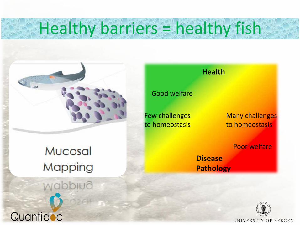
Health
DiseasePathology
Good welfare
Poor welfare
Few challengesto homeostasis
Many challengesto homeostasis
Healthy barriers = healthy fish
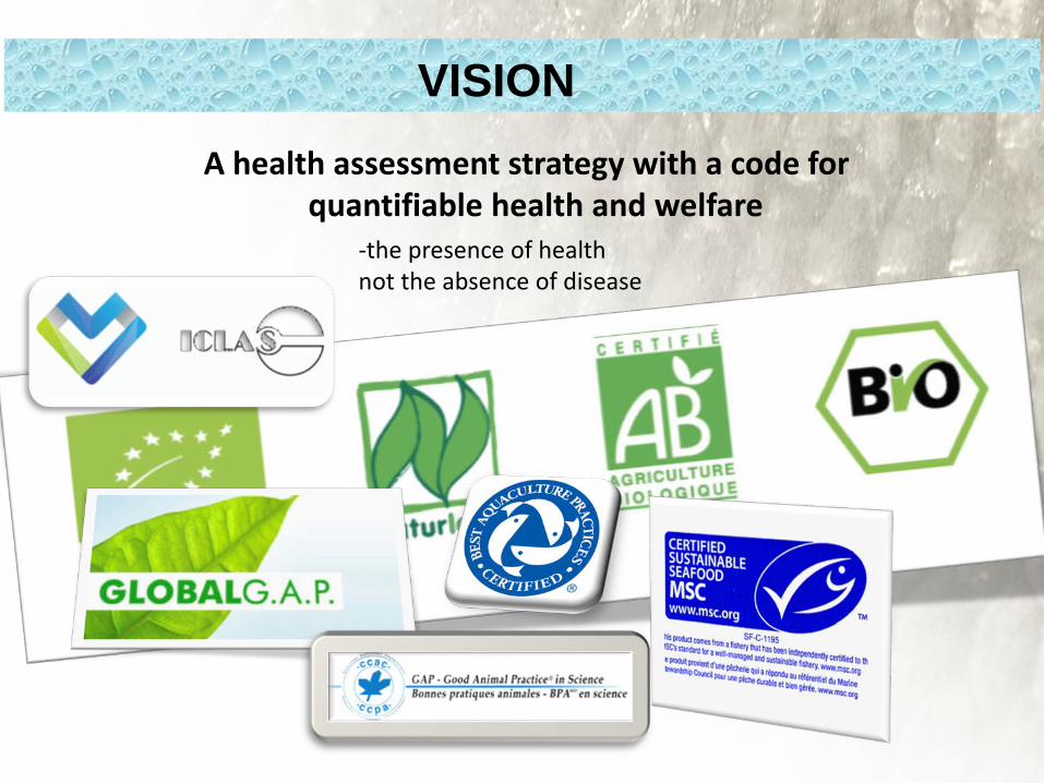
VISIONA health assessment strategy with a code for
quantifiable health and welfare-the presence of healthnot the absence of disease

Quantidoc owns the IP for this diagnostic method of quantitatively assessing mucous cells and is commercializing the product for industrial application
• Diagnostic – health status (skin, gills and guts)• Quantitative, objective & comparable• Statistically robust• Links diet & immunity• Summarises the effects of >200 genes• Important tool for monitoring and improving
fish health and welfare
MUCOSAL MAPPING (MUCOMAP)
Contact: [email protected]
Recommended