Role of In Vitro System as Continuous Betalain Sources
Retno Mastuti
Biology Department, Faculty of Mathematics and Natural Sciences, University of Brawijaya,
Malang, Indonesia ([email protected])
Abstract
Plants are sources of many important secondary metabolites including pigments. Betalain, one of plant major
pigments is of growing interest for colorant of food and pharmaceutical industries. This natural colorant has
been concerned as an alternative for synthetic colorants due to its positive effects on health. Recent reports show
that betalains have high anti-oxidative, free radical scavenging activities and potential for anti malaria. In
higher plants betalain occurrence is restricted to 13 families of the order Caryophyllales. Their occurence is
mutually exclusive to that of the anthocyanins. Although the major pigment group shows wide occurence in
different plant tissues, the production is very affected by environmental conditions. Therefore, an in vitro system
under controlled conditions is one promising alternative pigments source for all seasons. Plant cells and tissue
cultures can be established routinely under sterile conditions from explants, such as plant leaves, stems, and
roots for both multiplication and extraction of secondary metabolites including pigments. Some in vitro
strategies have been applied to improve betalain production. Strain improvement, methods for the selection of
high-producing cell lines, and medium optimizations could enhance secondary metabolite production. In
addition, this paper will discuss hairy root cultures for pigment production, precursor addition for betalain
improvement, elicitation of in vitro products, and bioreactors scaling up of production.
Keywords: betalains, plant in vitro systems, optimization strategies
I. INTRODUCTION
The plant major pigments are chlorophyll,
carotenoids, anthocyanins and betalains. Except
chlorophyll, the three families of pigments play
important ecological functions, for example in the
attraction of pollinators and seed dispersal animals.
All of them are secondary metabolite which
economically important such as in food and
medicinal industry. Interest in betalains has grown
since their antiradical activity was characterized (1),
and they are widely used as additives in the food
industry because of their natural colorant properties
and absence of toxicity (2). Consequently, the
sustain availability of betalain pigments become
important. However, the distribution of secondary
metabolites in plants is far more restricted than that
of primary metabolites. Also, the secondary
metabolites often accumulate in the plant in small
quantities, sometimes in specialized cells which
caused the difficulty in their extraction. Although
extraction of secondary metabolites from plants is
still commercially important a large number of these
metabolites are difficult or impossible to synthesize
at economic values (3). Therefore, development of
alternatives to the intact plant for the production of
plant secondary metabolites has been intensively
approached. In the past three decades, research has
been concentrated on the use of plant cell and tissue
cultures. Biotechnological approaches, specifically
plant tissue culture plays a vital role in search for
alternatives to production of desirable compounds
from plants (4). This method can provide a
continuous, reliable source of natural products. In
vitro system offers several advantages over field
cultivation: it is independent of geographical and
seasonal variations, environmental factors, and
political interference; in addition, it allows optimal
and stable growth conditions, voluntary modulation
of growth parameters, and constant quality control
(5-6). It also eliminates negative biological
influences (microorganisms and insects) that affect
secondary metabolites production in nature; and
possibility to select cultivars with higher production
of secondary metabolites (7). Moreover, colourants
produced in this way are classified as 'natural' rather
than 'nature-identical', which increases their
desirability to customers. In some cases, the yield
per gram fresh weight of secondary metabolites in
plant cell culture may exceed that which is found in
nature. Therefore, a priority in current and future
efforts towards sustainable conservation and rational
utilization of biodiversity is searching for new plant-
derived chemicals (8). Plant-produced secondary
compounds have been incorporated into a wide
range of commercial and industrial applications.
Different strategies using cell cultures systems have
been extensively studies with the objective of
improving the production of bioactive secondary
metabolites.
Plant tissue cultures were first established in
1939. The success in isolated visnagin (9) and
diosgenin (10) respectively from cell cultures in
larger quantities than from the whole plant showed
the potential of plant cell cultures to produce useful
compounds. Increasing the effort to obtain high
amount of secondary metabolites in plant cell
cultures was accompanied by emphasizing the
biochemical and molecular research on the
secondary metabolism of plants (11-16). These lead
to the successful manipulation of secondary
metabolism and significantly increase the amounts
of the compound(s) production. Now, it seems that
any substances of plant origin can be produced by
plant cell culture techniques. Identification of cell
lines that can produce amounts of compounds equal
or even higher than those in the plant from which
they derived has been possible.
II. BETALAIN – BIOCHEMISTRY, DISTRIBUTION AND
CHARACTERS
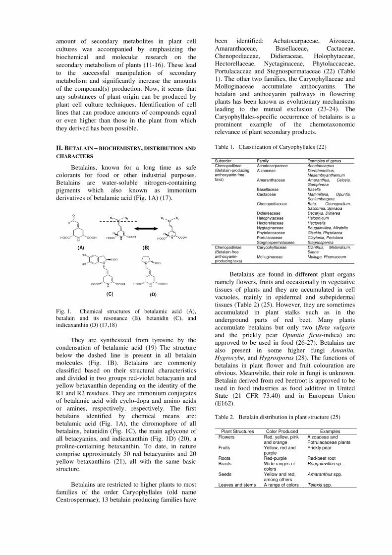
Betalains, known for a long time as safe
colorants for food or other industrial purposes.
Betalains are water-soluble nitrogen-containing
pigments which also known as immonium
derivatives of betalamic acid (Fig. 1A) (17).
Fig. 1. Chemical structures of betalamic acid (A),
betalain and its resonance (B), betanidin (C), and
indicaxanthin (D) (17,18)
They are synthesized from tyrosine by the
condensation of betalamic acid (19) The structure
below the dashed line is present in all betalain
molecules (Fig. 1B). Betalains are commonly
classified based on their structural characteristics
and divided in two groups red-violet betacyanin and
yellow betaxanthin depending on the identity of the
R1 and R2 residues. They are immonium conjugates
of betalamic acid with cyclo-dopa and amino acids
or amines, respectively, respectively. The first
betalains identified by chemical means are:
betalamic acid (Fig. 1A), the chromophore of all
betalains, betanidin (Fig. 1C), the main aglycone of
all betacyanins, and indicaxanthin (Fig. 1D) (20), a
proline-containing betaxanthin. To date, in nature
comprise approximately 50 red betacyanins and 20
yellow betaxanthins (21), all with the same basic
structure.
Betalains are restricted to higher plants to most
families of the order Caryophyllales (old name
Centrospermae); 13 betalain producing families have
been identified: Achatocarpaceae, Aizoacea,
Amaranthaceae, Basellaceae, Cactaceae,
Chenopodiaceae, Didieraceae, Holophytaceae,
Hectorellaceae, Nyctaginaceae, Phytolaccaceae,
Portulacaceae and Stegnospermataceae (22) (Table
1). The other two families, the Caryophyllaceae and
Molluginaceae accumulate anthocyanins. The
betalain and anthocyanin pathways in flowering
plants has been known as evolutionary mechanisms
leading to the mutual exclusion (23-24). The
Caryophyllales-specific occurrence of betalains is a
prominent example of the chemotaxonomic
relevance of plant secondary products.
Table 1. Classification of Caryophyllales (22)
Suborder Family Examples of genus
Chenopodiinae (Betalain-producing anthocyanin-free taxa)
Achatocarpaceae Achataocarpus
Aizoaceae Dorotheanthus, Mesembryanthemum
Amaranthaceae Amaranthus, Celosia, Gomphrena
Basellaceae Basella
Cactaceae Mammilaria, Opuntia, Schlumbergera
Chenopodiaceae Beta, Chenopodium, Salicornia, Spinacia
Didiereaceae Decaryia, Didierea
Halophytaceae Halophytum
Hectorellaceae Hectorella
Nygtaginaceae Bougainvillea, Mirabilis
Phytolaccaceae Gisekia, Phytolacca
Portulacaceae Claytonia, Portulaca
Stegnospermataceae Stegnosperma
Chenopodiinae (Betalain-free anthocyanin-producing taxa)
Caryophyllaceae Dianthus, Melandrium, Silene
Molluginaceae Mollugo, Pharnaceum
Betalains are found in different plant organs
namely flowers, fruits and occasionally in vegetative
tissues of plants and they are accumulated in cell
vacuoles, mainly in epidermal and subepidermal
tissues (Table 2) (25). However, they are sometimes
accumulated in plant stalks such as in the
underground parts of red beet. Many plants
accumulate betalains but only two (Beta vulgaris
and the prickly pear Opuntia ficus-indica) are
approved to be used in food (26-27). Betalains are
also present in some higher fungi Amanita,
Hygrocybe, and Hygrosporus (28). The functions of
betalains in plant flower and fruit colouration are
obvious. Meanwhile, their role in fungi is unknown.
Betalain derived from red beetroot is approved to be
used in food industries as food additive in United
State (21 CFR 73.40) and in European Union
(E162).
Table 2. Betalain distribution in plant structure (25)
Plant Structures Color Produced Examples
Flowers Red, yellow, pink and orange
Aizoaceae and Potrulacaceae plants
Fruits Yellow, red and purple
Prickly pear
Roots Red-purple Red-beet root Bracts Wide ranges of
colors Bougainvillea sp.
Seeds Yellow and red, among others
Amaranthus spp.
Leaves and stems A range of colors Teloxis spp.
III. ACCUMULATION OF BETALAIN AND CELL
GROWTH
Many secondary metabolites accumulate in
specific tissues and cell of higher plants, or at
specific stages during the growth of cultured cells.
The accumulation of most secondary metabolites in
cultured cells is maximum during the stationary
phase of growth. On the contrary, peaks of
accumulation of secondary products during the
logarithmic phase of growth have been observed in
only a few cases. Accumulation of betacyanin was
suppressed when growth of cells in suspension
cultures of Phytolacca americana was inhibited by
aphidicolin (an inhibitor of DNA synthesis) (29).
Betacyanin content in cell suspension culture of B.
vulgaris also increased in parallel with the cell
growth during the log phase (30). Peaks of
accumulation of betalains during the logarithmic
phase were also observed in suspension cultures of
Chenopodium rubrum (31). Close correlation
between growth and accumulation of betacyanin was
also reported in suspension cultures of P. americana
(32). Subsequently, it was suggested a positive
correlation between the accumulation of betacyanin
and cell proliferation (29).
A similar results has been reported for cell
suspension from the violet callus of B. vulgaris (35).
Lack of phosphate or addition of aphidicolin to the
medium suppressed both the division of cells and the
accumulation of betacyanin. Conversely, re-addition
of phosphate and removal of aphidicolin initiated
both process. Tracer experiments using labeled
tyrosine revealed that the incorporation of
radioactivity from tyrosine to betacyanin was
inhibited while that from DOPA was not. Therefore,
it is concluded that the step from DOPA to
betacyanin is not coupled with cell division, but that
from tyrosine to DOPA is coupled (34). Furthermore
such a relationship between the betacyanin
accumulation and cell growth has been observed in
cell suspension culture of B. vulgaris (30) and
Portulacca (35).
IV. MAXIMIZATION OF SYNTHESIS AND
PRODUCTION
Many efforts have focused on the stimulation
of biosynthetic activities of cultured cells using
various methods. Since the tissue culture cells
typically accumulate large amounts of secondary
compounds only under specific conditions the
maximization of accumulation secondary
metabolites requires: manipulating the parameters of
the environment and medium; selecting high
yielding cell clones precursor feeding and elicitation.
A. Optimization of medium composition
Since the economically viable is prerequisite, it
is important to develop methods that would allow
for consistent generation of high yields of products
(36). Several products were found to be
accumulating in cultured cells at a higher level than
those in native plants through optimization of
cultural conditions (37). There is no similar trend of
availability of phosphate ion in affecting betalain
content. In B. vulgaris cv Detroit Dark Red hairy
root cultures there is phosphate ions reduction from
initial levels of 120 to 10-15 mg L-1
after the start of
the exponential phase of cultivation (38). However
the same culture in phosphate-free medium resulted
in total betalain contents rising to 19 g L-1
: 4.8 times
higher than the amounts accumulated in a standard
MS medium (39).
The concentration of nitrate ions exhibited
different color phenotypes of B. vulgaris cv. Bikores
Monogerm cell lines (33). While combination of
nitrogen concentrations with some concentrations in
microelement increased betacyanin production in red
beet cell suspension (40), there was no significant
effect on betalain accumulation in various nitrogen
concentrations (39). Ammonium ions are utilized
more rapidly than nitrate ions but no significant
amount of either ammonium or nitrate ions are
consumed in the first 5 days of B. vulgaris hairy root
cultures (41). In suspension cultures of P. americana
L., betacyanin accumulation per cell increased with
increasing total nitrogen concentration (initial
NH+
4:NO−
3 ratio 1:2) in the range 0–40 mM and then
remained almost constant in the range 40–80 mM.
(42).
Microelements in medium also influenced
betalain production. Increasing Fe2+
concentration
up to 20-fold higher than that in the standard LS
medium markedly increased betacyanin content
without any affect on cell growth of B. vulgaris cell
suspension (30). Apparently, concentration of
microelement was significantly affected by basal
medium. Removing of boron, iodine, manganese and
molybdenum was found to decrease both the cell
growth and betacyanin content whereas absence of
copper and cobalt did not show any negative effect
on red beet cell suspension using LS basal medium
(43). On the other hand in Gamborg B5 medium
except manganese the addition high concentration of
copper, iron, molybdenum, zinc and cobalt showed
positive effect on betalain production.
Exogenous supply of a biosynthetic precursor
to culture medium may also increase the yield of the
desired product. Attempts to induce or increase the
production of plant secondary metabolites by
supplying precursor or intermediate compounds
have been effective in many cases. Betacyanin
accumulation in red-violet cell suspension cultures
of C. rubrum could be increased up to 1% or 100 mg
betacyanins/l by feeding tyrosine (44). Meanwhile
adding DOPA (44) and specific amino acids (45)
does not increase the betacyanin content.
The type of inoculums strongly influences
betacyanin production. The cell culture of Portulaca
grandiflora derived from the basipetal cut of a
hypocotyls segment is superior to the cell culture of
acropetal origin with regard to betacyanin
concentration. These differences indicate the
polarization of physiological conditions in the
hypocotyls segments (46).
B. Selection of highly productively cell lines
Plant cell cultures represent a heterogenous
population in which physiological characteristics of
individual plant cells are different (37) Proper
selection of productive cells and cultural conditions
results in accumulation of products in high level.
Not all cells express the pigmented phenotype,
giving rise to variegated patches (47). The pattern of
patch formation is different from one callus to
another, although all calluses stem from the same
original clone. Therefore, the selection of high-
producing strains is necessary. Secondary
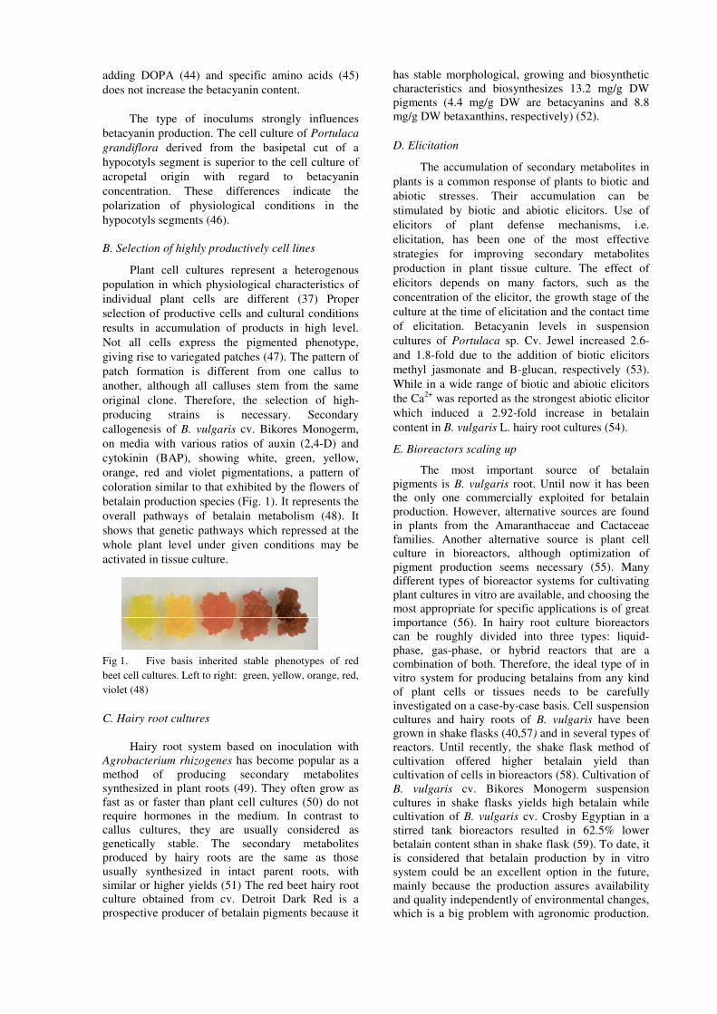
callogenesis of B. vulgaris cv. Bikores Monogerm,
on media with various ratios of auxin (2,4-D) and
cytokinin (BAP), showing white, green, yellow,
orange, red and violet pigmentations, a pattern of
coloration similar to that exhibited by the flowers of
betalain production species (Fig. 1). It represents the
overall pathways of betalain metabolism (48). It
shows that genetic pathways which repressed at the
whole plant level under given conditions may be
activated in tissue culture.
Fig 1. Five basis inherited stable phenotypes of red
beet cell cultures. Left to right: green, yellow, orange, red,
violet (48)
C. Hairy root cultures
Hairy root system based on inoculation with
Agrobacterium rhizogenes has become popular as a
method of producing secondary metabolites
synthesized in plant roots (49). They often grow as
fast as or faster than plant cell cultures (50) do not
require hormones in the medium. In contrast to
callus cultures, they are usually considered as
genetically stable. The secondary metabolites
produced by hairy roots are the same as those
usually synthesized in intact parent roots, with
similar or higher yields (51) The red beet hairy root
culture obtained from cv. Detroit Dark Red is a
prospective producer of betalain pigments because it
has stable morphological, growing and biosynthetic
characteristics and biosynthesizes 13.2 mg/g DW
pigments (4.4 mg/g DW are betacyanins and 8.8
mg/g DW betaxanthins, respectively) (52).
D. Elicitation
The accumulation of secondary metabolites in
plants is a common response of plants to biotic and
abiotic stresses. Their accumulation can be
stimulated by biotic and abiotic elicitors. Use of
elicitors of plant defense mechanisms, i.e.
elicitation, has been one of the most effective
strategies for improving secondary metabolites
production in plant tissue culture. The effect of
elicitors depends on many factors, such as the
concentration of the elicitor, the growth stage of the
culture at the time of elicitation and the contact time
of elicitation. Betacyanin levels in suspension
cultures of Portulaca sp. Cv. Jewel increased 2.6-
and 1.8-fold due to the addition of biotic elicitors
methyl jasmonate and B-glucan, respectively (53).
While in a wide range of biotic and abiotic elicitors
the Ca2+
was reported as the strongest abiotic elicitor
which induced a 2.92-fold increase in betalain
content in B. vulgaris L. hairy root cultures (54).
E. Bioreactors scaling up
The most important source of betalain
pigments is B. vulgaris root. Until now it has been
the only one commercially exploited for betalain
production. However, alternative sources are found
in plants from the Amaranthaceae and Cactaceae
families. Another alternative source is plant cell
culture in bioreactors, although optimization of
pigment production seems necessary (55). Many
different types of bioreactor systems for cultivating
plant cultures in vitro are available, and choosing the
most appropriate for specific applications is of great
importance (56). In hairy root culture bioreactors
can be roughly divided into three types: liquid-
phase, gas-phase, or hybrid reactors that are a
combination of both. Therefore, the ideal type of in
vitro system for producing betalains from any kind
of plant cells or tissues needs to be carefully
investigated on a case-by-case basis. Cell suspension
cultures and hairy roots of B. vulgaris have been
grown in shake flasks (40,57) and in several types of
reactors. Until recently, the shake flask method of
cultivation offered higher betalain yield than
cultivation of cells in bioreactors (58). Cultivation of
B. vulgaris cv. Bikores Monogerm suspension
cultures in shake flasks yields high betalain while
cultivation of B. vulgaris cv. Crosby Egyptian in a
stirred tank bioreactors resulted in 62.5% lower
betalain content sthan in shake flask (59). To date, it
is considered that betalain production by in vitro
system could be an excellent option in the future,
mainly because the production assures availability
and quality independently of environmental changes,
which is a big problem with agronomic production.

This technology can also produce novel metabolites
since new molecules which have not been found
previously in plants have been produced by cell
cultures. Plant tissue culture techniques offer the
rare opportunity to alter the chemical profile of a
phytochemical product, by manipulation of the
chemical or physical microenvironment, to produce
a compound of potentially more value for human
use.
REFERENCES
[1] J. Escribano, M. A. Pedreno, F. Garcıa-Carmona and R. Mun˜oz, “Characterization of the antiradical activity of betalains from Beta vulgaris L. roots”, Phytochem. Anal., no. 9, pp. 124–127, 1998.
[2] S. J. Schwartz, J. H. von Elbe, M. W. Pariza, T.
Goldsworthy and H. C. Pitot, “Inability of red beet
betalain pigments to initiate or promote
hepatocarcinogenesis”, Food Chem. Toxicol., no. 21,
pp. 531-535, 1983.
[3] A. Sasson, “Production of useful biochemicals by
higher-plant cell cultures: biotechnological and
economic aspects”. Options Méditerranéennes –
Sérié Séminaires, no. 14, pp. 59-74, 1991.
[4] R. S. Rao and G. A. Ravishankar, “Plant tissue
cultures; chemical factories of secondary
metabolites”, Biotechnol. Adv., no. 20, pp. 101-153,
2002.
[5] D.A. Moreno, C. García-Viguera, J.I. Gil, A. Gil-
Izquierdo, “Betalains in the era of global agri-food
science, technology and nutritional health”,
Phytochem. Rev., no. 7, pp. 261–280, 2008.
[6] S. Ramachandra Rao, G.A. Ravishankar, “Plant cell
cultures:Chemical factories of secondary
metabolites”, Biotechnol. Adv., no. 20, pp. 101–153,
2002.
[7] V. Mulabagal and H. Tsay, “Plant Cell Culture – An
Alternative and efficient source for the Production of
Biologically Important Secondary Metabolites”, Int.
J. Appl. Sci. Engineer., vol. 2, no. 1, pp. 29-48, 2004.
[8] Philipson JD (1990). Plants as source of valuable
products. In: B.V.Chalwood and M.J. Rhodes (Eds.),
Secondary products from plant tissue culture,
Oxford, Clarendon Press. pp.1-21.
[9] B. Kaul, and E. J. Staba, “Ammi visnaga L. Lam.
tissue cultures: multiliter suspension growth and
examination for furanochromones”, Planta Medica,
Journal of Medicinal Plant Research, pp.145-56,
1967.
[10] M. R. Heble, S. Narayanaswamy, and M.S. Chadha,
“Diosgenin and sitosterol isolation from Solanum
xanthocarpum tissue cultures”, Science, no. 161, pp.
1145, 1968. [11] M. Fowler, “Plant cell biotechnology to produce
desirable substances”, Chemistry and Industry, no. 7,
pp. 229-33, 1981.
[12] M. W. Fowler, “Time for plant cell culture?”, Nature,
vo1.307, no. 5951, pp. 504, 1984.
[13] M. W. Fowler, “Plant-cell culture: natural products
and industrial application”, Biotechnol. Genetic
Engineer. Rev., no. 2, pp. 41-67, 1984.
[14] M. Misawa, Production of useful metabolites. Adv.
Biochem. Eng./Biotech., no. 31, pp. 59-88, 1985.
[15] J. Berlin, S. Sieg, D. Strack, M. Bokern and H.
Harms, “Production ofbetalains by suspension
cultures of Chenopodium rubrum L.”, Plant Cell Tiss.
Org. Cult., no. 5, pp. 163–174, 1986.
[16] A. Stafford, P. Morris, M. W. Fowler, “Plant cell
biotechnology: a perspective”, Enzyme Microb.
Technol., no. 8, pp. 578-87, 1986.
[17] F. Delgado-Vargas and O. Paredes-López, Natural
colorants for food and nutraceutical uses. CRC
Press. Boca Raton, Florida, pp. 192 – 211, 2003.
[18] D. Strack, T. Vogt and W. Schliemann, “Recent
advances in betalain research”, hyrocem. No. 62,
pp.247-269, 2003 [19] E. Grotewold, “The genetics and biochemistry of
floral pigments”, Annual Review of Plant Biology, no.
57, pp. 761-780, 2006. [20] M. Piattelli, L. Minale, ”Pigments of centrospermae.
1. Betacyanins from Phyllocactus hybridus Hort. and
Opuntia ficus indica Mill.”, Phytochem., no. 3, pp.
307–311, 1964.
[21] F. J. Francis, Anthocyanins and betalains. In F.J
Francis (Ed.), Colorants (pp. 55–66). St Paul, MN:
Eagan Press. (1999).
[22] J. S. Clement and T. J. Mabry, “Pigment evolution in
the Caryophyllales: A systematic overview”, Bot
Acta, no. 109, pp. 360–367, 1996.
[23] Kimler L, Mears J, Mabry TJ, Roesler H (1970) On
thequestion of the mutual exclusiveness of betalains
andanthocyanis. Taxon 19:875–878
[24] H. A. Stafford, “Anthocyanin and betalain: evolution
of the mutually exclusive pathways”, Plant Science,
no. 101, pp. 91-98, 1994
[25] F. Delgado-Vargas, A. R. Jiménez, and O. Paredes-
López, “Natural pigments:carotenoids, anthocyanins,
and betalains — characteristics, biosynthesis,
processing and stability”, Critical Reviews in Food
Science and Nutrition, no. 40, pp. 173–289, 2000.
[26] R. L. Jackman, and J. L. Smith, Anthocyanins and
betalains, in Natural Food Colorants. G.A.F. Hendry
and J.D. Houghton, Eds. Chapman & Hall, New
York, pp. 244–310, 1996.
[27] T. J. Mabry, A. Taylor and B. I. Turner, “The
betacyanins and their distribution” Phytochemistry,
no. 2, pp. 61–64, 1963.
[28] D. Strack, W. Steglich, and V. Wray, “Betalains, in
Methods in Plant Biochemistry, Vol. 8. E.E. Conn,
Ed. Academic Press, Orlando, FL, pp. 421–450,
1993.
[29] M. Hirose, T. Yamakawa, T. Kodama and A.
Komamine, “Accumulation of Betacyanin in
Phytolacca americana Cells and of Anthocyanin in
Vitis sp. Cells in Relation to Cell Division in
Suspension Cultures”, Plant Cell Physiol., vol. 31,
no. 2, pp. 267-271, 1990.
[30] T. Akita, Y. Hina, T. Nishi, “Production of
betacyanins by a cellsuspension culture of table beet
(Beta vulgaris L.)”, BiosciBiotech Biochem no. 64,
pp.1807–1812, 2000.
[31] J. Berlin, S. Sieg, D. Strack, M. Bokern and H.
Harms, “Production of betalains by suspension
cultures of Chenopodium rubrum L.”, Plant Cell Tiss.
Org. Cult., no. 5, pp. 163-174, 1986.
[32] M. Sakuta, T. Takagi and A. Komamine, “Growth
related accumulation of betacyanin in suspension
cultures of Phytolacca Americana L.,” Journal of
Plant Physiol, no. 125, pp. 337-343, 1986.
[33] R. Leathers, C. Davin, and J. Zrÿd, “Betalain
producing cell cultures of Beta vulgaris L. (red
beet)”, In Vitro Cell Development Biology, no. 28,
pp. 39–45, 1992.
[34] M. Sakuta, H. Hirano, K. Kakegawa, J. Sunda, M.
Hirose, R. W. Joy, M. Sugyama and A. Komamine,
“Regulatory mechanism of biosynthesis of
betacyanin and anthocyanin in relation to cell
division activity in suspension cultures”, Plant Cell
Tis. Org. Cult., no. 38, pp. 167-169, 1994.
[35] M. Bhuiyan, K. Murakami and T. Adachi, “Variation
in betalain content and factors affecting the
biosynthesis in Portulaca sp. “Jewel’ ceel cultures”,
Plant Biotechnology, vol. 19, no. 5, pp. 369-376,
2002.
[36] J. Berlin and F. Sasse, “Selection and screening
techniques for plant cell cultures”, Advanced
Biochemistry and Engineering, no. 31, pp:99-132,
1985.
[37] V. Mulabagal and H. Tsay, “Plant Cell Culture – An
Alternative and efficient source for the Production of
Biologically Important Secondary Metabolites,
International Journal of Applied Science and
Engineering, vol. 2, no. 1, pp. 29-48, 2004
[38] A. Pavlov, V. Georgiev and M. Ilieva, “Betalain
biosynthesis by red beet (Beta vulgaris L.) hairy root
culture. Process Biochem., no. 40, pp. 1531-1533,
2005.
[39] M. Taya, K. Yakura, M. Kino-Oka and S. Tone,
Influence of medium constituent on enhancement of
pigment production by batch culture of red beet hairy
roots. J. Fermen Bioeng, no. 77, pp. 215-217, 1994.
[40] T. Akita, Y. Hina and T. Nishi, “New medium
composition for highbetacyanin production by a cell
suspension culture of table beet (Beta vulgaris L.)”,
Biosci Biotech Biochem., no. 66, pp. 902–905, 2002.
[41] K. S. Shin, D. Chakrabarty, Y. J. Ko, S. S. Han and
K. Y. Paek, “Sucrose utilization and mineral nutrient
uptake during hairy root growth of red beet (Beta
vulgaris L.) in liquid culture”, Plant Growth Reg. no.
39, pp. 187–193, 2003.
[42] M. Sakuta, T. Takagi and A. Komamine, “ Effects of
nitrogen source on betacyanin accumulation and
growth in suspension cultures of Phytolaca
americana”, Physiologia Plantarum, no. 71, pp. 459-
463., 1987
[43] T. Akita, Y. Hina and T. Nishi, “Effect of zinc
deficiency onbetacyanin production in a cell
suspension culture of table beet (Beta vulgaris L.)”,
Biosci Biotech Biochem., no. 65, pp. 962–965, 2001.
[44] J. Berlin, S. Sieg, D. Strack, M. Bokern and H.
Harms, “Production of betalains by suspension
cultures of Chenopodium rubrum L.”, Plant Cell Tiss.
Org. Cult., no. 5, pp. 163-174, 1986.
[45] J. Hempel and H. Bo¨hm, “Betaxanthin pattern of
hairy roots from Beta vulgaris var. lutea and its
alteration by feeding of amino acids”, Phytochem.,
no. 44, pp. 847–852, 1997.
[46] W. Schroeder and H. Bohm, “Betacyanin
concentrations in young cell cultures from Portulacca
grandiflora an analysis of variation. Plant Cell
Reports, no. 3, pp. 14-17, 1984
[47] P. A. Girod and J. P. Zryd, “Cloal variability and
light induction of betalain synthesisi in red beet cell
cultures. Plant Cell Rep., no. 6, pp. 27-30, 1987.
[48] P. A. Girod and J. Zrÿd, “Secondary metabolism in
cultured red beet (Beta vulgaris) cells: differential
regulation of betaxanthin and betacyanin
biosynthesis”, Plant Cell Tiss. Org. Cult., no. 25, pp.
1–12, 1991.
[49] J. Palazon, M. T. Pinol, R. M. Cusido, C. Morales
and M. Bonfill, “Application of transformed root
technology to the production of bioactive
metabolites. Recent Res. Dev. Plant Phys, no. 1, pp:
125-143, 1997.
[50] H. E, Flores, J. M. Vivanco, V. M. Loyola-Vargas,
“Radicle biochemistry: the biology of root-specific
metabolism”, Trends Plant Sci., no. 4, pp. 220–226,
1999.
[51] N. Sevon, K. M. Oksman-Caldentey, “Agrobacterium
rhizogenesmediates transformation: Root cultures as
a source of alkaloids”, Planta Med., no. 68, pp. 859-
868, 2002.
[52] A. Pavlov, P. Kovatcheva, V. Georgiev, I. Koleva
and M. Ilieva, “Biosynthesis and Radical Scavenging
Activity of Betalains during the Cultivation of Red
Beet (Beta vulgaris) Hairy root Cultures”, Z.
Naturforsch, no. 57c, pp. 640-644, 2022.
[53] N. H. Bhuiyan and T Adachi, “Stimulation of
betacyanin synthesis through exogenous methyl
jasmonate and other elicitors in suspension-cultured
cells of Portulaca”, J Plant Physiol., no. 160, pp.
1117–1124, 2003.
[54] Savitha BC, Thimmaraju R, Bhagyalakshmi N, G. A.
Ravishankar, “Different biotic and abiotic elicitors
influence betalain production in hairy root cultures of
Beta vulgaris in shake-flaskand bioreactor”, Process
Biochem., no. 41, pp. 50–60, 2006.
[55] D. Pavokovik and M. Krsnik-Rasol, “Complex
Biochemistry and Biotechnological Production of
Betalains”, Food Technol. Biotechnol., vo. 49, no. 2,
pp. 145–155, 2011.
[56] Eibl R, Eibl D (2002) Bioreactors for plant cell and
tissue cultures. In:Oksman-Caldentey KM, Barz WH
(eds) Plant Biotechnology and Transgenic Plants.
Marcel & Dekker, New York, pp 163–200.
[57] G. Trejo-Tapia, A. Jimenez-Aparicio, M. Rodriguez,
G. Sepulveda, G. Salcedo, B. Martinez, Influence of
medium constituents on growth and betalain
production in cell suspension cultures of Beta
vulgaris, Asia-Pacific J. Mol. Biol. Biotechnol., no. 7,
pp. 167–172, 1999.
[58] B. Neelwarne, R. Thimmaraju, “Bioreactor for
cultivation of red beet hairy roots and in situ recovery
of primary and secondary metabolites”, Eng. Life Sci.
no. 9, pp. 227–238, 2009.
[59] M. Rodriquez-Monroy and E. Galindo, “Broth
rheology, growth and metabolite production of Beta
vulgaris suspension culture: a comparative study
between cultre grown in shake flaks and in a stirred
tank”, Enzyme Mcrob. Technol., no. 24, pp. 687-693,
1999.
Recommended