Regulation of murine dendritic cell functions by calcium channels
Gabriela Mellado-Sánchez1, Héctor Vivanco-Cid1, and Adriana Sumoza-Toledo1*.
1.- Laboratorio Multidisciplinario de Ciencias Biomédicas, Instituto de
Investigaciones Medico-Biológicas, Universidad Veracruzana campus Veracruz,
Iturbide S/N. CP. 91700, Veracruz, Ver, México.
Running title: Dendritic cells and calcium channels
Key words: calcium, dendritic cells, ion channels, CRAC, TRPM2, TRPV1, RyR.
*Corresponding author
Address correspondence to Dr. Adriana Sumoza-Toledo, [email protected],
Laboratorio Multidisciplinario de Ciencias Biomédicas. Instituto de
Investigaciones Medico-Biológicas. Universidad Veracruzana campus Veracruz.
Iturbide S/N. Col. Centro CP. 91700. Veracruz, Ver., Mexico
Abstract
Dendritic cells (DCs) are highly potent antigen-presenting cells that have a key
role in mediating tolerance or immunity to self and non self-antigens. In their
immature stage DCs are highly phagocytic and undergo into a maturation
process after taking up an antigen. DC maturation is characterized by activation
of mechanisms of antigen presentation, increase expression of major
histocompatibility complex (MHC) class II and co-stimulatory molecules in the
plasma membrane, and secretion of cytokines and chemokines. Despite the role
of calcium (Ca2+) in DC function has clearly been established, regulation of Ca2+
signals in these cells is not well known. However, recently it has been
demonstrated that functional capacitative Ca2+ release-activated Ca2+ (CRAC),
transient receptor potential (TRP) melastatin-2 (TRPM2), and TRP vanilloid-1
(TRPV1) channels are critical for mouse DC maturation and migration. Also,
ryanodine receptor-1 (RyR1) signaling activated by L-type Ca2+ channel Cav1.2
cause rapid MHC-II expression in the plasma membrane of DCs. The
understanding of the regulation of Ca2+ signals in DCs is essential to potentially
modulate DC functions in disease conditions. Therefore, in this review, we
discuss recent studies on the expression and roles of Ca2+ channels in DC
biology and function.

Introduction
Dendritic cells (DCs) are antigen-presenting cells (APCs) that play a critical role
in the regulation of both: innate and adaptive immune responses. Initially, DCs
were described by Ralph Steinman in 1973, as a different immune cell population
in the spleen and lymph nodes of mice [1]. DCs are the only APCs that have the
ability to induce a primary immune response in naïve T lymphocytes, and
therefore they are considered the most potent APCs, influencing the type and
quality of the response [2]. DCs can exist in two main states: in a steady state
immature dendritic cells (iDCs) and fully mature DCs (mDCs). The distinction
between immature and mature DCs is based on phenotypic markers and
biological functions [3]. iDCs lack or have low levels of several important
accessory molecules that mediate binding and stimulation of T cells, such as
CD40, CD54, CD58, CD80, CD83 and CD86. They also express high levels of
intracellular major histocompatibility complex (MHC) class II molecules. On the
cell surface, iDCs express high levels of chemokines receptors such as CCR1,
CCR5, and CCR6. Functionally, iDCs are characterized by high endocytic activity
and low T-cell stimulation potential [4-7]. Phenotypic maturation is characterized
by down-regulation of the capacity to capture antigen and up regulation of
antigen processing and presentation functions. The mDCs phenotype is
characterized by expression of high levels on the surface of MHC II, CCR7,
CD40, CD54, CD80, CD83, CD86, CD58 and low expression of CCR1, CCR5,
CCR6 [4-7]. DCs are also able to interact with other cells besides T cells, such as
natural killer (NK) cells, neutrophils, and epithelial cells [8-10]. Other critical roles

of DCs in immunity are the maintenance of B cell function, the establishment of
immunological memory, and even in the maintaining of peripheral tolerance [11].
DC subsets
DCs are widely distributed in all tissues, especially in those that provide an
environmental interface, such as the skin and mucosal tissues. Similar to other
immune cells, DCs are divided in subsets, which have been shown to possess a
differential ontogeny, morphology, phenotype, transcriptional programs and
functions [12]. In mice, DCs can be subdivided into CD8+CD11b+ and
CD8− CD11b+ conventional DCs (cDCs), a lineage originated from a myeloid
progenitor in the bone marrow. cDCs characteristically express high levels of
MHC class II and the integrin CD11c, but not B220 markers [13-16]. cDC subsets
are activated by microbial products through cell surface Toll like receptors (TLRs)
to produce inflammatory cytokines such as interleukin (IL)-1, IL-6, IL-12, and
tumor necrosis factor-alpha (TNF-α) and they are specialized in the activation of
CD8+ and CD4+ T cells [17]. They have a predominant role in MHC-II
presentation and immunological tolerance, inducing clonal deletion of
autoreactive T cell or Treg differentiation [18, 19]. CD8+CD11b+ DCs are
specialized in the induction of CD8+ T cell immunity. They are the main source of
IL-12 and IL-15 [18], two cytokines involved in the differentiation of cytotoxic
CD8+ T cells and have the ability to prime CD8 T cell responses in a cross-
presentation dependent mechanism [20]. CD8-CD11b+ cDCs can sense
pathogens and migrate from non lymphoid tissues to regional lymph nodes
charged with self and foreign antigens. Other cDCs subsets include migratory

CD103+ CD11b− DCs, CD103− CD11b+ DCs, and Langerhans cells (LCs), which
are abundant in the intestinal mucosa and skin [21-24]. DCs can be also
originated from a lymphoid progenitor, plasmacytoid DCs (pDCs) are the
prominent subset of this group, which phenotypically express CD8α+ CD11b−
B220+ DC SING+ [25, 26]. Other pDCs specific surface marker is the murine
Siglec H [27]. pDCs are a specialized population that have the ability to produce
very large amounts of interferon alpha/beta (IFN-α/β) upon activation and a
limited ability to prime naïve CD4+ and CD8+ T cells. They are an important DCs
subset in viral and antitumoral immunity [26]. Other DCs subset includes in vitro
or in vivo inflammatory or infection-derived DCs, which develop from monocytes
in response to stimulation such as granulocyte-macrophage colony-stimulating
factor (GM-CSF), IL-4 and TNF-α [28]. A summary of DC subsets is showed in
Table 1.
Early studies using calcium (Ca2+) ionophores and chelators have shown that
Ca2+ signals can trigger DC maturation and functional properties [29-31].
Intracellular Ca2+ ions are crucial second messengers to initiate signaling
pathways for fundamental cellular functions such as cell cycle, survival,
apoptosis, migration, and gene expression [32, 33]. Regulation of intracellular
Ca2+ concentrations ([Ca2+]i; ~100 nM) involves both Ca2+ entry from the
extracellular space and calcium release from intracellular stores, such as
calciosomes, endoplasmic reticulum (RE), lysosomes, or mitochondria, by
specialized pumps and ion channels [32-34]. Although [Ca2+]i increase triggers
signaling pathways in the cell, the exquisite spatial and temporal organization of

Ca2+, oscillations, waves and sparks might also provides a code for selective
activation of signaling pathways and their duration. For example, a short [Ca2+]i
increase is observed in lymphocytes during immunological synapse, release of
lytic granules, and cytotoxicity. In contrast, prolonged [Ca2+]i increase regulates
cytokine production, cell differentiation, effector functions, etc. The present
review addresses the role of calcium channels in DC functions [33, 35].
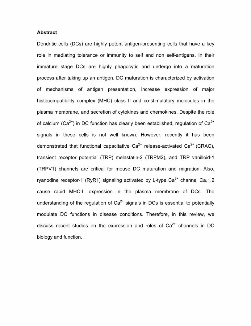
Calcium channels in DC
The main mechanism for Ca2+ entry in immune cells, including DCs, is the store-
operated Ca2+ entry (SOCE), which is activated by Ca2+ release from the
intracellular stores (Fig. 1) [34, 36, 37]. SOCE-mediated Ca2+ influx provides ions
not only for signaling purposes, but also for ER and calciosomes stores refilling.
SOCE activation can be initiated by stimulation of G protein-coupled receptors or
receptor protein tyrosine kinases by external signals (cytokines, chemokines,
bacterial peptides, etc), leading to activation of phospholipase C (PLC), which in
turn hydrolyzes phosphatidylinositol-3,4-bisphophate (PIP2) to release inositol-
1,4,5-triphosphate (IP3) and diacylglycerol (DAG) [33, 34]. Binding of IP3 to IP3
receptors in the ER and calciosomes cause a rapid and transient Ca2+ release
raising the [Ca2+]i (Figure 1). On the other hand, decrease in the luminal Ca2+ in
the ER is detected by the stromal interaction molecule 1/2 (STIM1/2) resulting in
its conformational change (oligomerization and aggregation) and activation of
store operated Ca2+ (SOC) channels in the plasma membrane, which then allow
influx of extracellular Ca2+ across the plasma membrane [33, 34]. SOC channels
include capacitative Ca2+ release-activated Ca2+ (CRAC) channel (Figure 1),

which pore is formed by CRACM/Orai 1-3 proteins. CRAC channels are highly
Ca2+-selective, low conductance channel with a characteristic inwardly rectifying
current-voltage relationship [33, 34]. Interestingly, Orai and STIM proteins may
have different tissue distribution, selectivity and conductivity for Ca2+. As result of
[Ca2+]i increase several signaling pathways and transcription factors are
activated, such as the calmodulin-calcineurin pathway to activate the nuclear
factor of activated T cells (NFAT), the Ca2+-dependent kinase-calmodulin (CaMK)
pathway which active cyclic-adenosine monophosphate-responsive element
binding protein (CREB), and the nuclear factor κB (NFκB) pathway. Moreover,
the DAG formed from PIP2 hydrolyzes can activate the protein kinase C pathway
(PCK), and Ras-mitogen-activated protein kinase, which ultimately activate
transcription factors such as activating protein-2 (AP-2) and NFκB [33, 34].
Although the presence of CRAC currents and its role in DC maturation have
previously been demonstrated in mouse DCs [36], only recently have been
shown that Orai2 and STIM2 are the most abundant in DCs [38]. Furthermore,
recruitment of Orai2 and STIM2 towards the immunological synapse have been
observed during antigen presentation of DC to T lymphocytes [38]. Studies using
CRAC blockers have also shown that this channel plays an important role in the
maturation, cytokine production (TNF-α and IL-6) and chemotaxis of DCs [37].
DC maturation can be triggered in vitro by [Ca2+]i increase by stimulating them
with peptoglycan (PNG), CpG DNA, microbial products such as
lipopolysaccharide (LPS) [39, 40], or ionophores [29-31]. It has been suggested
that LPS, PNG and CpG induce activation of PLCγ2 [39], which in turn acts on

PIP2 to produce IP3 that lead to Ca2+ release from intracellular stores; follow by
CRAC channel activation (reviewed in [34]) causing the nuclear translocation of
calcineurin-dependent NFAT factor and cytokine production, such as IL-2 [33,
41]. On other hand, DC maturation with Ca2+ ionophores are associated with
NFκB activation, likely by activating calcium/calmodulin-dependent kinase II
(CaMKII), which inactivates NFκB-inhibiting molecule IkB similar to in T cells [42].
DC chemotaxis also depends on Ca2+ influx. DC chemotactic response to
chemokines, such as (C-X-C motif) ligand 12 (CXCL12) and (C-C motif) ligand
21 (CCL21), also result in PLC activation, IP3 production, release of Ca2+ from
stores, and subsequent activation of CRAC channel and Ca2+ influx [31, 40, 43,
44]. In addition, our previous study shown that lysosomal Ca2+ release through
the transient receptor potential (TRP) melastatin-2 channel (TRPM2), a member
of the TRP channel family, plays an important role in DC maturation and
chemotaxis (Figure 1) [40]. DCs lacking TRPM2 channels express reduced levels
of costimulatory molecules CD80, CD86, MHC-II and CD83 in the plasma
membrane when they are stimulated with TNF-α and CpG, in comparison with
TRPM2 expressing-DCs [40]. They also show reduced Ca2+ signals in respond to
CXCL12 and CCL21, affecting the chemotaxis response towards these
chemokines [40]. TRPM2 channel is sinergically activated by ADPR and Ca2+,
and permeates sodium (Na+), Ca2+, potassium (K+) and caesium (Cs+) into the
cytosol. In addition to Ca2+, cyclic ADPR (cADPR), hydrogen peroxide (H2O2) and
nicotinic acid adenine dinucleotide phosphate (NAADP) may directly or indirectly
facilitate TRPM2 gating by ADPR [45]. DCs may produce ADPR by means of
CD38 activity, an ectoenzyme that use β-Nicotinamide adenine dinucleotide (β-
NAD+) as a substrate to catalyse the production of ADPR, cADPR, and NAADP,
and by activation of the poly(ADPR)-polymerase/poly(ADP-ribose)
glycohydrolase (PARP/PARG) pathway during DNA repair, replication and
transcription [45]. However, the mechanisms that link these pathways to TRPM2
and to chemokines receptors are still not clearly understood.
DCs also express other non-selective Ca2+ channel in the plasma membrane,
TRP vanilloid-1 (TRPV1) channel, which is activated by capsaicin. However,
there is controversial data on the expression and function of this channel in DCs.
Earlier studies from Basu and Srivastava showed that extracellular Ca2+ influx via
TRPV1 activation induces mouse DC maturation, as provoke increase in the
expression level of MHC clase II and CD86 in the surface [46]. Conversely,
O’Connell PJ, et al., did not detect TRPV1 transcripts and TRPV1 currents in
bone marrow derived-mouse DCs [47]. Recent study by Tóth BI, et al., have
shown molecular and functional expression of TRPV1 channels in monocyte
derived-human DCs [48]. However, although DC stimulation with capsaicin
induces Ca2+ mobilization, this reduces the expression level of maturation
markers in DCs, such as CD83 and CCR7 [48]. On the other hand, TRPM4, a
Ca2+-activated TRP channel that allow Na+ into the cell, indirectly regulates DC
migration but not maturation by decreasing the driving force for Ca2+ entry
through CRAC channels [43].
Furthermore, ryanodine receptor-1 (RyR1), an intracellular channel expressed in
intracellular Ca2+ stores, is also expressed in DCs [49, 50]. RyR1 signaling

coupled with L-type Ca2+ channels Cav1.2, which has been also detected in DCs,
cause rapid MHC class II expression in the plasma membrane of DCs [50].
Interestingly, RyRs are also activated by cADPR and NAADP+, and might
contribute trough these pathways to DC maturation [44, 51, 52]. Finally, DCs
express purinergic receptor (P2Rs), P2X (ligand-gated ion channels) and P2Y
(G-protein coupled receptors) on their surface, such as P2X1, P2X4, and P2X7,
and P2Y1, P2Y2, P2Y4, P2Y6, P2Y11, and P2Y14, respectively. DC stimulation
with adenosine triphosphate (ATP), a damage-associated molecular pattern
(DAMP) molecule released by injured cells during inflammation and necrosis, or
UTP result in characteristic Ca2+ signaling associated to P2X or P2Y, mainly
P2X7 [53-57].
Concluding remarks
Not much is known about Ca2+ channel expression and Ca2+ regulation in DC.
Recent studies have addressed the role of CRAC, TRPV1, TRPM2, RyR1 and
Cav 1.2 channels in DC maturation and migration. However, the mechanisms
that lead to activation of these channels during DC function are not well
understood. Moreover, future studies still need to address which channels
regulate Ca2+ signals during antigen presentation, immune synapse, apoptosis,
and other DC functions. The meaning of Ca2+ oscillations, frequency and
patterns are unknown, which might play and important role in establishing and/or
maintaining immunological tolerance or immunity to self and non self-antigens.

References
1. Steinman, R.M. and Z.A. Cohn, Identification of a novel cell type in
peripheral lymphoid organs of mice. I. Morphology, quantitation, tissue
distribution. J Exp Med, 1973. 137(5): p. 1142-62.
2. Guermonprez, P., et al., Antigen presentation and T cell stimulation by
dendritic cells. Annu Rev Immunol, 2002. 20: p. 621-67.
3. Merad, M., et al., The dendritic cell lineage: ontogeny and function of
dendritic cells and their subsets in the steady state and the inflamed setting.
Annu Rev Immunol, 2013. 31: p. 563-604.
4. Dalod, M., et al., Dendritic cell maturation: functional specialization
through signaling specificity and transcriptional programming. EMBO J, 2014.
33(10): p. 1104-16.
5. Dieu, M.C., et al., Selective recruitment of immature and mature dendritic
cells by distinct chemokines expressed in different anatomic sites. J Exp Med,
1998. 188(2): p. 373-86.
6. Sallusto, F., et al., Rapid and coordinated switch in chemokine receptor
expression during dendritic cell maturation. Eur J Immunol, 1998. 28(9): p. 2760-
9.
7. Yanagihara, S., et al., EBI1/CCR7 is a new member of dendritic cell
chemokine receptor that is up-regulated upon maturation. J Immunol, 1998.
161(6): p. 3096-102.

8. Ferlazzo, G. and B. Morandi, Cross-Talks between Natural Killer Cells and
Distinct Subsets of Dendritic Cells. Front Immunol, 2014. 5: p. 159.
9. Bennouna, S., et al., Cross-talk in the innate immune system: neutrophils
instruct recruitment and activation of dendritic cells during microbial infection. J
Immunol, 2003. 171(11): p. 6052-8.
10. Rescigno, M., Dendritic cell-epithelial cell crosstalk in the gut. Immunol
Rev, 2014. 260(1): p. 118-28.
11. Liu, Y.J., Dendritic cell subsets and lineages, and their functions in innate
and adaptive immunity. Cell, 2001. 106(3): p. 259-62.
12. Liu, K. and M.C. Nussenzweig, Origin and development of dendritic cells.
Immunol Rev, 2010. 234(1): p. 45-54.
13. Vremec, D., et al., The surface phenotype of dendritic cells purified from
mouse thymus and spleen: investigation of the CD8 expression by a
subpopulation of dendritic cells. J Exp Med, 1992. 176(1): p. 47-58.
14. Vremec, D., et al., CD4 and CD8 expression by dendritic cell subtypes in
mouse thymus and spleen. J Immunol, 2000. 164(6): p. 2978-86.
15. den Haan, J.M., S.M. Lehar, and M.J. Bevan, CD8(+) but not CD8(-)
dendritic cells cross-prime cytotoxic T cells in vivo. J Exp Med, 2000. 192(12): p.
1685-96.
16. Belz, G.T. and S.L. Nutt, Transcriptional programming of the dendritic cell
network. Nat Rev Immunol, 2012. 12(2): p. 101-13.
17. Kaisho, T., Pathogen sensors and chemokine receptors in dendritic cell
subsets. Vaccine, 2012. 30(52): p. 7652-7.

18. Maldonado, R.A. and U.H. von Andrian, How tolerogenic dendritic cells
induce regulatory T cells. Adv Immunol, 2010. 108: p. 111-65.
19. Steinman, R.M., D. Hawiger, and M.C. Nussenzweig, Tolerogenic
dendritic cells. Annu Rev Immunol, 2003. 21: p. 685-711.
20. Morelli, A.E., et al., Cytokine production by mouse myeloid dendritic cells
in relation to differentiation and terminal maturation induced by
lipopolysaccharide or CD40 ligation. Blood, 2001. 98(5): p. 1512-23.
21. Bedoui, S., et al., Cross-presentation of viral and self antigens by skin-
derived CD103+ dendritic cells. Nat Immunol, 2009. 10(5): p. 488-95.
22. Ng, S.C., et al., Intestinal dendritic cells: their role in bacterial recognition,
lymphocyte homing, and intestinal inflammation. Inflamm Bowel Dis, 2010.
16(10): p. 1787-807.
23. del Rio, M.L., et al., Development and functional specialization of CD103+
dendritic cells. Immunol Rev, 2010. 234(1): p. 268-81.
24. Milling, S., et al., Subsets of migrating intestinal dendritic cells. Immunol
Rev, 2010. 234(1): p. 259-67.
25. Asselin-Paturel, C., et al., Mouse type I IFN-producing cells are immature
APCs with plasmacytoid morphology. Nat Immunol, 2001. 2(12): p. 1144-50.
26. Reizis, B., et al., Plasmacytoid dendritic cells: recent progress and open
questions. Annu Rev Immunol, 2011. 29: p. 163-83.
27. Zhang, J., et al., Characterization of Siglec-H as a novel endocytic
receptor expressed on murine plasmacytoid dendritic cell precursors. Blood,
2006. 107(9): p. 3600-8.

28. Segura, E. and S. Amigorena, Inflammatory dendritic cells in mice and
humans. Trends Immunol, 2013. 34(9): p. 440-5.
29. Bagley, K.C., et al., Calcium signaling through phospholipase C activates
dendritic cells to mature and is necessary for the activation and maturation of
dendritic cells induced by diverse agonists. Clin Diagn Lab Immunol, 2004. 11(1):
p. 77-82.
30. Czerniecki, B.J., et al., Calcium ionophore-treated peripheral blood
monocytes and dendritic cells rapidly display characteristics of activated dendritic
cells. J Immunol, 1997. 159(8): p. 3823-37.
31. Koski, G.K., et al., Calcium mobilization in human myeloid cells results in
acquisition of individual dendritic cell-like characteristics through discrete
signaling pathways. J Immunol, 1999. 163(1): p. 82-92.
32. Chen, Y.F., et al., Remodeling of calcium signaling in tumor progression. J
Biomed Sci, 2013. 20: p. 23.
33. Izquierdo, J.H., et al., Calcium, channels, intracellular signaling and
autoimmunity. Reumatol Clin, 2014. 10(1): p. 43-7.
34. Shumilina, E., S.M. Huber, and F. Lang, Ca2+ signaling in the regulation
of dendritic cell functions. Am J Physiol Cell Physiol, 2011. 300(6): p. C1205-14.
35. Feske, S., Calcium signalling in lymphocyte activation and disease. Nat
Rev Immunol, 2007. 7(9): p. 690-702.
36. Hsu Sf, et al., Fundamental Ca2+ signaling mechanisms in mouse
dendritic cells: CRAC is the major Ca2+ entry pathway. J Immunol, 2001.
166(10): p. 6126-33.

37. Matzner, N., et al., Ion channels modulating mouse dendritic cell functions.
J Immunol, 2008. 181(10): p. 6803-9.
38. Bandyopadhyay, B.C., S.C. Pingle, and G.P. Ahern, Store-operated Ca²+
signaling in dendritic cells occurs independently of STIM1. J Leukoc Biol, 2011.
89(1): p. 57-62.
39. Aki, D., et al., Peptidoglycan and lipopolysaccharide activate
PLCgamma2, leading to enhanced cytokine production in macrophages and
dendritic cells. Genes Cells, 2008. 13(2): p. 199-208.
40. Sumoza-Toledo, A., et al., Dendritic cell maturation and chemotaxis is
regulated by TRPM2-mediated lysosomal Ca2+ release. FASEB J, 2011. 25(10):
p. 3529-42.
41. Zanoni, I., et al., CD14 regulates the dendritic cell life cycle after LPS
exposure through NFAT activation. Nature, 2009. 460(7252): p. 264-8.
42. Li, Q. and I.M. Verma, NF-kappaB regulation in the immune system. Nat
Rev Immunol, 2002. 2(10): p. 725-34.
43. Barbet, G., et al., The calcium-activated nonselective cation channel
TRPM4 is essential for the migration but not the maturation of dendritic cells. Nat
Immunol, 2008. 9(10): p. 1148-56.
44. Partida-Sanchez, S., et al., Chemotaxis of mouse bone marrow
neutrophils and dendritic cells is controlled by adp-ribose, the major product
generated by the CD38 enzyme reaction. J Immunol, 2007. 179(11): p. 7827-39.
45. Sumoza-Toledo, A. and R. Penner, TRPM2: a multifunctional ion channel
for calcium signalling. J Physiol, 2011. 589(Pt 7): p. 1515-25.

46. Basu, S. and P. Srivastava, Immunological role of neuronal receptor
vanilloid receptor 1 expressed on dendritic cells. Proc Natl Acad Sci U S A, 2005.
102(14): p. 5120-5.
47. O'Connell, P.J., S.C. Pingle, and G.P. Ahern, Dendritic cells do not
transduce inflammatory stimuli via the capsaicin receptor TRPV1. FEBS Lett,
2005. 579(23): p. 5135-9.
48. Tóth, B.I., et al., Transient receptor potential vanilloid-1 signaling inhibits
differentiation and activation of human dendritic cells. FEBS Lett, 2009. 583(10):
p. 1619-24.
49. O'Connell, P.J., V.A. Klyachko, and G.P. Ahern, Identification of functional
type 1 ryanodine receptors in mouse dendritic cells. FEBS Lett, 2002. 512(1-3):
p. 67-70.
50. Vukcevic, M., et al., Ryanodine receptor activation by Ca v 1.2 is involved
in dendritic cell major histocompatibility complex class II surface expression. J
Biol Chem, 2008. 283(50): p. 34913-22.
51. Lee, H.C., Multiplicity of Ca2+ messengers and Ca2+ stores: a
perspective from cyclic ADP-ribose and NAADP. Curr Mol Med, 2004. 4(3): p.
227-37.
52. Partida-Sánchez, S., et al., Regulation of dendritic cell trafficking by the
ADP-ribosyl cyclase CD38: impact on the development of humoral immunity.
Immunity, 2004. 20(3): p. 279-91.
53. Di Virgilio, F., Purinergic mechanism in the immune system: A signal of
danger for dendritic cells. Purinergic Signal, 2005. 1(3): p. 205-9.

54. Ferrari, D., et al., The P2 purinergic receptors of human dendritic cells:
identification and coupling to cytokine release. FASEB J, 2000. 14(15): p. 2466-
76.
55. Mutini, C., et al., Mouse dendritic cells express the P2X7 purinergic
receptor: characterization and possible participation in antigen presentation. J
Immunol, 1999. 163(4): p. 1958-65.
56. Schnurr, M., et al., ATP gradients inhibit the migratory capacity of specific
human dendritic cell types: implications for P2Y11 receptor signaling. Blood,
2003. 102(2): p. 613-20.
57. Shin, A., et al., P2Y receptor signaling regulates phenotype and IFN-alpha
secretion of human plasmacytoid dendritic cells. Blood, 2008. 111(6): p. 3062-9.

Figure 1. Calcium channels in DCs. Extracellular signals (chemokines,
cytokines, microbial peptides, etc) are recognized by DCs by means of G protein-
coupled receptors or receptor protein tyrosine kinases, activating the formation of
1,4,5-triphosphate (IP3) that in turn binds to IP3 receptors in the ER and
calciosomes, causing Ca2+ release. Decrease in the luminal Ca2+ in the ER is
detected by the stromal interaction molecule 1/2 (STIM1/2) resulting in the
activation of capacitative Ca2+ release-activated Ca2+ (CRAC) channels, allowing
Ca2+ influx across the plasma membrane. Chemokines also activates transient
receptor potential (TRP) melastatin-2 (TRPM2) channels in DC lysosomes. The
Ca2+ signals activate transcription factors such as nuclear factor of activated T
Activation
Migration Antigen presentation
Cytokines production
Ca2+
Ca2+ Ca2+
Calciosome
Endoplasmic reticulum
NF- κB
Na+
?
?
IP3
Receptor TRPM4 TRPV1?
IP3R
SERCA
STIM1/2
Lysosome
Ca2+
Ca2+
cADPR
CRAC
ADPR

cells (NFAT) or nuclear factor-κB (NF-κB) for gene expression. TRPM4, a Ca2+-
activated TRP channel that allow Na+ into the cell is expressed in the plasma
membrane of DCs and indirectly regulates DC functions by decreasing the
driving force for Ca2+ entry through CRAC channels. DCs also express TRP
vanilloid-1 (TRPV1) and ryanodine (RyR) channels but their functions are still not
clear.

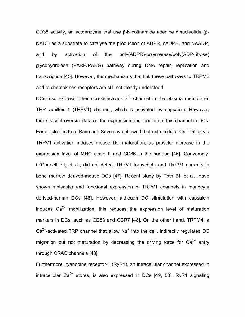
DC subsets CD8α CD103 CD205 CD11b B220 or CD45RA
DC-SING
Langerin (CD207)
MHC class
II
CD11c
pDCs + - - - + ++ - + + CD8α+ DCs + low + + - - +/- ++ +++ CD8α_
CD11b+ DCs - +/- + + - ND - ++ +++
CD103+ DCs Lung Intestine
-
+
++
-
-
-
+
++
++
- + - + - - - ++ ++ Langerhans cells
- - ++ + - - ++ ++ ++
Monocyte-derived inflammatory DCs
- - - + - + - ++ ++
Table1. Dendritic cell subsets.
Recommended