Parasitic Protozoa
One cell menaces

Protozoa Single Cell Asexual multiplication provides the mechanism for the development
of pathogenic protozoan populations. Pathology is generally seen as the dysfunction of host tissue
direct destruction of the host cells (Coccidiosis, Malaria, Piroplasms) indirect destruction of host cells (Entamoeba) barrier to tissue function (Giardia) excessive activation of host immune system (Trypanosomes) excretion of toxins

Pathology:Cellular traumaOrgan dysfunction

Protozoa Life Cycle Strategies
Direct life cycles using only a single host species (e.g. Eimeria)
Indirect Life cycle -- require 2 or more hosts (e.g. Sarcocystis, Trypanosoma)
Asexual stages only – thus “clonal” (e.g. Giardia, Entamoeba)
Alternation of sexual and asexual stages (all of the apicomplexans)
Continuous life cycle Without host immunity; organism would continue multiplying (e.g.
Plasmodium, Trypanosoma)
Single direction life cycle Once the life cycle is completed then all organisms are gone (except in
the case of re-infection) “all in – all out” (e.g. Eimeria).
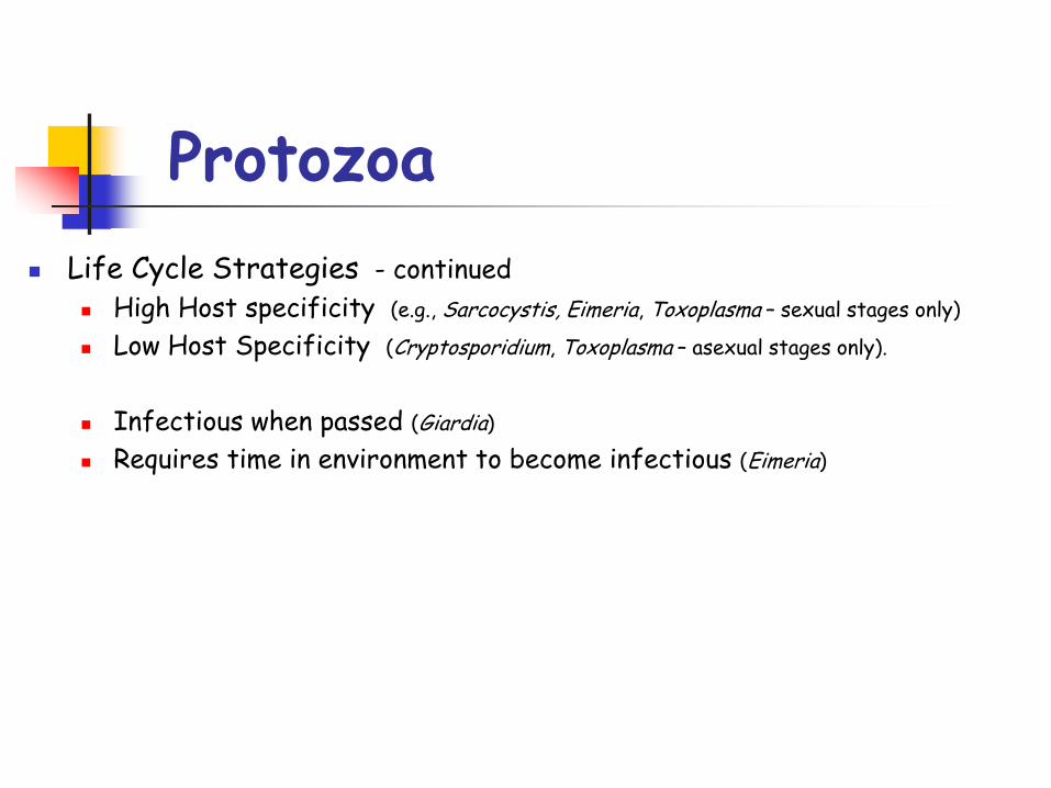
Protozoa Life Cycle Strategies - continued
High Host specificity (e.g., Sarcocystis, Eimeria, Toxoplasma – sexual stages only)
Low Host Specificity (Cryptosporidium, Toxoplasma – asexual stages only).
Infectious when passed (Giardia)
Requires time in environment to become infectious (Eimeria)
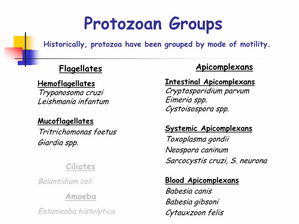
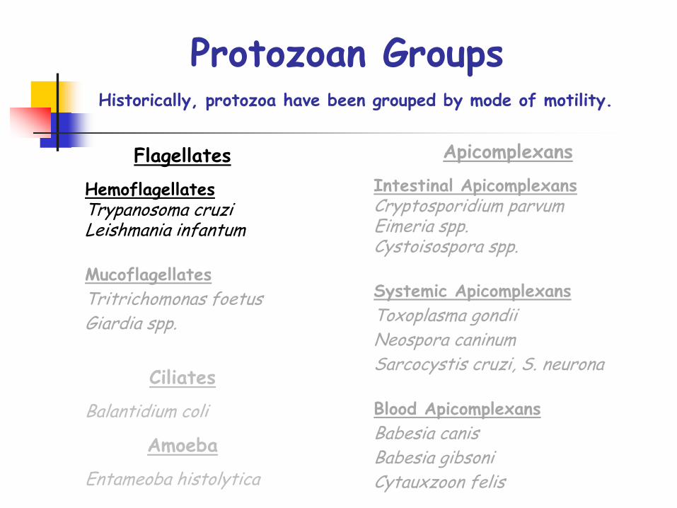
Protozoan Groups
Flagellates
HemoflagellatesTrypanosoma cruziLeishmania infantum
MucoflagellatesTritrichomonas foetusGiardia spp.
Historically, protozoa have been grouped by mode of motility.
Apicomplexans
Intestinal ApicomplexansCryptosporidium parvumEimeria spp.Cystoisospora spp.
Systemic ApicomplexansToxoplasma gondiiNeospora caninumSarcocystis cruzi, S. neurona
Blood ApicomplexansBabesia canisBabesia gibsoniCytauxzoon felis
Ciliates
Balantidium coli
Amoeba
Entameoba histolytica

Hemoflagellates
Trypanosoma cruzi
Leishmania infantum
Protozoan Groups
Flagellates
HemoflagellatesTrypanosoma cruziLeishmania infantum
MucoflagellatesTritrichomonas foetusGiardia spp.
Historically, protozoa have been grouped by mode of motility.
Apicomplexans
Intestinal ApicomplexansCryptosporidium parvumEimeria spp.Cystoisospora spp.
Systemic ApicomplexansToxoplasma gondiiNeospora caninumSarcocystis cruzi, S. neurona
Blood ApicomplexansBabesia canisBabesia gibsoniCytauxzoon felis
Ciliates
Balantidium coli
Amoeba
Entameoba histolytica
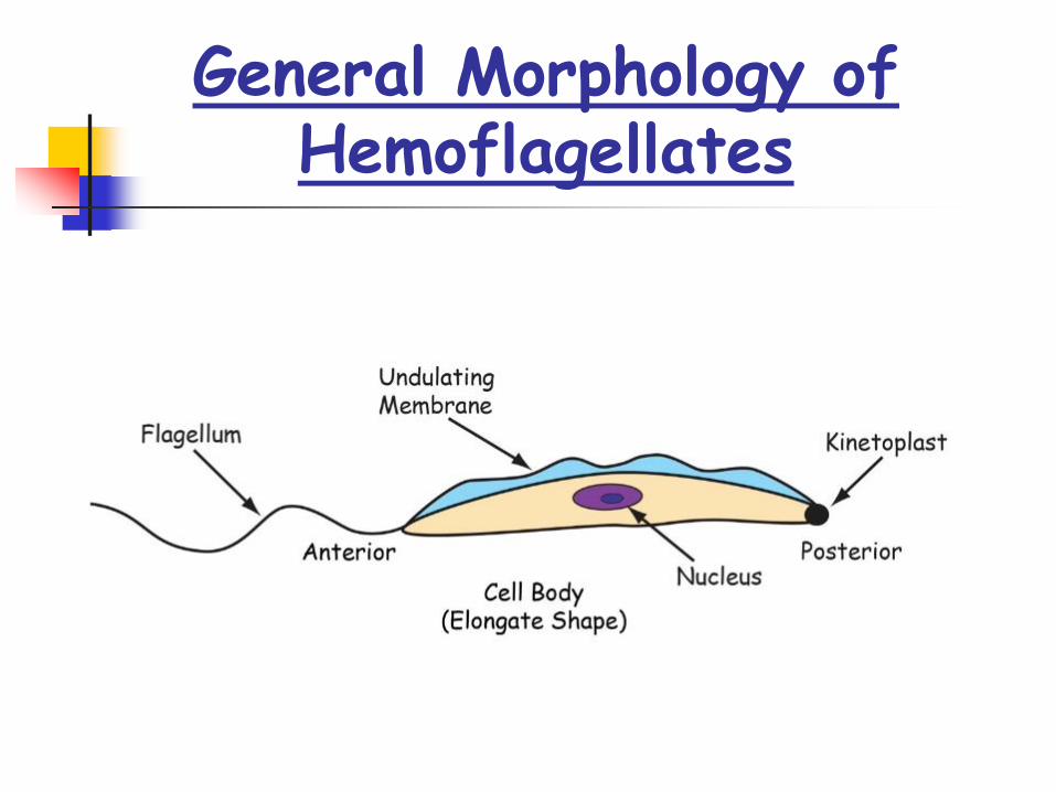
General Morphology of Hemoflagellates
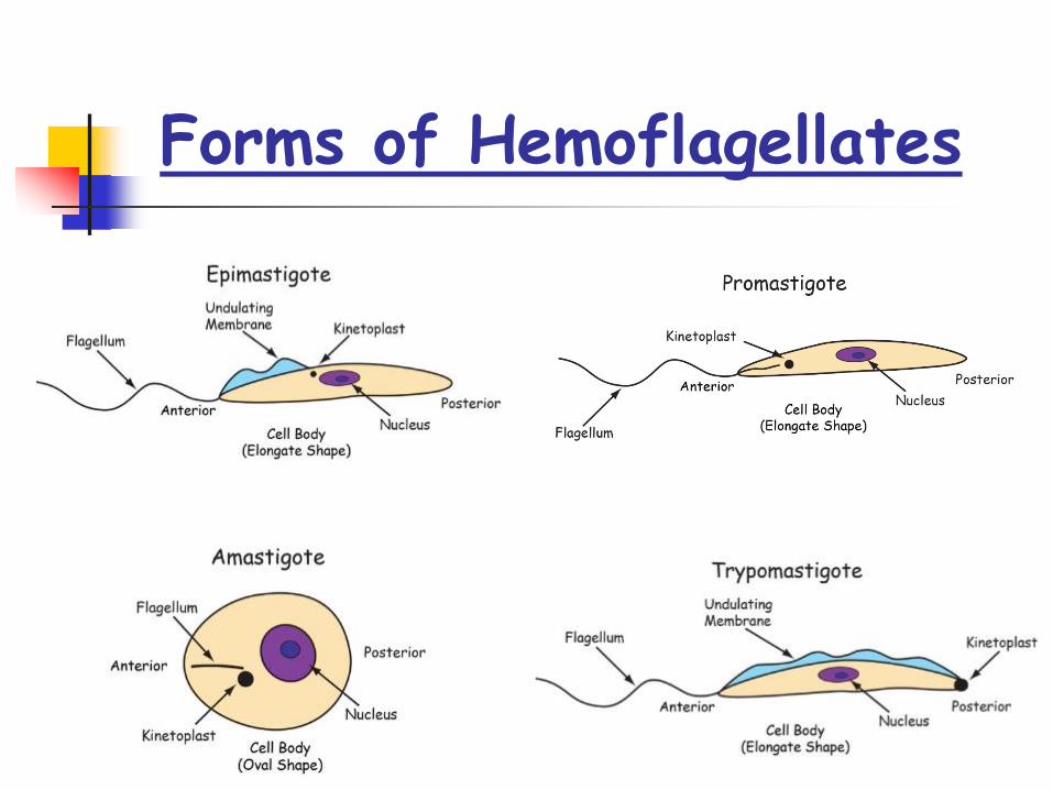
Forms of Hemoflagellates
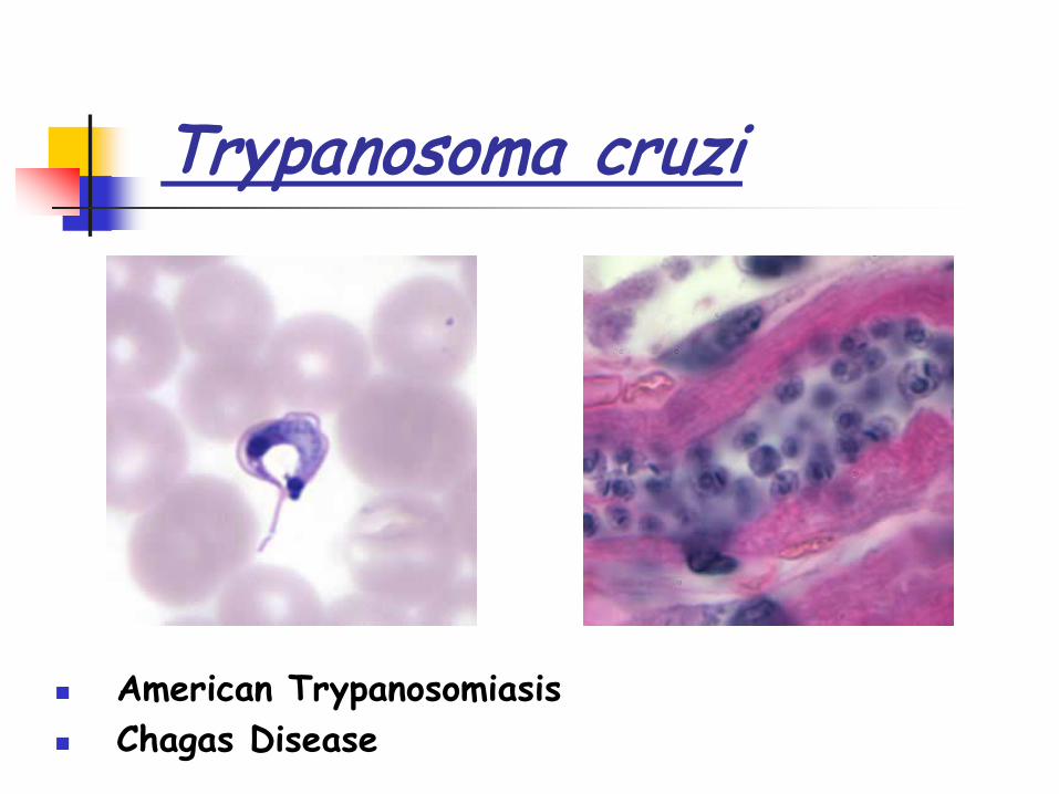
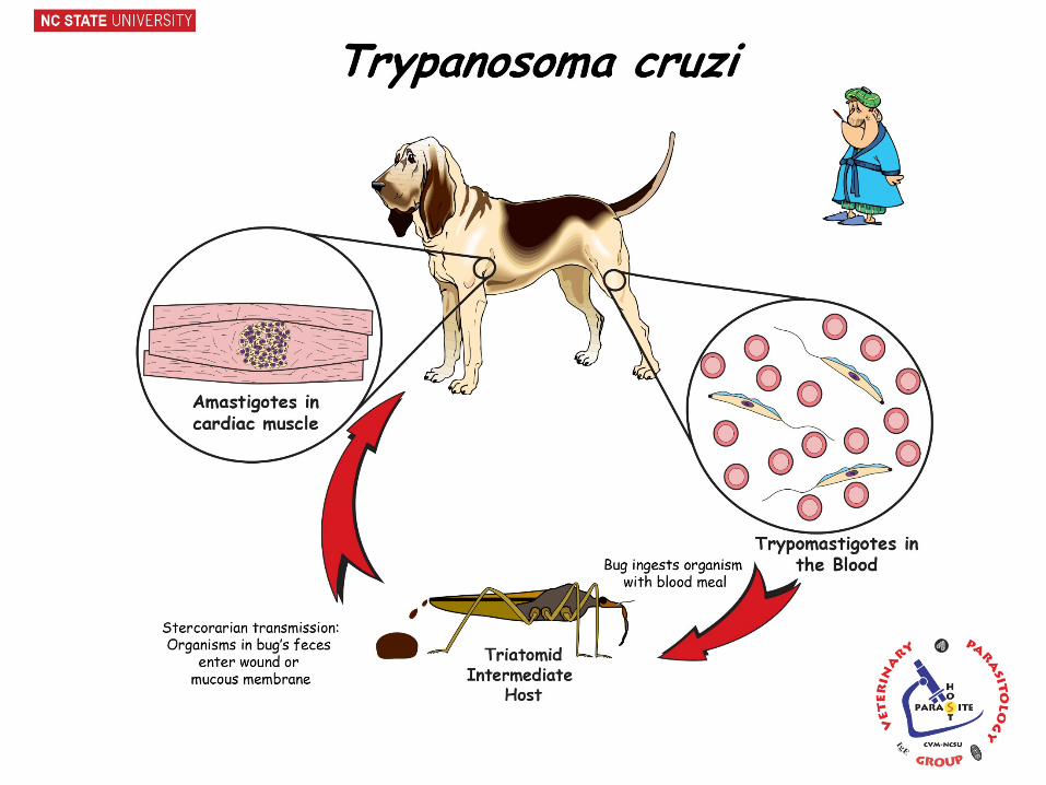
Trypanosoma cruzi
American Trypanosomiasis Chagas Disease
Life Cycle – Mammalian Hosts
Mammalian hosts Dogs (cardiac muscle) & humans Opossums, armadillos, raccoons, wood rats, etc. (>100 mammal species)
Metacyclic Trypomastigote – infective form – rubbed into bug bite, skin scratch, oral or ocular mucosae. Or via ingestion of bug.
Metacyclic Trypomastigotes invade local cells and macrophages, become amastigotes that multiply via binary division
Amastigotes turn into trypomastigotes, which burst from host cells Trypomastigotes travel to other cells in the body via blood stream and
invade host cells- usually cardiac muscles in dogs – turn into amastigotes and repeat multiplication and distribution cycle.
Some trypomastigotes in the blood change into metacyclic trypomastigotes, which may be ingested by vector.
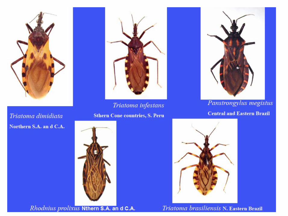
Arthropod hosts (vectors) Triatomine (Reduviid) Bugs (Triatoma, Rhodnius, Panstrongylus)
[kissing bug, assassin bug] Stercorarian transmission
(Infective metacyclic trypomastigotes in bug feces) Metacyclic trypomastigotes ingested by vector during blood meal In midgut, trypomastigotes transform to epimastigotes, which
multiply via binary division In hindgut, epimastigotes transform to metacyclic
trypomastigotes which are passed in the bug feces when the bug feeds on the mammalian host. (stercorarian transmission)
Life Cycle – Arthropod Hosts
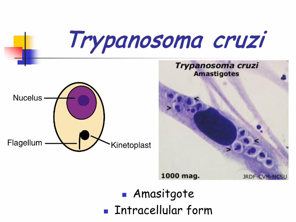
Trypanosoma cruzi
Amasitgote Intracellular form

Trypanosoma cruzi
Trypomastigote Blood form
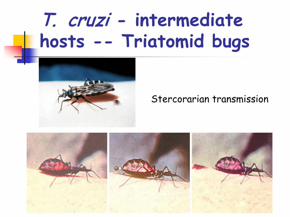
T. cruzi - intermediate hosts -- Triatomid bugs
Stercorarian transmission
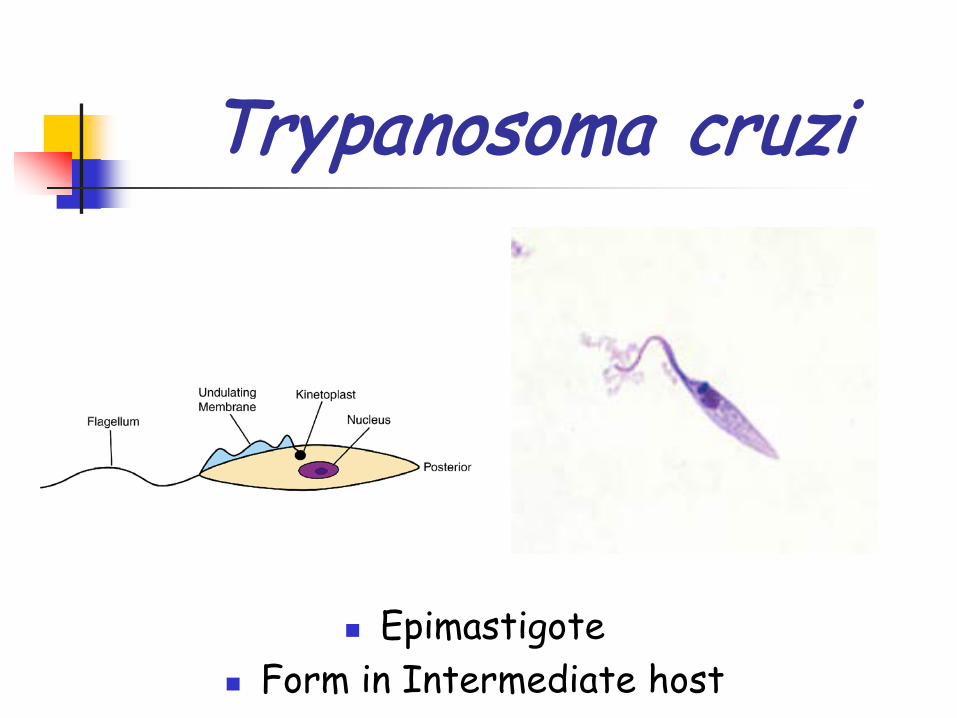
Trypanosoma cruzi
Epimastigote Form in Intermediate host

Vector-borne -- Triatomid bugs Transplacental Blood Transfusion
Transmission
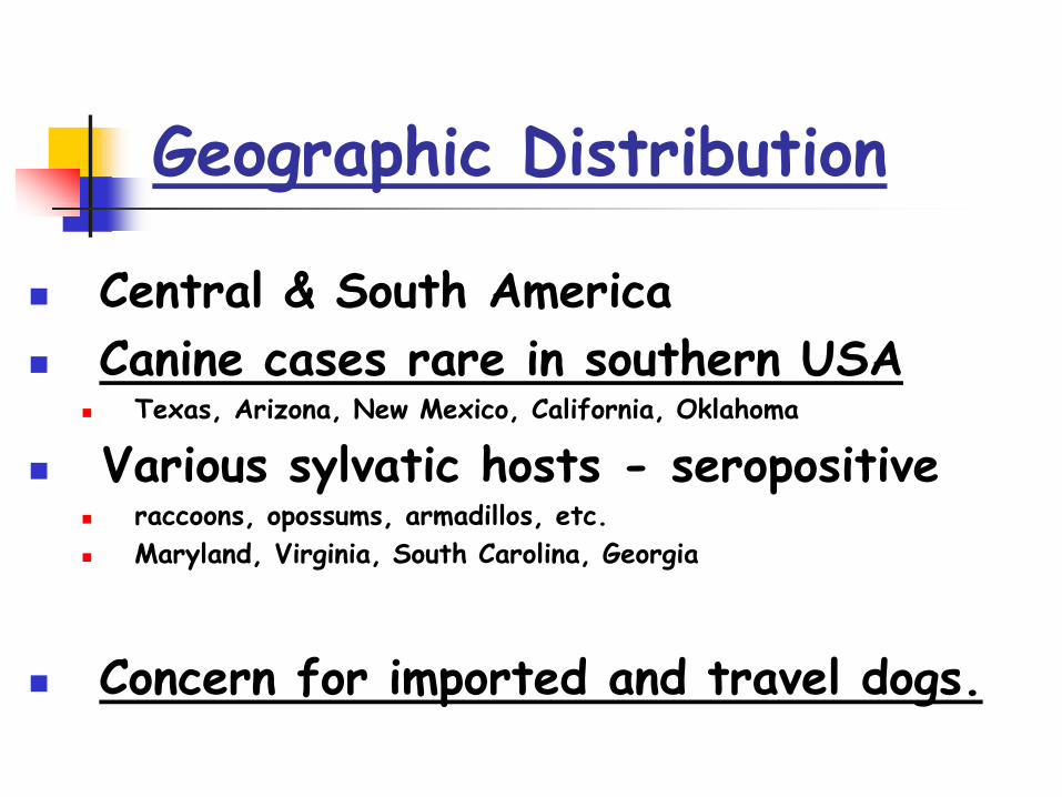
Geographic Distribution
Central & South America Canine cases rare in southern USA
Texas, Arizona, New Mexico, California, Oklahoma
Various sylvatic hosts - seropositive raccoons, opossums, armadillos, etc. Maryland, Virginia, South Carolina, Georgia
Concern for imported and travel dogs.
Pathology
Multi-System Disease Especially cardiac muscle in dogs
Repeat cycles of intracellular multiplication of parasites with destruction of host cells.
In Humans Cardiac, Smooth muscles, glial & nueural cells Also destruction of the myenteric plexus of esophagus
and colon, resulting in the dilatation of these structures (megesophagus, megacolon)

Clinical Disease – Acute Phase
Acute Phase (1st month) Inflammation at site of transmission lymphadenopathy and non-specific febrile
disease diarrhea, vomitus, anorexia, lethargy
rare cases: quickly develop to acute myocarditis and hepatosplenomegaly
Parasitemia (Trypomastigotes in blood)
Clinical Disease – Latent Phase
Latent Phase (months to years post-infection) usually asymptomatic immunosuppression (disease, therapy, age) may
cause relapse to acute phase quiescent in tissues
Clinical Disease – Chronic Phase
Chronic Phase (maybe years post-infection) Gradual decline to death, usually about 2 years after
diagnosis. Chronic general weakness with progressive heart
failure Right-side congestive heart failure w/ myocarditis and
arrhythmias leading to bilateral dilation and eventual death.
Active multiplication & destruction of host tissues Also important is the autoimmune destruction of host
tissues

Chagas Disease caused by
Trypanosoma cruziis a
multi-systemdisease.
In dogs the disease most often manifests as cardiac pathology.
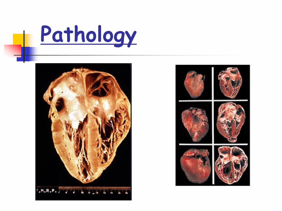
Pathology
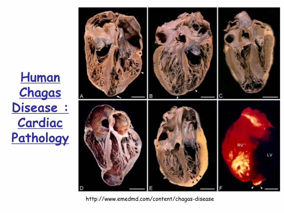
Human Chagas
Disease : Cardiac
Pathology
http://www.emedmd.com/content/chagas-disease

Normal
Pathology
Teixeira, Nascimento, Sturm. 2006. Evolution and pathology in Chagas disease - a review. Mem Inst Oswaldo Cruz, Rio de Janeiro. 101(5): 463-491.
Chicken - cardiac muscleInfected
Chicken - HeartNormalInfected
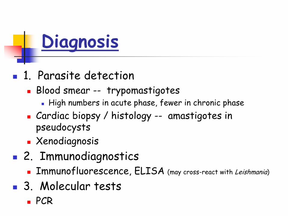
Diagnosis
1. Parasite detection Blood smear -- trypomastigotes
High numbers in acute phase, fewer in chronic phase Cardiac biopsy / histology -- amastigotes in
pseudocysts Xenodiagnosis
2. Immunodiagnostics Immunofluorescence, ELISA (may cross-react with Leishmania)
3. Molecular tests PCR
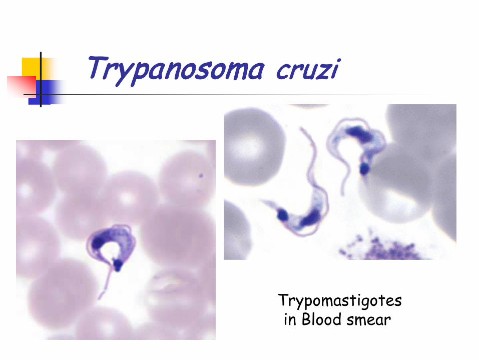
Trypanosoma cruzi
Trypomastigotesin Blood smear

Trypanosoma cruzi
Trypomastigotesin Blood smear
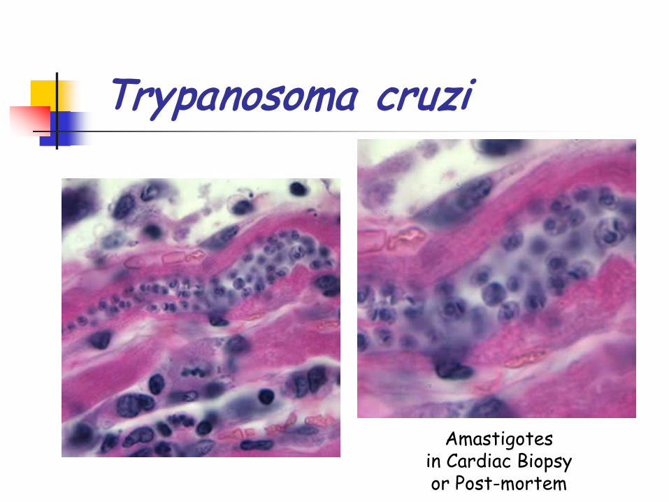
Trypanosoma cruzi
Amastigotesin Cardiac Biopsyor Post-mortem
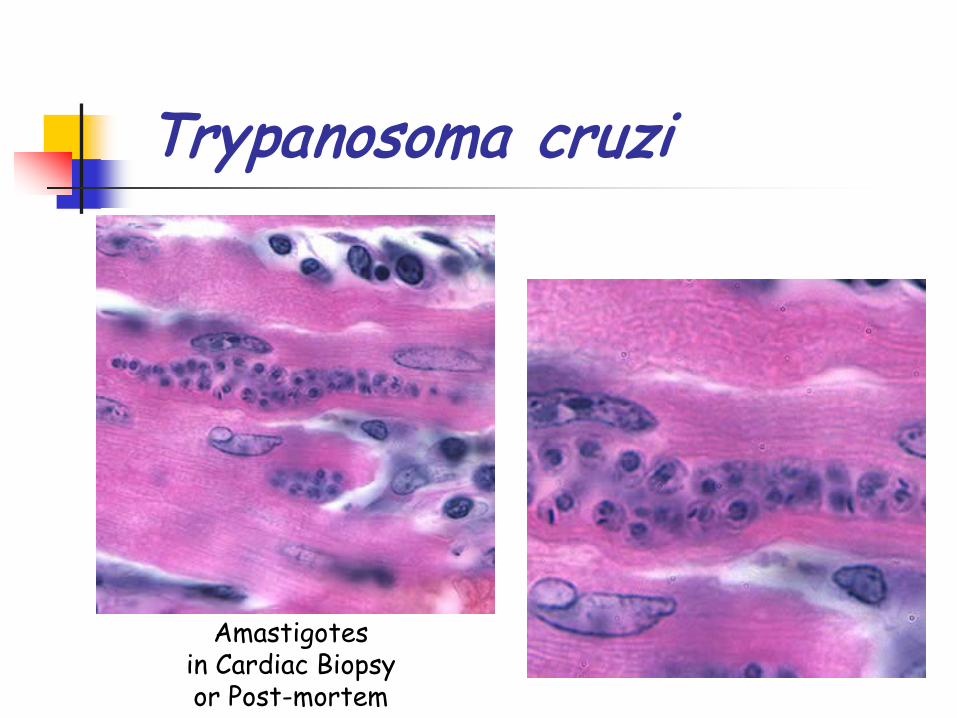
Trypanosoma cruzi
Amastigotesin Cardiac Biopsyor Post-mortem
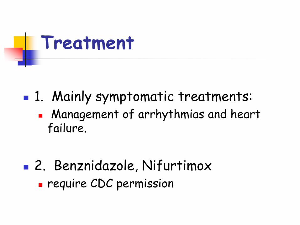
Treatment
1. Mainly symptomatic treatments: Management of arrhythmias and heart
failure.
2. Benznidazole, Nifurtimox require CDC permission

Control
Vector control Dx Triatomids & their habitat
Breeding control v/s Transplacental Transmission
Screen Blood donors v/s Transfusion Transmission
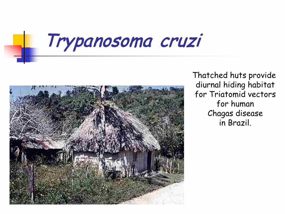
Trypanosoma cruziThatched huts provide diurnal hiding habitatfor Triatomid vectors
for humanChagas disease
in Brazil.
Zoonosis
Human Chagas Disease Endemic areas:
Mexico, Central America, South America Triatomids thrive in poor housing conditions
Mud Walls, Thatched Roofs Dogs are important reservoirs for human
infections
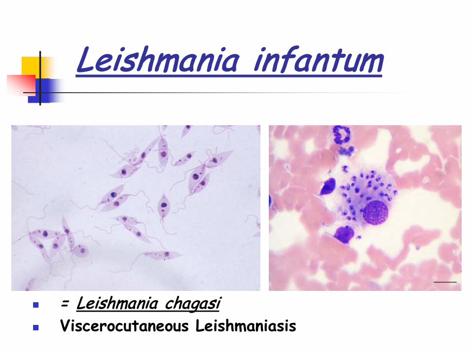
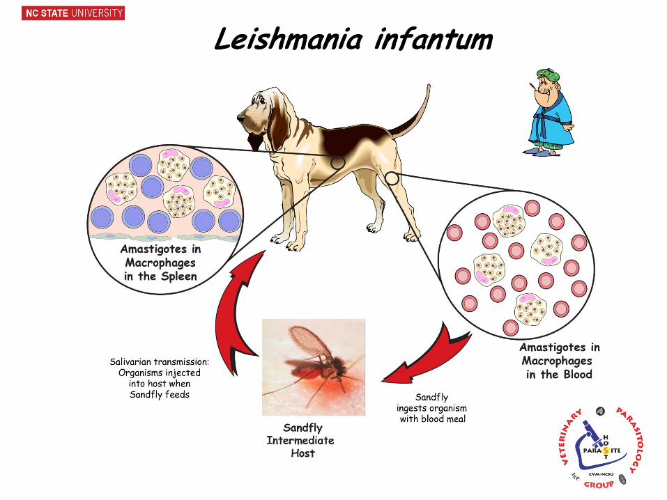
Leishmania infantum
= Leishmania chagasi Viscerocutaneous Leishmaniasis
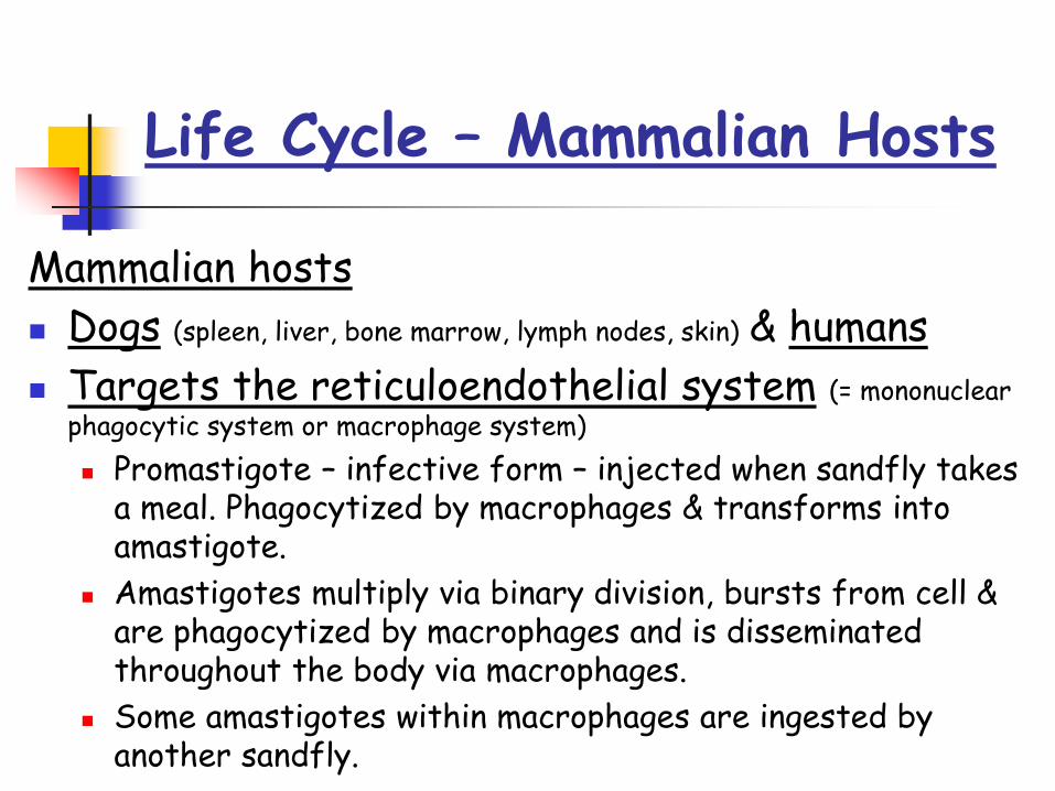
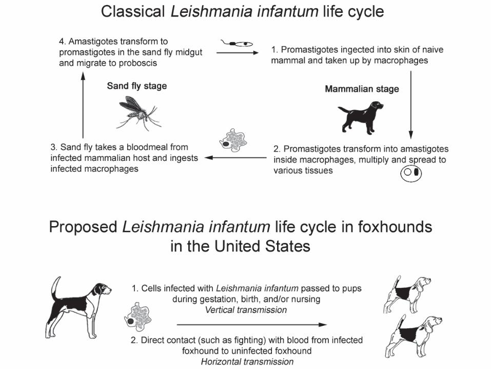
Life Cycle – Mammalian Hosts
Mammalian hosts Dogs (spleen, liver, bone marrow, lymph nodes, skin) & humans Targets the reticuloendothelial system (= mononuclear
phagocytic system or macrophage system) Promastigote – infective form – injected when sandfly takes
a meal. Phagocytized by macrophages & transforms into amastigote.
Amastigotes multiply via binary division, bursts from cell & are phagocytized by macrophages and is disseminated throughout the body via macrophages.
Some amastigotes within macrophages are ingested by another sandfly.
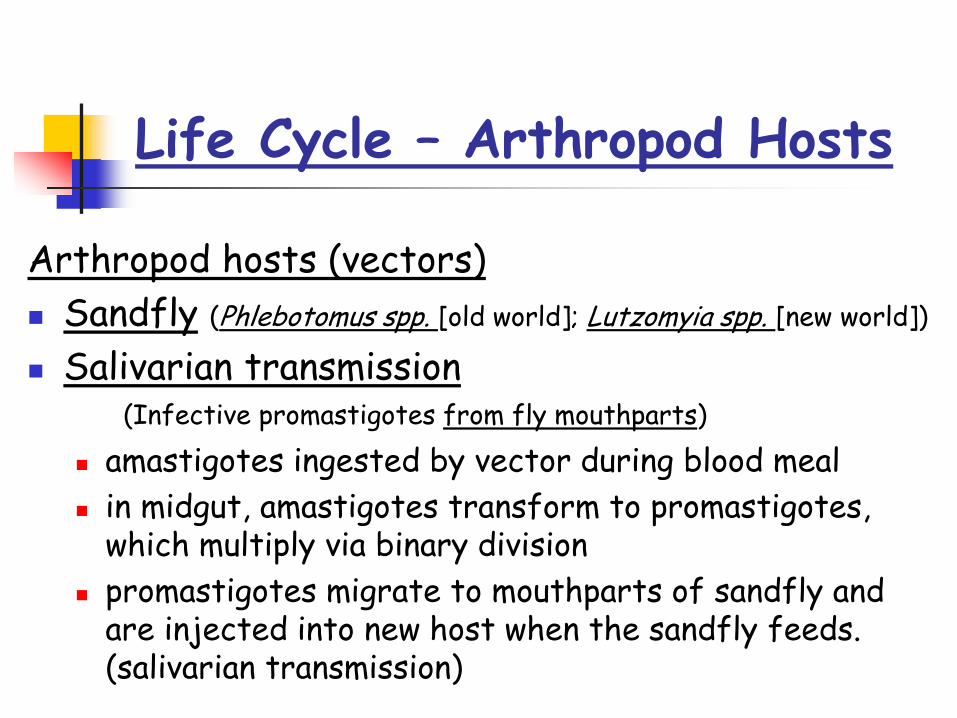
Arthropod hosts (vectors) Sandfly (Phlebotomus spp. [old world]; Lutzomyia spp. [new world])
Salivarian transmission(Infective promastigotes from fly mouthparts)
amastigotes ingested by vector during blood meal in midgut, amastigotes transform to promastigotes,
which multiply via binary division promastigotes migrate to mouthparts of sandfly and
are injected into new host when the sandfly feeds. (salivarian transmission)
Life Cycle – Arthropod Hosts

Sandfly Vectors
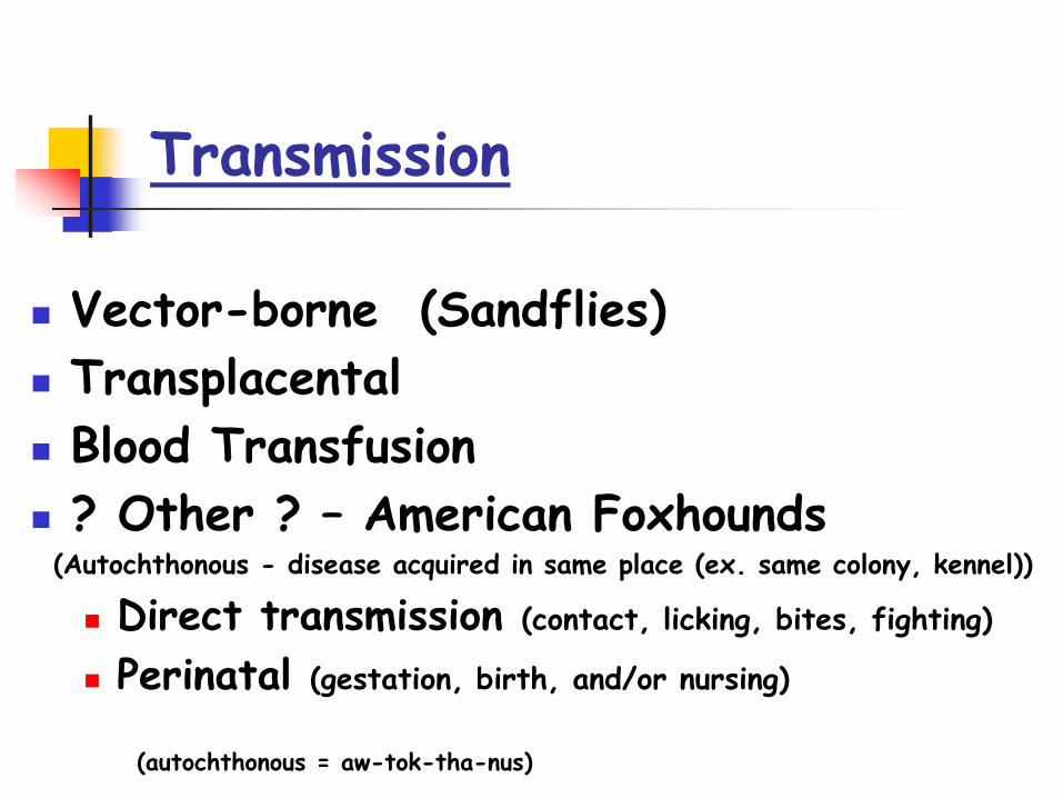
Vector-borne (Sandflies) Transplacental Blood Transfusion ? Other ? – American Foxhounds(Autochthonous - disease acquired in same place (ex. same colony, kennel))
Direct transmission (contact, licking, bites, fighting) Perinatal (gestation, birth, and/or nursing)
(autochthonous = aw-tok-tha-nus)
Transmission

Geographic Distribution
>70 countries: Southern Europe, Africa, Asia, Caribbean, Central & South America Concern for imported and travel dogs.
Sporadic in US Foxhound colonies. (Oklahoma, Kansas, NY, Ohio, NC)
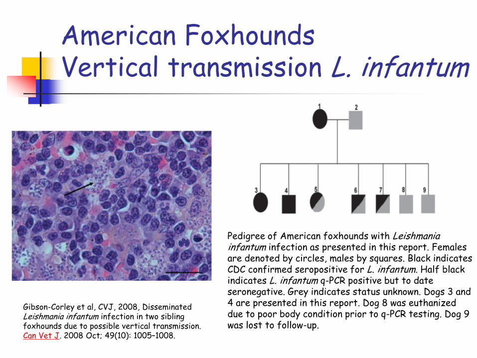
American FoxhoundsVertical transmission L. infantum
Pedigree of American foxhounds with Leishmania infantum infection as presented in this report. Females are denoted by circles, males by squares. Black indicates CDC confirmed seropositive for L. infantum. Half black indicates L. infantum q-PCR positive but to date seronegative. Grey indicates status unknown. Dogs 3 and 4 are presented in this report. Dog 8 was euthanized due to poor body condition prior to q-PCR testing. Dog 9 was lost to follow-up.
Gibson-Corley et al, CVJ, 2008, Disseminated Leishmania infantum infection in two sibling foxhounds due to possible vertical transmission. Can Vet J. 2008 Oct; 49(10): 1005–1008.
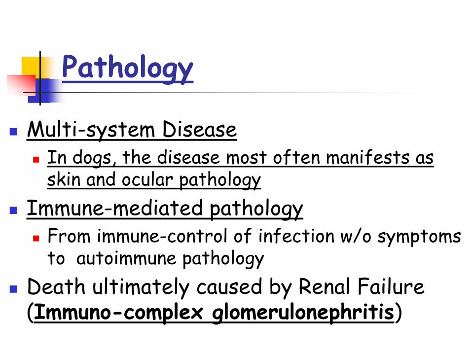
Pathology
Multi-system Disease In dogs, the disease most often manifests as
skin and ocular pathology Immune-mediated pathology
From immune-control of infection w/o symptoms to autoimmune pathology
Death ultimately caused by Renal Failure (Immuno-complex glomerulonephritis)

Clinical Disease in Canines
Various issues - vary by case Incubation period from 3 months to several years
Client complaint Skin lesions, ocular abnormalities, epistaxis (nose
bleed), weight loss, lethargy Clinical findings
Dermal lesions, lymphadenopathy, fever, ocular dz(uveitis), splenomegaly, signs of liver dz(hyperglobulinemia, hypoalbuminemia), signs of anemia (non-regenerative anemia), signs of kidney dz(proteinuria)

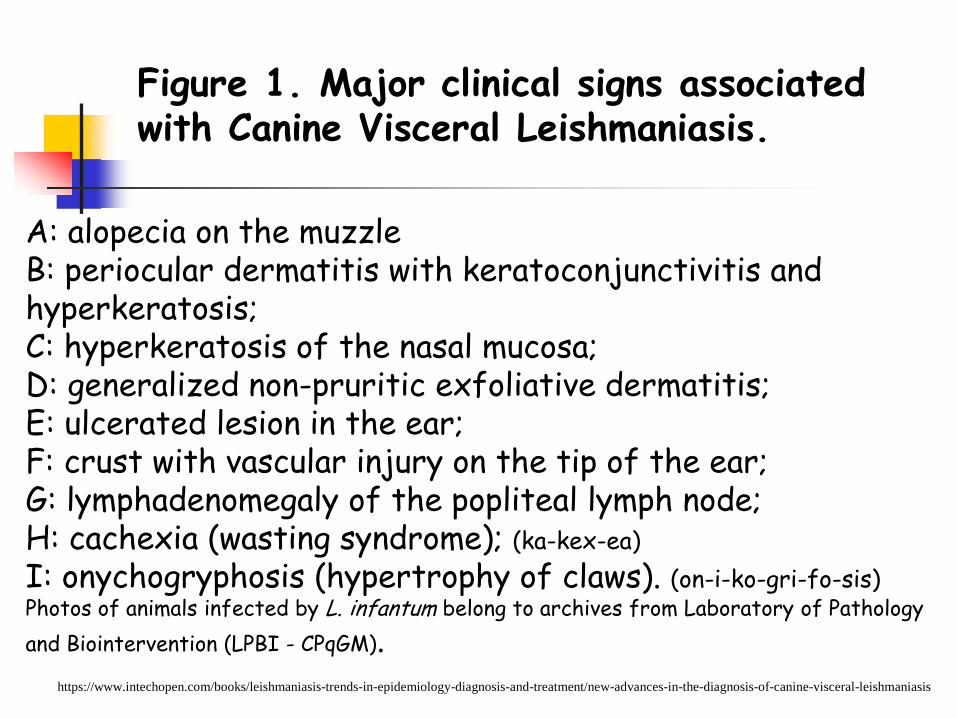
Figure 1. Major clinical signs associated with Canine Visceral Leishmaniasis.
https://www.intechopen.com/books/leishmaniasis-trends-in-epidemiology-diagnosis-and-treatment/new-advances-in-the-diagnosis-of-canine-visceral-leishmaniasis
A: alopecia on the muzzleB: periocular dermatitis with keratoconjunctivitis and hyperkeratosis;C: hyperkeratosis of the nasal mucosa;D: generalized non-pruritic exfoliative dermatitis;E: ulcerated lesion in the ear;F: crust with vascular injury on the tip of the ear;G: lymphadenomegaly of the popliteal lymph node;H: cachexia (wasting syndrome); (ka-kex-ea)I: onychogryphosis (hypertrophy of claws). (on-i-ko-gri-fo-sis)Photos of animals infected by L. infantum belong to archives from Laboratory of Pathology and Biointervention (LPBI - CPqGM).
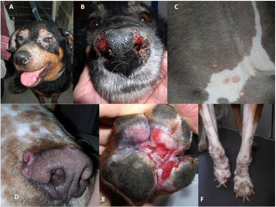
Figure 5: Different patterns of cutaneous lesions in CanL
A) Exfoliative periocular alopecia and blepharitis;B) Ulcerative nasal mucocutaneous lesions;C) Papular dermatitis in the inguinal region;D) Nodular crateriform lesions bordering the muzzle;E) Ulcerative erythematous lesions on the plantar surface of the paw and between pads;F) Onychogryphosis. (on-i-ko-gri-fo-sis)
https://parasitesandvectors.biomedcentral.com/articles/10.1186/1756-3305-4-86

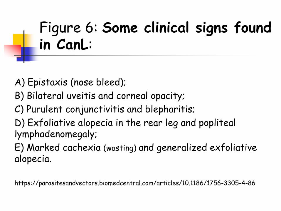
A) Epistaxis (nose bleed); B) Bilateral uveitis and corneal opacity;C) Purulent conjunctivitis and blepharitis;D) Exfoliative alopecia in the rear leg and popliteal lymphadenomegaly;E) Marked cachexia (wasting) and generalized exfoliativealopecia.
https://parasitesandvectors.biomedcentral.com/articles/10.1186/1756-3305-4-86
Figure 6: Some clinical signs found in CanL:
Diagnosis
Combination of findings Clinical Findings (physical exam, CBC,
Biochemical profile, urinalysis) Serology, Immunofluorescence, ELISA
(may cross-react with T. cruzi) PCR Amastigotes in cytology specimens
Lymph nodes, skin, spleen, etc. unreliable due to low numbers of amastigotes
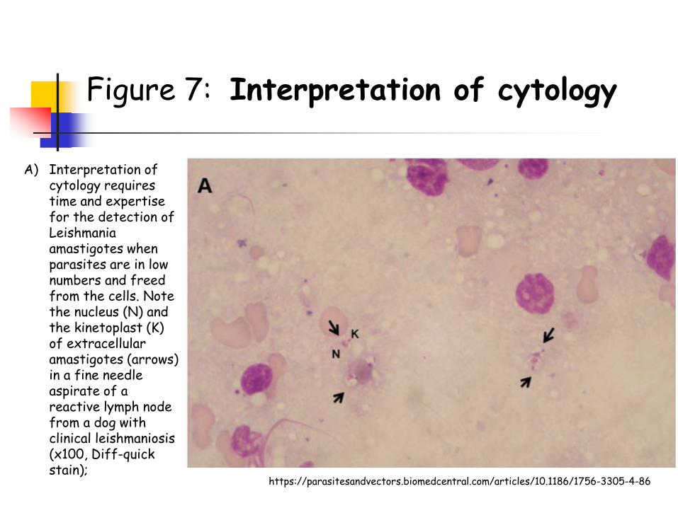
A) Interpretation of cytology requires time and expertise for the detection of Leishmania amastigotes when parasites are in low numbers and freed from the cells. Note the nucleus (N) and the kinetoplast (K) of extracellular amastigotes (arrows) in a fine needle aspirate of a reactive lymph node from a dog with clinical leishmaniosis (x100, Diff-quick stain);
Figure 7: Interpretation of cytology
https://parasitesandvectors.biomedcentral.com/articles/10.1186/1756-3305-4-86

B) High numbers of intracellular and extracellular Leishmania amastigotes in a fine needle aspirate of a reactive lymph node from a dog with clinical leishmaniosis (x100, modified Giemsa stain).
Figure 7: Interpretation of cytology
https://parasitesandvectors.biomedcentral.com/articles/10.1186/1756-3305-4-86
Treatment
1. Antimonial drugs and Purine analogues 2. Some only available through the CDC 3. Temporary clinical improvement, but
none can eradicate infection
Control Vector Control
Insect repellants (sandflies) collars, spot-on’s, etc.
Breeding control v/s Transplacental Transmission
Screen Blood donors v/s Transfusion Transmission
Vaccines have been developed in Brazil & Europe (Leishmune, Leish-Tec, CaniLeish)

Zoonosis Dogs are very important reservoir for human infections
Visceral Leishmaniasis (Kala-azar) Human - L. donovani, L. infantum = L. chagasi
Viscerocutaneous Leishmaniasis Dog - L. infantum = L. chagasi
Mucocutaneous Leishmaniasis (Espundia) Human – L. brasiliensis
Cutaneous Leishmaniasis (Oriental Sore) Human – L. tropica, L mexicana Cat – L. mexicana
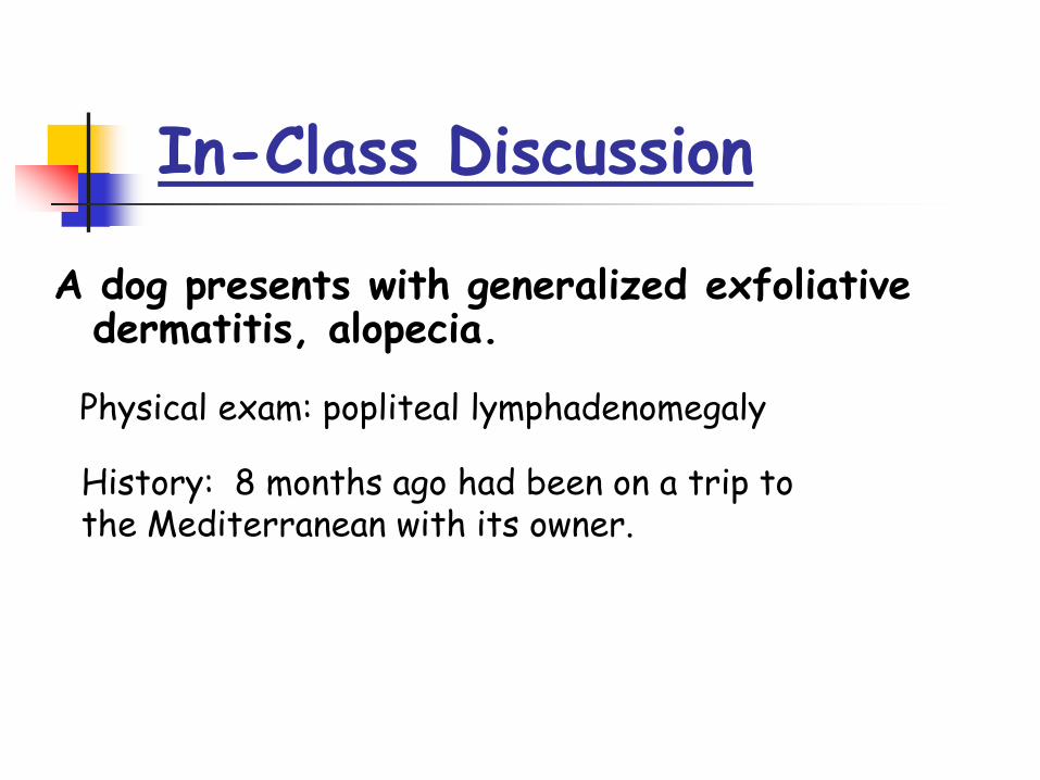
A dog presents with generalized exfoliativedermatitis, alopecia.
In-Class Discussion
History: 8 months ago had been on a trip to the Mediterranean with its owner.
Physical exam: popliteal lymphadenomegaly
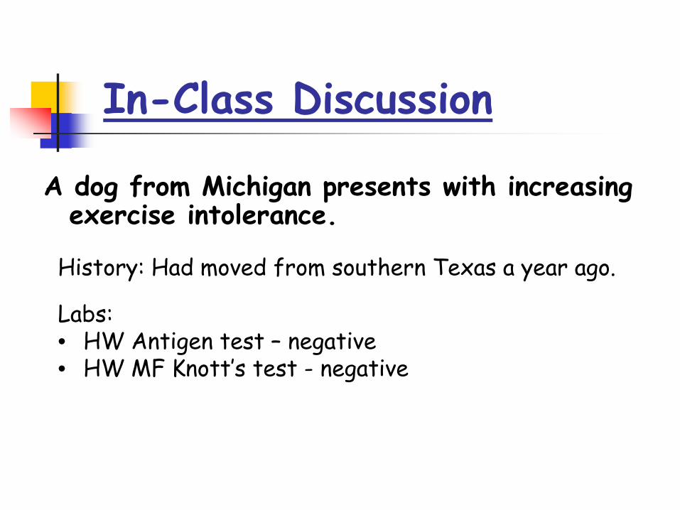
A dog from Michigan presents with increasing exercise intolerance.
In-Class Discussion
History: Had moved from southern Texas a year ago.
Labs:• HW Antigen test – negative• HW MF Knott’s test - negative

Contrast the life cycles, pathology, ecology, and diagnosis of the 2 Hemoflagellates of dogs.
In-Class Discussion
Recommended