Neurons


Copyright © The McGraw-Hill Companies, Inc. Permission required for reproduction or display.
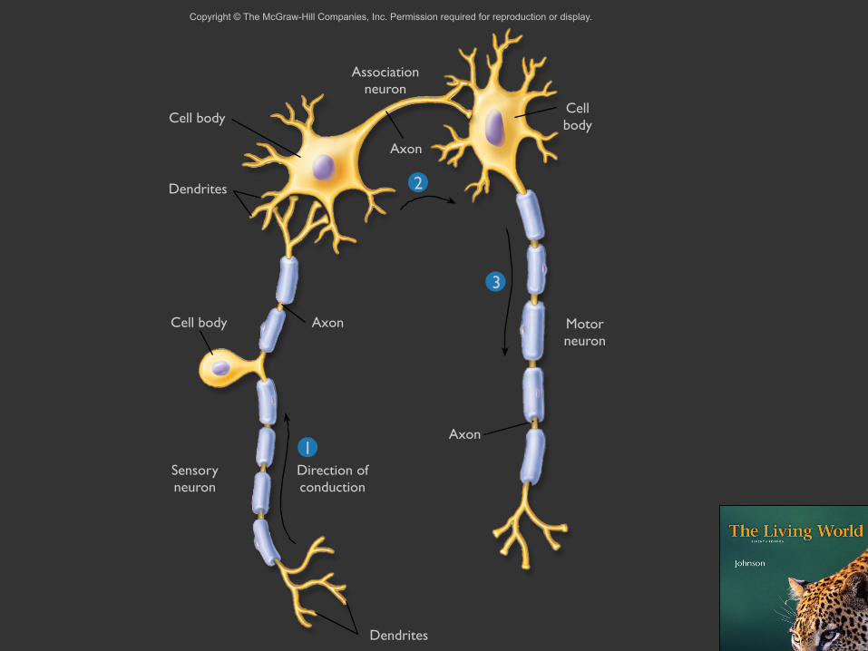
1
2
3
Associationneuron
Cellbody
Motorneuron
Axon
Dendrites
Sensoryneuron
Direction ofconduction
Cell body
Dendrites
Cell body
Axon
Axon

Dendrites


Axons
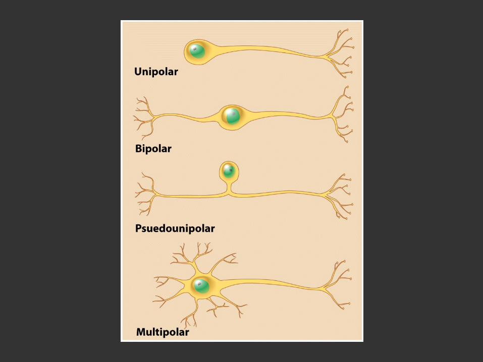

Figure 12-31 Essential Cell Biology (© Garland Science 2010)

Glial Cells


Roughly 3 times more glia than neurons

Astrocytes


StructuralGlycogen fuel reserve bufferMetabolic supportBlood–brain barrier?Transmitter uptake and releaseRegulation of ion concentration
in the extracellular spaceModulation of synaptic transmissionVasomodulationPromotion of the myelinating activity
of oligodendrocytesNervous system repairLong-term potentiation

Text

Maintain an appropriate chemical environment for neuronal signaling

Microglia

Macrophagesof the CNS


ScavengingPhagocytosisCytotoxicityAntigen presentationSynaptic strippingPromotion of repairExtracellular signaling


Oligodendrocytes

A.M. BUTT, K. COLQUHOUN, AND M. BERRY


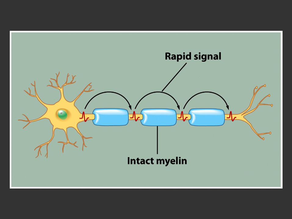
Schwann Cells

Copyright © The McGraw-Hill Companies, Inc. Permission required for reproduction or display.
Dendrites
Schwanncell
Schwanncell
Axon
Nucleus
Schwanncell
Axon
Myelin sheath
Myelinsheath
Axon

Myelin


“...fatty substance that surrounds the axons of many neurons.”

“...wrapping their cell membranes around the axon in a concentric manner.”

Water: 40%
Remainder: 70 - 85% Lipids15 - 30% Proteins


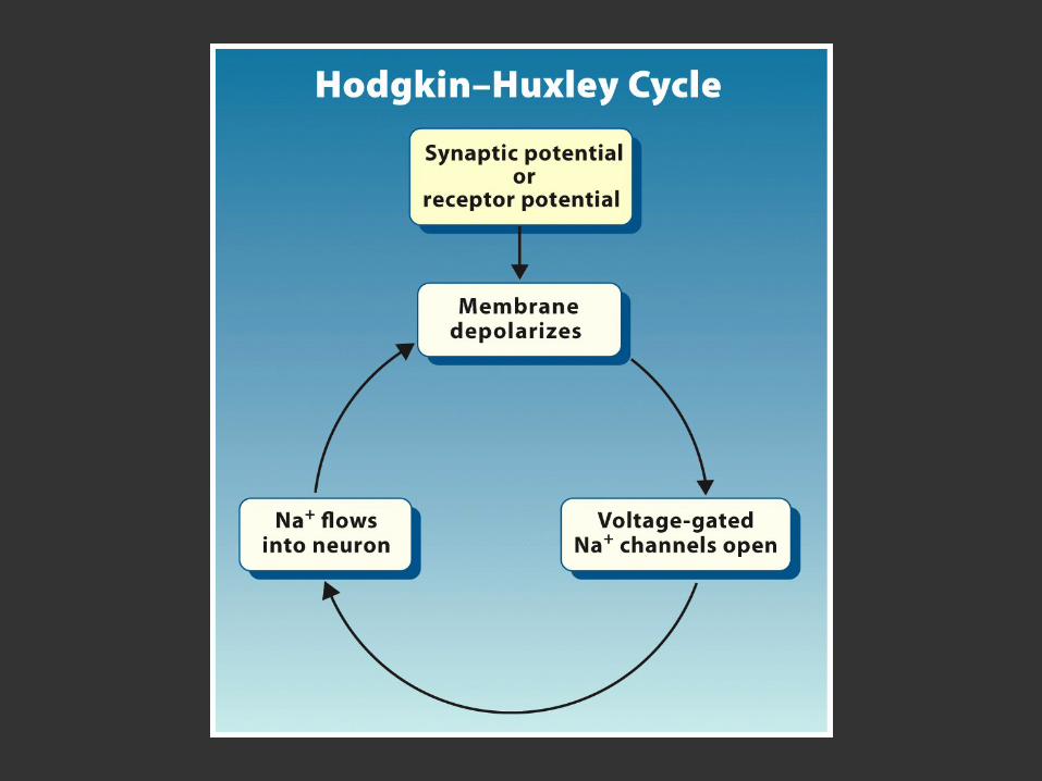
Action Potential


MembraneDepolarization

Figure 12-33 Essential Cell Biology (© Garland Science 2010)

Voltage-GatesSodium Channels


Figure 12-34 Essential Cell Biology (© Garland Science 2010)

Figure 12-35 Essential Cell Biology (© Garland Science 2010)

MembraneRepolarization


Voltage-GatedPotassium Channels


Figure 12-29 Essential Cell Biology (© Garland Science 2010)

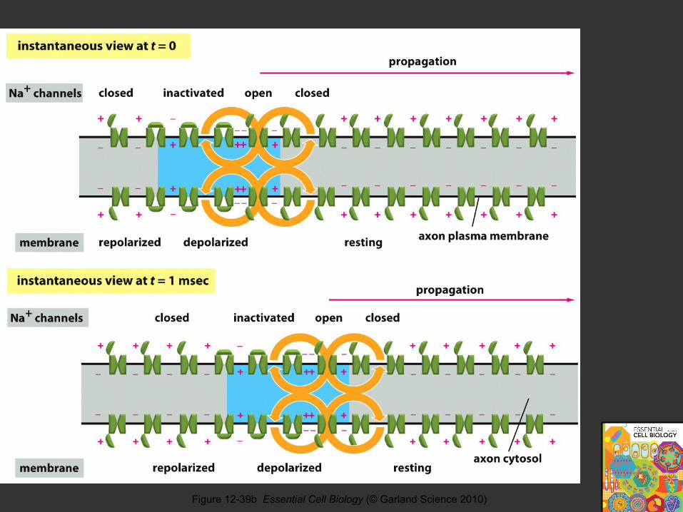
Figure 12-39b Essential Cell Biology (© Garland Science 2010)


SynapticTransmission
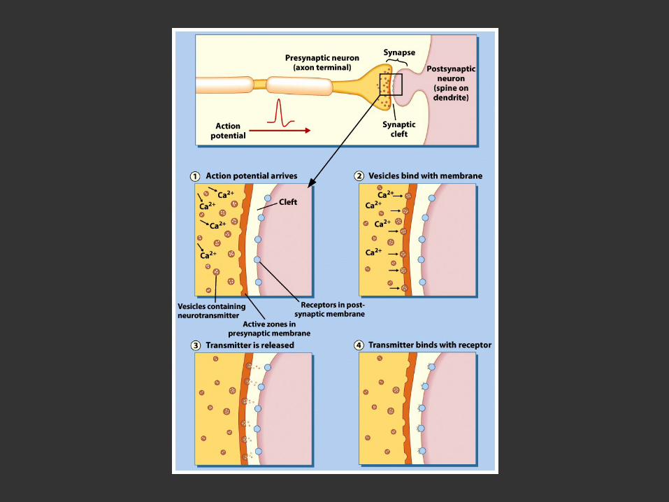

Figure 12-40a Essential Cell Biology (© Garland Science 2010)

Figure 12-40b Essential Cell Biology (© Garland Science 2010)

Voltage-GatedCalcium Channels

Figure 12-41 Essential Cell Biology (© Garland Science 2010)

Figure 12-42 Essential Cell Biology (© Garland Science 2010)
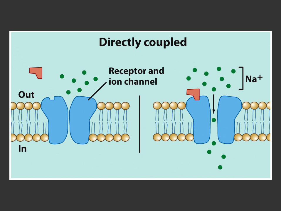


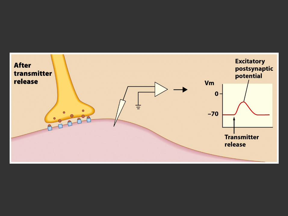


Figure 5.14 Molecular mechanisms of exocytosis during neurotransmitter release (Part 2)

Figure 5.9 Local recycling of synaptic vesicles in presynaptic terminals (Part 2)

Neurotransmitters


Excitatory


Inhibitory

Figure 5.3 Sequence of events involved in transmission at a typical chemical synapse

Figure 5.23 Events from neurotransmitter release to postsynaptic excitation or inhibition

Figure 12-45b Essential Cell Biology (© Garland Science 2010)

Figure 12-45a Essential Cell Biology (© Garland Science 2010)

NeurotransmitterBiosynthesis

Small Molecules

Figure 5.5 Metabolism of small-molecule and peptide transmitters (Part 1)

Figure 5.5 Metabolism of small-molecule and peptide transmitters (Part 2)

Neuropeptides(Peptide Neurotransmitters)
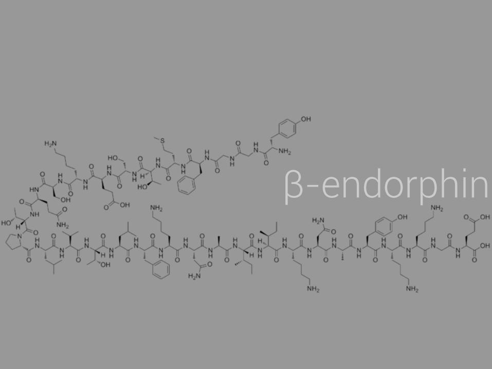
β-endorphin

Figure 5.5 Metabolism of small-molecule and peptide transmitters (Part 3)

Figure 5.5 Metabolism of small-molecule and peptide transmitters (Part 4)

Electrical Synapses

Figure 5.1 Structure of electrical synapses (Part 1)


Gap Junctions


Figure 5.1 Structure of electrical synapses (Part 3)

Figure 5.1 Structure of electrical synapses (Part 2)


Axon

Glial Cell
Axon

Axon

Myelin
Axon

Recommended