
Molecular Pathology Using Paraffin Embedded
Tissue and Cytological Samples
Gregory J. Tsongalis, Ph.D.
Professor of Pathology
Director, Molecular Pathology
Dartmouth Medical School
Dartmouth Hitchcock Medical Center
Norris Cotton Cancer Center
Lebanon, NH

DHMC Molecular Test Menu 2011
GENETICS HEMEPATH IDENTITY INFECTIOUS
DISEASES ONCOLOGY
Personalized
Medicine
AAT
aCGH
CF
FII
FRAX
FV
HFE
MTHFR
SRY
Bcl-2
Bcr/Abl
Chimerism
FLT3
IgH
JAK2
NPM1
PML-RARA
TCR
BKV
B. holmesii
B. parapertussis
B. pertussis
GBS
HCV Quant
HCV Geno
HIV Quant
HPV
MRSA
Parvo B19
BRAF
C-kit
Colon MSI
EGFR
ELM4-ALK1
HER2
KRAS
MGMT
N-Myc
Pancreas miRNA
TTF-1
UGT1A1
CYP2C9
CYP2C19
CYP2D6
GSTP1
IL28B
VKORC1
M/F Contam.
Spec. ID
Twinship
Rapid increase in demand for tissue-based molecular diagnostics
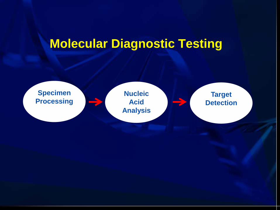
Molecular Diagnostic Testing
Specimen
Processing Nucleic
Acid
Analysis
Target
Detection

Handling of All Clinical Samples
• Observe universal precautions for
biohazards.
• Use protective gowns, gloves, face
and eye shields.
• Decontaminate all spills and work
areas with 10% bleach.
• Dispose of all waste in appropriate
biologic waste containers
• Gloves on - your RNA depends on it!

SPECIMEN TYPES
• Whole blood
• Bone marrow
• PBSC (Phoresis Product)
• Serum/plasma
• Buccal cells
• Cultured cells
• Blood spots
• Liquid cytology
• FNA
• BODY FLUIDS – CSF
– BRONCHIAL LAVAGE
– AMNIOTIC
– SEMEN
– URINE
• TISSUE – FRESH/FROZEN
– PARAFFIN-EMBEDDED
• HAIR (SHAFT/ROOT)
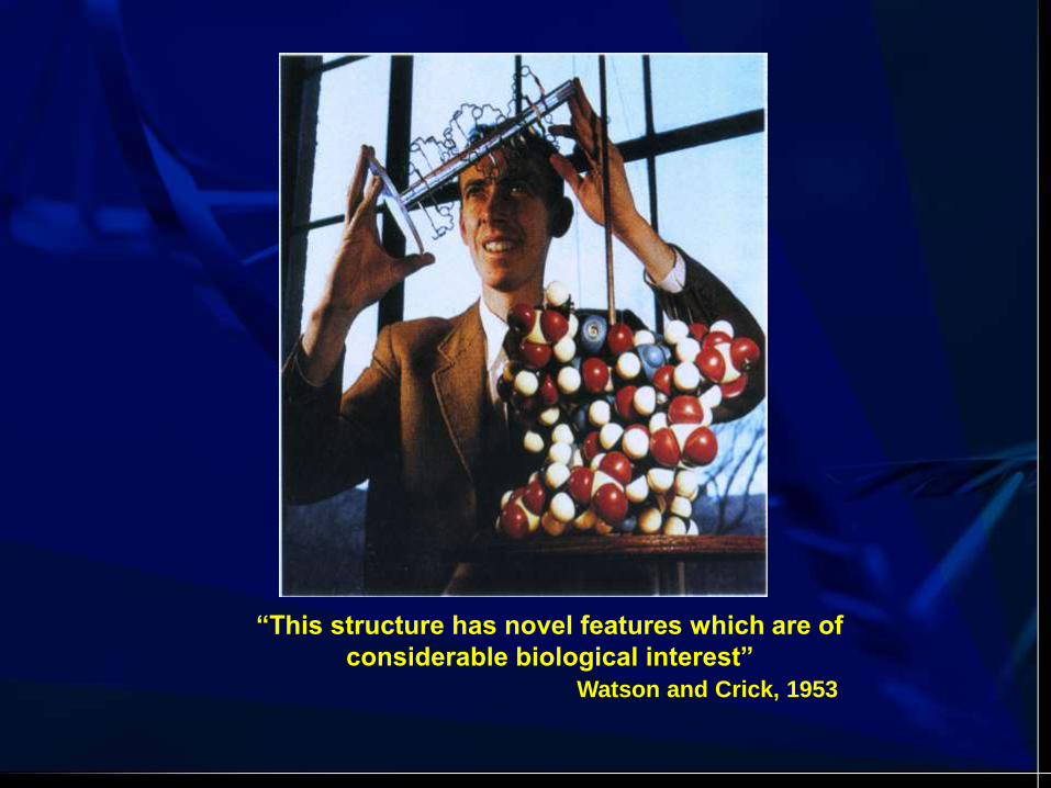
“This structure has novel features which are of
considerable biological interest”
Watson and Crick, 1953
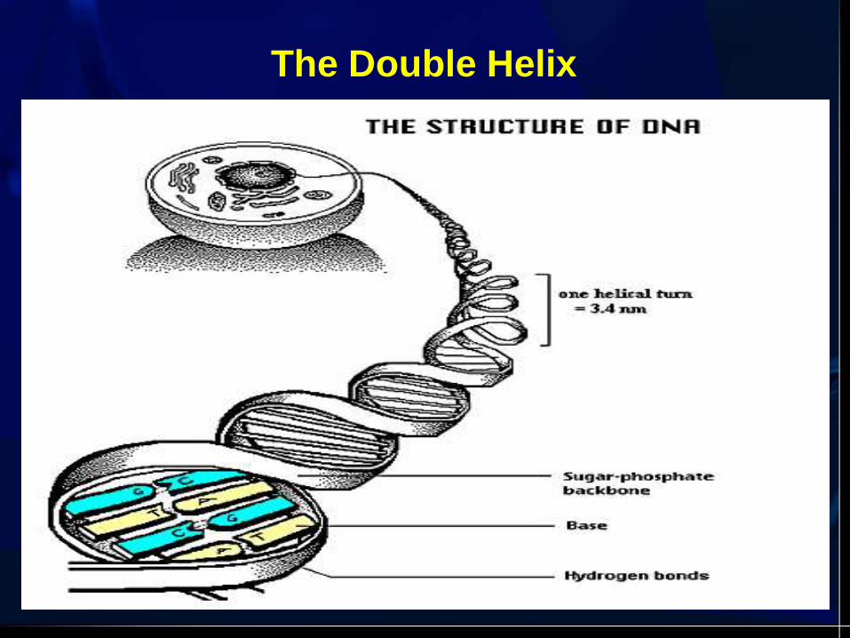
The Double Helix
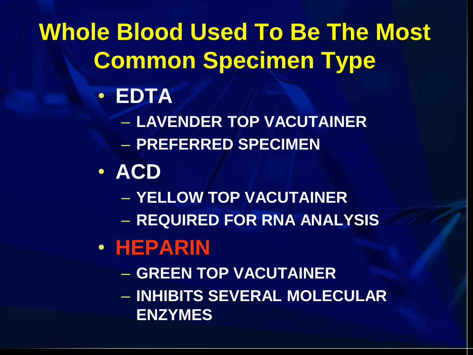
Whole Blood Used To Be The Most
Common Specimen Type
• EDTA – LAVENDER TOP VACUTAINER
– PREFERRED SPECIMEN
• ACD – YELLOW TOP VACUTAINER
– REQUIRED FOR RNA ANALYSIS
• HEPARIN – GREEN TOP VACUTAINER
– INHIBITS SEVERAL MOLECULAR
ENZYMES

DNA Isolation Methods
Method #1: Liquid Phase Organic Extraction
• Phenol (50):chloroform(49): isoamyl alcohol (1)
• Lysed samples mixed with above; 2 layers form
• Proteins remain at interface
• DNA is removed with top aqueous layer
• DNA is precipitated with alcohol and rehydrated
• Disadvantages: slow, labor-intensive, toxic
(phenol, chloroform), fume hood required,
disposal issues
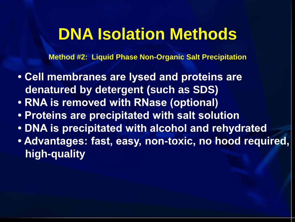
DNA Isolation Methods
Method #2: Liquid Phase Non-Organic Salt Precipitation
• Cell membranes are lysed and proteins are
denatured by detergent (such as SDS)
• RNA is removed with RNase (optional)
• Proteins are precipitated with salt solution
• DNA is precipitated with alcohol and rehydrated
• Advantages: fast, easy, non-toxic, no hood required,
high-quality

Step 1) Lyse RBCs
Step 2) Lyse WBCs
(+Opt. RNA Digest)
Step 3) Precipitate Proteins
Step 4) Precipitate DNA
Step 5) Wash
Step 6) Rehydrate DNA
PUREGENE® Purification Process
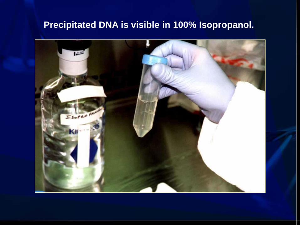
Precipitated DNA is visible in 100% Isopropanol.

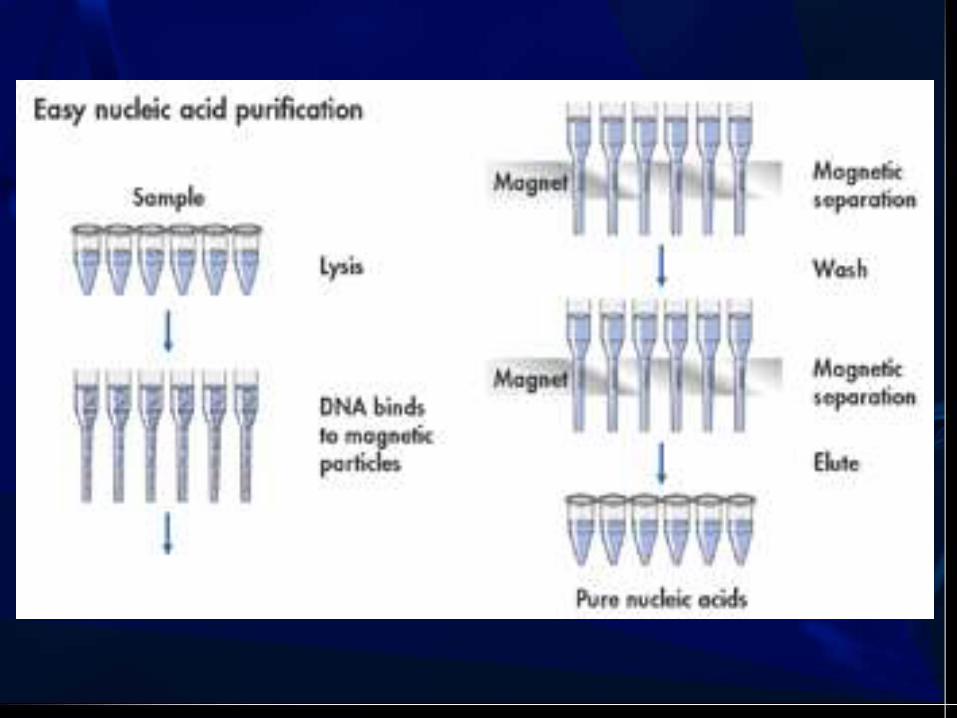
DNA Isolation Methods
Method #3: Solid Phase Procedures
Solid support columns
Fibrous or silica matrices bind DNA
Magnetic beads
DNA binds to beads; separated with magnet
Chelating resins
Advantages: fast, easy, no precipitation

Qiagen Biorobot EZ1
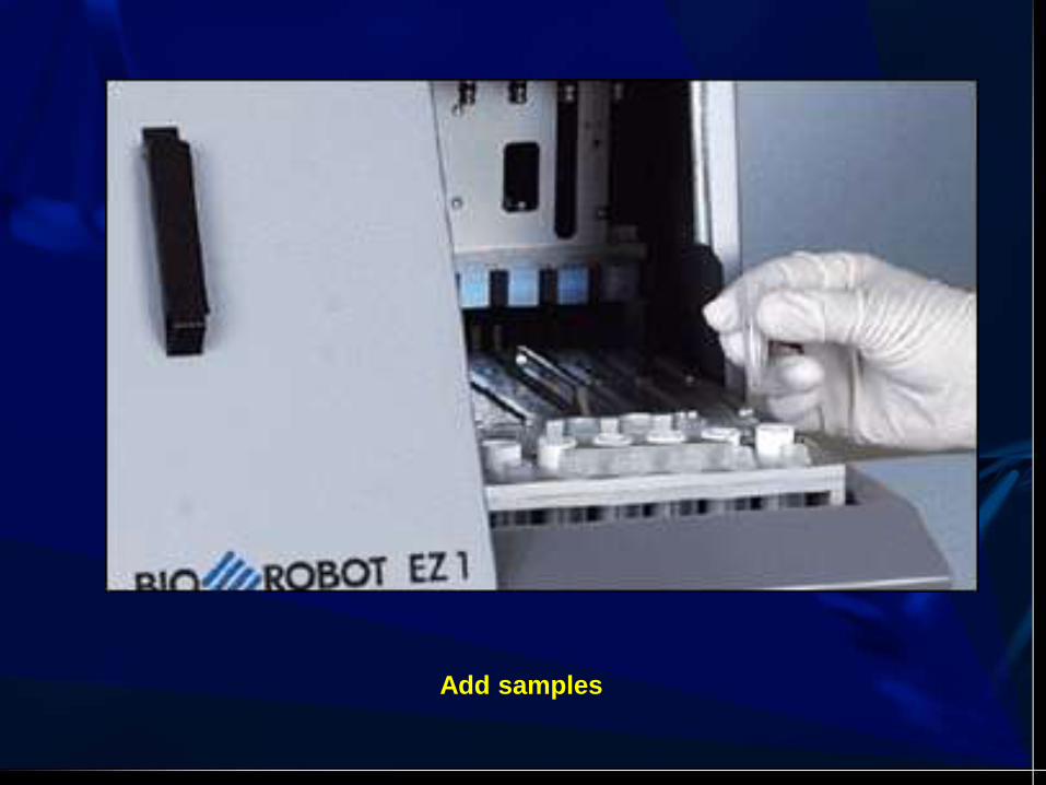
Add samples

Insert protocol card

Separation

Tissue-based Molecular Diagnostic
Testing
Pre-analytic Post-analytic Analytic
Qualitative
Quantitative Therapeutic
Genotyping
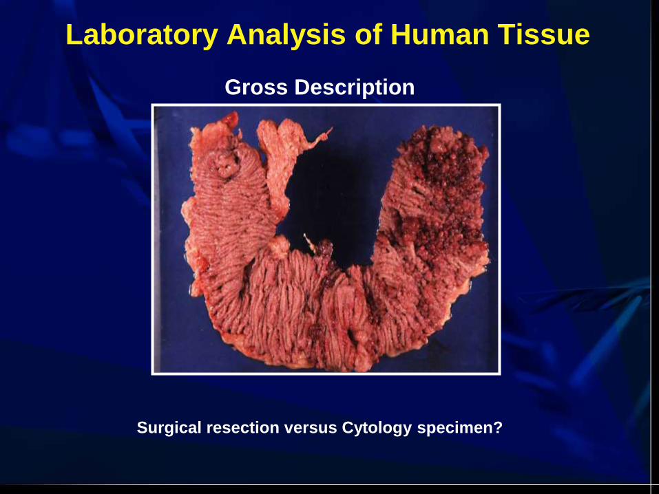
Laboratory Analysis of Human Tissue
Gross Description
Surgical resection versus Cytology specimen?

Gross Examination
1. Label or identification
2. Correct tissue or diseased tissue
3. Cross contamination
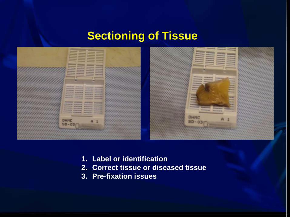
Sectioning of Tissue
1. Label or identification
2. Correct tissue or diseased tissue
3. Pre-fixation issues

Tissue Processing
1. Types of fixatives
2. Time of fixation

Tissue Embedding
1. Label or identification
2. Correct positioning of tissue

Tissue Cutting
1. Label or identification
2. Cross contamination
-Blade or floaters
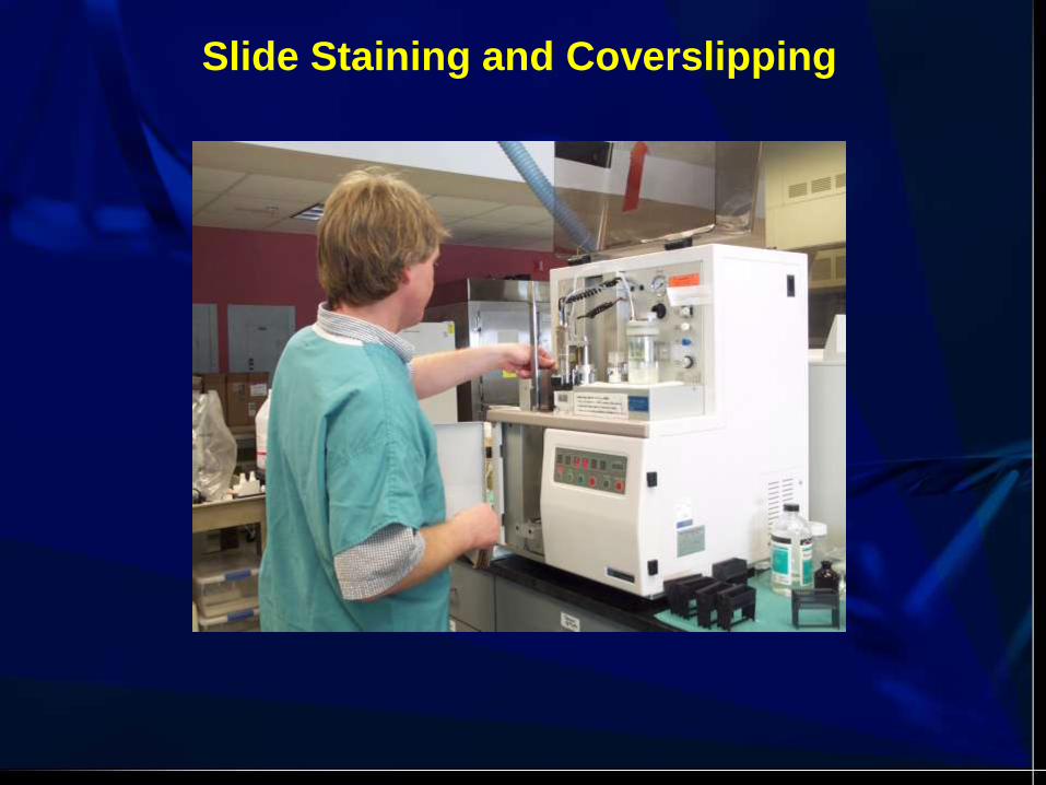
Slide Staining and Coverslipping

Pathologist Review


• Can be used for most molecular testing
• Various thickness sections
• 3-6 10 uM sections
• Mercury or heavy metal fixatives not
acceptable
• Unstained slides can be used for in situ
applications or for macro- and micro-
dissections
Formalin Fixed, Paraffin
Embedded Tissues (FFPE)

Fluorescecne In Situ Hybridization (FISH)
HER2 Gene Amplification EGFR Gene Amplification
N-myc Gene Amplification
1. Tissue fixation
2. Morphology
3. Permeabilization
4. Hybridization

Nucleic Acid Extraction from FFPE
• Requires deparaffinization (chemical, heat,
physical)
• Amplification technologies allow for smaller
targets and smaller specimen size
• Tissue can be scraped off of stained slides
• FFPE rolls can be used
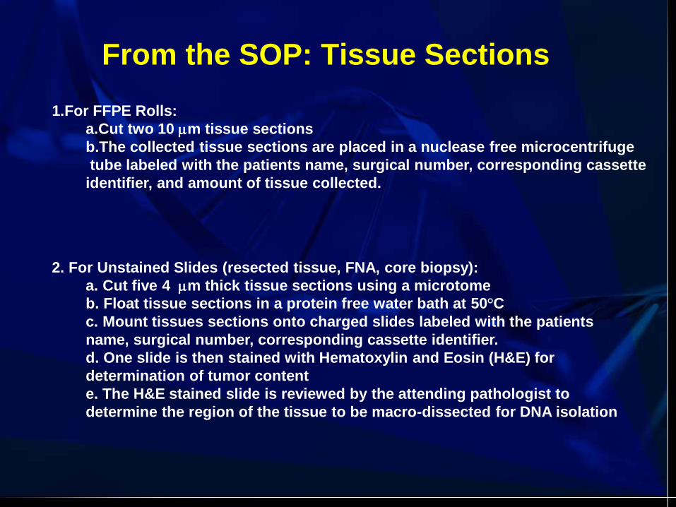
1.For FFPE Rolls:
a.Cut two 10 m tissue sections
b.The collected tissue sections are placed in a nuclease free microcentrifuge
tube labeled with the patients name, surgical number, corresponding cassette
identifier, and amount of tissue collected.
2. For Unstained Slides (resected tissue, FNA, core biopsy):
a. Cut five 4 m thick tissue sections using a microtome
b. Float tissue sections in a protein free water bath at 50°C
c. Mount tissues sections onto charged slides labeled with the patients
name, surgical number, corresponding cassette identifier.
d. One slide is then stained with Hematoxylin and Eosin (H&E) for
determination of tumor content
e. The H&E stained slide is reviewed by the attending pathologist to
determine the region of the tissue to be macro-dissected for DNA isolation
From the SOP: Tissue Sections

From the SOP: DNA Extraction
Paraffin Extraction Day 1-For FFPE Tissue Rolls
1.Add 1.0 mL of Xylene to the microcentrifuge tube containing tissue rolls
2. Place samples on the Nutator mixer for 5-10 minutes; Vortex vigorously
3.Centrifuge samples at maximum speed for 3 minutes
4.Completely remove supernatant without disturbing the tissue
5.Repeat Xylene wash steps 1-5 until all paraffin is removed from the sample
6.Add 1.0 mL of absolute ethanol to the sample tube; Vortex vigorously
7.Centrifuge samples at maximum speed for 3 minutes
8.Completely remove supernatant without disturbing the pelleted tissue
9.Repeat absolute ethanol wash steps 7-10
10.Add 1.0 mL of 95% ethanol to the sample tube; Vortex vigorously
11.Centrifuge samples at maximum speed for 3 minutes
12.Completely remove supernatant without disturbing the tissue
13.Add 1.0 mL of Phosphate Buffered Saline (PBS) to the sample
If small tissue specimens are being processed, it is best to forgo the wash with PBS and
instead, allow tissue to completely dry prior to the addition of Cell Lysis Solution
14. Vortex vigorously; Centrifuge samples at maximum speed for 3 minutes
15. Completely remove supernatant without disturbing the tissue
16.Briefly centrifuge tubes and pipette residual buffer from bottom of tube
17. Add 300 L of Cell Lysis Solution and 10 L of Proteinase K to the sample
(May need to scale up the volume of Cell Lysis Solution and Proteinase K)
18. Completely seal sample tubes (O-ring seal or parafilm)
19. Place in a 55ºC incubation oven on a Nutator mixer overnight
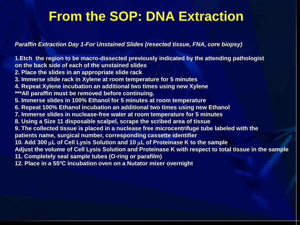
Paraffin Extraction Day 1-For Unstained Slides (resected tissue, FNA, core biopsy)
1.Etch the region to be macro-dissected previously indicated by the attending pathologist
on the back side of each of the unstained slides
2. Place the slides in an appropriate slide rack
3. Immerse slide rack in Xylene at room temperature for 5 minutes
4. Repeat Xylene incubation an additional two times using new Xylene
***All paraffin must be removed before continuing.
5. Immerse slides in 100% Ethanol for 5 minutes at room temperature
6. Repeat 100% Ethanol incubation an additional two times using new Ethanol
7. Immerse slides in nuclease-free water at room temperature for 5 minutes
8. Using a Size 11 disposable scalpel, scrape the scribed area of tissue
9. The collected tissue is placed in a nuclease free microcentrifuge tube labeled with the
patients name, surgical number, corresponding cassette identifier
10. Add 300 L of Cell Lysis Solution and 10 L of Proteinase K to the sample
Adjust the volume of Cell Lysis Solution and Proteinase K with respect to total tissue in the sample
11. Completely seal sample tubes (O-ring or parafilm)
12. Place in a 55ºC incubation oven on a Nutator mixer overnight
From the SOP: DNA Extraction

Paraffin Extraction Day 2-For FFPE or Unstained Slides
1.Evaluate samples to determine if all tissue particulates have dissolved
***If cell lysate is not homogeneous add additional proteinase K and incubate at 55ºC for
additional time until all tissue particulates have dissolved
2. Add 1.5 L of RNase A solution to the lysate
3. Mix by inverting 25 times; Incubate lysate at 37ºC for 30 minutes
4. Cool sample to room temperature on ice for approximately 5 minutes
5. Add 100 L protein precipitation solution to the lysate; Vortex vigorously for 20 seconds
***The protein precipitation solution must be mixed uniformly with the cell lysate before continuing
6. Chill the sample on ice for 5 minutes; Vortex vigorously for 20 seconds
7. Centrifuge at 13,000-16,000 RPM for 3 minutes (The precipitate proteins should form a tight pellet)
8. Pipette the supernatant into a clean microcentrifuge tube containing 300 L of 100% isopropanol
and 1.0 L of glycogen at a concentration of 20 mg/mL (Completely thaw glycogen before use).
9. Mix the sample by vortexing; Centrifuge at 13,000-16,000 RPM for 5 minutes
10. Pipette off and discard the supernatant; Add 300 L of 70% ethanol to the sample
11. Invert the tube several times to wash the DNA pellet; Centrifuge at 13,000-16,000 RPM for 1 minute
12. Carefully pipette off and discard the ethanol; quickspin and remove the remaining ethanol
13. Allow to air dry for ≥5 minutes; There should be no visible evidence of alcohol before rehydrating
14. Add 20-50 L of DNA hydration solution to the cell pellet (final concentration = 100-300 g/mL)
15. Allow DNA to rehydrate overnight or heat the tube to 65 ºC for 1 hour.
16. Quantify each sample using the Nanodrop-1000
From the SOP: DNA Extraction

Troubleshooting Nucleic Acid
Preparation Methods
• Problem: No or low nucleic acid yield.
– Make sure that ample time was allowed for
resuspension or rehydration of sample.
– Repeat isolation from any remaining original
sample (adjust procedure for possible low cell
number or poorly handled starting material).
– Concentrate dilute nucleic acid using ethanol
precipitation.
Troubleshooting Nucleic Acid
Preparation Methods
• Problem: Poor nucleic acid quality
– If sample is degraded, repeat isolation from
remaining original sample, if possible.
– If sample is contaminated with proteins or other
substances, clean it up by re-isolating
(improvement depends on the extraction
procedure used).

The Molecular Touch Prep Getting An Upfront Specimen
M.L. Petras M.D. et al. Diagn Molec Pathol, 2011, in press.
The mid-’80s and PA Training
• Southern blots were going to revolutionize the
industry
• Plasmids were a threat to human life
• Surgery and chemo weren’t curing anybody
• The first oncogenes and tumor suppressor
genes were identified
• Tumors were gigantic
New imaging modalities and diagnostic procedures
are detecting cancers earlier.

• Surgical vs. cytologic specimens?
• What is the state of “molecular cytopathology”?
• What are the impediments to using cytology
specimens?
• Is there a future and utility of FNA specimens for
molecular diagnostics?
Questions we should be asking:
Surgical vs. Cytologic specimens Two most common modalities for collecting tissue samples
• Surgical specimens typically are fixed and thus compromise the quality of nucleic acids via formalin induced cross-linking and fragmentation
• FNA procedures are used for rapid, cost-effective and accurate diagnosis with reduced patient morbidity
• Cytologic preparations represent and average of 10-20% of archival hospital specimens
• Two standard preparations of FNA materials: Air-dried Diff-Quik, Alcohol fixed Pap (no formalin fixation, alcohol fixation preserves DNA)

Impediments to using Cytology Specimens
• FNA samples under-utilized for molecular profiling
• Cell numbers required for analysis (20-30) are compatible with typical FNA yields, but because of the intra-tumoral heterogeneity , sampling artifacts may occur
• To avoid false negative results, sequencing-based tests may require up to 50-70% tumor cells in a sample.
• Preanalytical recommendations in report from Molecular Assay in NSCLS Working Group of ASCO :
“ cytology smears are not acceptable for IHC and FISH”
“at least 3 representative areas should be assessed per tumor section”
“tissue block preferred”
“FFPE is standard”
• Poor quality smears, poor quality FNA harvests
• Past and current concept of adequacy
• Maintaining records of cytomorphology
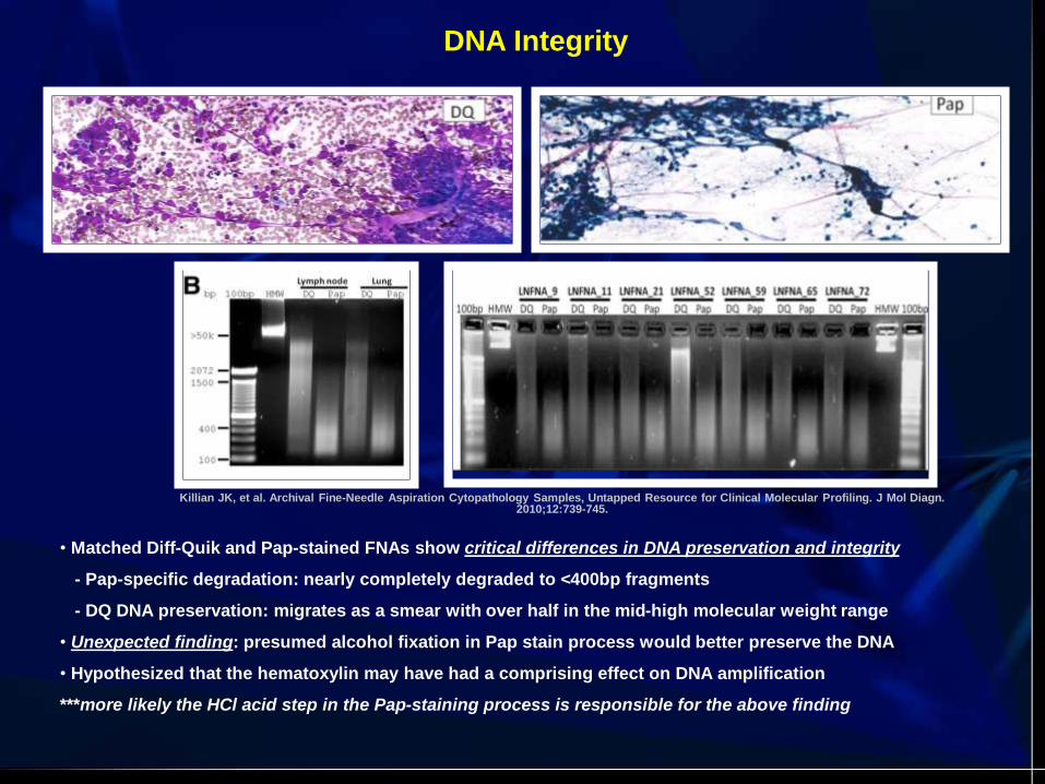
• Matched Diff-Quik and Pap-stained FNAs show critical differences in DNA preservation and integrity
- Pap-specific degradation: nearly completely degraded to <400bp fragments
- DQ DNA preservation: migrates as a smear with over half in the mid-high molecular weight range
• Unexpected finding: presumed alcohol fixation in Pap stain process would better preserve the DNA
• Hypothesized that the hematoxylin may have had a comprising effect on DNA amplification
***more likely the HCl acid step in the Pap-staining process is responsible for the above finding
Killian JK, et al. Archival Fine-Needle Aspiration Cytopathology Samples, Untapped Resource for Clinical Molecular Profiling. J Mol Diagn. 2010;12:739-745.
DNA Integrity

1 2 3 4 5 6 7 8 9 10 11 12 13
HSV 1-2 PCR Gel
Fiel-Gan M et al. Rapid detection and typing of HSV from cytology specimens collected into thinprep fixative.
Acta Cytologica 43:1034-8, 1999.

BK/JC Melt Derivative Plot
Gonzalez JL, et al. Polyomavirus infection of the urinary tract presenting as hemorrhagic cystitis in
an immunocompetent five-year-old boy. Diagn Cytopathol 36(6):375-378, 2008.
Utility of FNA Specimens for Molecular Diagnostics
Reasons for FNA specimens to receive greater attention in future studies:
• Relative tumor purity in FNA specimens versus tissue extracts
• FNA samples contain copious high quality gDNA suitable for high-resolution genomic and epigenomic profiling
• Excellent potential source of patient materials for clinical molecular profiling, including:
-retrospective genomic analysis
-prospective collection for individual therapy or eligibility review for clinical trial enrollment
• Potential enhancement for biomarker detection
• Abundance of archival FNA smears

DHMC Molecular Pathology Laboratory and
Translational Research Program
Samantha Allen
Claudine Bartels, Ph.D.
Heather Bentley
Betty Dokus
Susan Gallagher
Carol Hart
Arnold Hawk
Joel Lefferts, Ph.D.
Rebecca O’Meara
Elizabeth Reader
Mary Schwab
Laura Tafe, M.D.
Brian Ward
Brendan Wood
Eric York
Recommended


