MALDI-IMS (MATRIX-ASSISTED LASER DESORPTION/IONIZATION
IMAGING MASS SPECTROMETER) IN TISSUE STUDY
YANXIAN CHEN
MARCH 8TH , WEDNESDAY.
SEMINAR FOCUSING ON
• What is MALDI imaging mass spectrometer?
• How does MALDI imaging work for tissue study?
• Applications of MALDI imaging.
• Case study: N-glycans MALDI imaging to FFPE tissues.
• Challenges to deal with human tissues.

WHAT IS MALDI IMAGING MASS SPECTROMETER (MALDI-IMS)?
• Matrix-assisted laser desorption/ionization (MALDI) imaging mass spectrometry (IMS) is a powerful tool for investigating the distribution of proteins and small molecules within biological systems through the in situ analysis of tissue sections.
• MALDI-IMS can determine the distribution of hundreds of unknown compounds in a single measurement and enables the acquisition of cellular expression profiles while maintaining the cellular and molecular integrity.
• MALDI images are often used to identify, characterize, and comparatively quantify the relative level of expression of hundreds of proteins that are co-expressed in a given cell type or tissue.

WHAT IS MALDI IMAGING MASS SPECTROMETER?
• MALDI imaging MS uses Matrix-assisted laser desorption/ionization to generate (M+H)+ or (M+Na)+ ions.
• MALDI-IMS is often coupled with TOF analyzer to generate the mass spectrum.
• Displaying the intensity of selected mass signals at each measurement point generates images that depict
distribution and abundance of specific compounds within the sample.
The technique that uses matrix to ionize samples, utilizes mass spectrometer to generate the mass spectrum and image generation based on mass spectrum is called ”MALDI Imaging”.
HOW DOES MALDI IMAGING WORK FOR TISSUE STUDY?
• Sample preparation: freeze the tissue in liquid nitrogen immediately after procurement. The frozen tissues can be stored below -80oC up to 1 year.
• Sectioning: Cryosectioning is often performed directly to the frozen tissue sample. Sometimes, embedding in gelatine and agarose has been used to facilitate handling of small or fragile samples. The thickness of tissue sectioning varies depending on the type of the tissue. Normally, 10-20µm thick sections are optimal for handling and analyzing in the high vacuum environment of mass spectrometer.
• Transfer sample section to a transparent conductive slide and get a optical microscopic image.

HOW DOES MALDI IMAGING WORK FOR TISSUE STUDY?
-Histological staining of tissues: common use a Hematoxylin-Eosin (H&E) staining.
-Matrix desorption: composition of matrix for tissue analysis, sinapinic acid provides the best signals for higher molecular weight proteins, while ª-cyano-4-hydroxycinnamic acid is more suitable for lower molecular weight peptides.
-Dry analyte ions (M+H)+/(M+Na)+ are transferred into a mass spectrometry.
-Mass spectrum generation and pick up the interesting peaks to construct the MALDI images.

APPLICATION OF MALDI-IMS TO TISSUE ANALYSIS
• (1) Diagnostic and prognostic assessments in clinical pathology
• (2) Drug discovery and drug metabolism
• (3) Developmental biology
• (4) Imaging of phospholipids

(1) DIAGNOSTIC AND PROGNOSTIC ASSESSMENTS IN CLINICAL PATHOLOGY
• Ex: Diagnose ADHD (attention-deficit hyperactivity disorder).
• Comparing their MALDI images, doctors can diagnose whether the person is ADHD.
• Brain for ADHD people have smaller frontal lobes, temporal grey matter, caudate nucleus and cerebellum than normal brain.
MALDI-IMS can be used to measure the response of tumor and surrounding tissues to therapeutic agents, so the site of origin for tumor can be identified. The doctors can make a proper treatment for patients based on their MALDI images.Also, doctors can predict the development trend of diseases depending on MALDI images to make further treatments.

(2) DRUG DISCOVERY AND DRUG METABOLISM
• MALDI-IMS opens the possibility to directly determine the distribution of pharmaceuticals in tissue sections which might unravel their disposition or biotransformation pathway for new drug.
• MALDI-IMS doesn’t require radioactive labelling, comparing with WBA (Whole-body autoradiography-—common method).
• The difference between the drug and its metabolites can be distinguish from MALDI image.

(3) DEVELOPMENTAL BIOLOGY
• Protein imaging and profiling using MALDI-IMS have become a powerful method for analyzing changes in global protein expression patterns in cells and tissues as a function of development processes (Cornett et al. 2007).
• Proteomic studies using MALDI-IMS to monitor some complex biological processes.
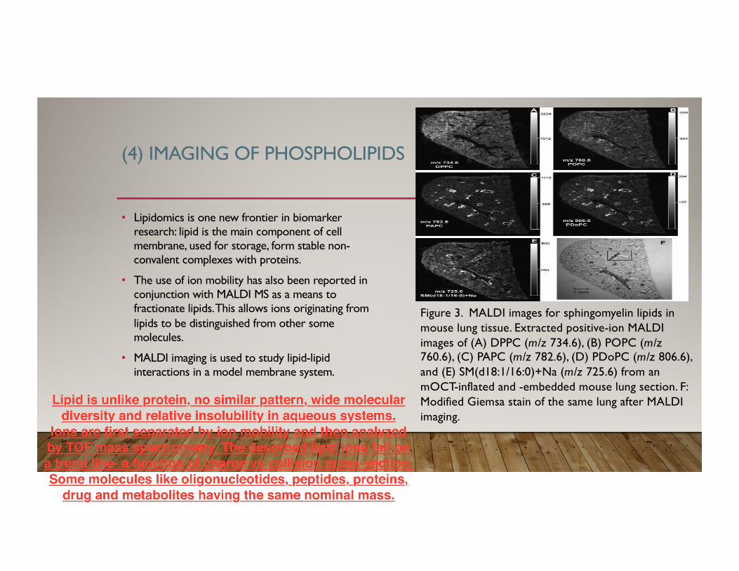
(4) IMAGING OF PHOSPHOLIPIDS
• Lipidomics is one new frontier in biomarker research: lipid is the main component of cell membrane, used for storage, form stable non-convalent complexes with proteins.
• The use of ion mobility has also been reported in conjunction with MALDI MS as a means to fractionate lipids. This allows ions originating from lipids to be distinguished from other some molecules.
• MALDI imaging is used to study lipid-lipid interactions in a model membrane system.
Figure 3. MALDI images for sphingomyelin lipids in mouse lung tissue. Extracted positive-ion MALDI images of (A) DPPC (m/z 734.6), (B) POPC (m/z760.6), (C) PAPC (m/z 782.6), (D) PDoPC (m/z 806.6), and (E) SM(d18:1/16:0)+Na (m/z 725.6) from an mOCT-inflated and -embedded mouse lung section. F: Modified Giemsa stain of the same lung after MALDI imaging.

CASE STUDY:
•MALDI imaging mass spectrometry of N-linked glycans on formalin-fixed paraffin-embedded murine kidney
• Ove J. R. Gustafsson & Matthew T. Briggs & Mark R. Condina & Lyron J. Winderbaum & Matthias Pelzing & Shaun R. McColl & Arun V. Everest-Dass & Nicolle H. Packer &Peter Hoffmann
• Received: 15 August 2014 / Revised: 20 October 2014 / Accepted: 21 October 2014 / Published online: 2 December 2014 # The Author(s) 2014. This article is published with open access at Springerlink.com
MATERIAL AND METHODS
• Glycerol-free PNGase F: used to cleave the linkage between glycans and asparagine residues.
• FFPE blocks were sectioned to 6µm on a Microm HM 325 microtome.
• Matrix was composed of 10mg/mL 2,5-DHB in 0.1%TFA and 1mM NaCl into multiple layers.
• The released N-linked glycans were reduced and desalted: reduced by acidification with 100mM NH4COOH; desalted by cation-exchange chromatography with 30µL AG50W-X8 cation-exchange resin.
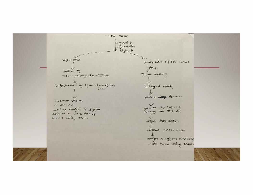
RESULTS
• In totally, 79 peaks existed in >=2 of the PNGase F spots and <=1 of the buffer control spots and were larger than m/z 1000. required >=4 counts in PNGase F spots, N-glycan candidates decreased to 43. MS/MS resulted in the identification of 34 N-glycan peaks.
• Reduced “ sweet spot” formation improved mass accuracy, reproducibility and signal intensity.
• The isotopic profile of [M+Na]+ on target indicated the overlap of two species at roughly 2303.8 and 2304.8 (there are two different N-glycan species with a difference in mass od roughly 1Da; there are two forms of the same N-glycan)

CHALLENGES TO DEAL WITH HUMAN TISSUES
• Humans are more genetically diverse than mice.
• People from different backgrounds, lived in different environments or accepted different level of education lead to high variability with the same nominal disease.
• A person’s lifestyle, diet and therapeutic regimen can have a marked effect on protein expression. For example, a woman took oral contraceptives can change the breast proliferation index. Because the medicine alters the level of estrogen and progestrogen.
• Therefore, human samples must be controlled for a variety of factors, such as gender, race, age and menopausal status, to achieve a uniform patient population.
• Also, permission must be obtained from the patient for use of their tissue.

CONCLUSION
• MALDI imaging is a very useful tool to analyze animal tissues, but still need to be improved the techniques for human tissue.
• Formalin fixed paraffin embedded tissue can be stored for long time, but has limited techniques for study.
• Sample preparation is a very important step in tissue study, it affects the signal in mass spectrum directly.
REFERENCE
• 1.Axel Walch, Sandra Rauser, Soren-Oliver Deininger & Heinz Hofler.” MALDI imaging mass spectrometry for direct tissue analysis: a new frontier for molecular histology.” Histochem Cell Biol. (2008) Sep; 130(3): 421-434.
• 2. Seeley, Erin H., and Richard M. Caprioli. "MALDI imaging mass spectrometry of human tissue: method challenges and clinical perspectives." Trends in biotechnology 29.3 (2011): 136-143.
• 3. Gustafsson, Ove JR, et al. "MALDI imaging mass spectrometry of N-linked glycans on formalin-fixed paraffin-embedded murine kidney." Analytical and bioanalytical chemistry 407.8 (2015): 2127-2139.
• 4. Norris, Jeremy L.; Caprioli, Richard M. (10 April 2013). "Analysis of Tissue Specimens by Matrix-Assisted Laser Desorption/Ionization Imaging Mass Spectrometry in Biological and Clinical Research". Chemical Reviews. 113 (4): 2309–2342
Recommended