Liver masses: how to workup a liver mass and update on liver cancer
Alice C. Wei, MD, MSc, FRCSC, FACS Princess Margaret Cancer Centre
HPB Surgical Oncology and General Surgery Associate Professor of Surgery, University of Toronto
Lead, Quality and KT, Surgical Oncology, Cancer Care Ontario BC Surgical Oncology Network, Oct 22 2016
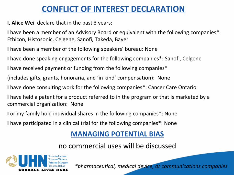
CONFLICT OF INTEREST DECLARATION I, Alice Wei declare that in the past 3 years:
I have been a member of an Advisory Board or equivalent with the following companies*: Ethicon, Histosonic, Celgene, Sanofi, Takeda, Bayer
I have been a member of the following speakers’ bureau: None
I have done speaking engagements for the following companies*: Sanofi, Celgene
I have received payment or funding from the following companies*
(includes gifts, grants, honoraria, and ‘in kind’ compensation): None
I have done consulting work for the following companies*: Cancer Care Ontario
I have held a patent for a product referred to in the program or that is marketed by a commercial organization: None
I or my family hold individual shares in the following companies*: None
I have participated in a clinical trial for the following companies*: None
MANAGING POTENTIAL BIAS no commercial uses will be discussed
*pharmaceutical, medical device, or communications companies

Learning Objectives
1. review approach for diagnosing liver masses 2. review management of benign lesions 3. review management of malignant tumors
3

Question: Which of the following statements are true?
1. Liver cysts should be resected if they grow rapidly? 2. Hepatic adenomas should only be resected if
symptomatic 3. MRI should be used to assess all liver masses 4. Liver biopsy should be used to confirm diagnosis in all
suspected liver cancers 5. Focal nodular hyperplasia can be confused with
malignant liver tumours
4
5
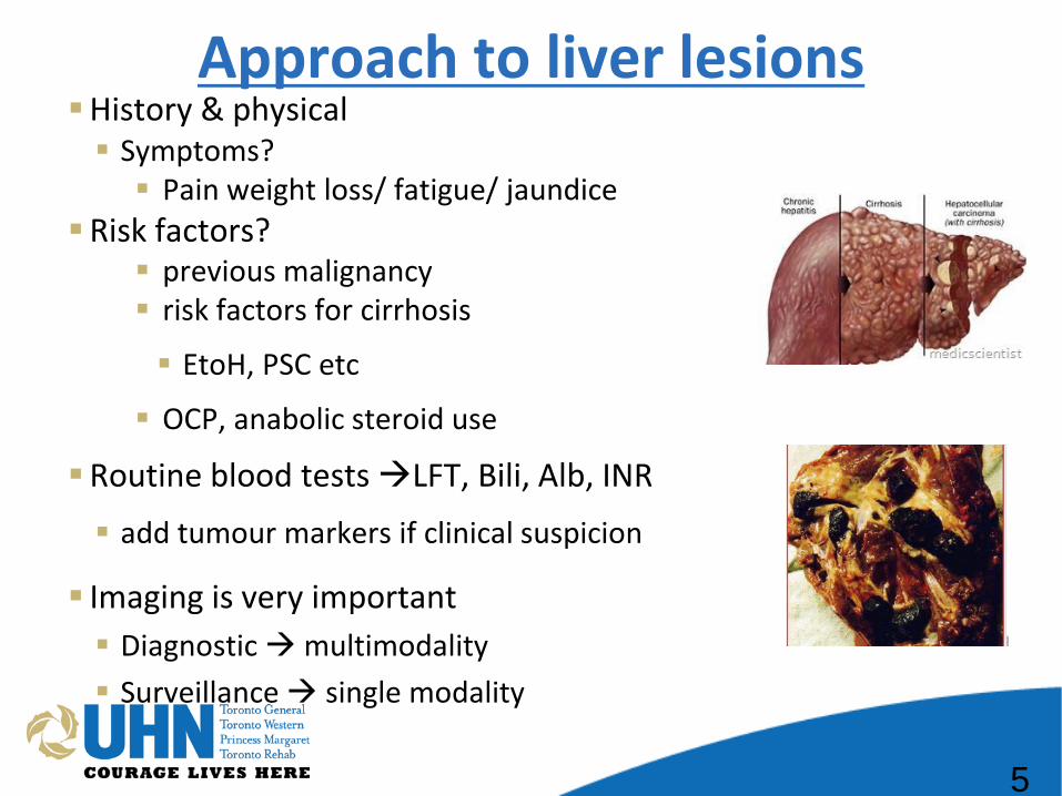
Approach to liver lesions History & physical Symptoms? Pain weight loss/ fatigue/ jaundice
Risk factors? previous malignancy risk factors for cirrhosis
EtoH, PSC etc
OCP, anabolic steroid use
Routine blood tests LFT, Bili, Alb, INR
add tumour markers if clinical suspicion
Imaging is very important Diagnostic multimodality Surveillance single modality

Imaging modalities • US CT MRI
• Special tests – contrast enhanced US – CT/PET
• Nuclear medicine scans – RBC scans/sulfur colloid scans obsolete
• Biopsy – For indeterminate lesions
6
What to look for on imaging reports • Important features
– lesion consistency was it there before – imaging characteristics enhancement pattern – number/ location – evaluate non-tumour liver
• Get to know your radiologists – different sensitivity/ specificity thresholds – variety of area of interest/training – dictating styles differ
• Modifiers used: suggestive, worrisome, cannot exclude…
• if dictation is not clear • call radiologist for clarification or advice
7

Ultrasound Useful for Screening exam assess for biliary obstruction surveillance of established lesions
Disadvantages Additional tests required for
confirmation Quality is operator dependent Limited visualization in fatty livers
8

CT scan excellent size and anatomic
resolution IV contrast required Dye protocol depends on pathology
Dedicated liver protocol CT needed
Contraindications: impaired renal function dye allergies can be pre medicated
radiation exposure
9

MRI scan Use MRI as confirmatory test
for ’doubtful’ cases
No required for surveillance of known lesion
helpful for ‘indeterminate’ lesions
Primovist and/or gadolinium dye
superior for fatty livers
MRCP to assess biliary system
10

When to biopsy Biopsy selectively
Indicated if tissue needed to guide Rx to establish initial diagnosis
of malignancy Distinguish primary cancer
site Non-tumour liver if liver
function an issue
1

12
Liver lesions
Liver cyst
Liver metastases

13
Liver cysts >90% asymptomatic >50% multiple Vast majority are benign If symptoms Intra-cystic bleeding/mass effect Consider drainage If ↑ growth or complexity consider MRI to
characterize Polycystic liver disease Assess for extra-hepatic disease
PLD
liver abscess

14
Complex liver cysts
Often involuted simple cysts appear complex
Infectious cysts Hydatid cysts Echinococcal cysts Exposure to sheep/dogs
Fever and pain may be present
Neoplastic cysts biliary cystic neoplasms rare Cystic metastases occasionally
hydatid disease
neoplastic cyst

Solid benign lesion: Hemangioma AP
PVP
Delayed
most common liver neoplasm
20% population
F:M 5:1
always asymptomatic
20-30% multiple
Typical features characteristics sharply demarcated peripheral nodular enhancement centripetal filling

Hemangioma: Work up and treatment Ultrasound diagnostic if healthy patient
and no risk factors
CT – liver contrast often diagnostic If classic features present no F/U needed
Beware of the atypical hemangioma
MRI accuracy 85-95% confirmatory test for
atypical lesions
Rx: NO F/U required

Solid benign lesion: Focal Nodular Hyperplasia
benign, hyperplastic lesion hamartoma?
3% population
female: male 6:1
FNH has central stellate scar tortuous feeding artery homogenous arterial
enhancement
Pre AP PVP

Focal Nodular Hyperplasia FNH can be confused adenoma fibrolamellar HCC Typical HCC
US and CT are NOT diagnostic
FNH must be confirmed with MRI
MRI accuracy 70-90%
sometimes biopsy needed

19
Solid benign lesion: Hepatic adenoma benign hepatocyte tumour uncommon 1/106 - 4/105 44% have symptoms
30% multiple Premalignant (𝛽𝛽- catenin mutation)
associated with OCP use / anabolic steroids obesity storage diseases (Glyogen Storage Types 1 And 3)
** potential for rupture and malignant transformation
MRI to characterize
biopsy usually required

20
Solid benign lesion: Hepatic adenoma
Treatment • Stop exogenous hormones • Refer for surgical resection If bleeding urgent embolization +/- surgery
Expectant management an option for small adenomas1 < 3 cm, no high risk features (beta-catenin mutated, inflammatory,
undifferentiated subtype, hypervascular) Surveillance Imaging and AFP q6 mo X 2 yrs, then qyr
Treatment for > 3cm Ablation, RFA, resection
Meyer C, Curr Hepatol Rep, 2015 doi:10.1007/s11901-015-0265-7

Solid malignant lesion: Metastases most common malignancy in liver
often multiple
appearance depends on primary most hypoattenuated: adenocarcinoma
hypervascular: neuroendocrine, renal, melanoma, other
workup depends on clinical setting often biopsy NOT required for new lesions in
recent cancer patient
Treatment depends on primary cancer

Solid malignant lesion: Hepatocellular carcinoma Increasing incidence due to Hep C, fatty liver disease (NASH) Improved screening for cirrhosis
majority have liver disease
hyperplastic dysplastic malignant
difficult to differentiate between dysplastic nodule and HCC
Usual variants have atypical imaging HCC-cholangicarcinoma variant
atypical imaging Worse prognosis
Fibrolamellar HCC

Hepatocellular carcinoma Imaging characteristics Signs of cirrhosis Arterial enhancement with venous
‘washout’ Often multifocal
Typical imaging + AFP are definitive
Biopsy often NOT required
Rx: depends on liver function and tumour stage Surgery only for Childs A, no portal
hypertension
AP
PVP

24
Management of HCC: BCLC algorithm1
Nature Reviews Clinical Oncology 11, 525–535 (2014) doi:10.1038/nrclinonc.2014.122 The Lancet, 379, Forner, A., Llovet, J. M. &Bruix, J. Hepatocellular carcinoma, 1245–1255

Solid malignant lesion: cholangiocarcinoma
usually singe hypoattenuated lesion
Klatskin type if jaundice and biliary obstruction at bifurcation
US/CT/MRI suggest adenoCA
metastatic W/U usually required
CT chest, OGD/, colonoscopy, mammogram
biopsy may be required IHC CK-7 + positive, CK 20-

Cholangiocarcinoma treatment Treat jaundice +/- cholangitis if present
Surgical resection if resectable
Role of adjuvant chemo +/- XRT No good evidence Gem/Cis considered for Stage III XRT if positive margins
Liver transplantation (Mayo protocol) < 3cm localized klatskin tumour Unresectable or liver disease

Assy N, World J Gastro, 2009
Radiologic features of common liver lesions

28
HPB sub-specialization Surgeons now better oncologists collaboration with cancer centers
use of neo-adjuvant therapy
advanced technical toolbox high volume experience vascular resections now routine minimization of complication rates
Treatment with liver surgery
Finley C, CPAC 2015, http://www.partnershipagainstcancer.ca/disparities-in-care-point-to-need-for-complex-cancer-surgical-centres/

29
HPB surgery in Ontario
29
0
50
100
150
200
Surg
ery
Volu
me
HPB Cancer Surgery Standards Number of liver cancer surgeries by hospital corporation, fiscal years 2004/05 vs. 2014/15
FY2004/05
Ontario volume 2004/05: 385 Ontario volume 2014/15: 770
Designated centre volume requirements: 30 liver resections (50 total HPB surgeries of which a minimum of 20 must be pancreatic )
Report date: July 2015 Data source: CSQI methodology, Discharge Abstract Database (CIHI) and National Ambulatory Care Reporting System (CIHI) Prepared by: Cancer Care Ontario, Cancer Informatics Notes: * indicates designated centre ^ In 2010/11 St. Joseph's Healthcare Hamilton performed HPB surgery in partnership with Hamilton Health Sciences as a temporrary measure until the standards could be fully implemented at one site. In 2011/12 HPB surgery has been consolidated
Liver surgery target

30
Conclusions Liver masses are commonly identified Imaging usually distinguishes between benign/malignant Selective biopsy for indeterminant lesions or when tissue is required for Rx Liver resection should be performed at an experienced centre

Questions?
10EN-215 Division of General Surgery University Health Network 200 Elizabeth St | Toronto ON M5G 2C4 Tel: 416-340-4232 | Fax: 416-340-3808 Email: [email protected]
Recommended