KNEE
Intra-articular lateral femoral condyle fracture following an ACLrevision reconstruction
Benjamin R. Coobs • Stanislav I. Spiridonov •
Robert F. LaPrade
Received: 17 September 2009 / Accepted: 6 November 2009! Springer-Verlag 2009
Abstract Lateral femoral condyle fractures following anACL reconstruction are rare. To our knowledge, this is
the first case report of a lateral femoral condyle fracture
following a revision ACL reconstruction. The patient’sfracture was intra-articular, had a significant amount of
soft tissue damage, and was further complicated by a large
defect involving the bone tunnel from the ACL revisionreconstruction. The patient was treated with an open
reduction and internal fixation and recovered well.
Keywords Anterior cruciate ligament !Lateral femoral condyle fracture ! Revision reconstruction
Introduction
Anterior cruciate ligament (ACL) reconstruction is a
common procedure among young and active patients.However, a small subset of patients may have surgical
complications such as fractures of the patella, patellar
tendon rupture, loss of motion, and hardware failure, whichcan significantly affect their outcomes [7]. Lateral femoral
condyle fracture is a rare complication of primary ACLreconstruction [1, 2, 6, 8, 10] through drill holes that may
have served as stress concentrators and reduced the energy
absorbing capacity of the bone [3]. No cases of lateralfemoral condyle fracture following a revision ACL
reconstruction were found in the literature. This case report
presents a complex fracture of the lateral femoral condylefollowing an ACL revision reconstruction.
Case report
A 33-year-old healthy Caucasian male was referred to our
tertiary care center for evaluation and treatment of a lateralfemoral condyle fracture that occurred approximately
3 weeks after undergoing a left ACL revision reconstruc-
tion at an outside institution. Six years previously, thepatient had an index one incision endoscopic left knee ACL
reconstruction using a bone–patellar tendon–bone allograft
and a partial medial and lateral meniscectomy after sus-taining an ACL tear. He reported that his graft never
functioned well, and he had continued instability through-
out these 6 years. Therefore, he underwent a two incisionrevision ACL reconstruction using an autogenous bone–
patellar tendon–bone graft placed into a separate femoral
tunnel than the original one incision ACL reconstruction.He initiated a postoperative rehabilitation program, which
additionally included physical therapy for low back pain.
Three weeks following his revision ACL reconstruction, heunderwent a manipulation of his left SI joint that involved
axial distraction of his left lower extremity. Immediatelyfollowing the manipulation, the patient experienced sig-
nificant left knee pain and swelling. A left knee MRI scan
showed an intra-articular lateral femoral condyle fracture.He was placed into a knee immobilizer and referred to our
center for further treatment.
His initial physical examination, 6 weeks following hisrevision ACL reconstruction, revealed a 2? left knee
effusion and significant tenderness along the lateral joint
line. Passive knee flexion resulted in significant pain, withlimited motion from 30" to 75". Lachman, posterior drawer,
varus, and valgus stress were not evaluated secondary
to pain.Radiographs demonstrated a displaced lateral femoral
condyle fracture. A thin slice CT scan was also obtained to
B. R. Coobs ! S. I. Spiridonov ! R. F. LaPrade (&)Department of Orthopaedic Surgery, University of Minnesota,2450 Riverside Ave, R-200, Minneapolis, MN 55454, USAe-mail: [email protected]
123
Knee Surg Sports Traumatol Arthrosc
DOI 10.1007/s00167-009-0995-6

evaluate the fracture pattern in relation to his revision ACLfemoral tunnel, which showed intra-articular involvement
of the fracture (Fig. 1).
A lateral parapatellar arthrotomy incision was made toidentify the fracture fragment (Fig. 2). The fracture plane
was complex and involved the entire weight bearing sur-
face of the central aspect of the lateral femoral condylefrom anterior to posterior. The ACL graft was nonfunc-
tional and displaced into the lateral gutter because the
fracture involved the revision reconstruction tunnel, andthe graft was subsequently removed. Other than the lateral
capsule and soft tissues around the popliteus tendon and
fibular collateral ligament, there was little soft tissue stillattached to the intra-articular fracture fragment. Reduction
was obtained with the use of Kirschner wires and evaluated
with intraoperative fluoroscopic imaging. Final fixation ofthe fracture fragment was performed using three 4.5-mm
cannulated titanium screws with washers. It was not pos-
sible to use a plate for fixation because the fracture frag-ment was so far distal. In addition, compression of the
fracture fragment medially was not possible due to the
large defect from the reconstruction tunnel which wouldhave displaced the fracture fragment into varus. Allograft
bone graft was used to fill the large defect involving the
revision ACL reconstruction tunnel.Radiographs and thin slice CT scan images obtained
6 months postoperatively demonstrated interval healing ofhis fracture. Since sufficient interval healing of his fracture
had occurred, he underwent arthroscopy, debridement, and
manipulation.Thirteen months postoperatively the patient had returned
to work and was participating in a low-impact exercise
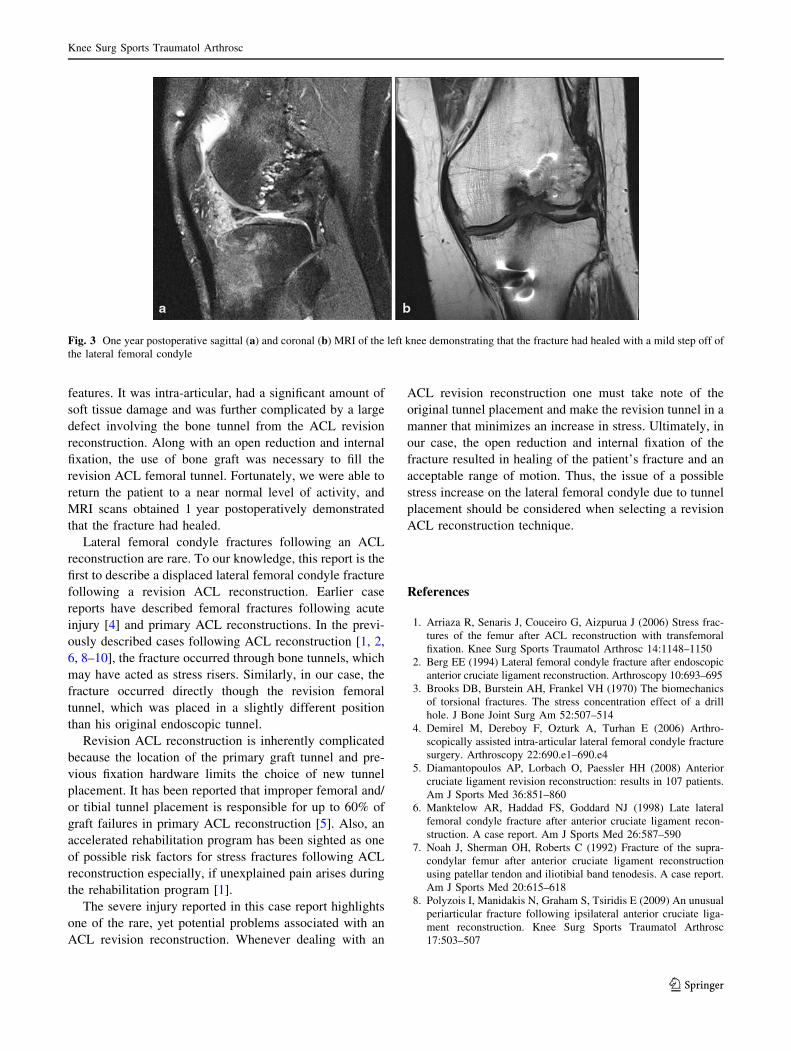
program. At this time, a 3-Tesla MRI scanner was used toobtain an MRI scan of the patient’s knee (Fig. 3). The
evaluation of his lateral compartment revealed that thelateral femoral condyle fracture had healed with a mild step
off of the lateral femoral condyle. The patient’s range of
motion was 3" to 115" of knee flexion.
Discussion
The key finding of the present study was that it brought to
light the issue of a possible stress increase on the lateralfemoral condyle due to a second tunnel placement during a
revision ACL reconstruction. The complex fracture pre-
sented in this case report had several distinct and unique
Fig. 1 Preoperative sagittal (a) and coronal (b) CT of the left knee demonstrating evidence of an intra-articular lateral femoral condyle fracture
Fig. 2 Intraoperative photograph of the left knee demonstrating thecomplex fracture plane involving the entire weight bearing surface ofthe central aspect of the lateral femoral condyle
Knee Surg Sports Traumatol Arthrosc
123
features. It was intra-articular, had a significant amount ofsoft tissue damage and was further complicated by a large
defect involving the bone tunnel from the ACL revision
reconstruction. Along with an open reduction and internalfixation, the use of bone graft was necessary to fill the
revision ACL femoral tunnel. Fortunately, we were able to
return the patient to a near normal level of activity, andMRI scans obtained 1 year postoperatively demonstrated
that the fracture had healed.
Lateral femoral condyle fractures following an ACLreconstruction are rare. To our knowledge, this report is the
first to describe a displaced lateral femoral condyle fracturefollowing a revision ACL reconstruction. Earlier case
reports have described femoral fractures following acute
injury [4] and primary ACL reconstructions. In the previ-ously described cases following ACL reconstruction [1, 2,
6, 8–10], the fracture occurred through bone tunnels, which
may have acted as stress risers. Similarly, in our case, thefracture occurred directly though the revision femoral
tunnel, which was placed in a slightly different position
than his original endoscopic tunnel.Revision ACL reconstruction is inherently complicated
because the location of the primary graft tunnel and pre-
vious fixation hardware limits the choice of new tunnelplacement. It has been reported that improper femoral and/
or tibial tunnel placement is responsible for up to 60% of
graft failures in primary ACL reconstruction [5]. Also, anaccelerated rehabilitation program has been sighted as one
of possible risk factors for stress fractures following ACL
reconstruction especially, if unexplained pain arises duringthe rehabilitation program [1].
The severe injury reported in this case report highlights
one of the rare, yet potential problems associated with anACL revision reconstruction. Whenever dealing with an
ACL revision reconstruction one must take note of theoriginal tunnel placement and make the revision tunnel in a
manner that minimizes an increase in stress. Ultimately, in
our case, the open reduction and internal fixation of thefracture resulted in healing of the patient’s fracture and an
acceptable range of motion. Thus, the issue of a possible
stress increase on the lateral femoral condyle due to tunnelplacement should be considered when selecting a revision
ACL reconstruction technique.
References
1. Arriaza R, Senaris J, Couceiro G, Aizpurua J (2006) Stress frac-tures of the femur after ACL reconstruction with transfemoralfixation. Knee Surg Sports Traumatol Arthrosc 14:1148–1150
2. Berg EE (1994) Lateral femoral condyle fracture after endoscopicanterior cruciate ligament reconstruction. Arthroscopy 10:693–695
3. Brooks DB, Burstein AH, Frankel VH (1970) The biomechanicsof torsional fractures. The stress concentration effect of a drillhole. J Bone Joint Surg Am 52:507–514
4. Demirel M, Dereboy F, Ozturk A, Turhan E (2006) Arthro-scopically assisted intra-articular lateral femoral condyle fracturesurgery. Arthroscopy 22:690.e1–690.e4
5. Diamantopoulos AP, Lorbach O, Paessler HH (2008) Anteriorcruciate ligament revision reconstruction: results in 107 patients.Am J Sports Med 36:851–860
6. Manktelow AR, Haddad FS, Goddard NJ (1998) Late lateralfemoral condyle fracture after anterior cruciate ligament recon-struction. A case report. Am J Sports Med 26:587–590
7. Noah J, Sherman OH, Roberts C (1992) Fracture of the supra-condylar femur after anterior cruciate ligament reconstructionusing patellar tendon and iliotibial band tenodesis. A case report.Am J Sports Med 20:615–618
8. Polyzois I, Manidakis N, Graham S, Tsiridis E (2009) An unusualperiarticular fracture following ipsilateral anterior cruciate liga-ment reconstruction. Knee Surg Sports Traumatol Arthrosc17:503–507
Fig. 3 One year postoperative sagittal (a) and coronal (b) MRI of the left knee demonstrating that the fracture had healed with a mild step off ofthe lateral femoral condyle
Knee Surg Sports Traumatol Arthrosc
123

9. Thangamani VB, Flanigan DC, Merk BR (2009) Intra-articulardistal femur fracture extending from an expanded femoral tunnelin an anterior cruciate ligament (ACL) reconstructed knee: a casereport. J Trauma. doi:10.1097/TA.0b013e3181469f42
10. Wilson TC, Rosenblum WJ, Johnson DL (2004) Fracture of thefemoral tunnel after an anterior cruciate ligament reconstruction.Arthroscopy 20:e45–e47
Knee Surg Sports Traumatol Arthrosc
123
Recommended