Embed Size (px)
Citation preview
12/13/15
1
The Knee
12/13/15
2
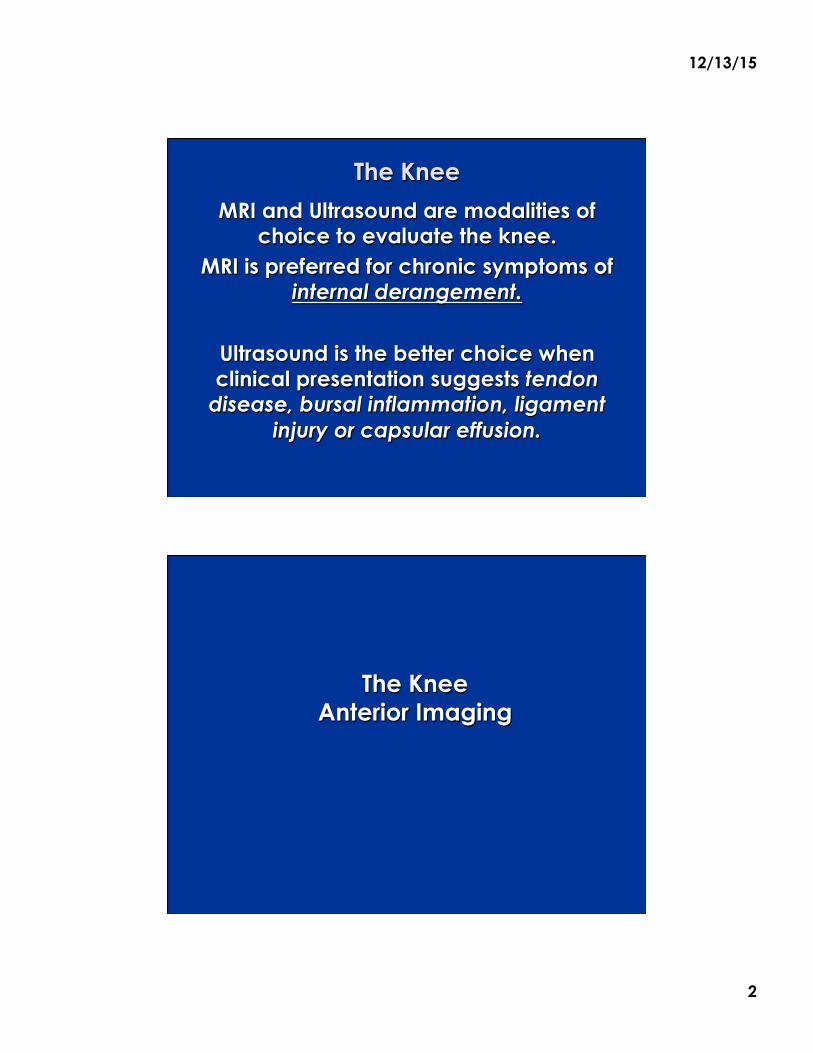
The Knee
MRI and Ultrasound are modalities of choice to evaluate the knee.
MRI is preferred for chronic symptoms of internal derangement.
Ultrasound is the better choice when clinical presentation suggests tendon
disease, bursal inflammation, ligament injury or capsular effusion.
The Knee Anterior Imaging

12/13/15
3
The Knee Quadriceps Tendon Longitudinal
Identifying three interfaces is helpful in using
supra-patellar pouch/bursa for intra-articular
injections.
OA with minimal bursal effusion
Ultrasound guidance adds increased accuracy
Check tendon pattern !
J Clin Ultrasound. 2012 Jan;40(1):20-5. doi:
10.1002/jcu.20890. Epub 2011 Oct 28
Articularis Genu + Fat Pad
The Knee Quadriceps Tendon Longitudinal
Supine patient & LAX probe A bolus for 30⁰ flexion.
Landmarks : patella and femur
3 interfaces identified 1 = Femur/Fat Pad
2 = Suprapatellar Pouch 3 = Quad Tendon
Quad Contraction
enhances bursal interface
3
2
1
12/13/15
4
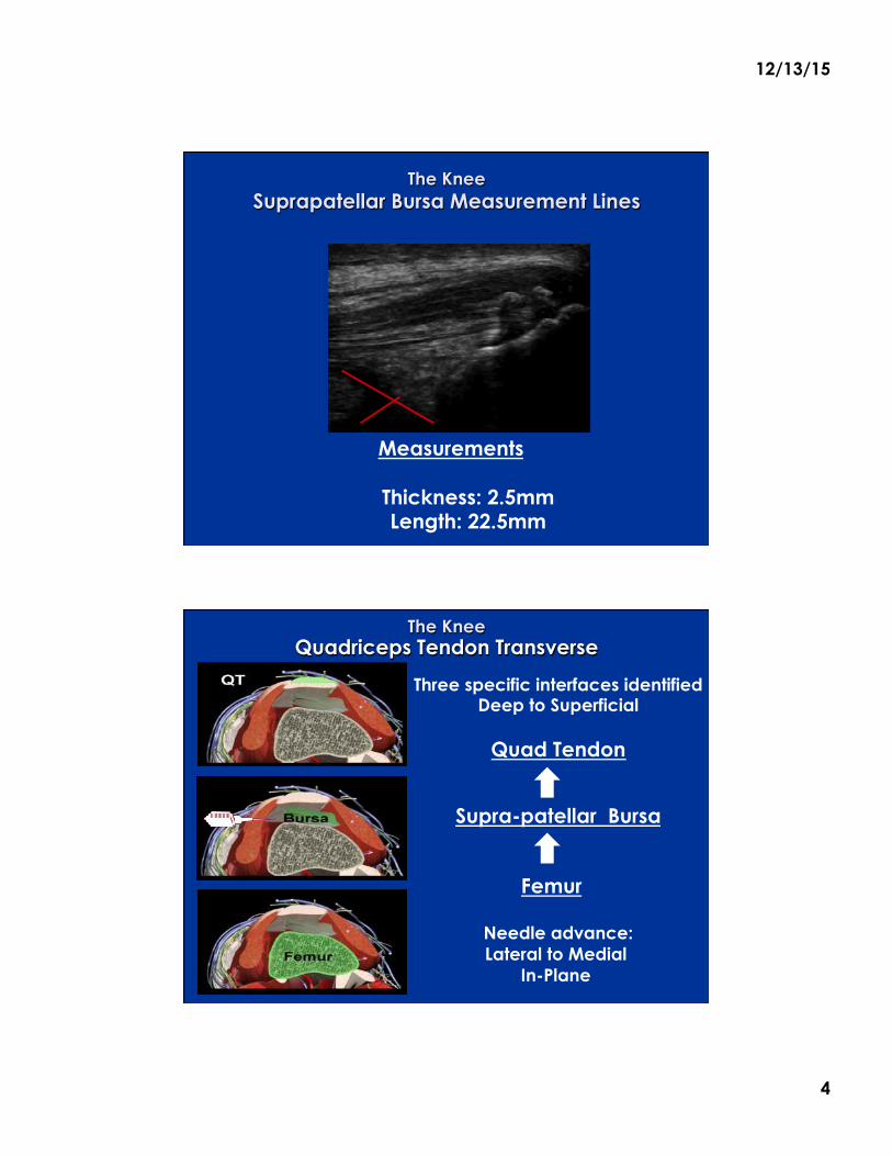
The Knee Suprapatellar Bursa Measurement Lines
Measurements
Thickness: 2.5mm Length: 22.5mm
The Knee Quadriceps Tendon Transverse
Three specific interfaces identified Deep to Superficial
Quad Tendon
Supra-patellar Bursa
Femur
Needle advance: Lateral to Medial
In-Plane
12/13/15
5
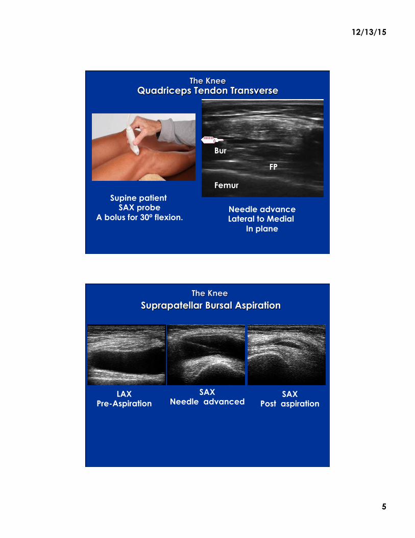
The Knee Quadriceps Tendon Transverse
Needle advance Lateral to Medial
In plane
Bur
Femur
FP
Supine patient SAX probe
A bolus for 30⁰ flexion.
The Knee
Suprapatellar Bursal Aspiration
LAX Pre-Aspiration
SAX Needle advanced
SAX Post aspiration
12/13/15
6
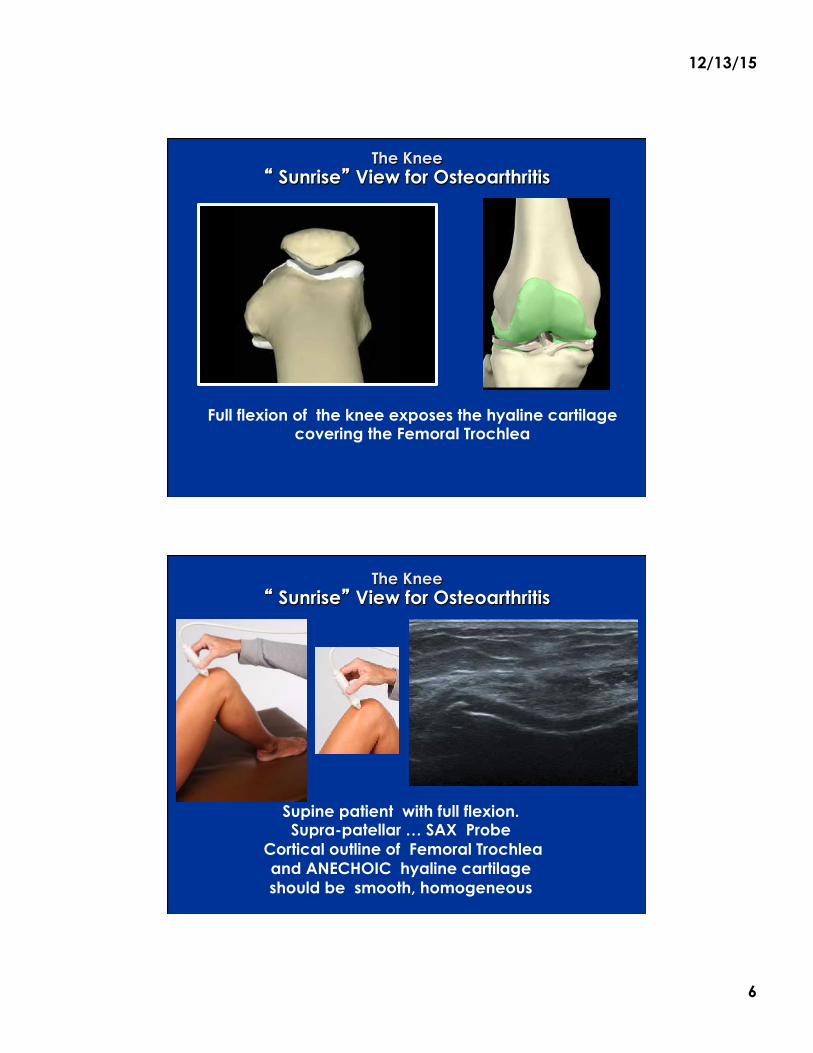
The Knee � Sunrise� View for Osteoarthritis
Full flexion of the knee exposes the hyaline cartilage covering the Femoral Trochlea
The Knee � Sunrise� View for Osteoarthritis
Supine patient with full flexion. Supra-patellar … SAX Probe
Cortical outline of Femoral Trochlea and ANECHOIC hyaline cartilage should be smooth, homogeneous
12/13/15
7
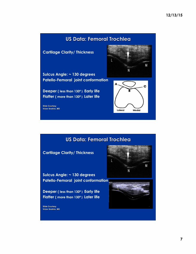
US Data: Femoral Trochlea
Cartilage Clarity/ Thickness
Sulcus Angle: ~ 130 degrees Patello-Femoral joint conformation Deeper ( less than 130⁰)Early life Flatter ( more than 130⁰) Later life Slide Courtesy Victor Ibrahim, MD
US Data: Femoral Trochlea
Cartilage Clarity/ Thickness
Sulcus Angle: ~ 130 degrees Patello-Femoral joint conformation Deeper ( less than 130⁰)Early life Flatter ( more than 130⁰) Later life Slide Courtesy Victor Ibrahim, MD
12/13/15
8
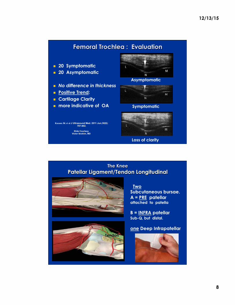
Femoral Trochlea : Evaluation
! 20 Symptomatic ! 20 Asymptomatic
! No difference in thickness ! Positive Trend: ! Cartilage Clarity ! more indicative of OA
Kazam JK et al J Ultrasound Med. 2011 Jun;30(6):797-802.
Slide Courtesy
Victor Ibrahim, MD
Asymptomatic
Symptomatic
Loss of clarity
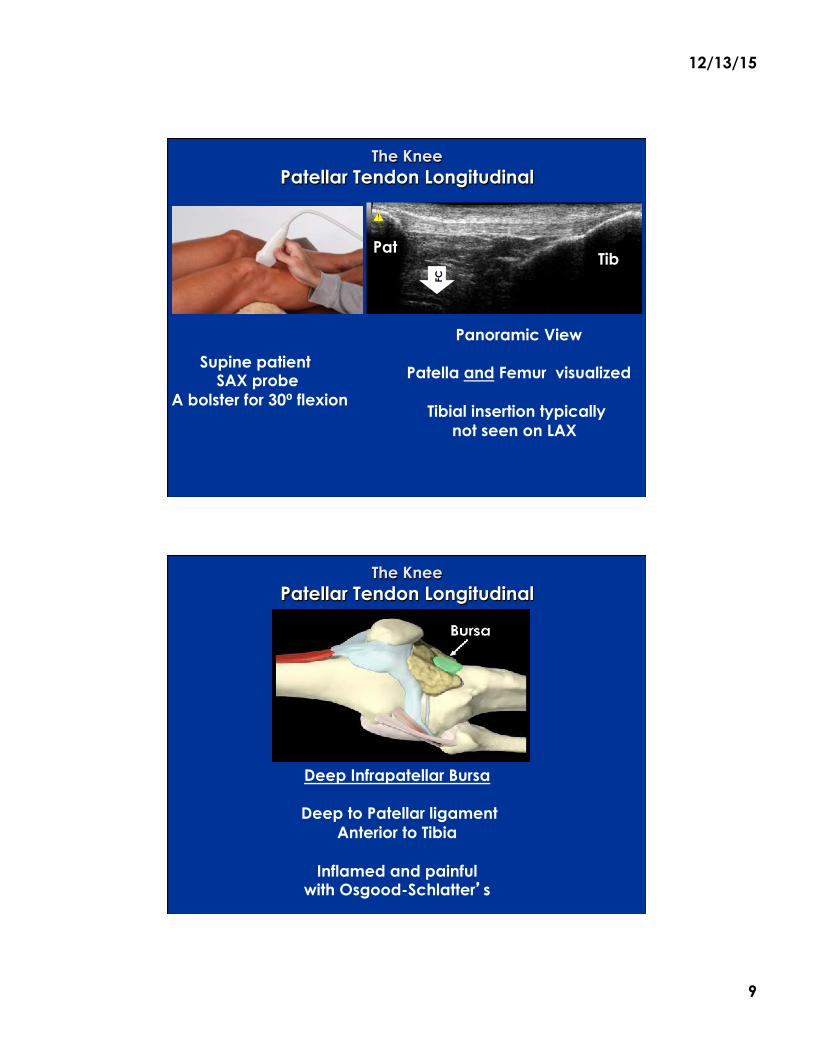
The Knee Patellar Ligament/Tendon Longitudinal
Two Subcutaneous bursae. A = PRE patellar attached to patella B = INFRA patellar Sub-Q, but distal. one Deep Infrapatellar
A B
Tendon
12/13/15
9
The Knee Patellar Tendon Longitudinal
Panoramic View
Patella and Femur visualized
Tibial insertion typically not seen on LAX
Supine patient SAX probe
A bolster for 30⁰ flexion
Pat Tib
FC
The Knee Patellar Tendon Longitudinal
Deep Infrapatellar Bursa
Deep to Patellar ligament Anterior to Tibia
Inflamed and painful
with Osgood-Schlatter�s
Supine patient SAX probe
A bolus for 30⁰ flexion Pat
Tib
12/13/15
10
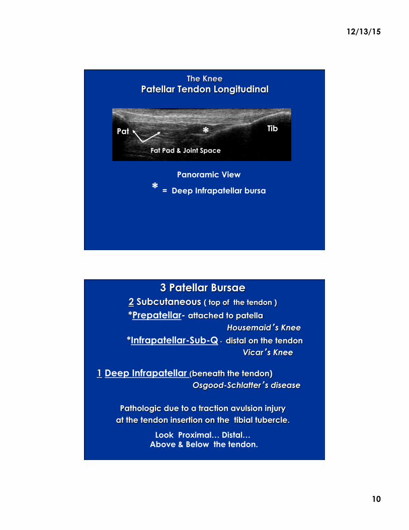
The Knee Patellar Tendon Longitudinal
Panoramic View
* = Deep Infrapatellar bursa
Tib Pat
Fat Pad & Joint Space
Tib *
3 Patellar Bursae 2 Subcutaneous ( top of the tendon )
*Prepatellar- attached to patella Housemaid�s Knee
*Infrapatellar-Sub-Q - distal on the tendon Vicar�s Knee
1 Deep Infrapatellar (beneath the tendon) Osgood-Schlatter�s disease
Pathologic due to a traction avulsion injury
at the tendon insertion on the tibial tubercle.
Look Proximal… Distal…
Above & Below the tendon.
12/13/15
11
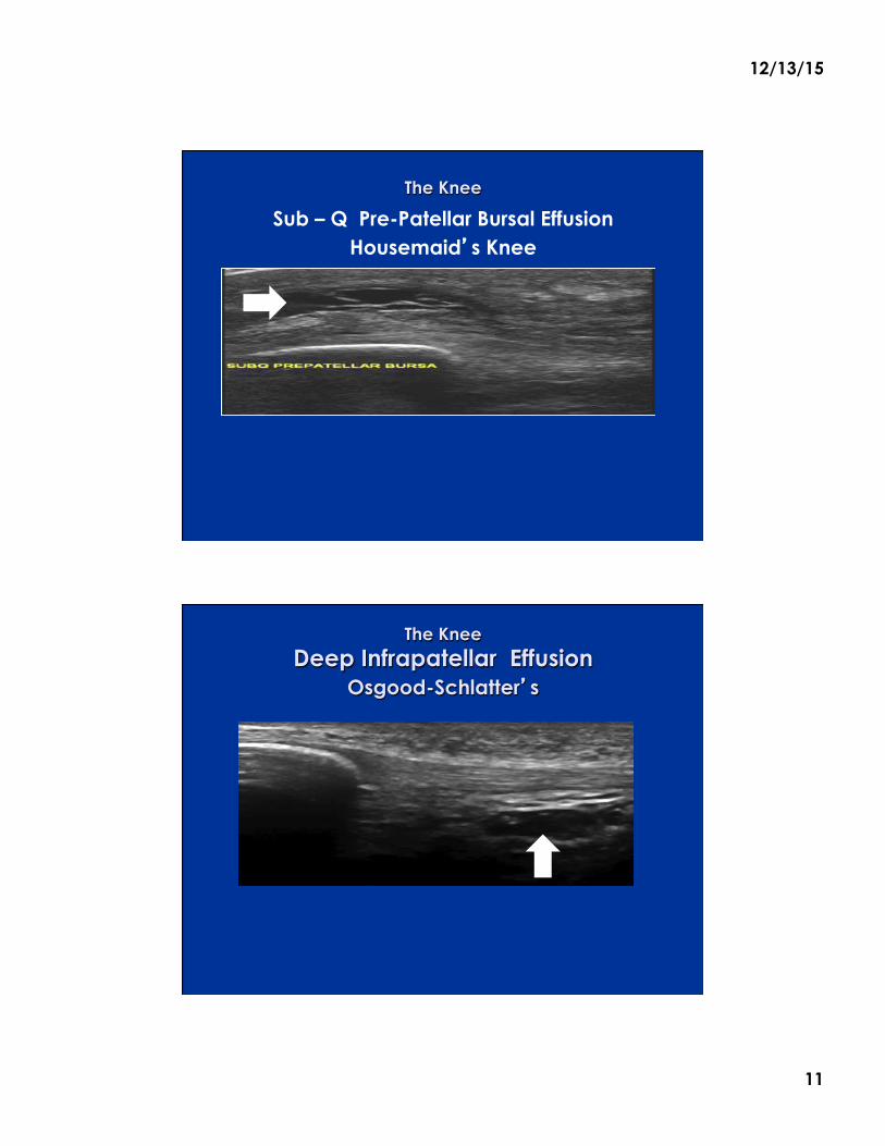
The Knee
Sub – Q Pre-Patellar Bursal Effusion Housemaid�s Knee
The Knee Deep Infrapatellar Effusion
Osgood-Schlatter�s
12/13/15
12
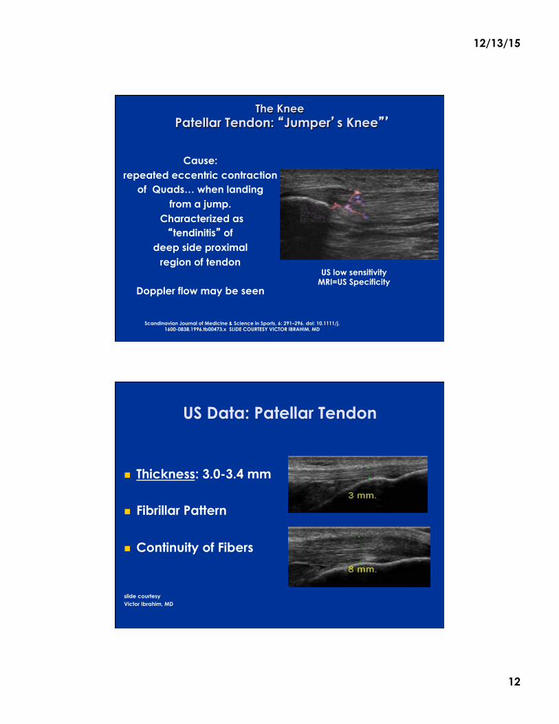
The Knee Patellar Tendon: �Jumper�s Knee��
Cause:
repeated eccentric contraction of Quads… when landing
from a jump. Characterized as �tendinitis� of
deep side proximal region of tendon
Doppler flow may be seen
Scandinavian Journal of Medicine & Science in Sports, 6: 291–296. doi: 10.1111/j.
1600-0838.1996.tb00473.x SLIDE COURTESY VICTOR IBRAHIM, MD
US low sensitivity MRI=US Specificity
US Data: Patellar Tendon
! Thickness: 3.0-3.4 mm
! Fibrillar Pattern
! Continuity of Fibers
slide courtesy Victor Ibrahim, MD
12/13/15
13
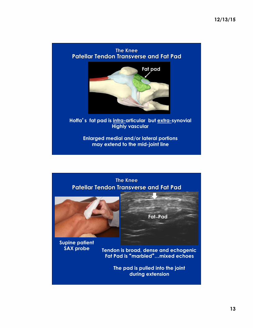
The Knee Patellar Tendon Transverse and Fat Pad
Hoffa�s fat pad is intra-articular but extra-synovial Highly vascular
Enlarged medial and/or lateral portions
may extend to the mid-joint line
The Knee Patellar Tendon Transverse and Fat Pad
Supine patient SAX probe
Tendon is broad, dense and echogenic
Fat Pad is �marbled�…mixed echoes
The pad is pulled into the joint during extension
Fat Pad
12/13/15
14
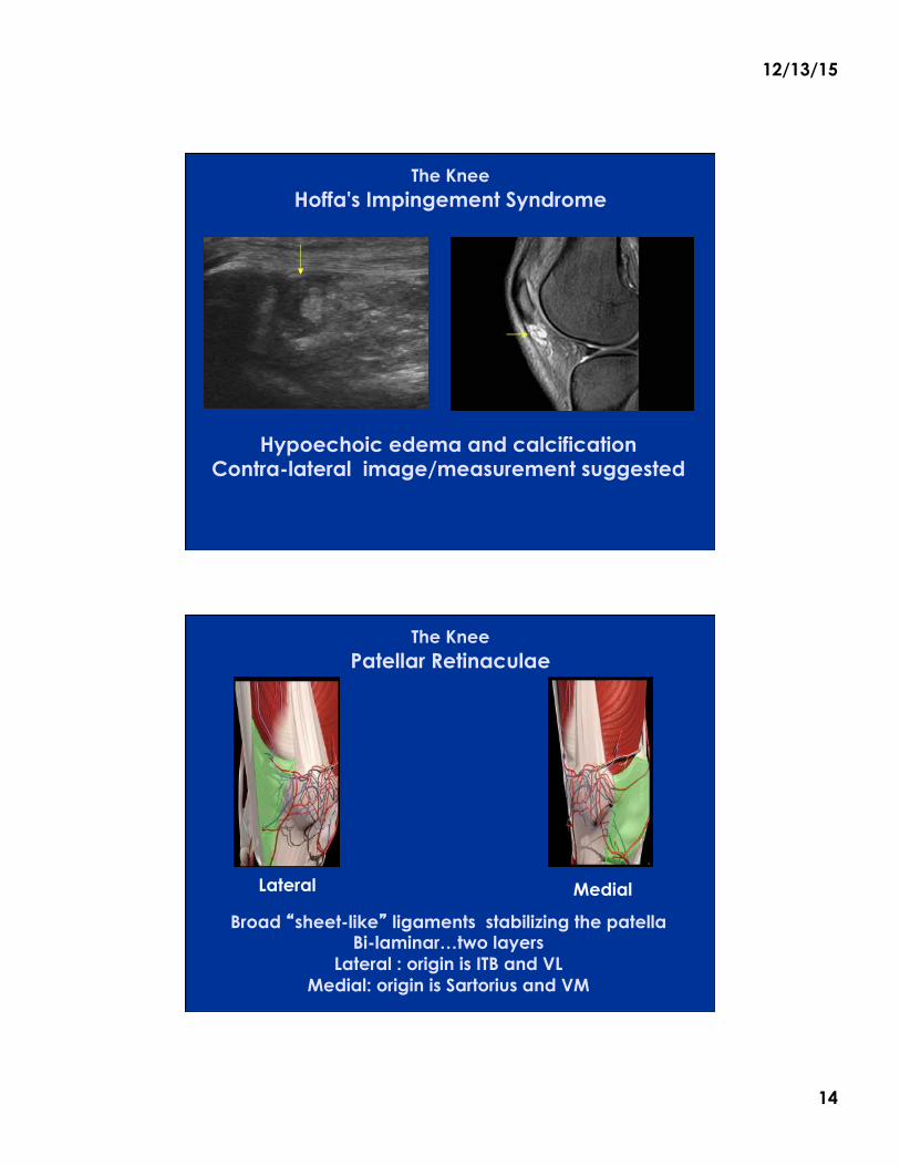
The Knee
Hoffa's Impingement Syndrome
Hypoechoic edema and calcification Contra-lateral image/measurement suggested
Hoffa’s fat pad is mobile with observable tracking on flexion/extension. Lack of observed mobility is suggestive of impingement .
The Knee Patellar Retinaculae
Broad �sheet-like� ligaments stabilizing the patella Bi-laminar…two layers
Lateral : origin is ITB and VL Medial: origin is Sartorius and VM
Medial Lateral
12/13/15
15
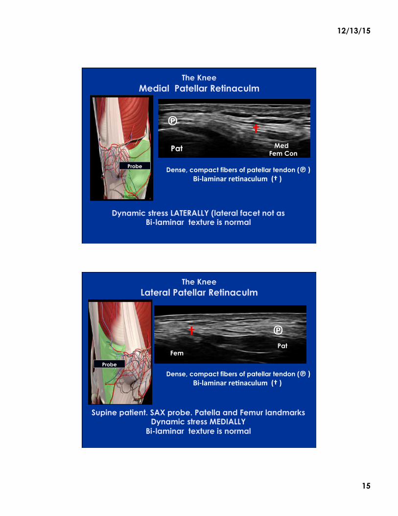
The Knee
Medial Patellar Retinaculm
Dynamic stress LATERALLY (lateral facet not as Bi-laminar texture is normal
Med Fem Con
Pat
Dense, compact fibers of patellar tendon (℗)Bi-laminarre.naculum(†)
† ℗
Probe
The Knee
Lateral Patellar Retinaculm
Supine patient. SAX probe. Patella and Femur landmarks Dynamic stress MEDIALLY
Bi-laminar texture is normal
Probe
Fem
Pat
† ℗
Dense, compact fibers of patellar tendon (℗)Bi-laminarre.naculum(†)
12/13/15
16
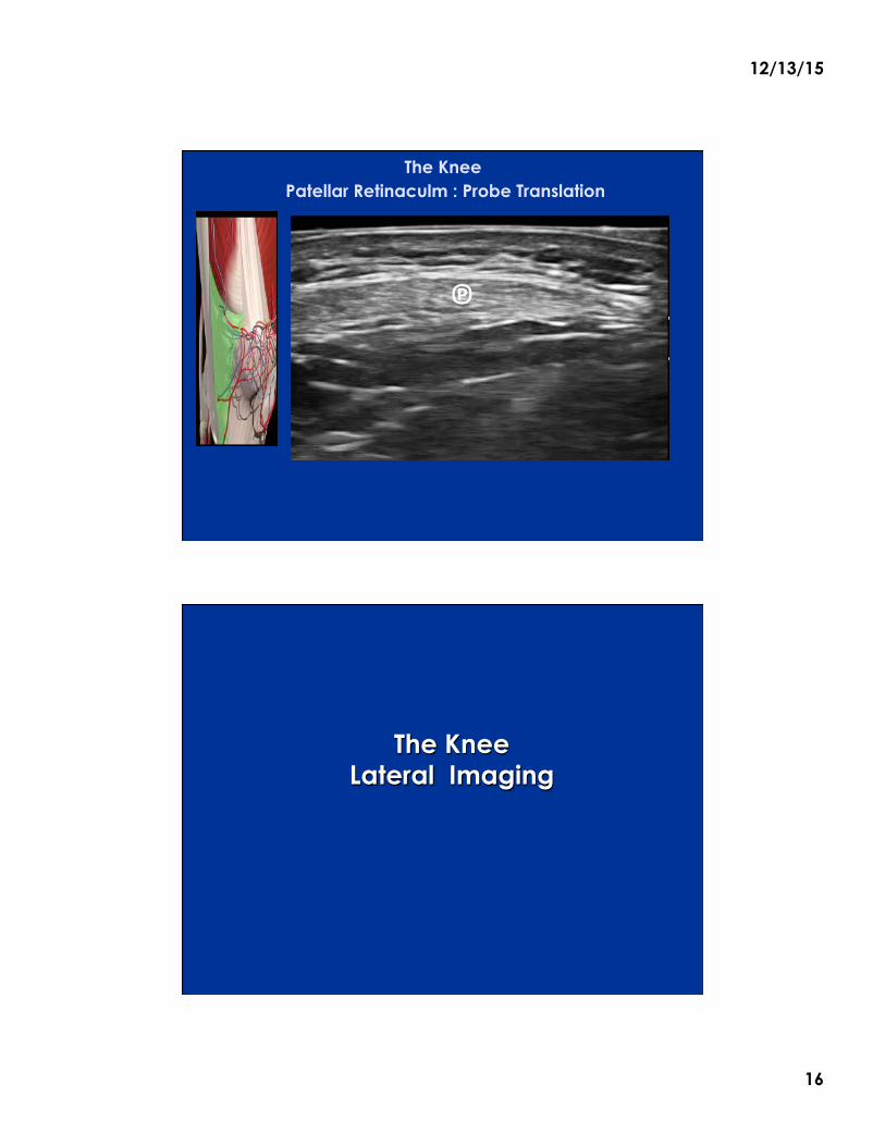
The Knee
Patellar Retinaculm : Probe Translation
℗
The Knee Lateral Imaging
12/13/15
17
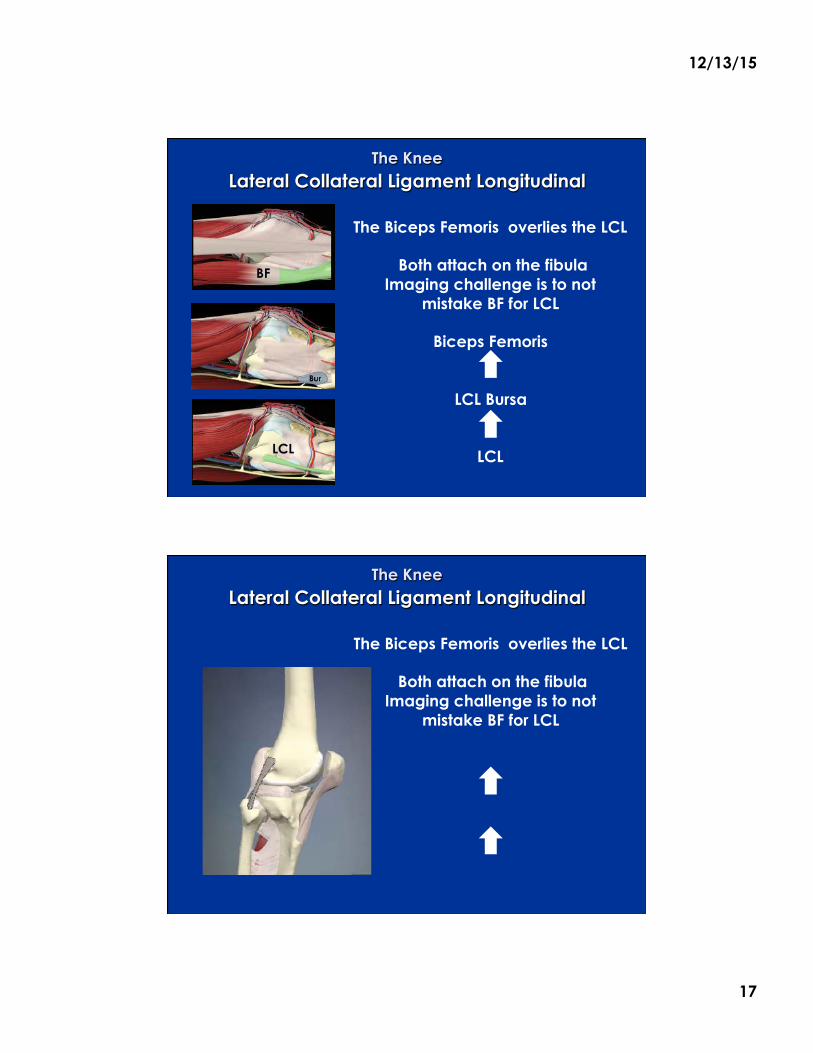
The Knee Lateral Collateral Ligament Longitudinal
IT Band BF
The Biceps Femoris overlies the LCL
Both attach on the fibula Imaging challenge is to not
mistake BF for LCL
Biceps Femoris
LCL Bursa
LCL
2
LCL
The Knee Lateral Collateral Ligament Longitudinal
IT Band BF
The Biceps Femoris overlies the LCL
Both attach on the fibula Imaging challenge is to not
mistake BF for LCL 2
LCL
12/13/15
18
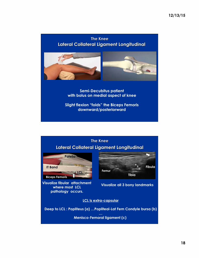
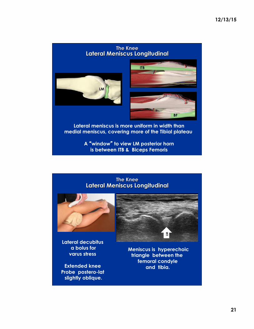
The Knee Lateral Collateral Ligament Longitudinal
Semi-Decubitus patient with bolus on medial aspect of knee
Slight flexion “folds” the Biceps Femoris
downward/posteriorward
1
IT Band LCL
Pat
The Knee
Lateral Collateral Ligament Longitudinal
IT Band
Biceps Femoris LCL
Visualize fibular attachment where most LCL
pathology occurs.
Patella
Femur
Visualize all 3 bony landmarks
Tibia
Fibula
LCL is extra-capsular
Deep to LCL : Popliteus (a) …Popliteal-Lat Fem Condyle bursa (b)…
Menisco-Femoral ligament (c)
a b
c
12/13/15
19
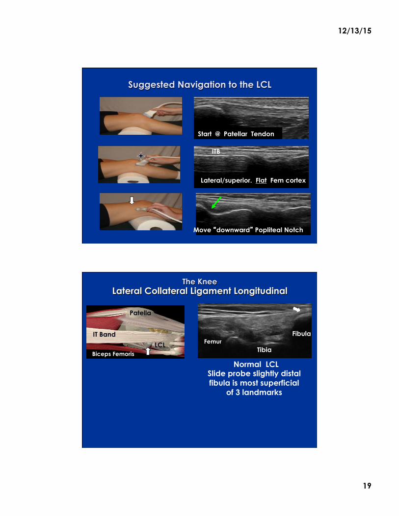
Suggested Navigation to the LCL
1
2
Start @ Patellar Tendon
Lateral/superior. Flat Fem cortex
Move �downward� Popliteal Notch
ITB
The Knee Lateral Collateral Ligament Longitudinal
Normal LCL Slide probe slightly distal fibula is most superficial
of 3 landmarks
IT Band
Biceps Femoris LCL
Patella
Tibia
Fibula Femur
12/13/15
20
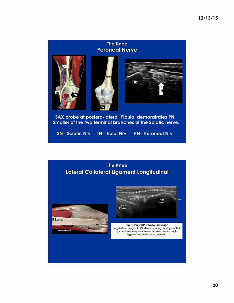
The Knee Peroneal Nerve
SAX probe at postero-lateral Fibula demonstrates PN Smaller of the two terminal branches of the Sciatic nerve.
SN= Sciatic Nrv TN= Tibial Nrv PN= Peroneal Nrv
PN
Tibia
Femur
Fib
The Knee Lateral Collateral Ligament Longitudinal
IT Band
Biceps Femoris
LCL
Femur
Tibia
Fibula
12/13/15
21
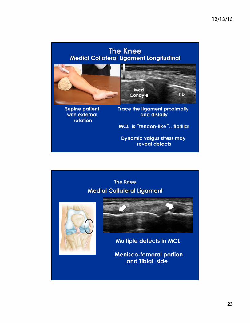
The Knee Lateral Meniscus Longitudinal
Lateral meniscus is more uniform in width than medial meniscus, covering more of the Tibial plateau
A �window� to view LM posterior horn
is between ITB & Biceps Femoris
Anterior
ITB
LM
BF
The Knee Lateral Meniscus Longitudinal
Lateral decubitus a bolus for varus stress
Extended knee
Probe postero-lat slightly oblique.
Meniscus is hyperechoic triangle between the
femoral condyle and tibia.
LM

12/13/15
22
The Knee Medial Imaging
The Knee Medial Collateral Ligament Longitudinal
A flat band-like ligament nearly 9cm in length
Anterior and posterior portions give it a �tri-laminar� appearance, best seen at it’s proximal portion
Sartorius is adjacent posteriorly to MCL in LAX
Sartorius
12/13/15
23
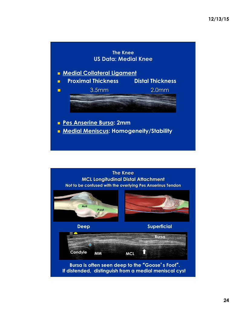
The Knee Medial Collateral Ligament Longitudinal
Supine patient with external
rotation
Med Condyle Tib
Trace the ligament proximally and distally
MCL is �tendon-like�…fibrillar
Dynamic valgus stress may
reveal defects
The Knee
Medial Collateral Ligament
Multiple defects in MCL
Menisco-femoral portion and Tibial side
12/13/15
24
The Knee US Data: Medial Knee
! Medial Collateral Ligament ! Proximal Thickness Distal Thickness
! 3.5mm 2.0mm
! Pes Anserine Bursa: 2mm ! Medial Meniscus: Homogeneity/Stability
The Knee MCL Longitudinal Distal Attachment
Not to be confused with the overlying Pes Anserinus Tendon
Deep Superficial
Bursa is often seen deep to the �Goose�s Foot�. If distended, distinguish from a medial meniscal cyst
Condyle MM MCL
Bursa
12/13/15
25
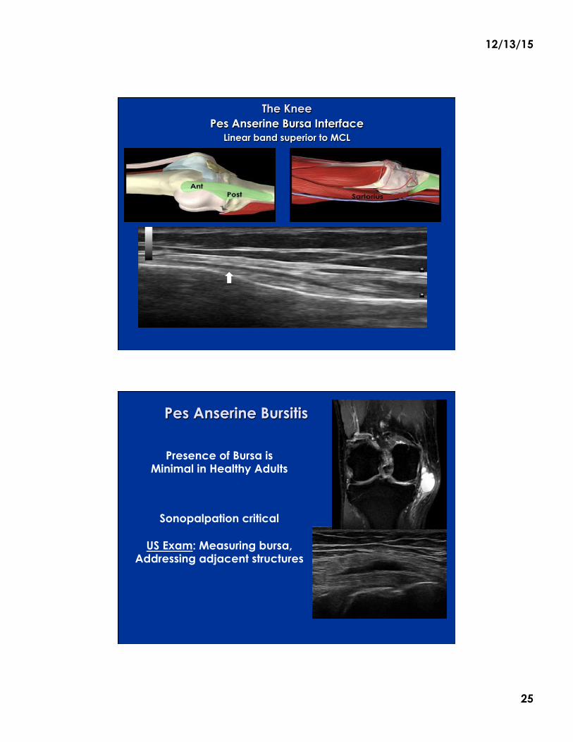
The Knee Pes Anserine Bursa Interface
Linear band superior to MCL
Pes Anserine Bursitis
Presence of Bursa is Minimal in Healthy Adults
Sonopalpation critical
US Exam: Measuring bursa, Addressing adjacent structures
12/13/15
26
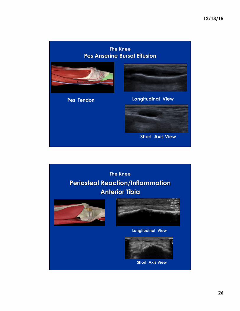
The Knee Pes Anserine Bursal Effusion
Pes Tendon Longitudinal View
Short Axis View
The Knee
Periosteal Reaction/Inflammation Anterior Tibia
Longitudinal View
Short Axis View
12/13/15
27
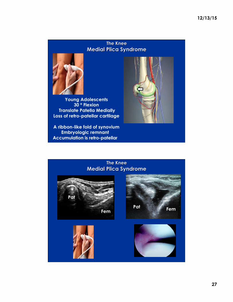
The Knee Medial Plica Syndrome
Young Adolescents 30 ⁰ Flexion
Translate Patella Medially Loss of retro-patellar cartilage
A ribbon-like fold of synovium
Embryologic remnant Accumulation is retro-patellar Pat Fem
The Knee Medial Plica Syndrome
Pat Fem
Pat
Fem
12/13/15
28
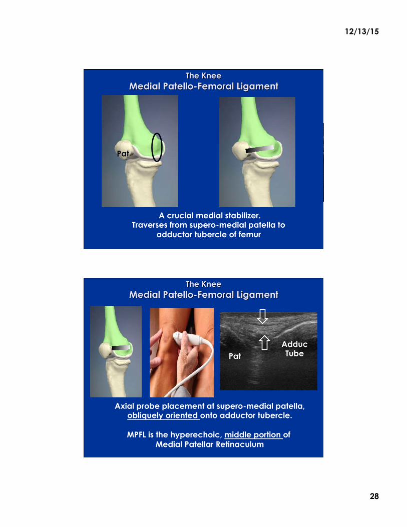
The Knee Medial Patello-Femoral Ligament
Pat
Fem
Pat
A crucial medial stabilizer. Traverses from supero-medial patella to
adductor tubercle of femur
The Knee Medial Patello-Femoral Ligament
Adduc Tube Pat
Axial probe placement at supero-medial patella, obliquely oriented onto adductor tubercle.
MPFL is the hyperechoic, middle portion of
Medial Patellar Retinaculum
12/13/15
29
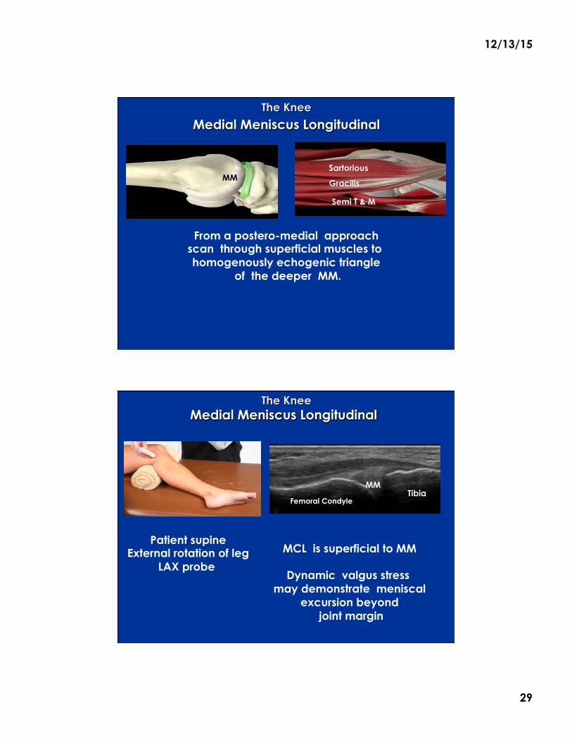
The Knee
Medial Meniscus Longitudinal
From a postero-medial approach scan through superficial muscles to homogenously echogenic triangle
of the deeper MM.
Med Condyle
MM
Femoral Condyle
Tibia
Sartorious
Gracilis
Semi T & M
The Knee Medial Meniscus Longitudinal
Patient supine External rotation of leg
LAX probe
Femoral Condyle Tibia
MCL is superficial to MM
Dynamic valgus stress may demonstrate meniscal
excursion beyond joint margin
MM
12/13/15
30
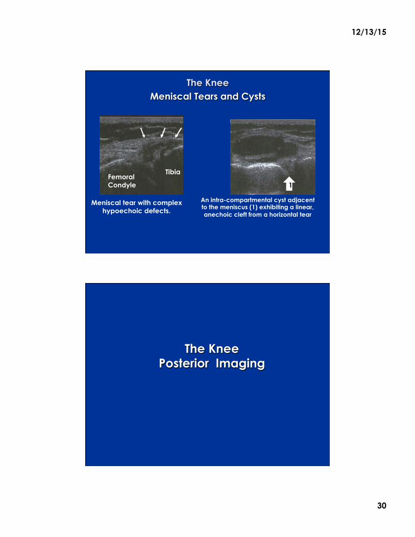
The Knee Meniscal Tears and Cysts
Meniscal tear with complex hypoechoic defects.
An intra-compartmental cyst adjacent to the meniscus (1) exhibiting a linear, anechoic cleft from a horizontal tear
Femoral Condyle
Tibia
1
The Knee Posterior Imaging
12/13/15
31
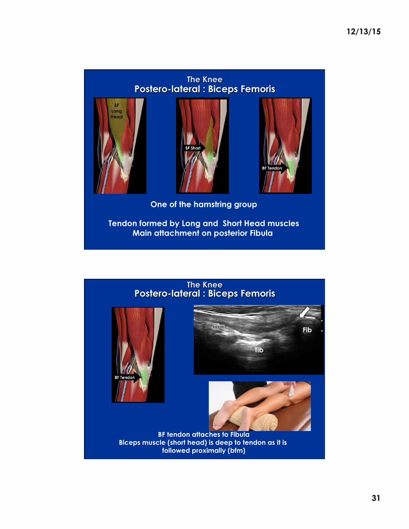
The Knee Postero-lateral : Biceps Femoris
Med Gastr
One of the hamstring group
Tendon formed by Long and Short Head muscles Main attachment on posterior Fibula
The Knee Postero-lateral : Biceps Femoris
BF tendon attaches to Fibula Biceps muscle (short head) is deep to tendon as it is
followed proximally (bfm)
Fib
Tib
12/13/15
32
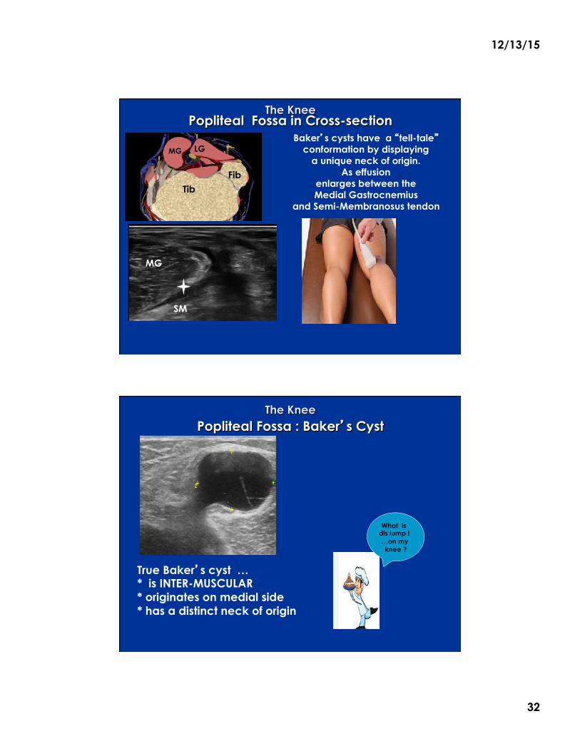
The Knee Popliteal Fossa in Cross-section
Baker�s cysts have a �tell-tale� conformation by displaying
a unique neck of origin. As effusion
enlarges between the Medial Gastrocnemius
and Semi-Membranosus tendon
SM
Tib
LG
MG
MG
Fib
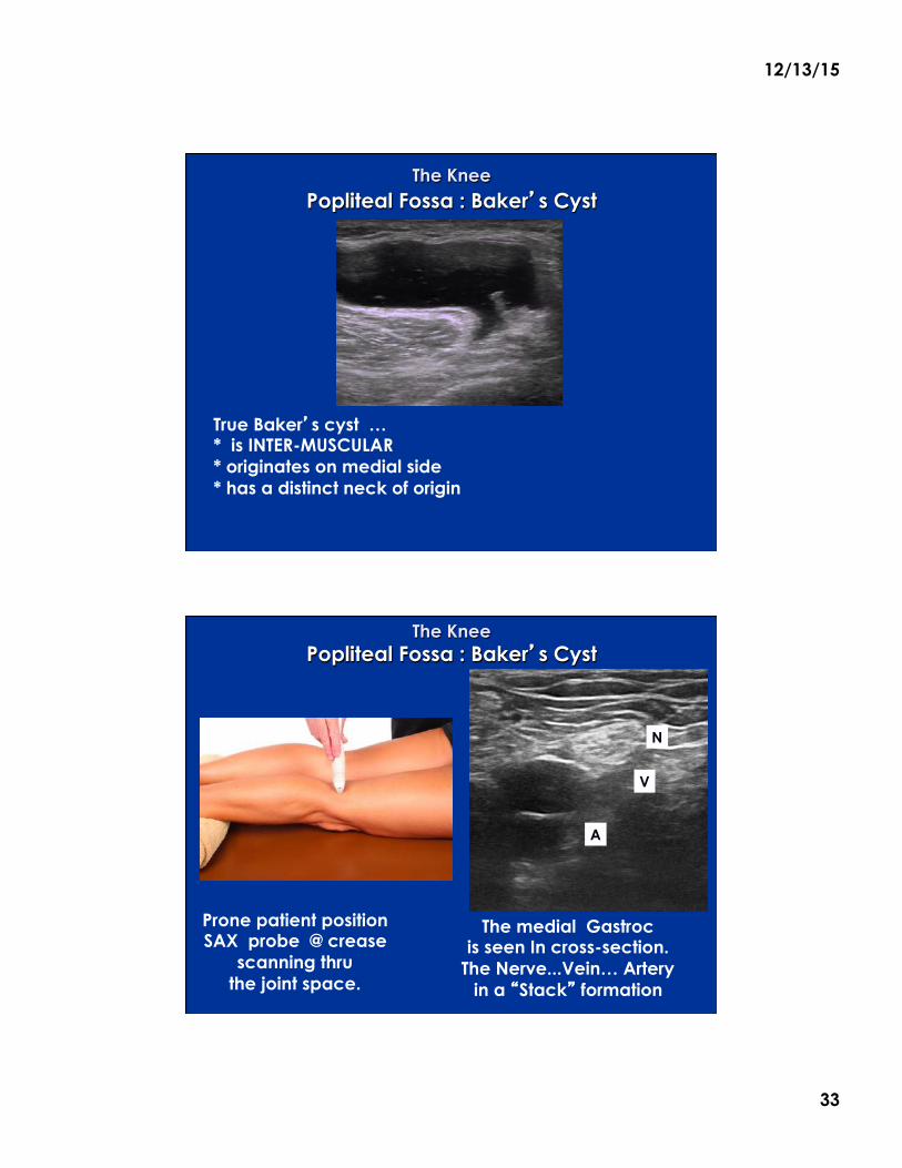
The Knee Popliteal Fossa : Baker�s Cyst
True Baker�s cyst … * is INTER-MUSCULAR * originates on medial side * has a distinct neck of origin
What is dis lump ! …on my knee ?
12/13/15
33
The Knee Popliteal Fossa : Baker�s Cyst
True Baker�s cyst … * is INTER-MUSCULAR * originates on medial side * has a distinct neck of origin
The Knee Popliteal Fossa : Baker�s Cyst
Prone patient position SAX probe @ crease
scanning thru the joint space.
The medial Gastroc is seen In cross-section.
The Nerve...Vein… Artery in a �Stack� formation
N
A
V
12/13/15
34
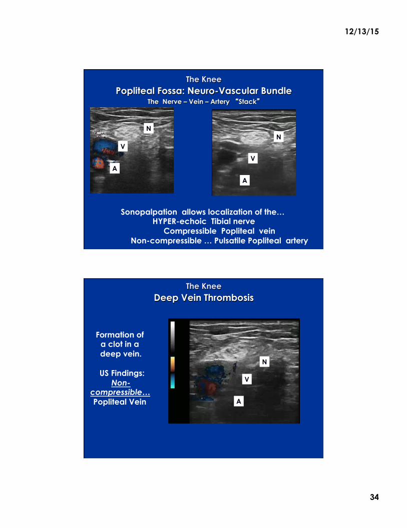
Popliteal Fossa: Neuro-Vascular Bundle The Nerve – Vein – Artery �Stack�
The Knee
Sonopalpation allows localization of the… HYPER-echoic Tibial nerve
Compressible Popliteal vein Non-compressible … Pulsatile Popliteal artery
N
A
V N
A
V
The Knee Deep Vein Thrombosis
Formation of a clot in a deep vein.
US Findings:
Non-compressible… Popliteal Vein
N
A
V
12/13/15
35
Thank you !