In Situ Fluoride Response of Caries Lesions with Different Mineral Distributions at Baseline
F Lippert, R.J.M. Lynch, G.J. Eckert, S.A. Kelly, A.T. Hara, D.T. Zero
Department of Preventive and Community Dentistry, Oral Health Research Institute, Indiana
University School of Dentistry, Indianapolis, Ind., USA
Short title: In Situ Study on Low- and High-R Lesions
Keywords: Fluoride, Caries, Demineralization, Remineralization, in situ
Corresponding author:
Frank Lippert
Department of Preventive and Community Dentistry
Oral Health Research Institute, Indiana University School of Dentistry
415 Lansing Street, Indianapolis, IN 46202 (USA)
Tel. +1 317 274 3983, Fax +1 317 274 5425, E-Mail [email protected]
_________________________________________________________________________________ This is the author's manuscript of the article published in final edited form as: Lippert, F., Lynch, R. J. M., Eckert, G. J., Kelly, S. A., Hara, A. T., & Zero, D. T. (2011). In situ fluoride response of caries lesions with different mineral distributions at baseline. Caries research, 45(1), 47-55. https://doi.org/10.1159/000323846

2
Declaration of Interests
This study was supported in part by a grant from GlaxoSmithKline.
D.T. Zero has on occasion received compensation from GlaxoSmithKline as a consultant.
IUPUI’s Conflict of Interest Committee and the Institutional Review Board have reviewed and
approved this financial interest, concluding that it does not pose any additional significant risk to
research subjects or the integrity of the research. Further information regarding these policies and
procedures will be provided upon request.
3
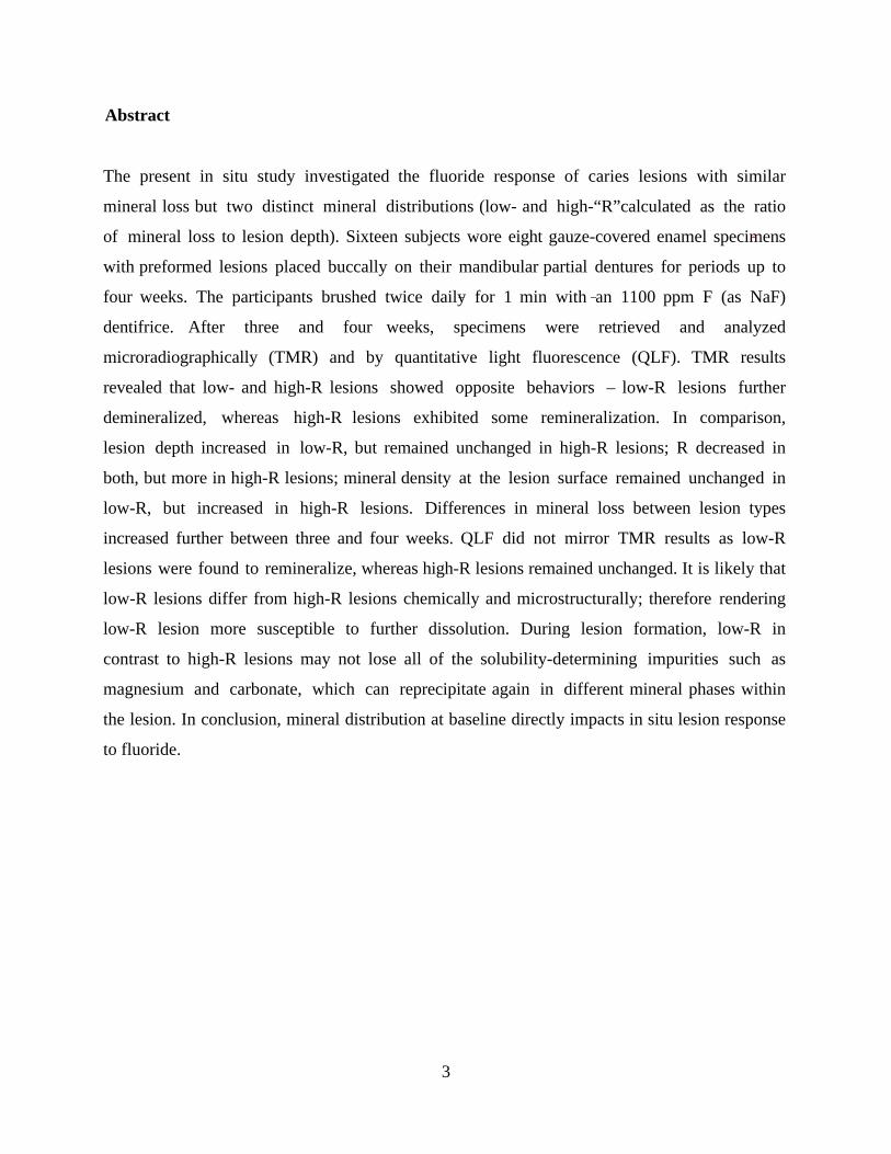
Abstract
The present in situ study investigated the fluoride response of caries lesions with similar
mineral loss but two distinct mineral distributions (low- and high-“R”calculated as the ratio
of mineral loss to lesion depth). Sixteen subjects wore eight gauze-covered enamel specimens
with preformed lesions placed buccally on their mandibular partial dentures for periods up to
four weeks. The participants brushed twice daily for 1 min with an 1100 ppm F (as NaF)
dentifrice. After three and four weeks, specimens were retrieved and analyzed
microradiographically (TMR) and by quantitative light fluorescence (QLF). TMR results
revealed that low- and high-R lesions showed opposite behaviors – low-R lesions further
demineralized, whereas high-R lesions exhibited some remineralization. In comparison,
lesion depth increased in low-R, but remained unchanged in high-R lesions; R decreased in
both, but more in high-R lesions; mineral density at the lesion surface remained unchanged in
low-R, but increased in high-R lesions. Differences in mineral loss between lesion types
increased further between three and four weeks. QLF did not mirror TMR results as low-R
lesions were found to remineralize, whereas high-R lesions remained unchanged. It is likely that
low-R lesions differ from high-R lesions chemically and microstructurally; therefore rendering
low-R lesion more susceptible to further dissolution. During lesion formation, low-R in
contrast to high-R lesions may not lose all of the solubility-determining impurities such as
magnesium and carbonate, which can reprecipitate again in different mineral phases within
the lesion. In conclusion, mineral distribution at baseline directly impacts in situ lesion response
to fluoride.
4
Introduction
The overall effectiveness of fluoride dentifrices in the reduction of caries incidence in vivo is
well documented [e.g. Marinho et al., 2009]. As clinical caries trials are not only time- but also
resource-consuming, a vast range of in situ caries models have been developed as surrogate
measures for fluoride efficacy, dating back to pioneer work conducted by Koulourides and
Volker [1964]. Ideally, these models should serve as bridges between the natural uncontrolled
clinical situation and the highly controlled laboratory situation [Zero, 1995]. Reviews [ten Cate,
1994; Zero, 1995] highlighted the importance of various model parameters from different
aspects. The mineral distribution of caries lesions at baseline has received only limited attention
in in situ caries research given the widely accepted importance of this parameter [Strang et al.,
1987; Mellberg, 1992; Schäfer et al., 1992; ten Cate, 1992]. Typically, studies were either
limited to artificial caries lesions created using one particular demineralization protocol or
lesions with different severities but similar mineral distributions.
Arends et al. [1987] first introduced the concept of describing the average amount of mineral loss
in caries lesions, and therefore mineral distribution characteristics, using the “R” value which
can be calculated for each lesion as the ratio of mineral loss (ΔZ) to lesion depth (L). More
recently, Lynch et al. [2007] were able to divide artificially created lesions into low- and high-R
lesions, depending on the demineralization protocol being used. More importantly, it was shown
on in vitro lesions with similar ΔZbase that high-R lesions were more responsive to remineralizing
solutions than low-R lesions, possibly driven by the greater porosity of high- vs. low-R lesions.
In caries research in general, the use of artificially created lesions is (for obvious reasons)
preferred over natural white spot lesions as a study substrate. While this allows for better control,
it is somewhat removed from the in vivo situation, especially since very little is known about the
mineral distribution characteristics of naturally occurring white spot lesions. To the authors’
knowledge, only one study [Lynch et al., 2007] actually reported R values for these lesions (R =
16 vol%), whereas two other studies [Iijima et al., 1999; Iijima and Takagi, 2000] provided
sufficient information that R values of 32 and 25 vol%, respectively, could be calculated. Several
other studies were concerned with natural white spot lesions in general; however, the lack of
information provided did not allow for R values to be retrieved. This information would be vital
in designing more appropriate laboratory and clinical models for the study of caries and
5
consequently for the development of strategies to reduce caries prevalence. As considerable
variations in R values were found in the aforementioned studies, it would be advantageous
to build on a previous study [Lynch et al., 2007] by investigating the effects of fluoride on
lesions with different mineral distributions simultaneously and under more dynamic
conditions, with potential for periods of undersaturation with respect to enamel as well as
supersaturation, typical of the caries process.
Therefore, the aim of the present study was to investigate the response of low- and high-R
lesions to a twice daily fluoride application using an established in situ caries model.
Subjects and Methods
Ethical Aspects
The study protocol was reviewed and approved by the IUPUI Institutional Review Board, #1002-
59. It was conducted at the Oral Health Research Institute of the Indiana University School of
Dentistry. All subjects signed a written informed consent prior to screening. Seventeen adult
volunteers met the inclusion criteria which included having stimulated and unstimulated salivary
flow rates equal or greater than the minimum requirement of 0.8 and 0.2 ml/min, respectively.
Experimental Design
The study was a randomized, single-center, single-product study design. Seventeen subjects
between the ages of 52 and 79 years undertook the study, with 16 completing the study. Two to
three days following a dental cleaning, eight partially demineralized specimens with two
different mineral distributions (four low-R and four high-R lesions) but similar lesion volume at
baseline (ΔZbase) were placed in the buccal flange areas of the subject’s mandibular partial
denture, four specimens with low-R lesions on one side and four with high-R lesions on the
opposite side. The side position (i.e. left or right) of specimens with low- and high-R lesions was
randomized among subjects. Specimens were wrapped in gauze in pairs of two to facilitate
plaque growth. Wrapped specimen parcels were mounted side by side and flush with the denture
surface. As four specimens (or two specimen parcels containing two specimens each) were
6
mounted on each side of the partial denture; one parcel had to be mounted in the mesial and one
in the distal position. The parcel position, lesion type and treatment period were all randomized.
During the four week test period, subjects brushed with a full ribbon of the study toothpaste
(Crest Cavity Protection, Procter & Gamble, USA; 1100 ppm F as NaF, silica abrasive) twice
daily for one timed minute. Subjects brushed their teeth with their partial denture in place, taking
care to not brush the specimen sites. After brushing, subjects were instructed to expectorate the
toothpaste slurry and rinse with 15 ml of water for 10 seconds and expectorate. Subjects wore
their partial dentures 24 hours a day during the test period. At the end of three weeks, four
specimens (two of each lesion type) were removed and analyzed. The remaining four specimens
were removed after four weeks. During specimen removal from the partial denture, care was
taken not to damage any part of the specimen, including the lesion area and the nail varnish-
covered sound enamel reference areas. Immediately after specimen removal, any plaque residue
was carefully removed from the specimens using a deionized water-moistened cotton bud, before
the specimens were placed, individually, into Eppendorf vials containing a moist cotton pellet.
Three and four week treatment periods were chosen to obtain information on potential
lesion reversal or progression rates.
Changes in the mineral content of the experimental caries lesions were measured using
transverse microradiography (TMR) and quantitative light fluorescence (QLF). The primary
response variable was change in mineral content of the lesions from baseline (ΔΔZ). Secondary
response variables were change in lesion depth (ΔL), R value (ΔR), degree of surface zone
mineralization (ΔSZmax), and lesion fluorescence (ΔΔF).
A washout period of two to three days was observed before starting the study. During this period
subjects were instructed to brush their teeth with a standard non-fluoridated dentifrice (Silly
Strawberry Fluoride Free Toothpaste, Tom’s of Maine, USA).
Specimen Preparation
Enamel specimens were obtained from human permanent teeth. Tooth crowns were cut into 4 × 4
mm specimens using a Buehler Isomet low-speed saw. The teeth were stored in thymol during
the sample preparation process. Specimens were ground and polished to create flat, planar
parallel dentin and enamel surfaces using a Struers Rotopol 31/Rotoforce 4 polishing unit
(Struers Inc., Cleveland, Pa., USA). The dentin side of the specimens was ground flat to a
7
uniform thickness with 500-grit silicon carbide grinding paper. The enamel side of the specimen
was serially ground using 1,200, 2,400 and 4,000 grit paper. The specimens were then polished
using a 1 µm diamond polishing suspension on a polishing cloth until the enamel surface had a
minimum of a 2 × 4 mm highly polished facet across the specimen. Resulting specimens had a
thickness range of 1.7 – 2.2 mm. The specimens were assessed under the Nikon SMZ 1500
stereomicroscope at 20 × magnifications for cracks, hypomineralized (white spots) areas or other
flaws in the enamel surface that would exclude them from use in the study. An experimental
window, measuring approximately 1.7 × 4 mm, was created on the specimens using acid-
resistant, clear nail varnish (Sally Hansen Advanced Hard As Nails Nail Polish, Natural, USA),
leaving sound enamel (reference) areas on either side. Prepared specimens were stored at 100%
relative humidity at 4 °C until use.
Artificial Caries Lesion Creation
In vitro incipient caries lesions were prepared using two methods described in detail elsewhere.
A modification of the method described by White [1987] was used to create low-R lesions,
whereas a modification of the method used by Laboratory D as described by ten Cate et al.
[1996] was used to create high-R lesions.
To create low-R lesions, sound enamel specimens were immersed in a demineralization solution
containing 0.1 M lactic acid, 4.1 mM Ca (as CaCl2 × 2 H2O), 8 mM PO4 (as KH2PO4) and 0.2
% w/v Carbopol 907 (BF Goodrich Co., USA), pH adjusted to 5.0 using KOH at 37 °C for 11 d.
Specimens were demineralized in groups of 50 using 10 ml demineralization solution per
specimen. The solution was not stirred or replaced during the demineralization period.
To create high-R lesions, sound enamel specimens were demineralized at pH 4.6 and 37 °C in
8% methylcellulose (aqueous, 1,500 cP, 63 kDa) covered with an equal volume of 0.1 M lactic
acid, pH adjusted with KOH, for 7 d. Specimens were demineralized in groups of 50 using 10 g
of gel and 10 g of demineralization solution per specimen. Neither the gel nor the
demineralization solution was replaced during the demineralization period.
Sodium azide (3 mM) was added to all demineralization solutions as a bacteriostat. After
demineralization, specimens were rinsed with deionized water. Specimens were then stored
individually in Eppendorf vials containing a moist cotton pellet and at room temperature.
8
A total of 400 specimens, 200 with a low- and 200 with a high-R lesion, were prepared for this
study. Before clinical use, all enamel specimens were sterilized by ethylene oxide gas.
Microradiography
Sections, approximately 100 µm in thickness, were cut from one side of the specimens and
across the lesion window and sound enamel (reference) areas after lesion creation (lesion
baseline) using a Silverstone-Taylor Hard Tissue Microtome (Scientific Fabrications
Laboratories, USA). After sectioning, a colored nail varnish was used to cover the cut surface of
the specimens, serving as a reference point. Specimens were wrapped in gauze in such a way that
the colored sides were facing each other. Post-treatment, another section was cut from each
specimen and from the same side the baseline section was cut from (i.e. the colored side). The
sections were mounted, with an aluminum step wedge, on high resolution glass plate Type I A
(Microchrome Technology Inc., San Jose, CA) and X-rayed at 20 kV and 30 mA at a distance of
42 cm for 65 min. The film was developed in Kodak d-19 developer for 3 min, placed in a stop
bath (Kodak 146-4247) for 45 s, and then fixed (Kodak 146-4106) for 3 min. All plates were
then rinsed in deionized water for 15 min and air-dried. Microradiographs were examined with a
Zeiss EOM microscope in conjunction with the TMR software v.3.0.0.11. Sound enamel was
assumed to be 87% v/v mineral.
Only lesions exhibiting an intact surface zone were included in the study. Further inclusion
criteria were based on ΔZbase [vol%min ∙ µm]: 1900 ≤ ΔZbase ≤ 3000 for low-R lesions, and 2200
≤ ΔZbase ≤ 2800 for high-R lesions, to achieve a similar average ΔZbase for both lesions of
approximately 2500. To allow for appropriate study comparisons, lesions were randomized
among subjects based on ΔZbase with the four low- and the four high-R lesions exhibiting the
lowest ΔZbase being allocated to subject 1; the next set of four low- and four high-R lesions
exhibiting the second lowest ΔZbase was allocated to subject 2 and so on. Furthermore, lesions
were also randomized to achieve similar ΔZbase for three and four week treatment periods, for
left and right sides and for specimen parcels in distal and mesial positions for each lesion type.
QLF Measurements
All specimens were air-dried for at least 30 min before QLF measurements were performed
using the QLF Clin System and the QLF Patient software v.3.0.0.35 (Inspektor Research,

9
Netherlands). The clear nail varnish used to protect sound enamel reference areas was not
removed, renewed or otherwise altered prior to QLF measurements.
Acquired QLF images were analyzed using the QLF Analysis software v.2.00f. ΔF values were
recorded and at a threshold level of 5 %, i.e. a minimum of 5 % fluorescence loss between sound
and demineralized enamel. The distance between the camera and the surface of the specimen was
kept constant throughout the experiment to facilitate repeat measurements. Lesion baseline and
post-treatment measurements were performed before sectioning.
Study Variables
For better clarity, all reported variables are summarized below:
ΔZ – lesion volume (product of lesion depth and the mineral loss over that depth)
L – lesion depth (83% mineral; i.e. 95% of the mineral content of sound enamel)
R – ratio of lesion volume to lesion depth (ΔZ/L)
SZmax – maximum mineral density at the lesion surface zone
ΔF – fluorescence of lesion area in relation to sound enamel
Changes in variables were calculated as follows:
ΔΔZ* = ΔZbase – ΔZpost ΔL =
Lpost – Lbase
ΔR = Rpost – Rbase
ΔSZmax = SZmax,post – SZmax,base
ΔΔF* = ΔFpost – ΔFbase * – indicative of remineralization if parameter > 0, or further demineralization if < 0
Statistical Analysis
ANOVA was used to compare the effects of lesion type (low- and high-R) and treatment duration
(three and four weeks) on ΔΔZ, Δ L , Δ R , Δ S Z max, and ΔΔF. The ANOVA models included
terms for lesion type, treatment duration, lesion type – by – treatment duration interaction, side of
mouth, and location within side (mesial or distal). The models also included a random effect to
correlate multiple measurements within a subject. Correlation coefficients (Pearson) were
calculated to evaluate the associations among the measurements and calculated changes from
baseline. Analyses were performed with SAS statistical software, version 9.1 (SAS
10
Institute Inc., Cary, N.C., USA), at a significance level of 5%, with no adjustment for multiple
comparisons.
Results
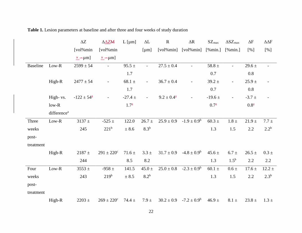
Baseline findings
Figure 1 shows the average mineral distributions of low- and high-R lesions at baseline, whereas
table 1 provides baseline data for all variables. All variables were found to be statistically
significantly different between lesion types at baseline. Percentage differences in variables
between low- and high-R lesions at baseline were smallest for ΔZbase (< 5 %). No statistically
significant difference in variables was observed between lesions assigned for the three or four
week study duration for each lesion type (data not shown). ΔZbase was found to only weakly
correlate with ΔFbase (r = 0.36).
Subject effects
Individual, mean subject responses (ΔΔZ) to the study treatment with respect to lesion type and
treatment duration are shown in table 2. Several observations can be made. There was a trend for
high-R lesions to remineralize, whereas low-R lesions tended to demineralize further. Low-R
lesions exhibited greater variability than high-R lesions which was also observed at lesion
baseline (data not shown). Subject 16 showed a very distinctive behavior which cannot be
sufficiently explained by a protocol deviation associated with this subject. Therefore, these data
were included in the analysis. For the interested reader, it is worth noting that exclusion of
subject 16 would have yielded a statistically significant change in ΔΔZ for high-R lesions at both
the three- and four-week time points. Other comparisons were unaffected by the in/exclusion of
this subject; likewise, the use of nonparametric statistics to reduce the influence of potential
outliers would have yielded similar results for all variables (data not shown).
Lesion type characterization
Figure 1 shows the average mineral distributions of low- and high--R lesions after the four-
week study duration in comparison to baseline. In the low-R lesions, a widening of the surface
11
zone and some remineralization in the original lesion body can be seen in addition to
demineralization beyond the original lesion. No changes in the maximum surface zone
mineralization can be observed. Furthermore, about half of the lesions showed lamination,
with some showing multiple laminations; a feature that was lost by showing average mineral
profiles. Figure 2 shows a representative microradiographic image of a low-R lesion after the
three-week study duration. The original lesion (OL) can be seen as well as the lamination
(Lam), separating the original lesion from the demineralization zone beyond the original lesion.
In contradiction to the results obtained on low-R lesions, remineralization occurred throughout
the entire lesion and including the surface zone in high-R lesions. Demineralization beyond
the original lesion can also be seen; however, this was predominantly due to the results
obtained for subject 16. Laminations occurred in only about 20 % of the high-R lesions. For
both lesion types, similar results were obtained after three weeks in comparison to the four
week study duration. Thus, three week results were omitted from figure 1 for better clarity.
Lesion type vs. treatment duration
Table 1 highlights results for all study variables by treatment duration and lesion type.
Statistically significant changes from baseline are highlighted. The lesion type – by – duration
interaction was not statistically significant for any of the outcomes (p = 0.13 for ΔΔZM, p = 0.20
for ΔL, p = 0.26 for ΔR, p = 0.36 for ΔSZmax, p = 0.31 for ΔΔF) so the lesion type comparisons
are valid for both durations, and the duration comparisons are valid for both lesion types.
Low- and high-R lesions had significantly different ΔΔZM (p = 0.0001). The numerical
difference in ΔΔZM between low- and high-R lesions was 816 at the three-week study duration
and increased further to 1227 after four weeks. High-R lesions had significantly less increase in
L (p = 0.0001), more decrease in R (p = 0.0001), more increase in SZmax (p = 0.0001), and less
decrease in ΔF (p = 0.0001) than low-R lesions. Worth pointing out is the discrepancy between
QLF and TMR measurements, as ΔΔF showed opposite results in relation to ΔMΔZ. The three
week duration had significantly less increase in L than the four week duration (p = 0.0347).
However duration did not significantly affect ΔΔZM (p = 0.09), ΔR (p = 0.12), ΔSZmax (p =
0.96), or ΔΔF (p = 0.11).
12
Side of mouth effect
Side of the mouth and position of specimen parcels in either distal or mesial position were also
examined to evaluate their influence on the study outcomes. The left side of the mouth had a
significantly more negative ΔΔZ (p = 0.0108) and significantly less increase in ΔL (p = 0.0024)
than the right side, while side did not significantly affect ΔR (p = 0.13), ΔSZmax (p = 0.42), or
ΔΔF (p = 0.53). Position of specimen parcels in either distal or mesial position did not
significantly affect any of the outcomes (p = 0.08 for ΔΔZ, p = 0.07 for ΔL, p = 0.83 for ΔR, p =
0.92 for ΔSZmax, and p = 0.21 for ΔΔF).
Correlation tests
Only weak correlations were found between ΔZbase and ΔΔZ for high- (r = – 0.236) and low-R (r
= – 0.30) lesions, respectively. The only high correlation found was between ΔΔZ and ΔL (r =
0.92). While several other correlations were statistically significantly greater than zero, none of
these correlations would be considered as indicating anything other than a weak association.
Discussion
The present in situ study aimed to gain a better understanding of the relative fluoride response of
caries lesions with different mineral distributions at baseline under physiologically
relevant conditions typical for the caries process. An established in situ caries model [Zero
et al., 2004] was used in the present study, with the key features being the use of gauze-
covered specimens to facilitate plaque growth and to simulate a caries prone
stagnation area. Furthermore, no diet restrictions were imposed on the study subjects, and
the twice-daily, one minute, 1100 ppm F dentifrice application resembled a typical oral
hygiene regimen. Therefore, this model/study can be considered of high clinical relevance.
Although model parameters have been refined over the years, leading to a better
understanding of the caries process, the impact of mineral distribution of caries lesions at
baseline remains poorly understood. Lynch et al. [2007] found in vitro that high-R lesions show
greater ability to remineralize than low-R lesions, which can possibly be attributed to
differences in porosity or SZmax with low-R lesions reaching inhibitory (for further
remineralization) SZmax

13
values faster than high-R lesions. Another possible factor which was discussed is the difference
in area of enamel per unit volume of remineralizing solution within the low- and high-R lesions.
The present study evaluated the behavior of these lesions under more dynamic in situ conditions,
and for their relative responsiveness to fluoride.
It must be mentioned that low- and high-R lesions were also found to differ in ΔZbase; therefore
somewhat compromising on the validity of study comparisons, especially considering that ΔZbase
was shown to directly impact the tendency of lesions to either de- or remineralize in situ
[Mellberg, 1991; Schäfer et al., 1992]. However, the difference in ΔZbase between lesion types
was relatively small (< 5%) and can be considered negligible, especially bearing in mind an
earlier reported measurement error for TMR alone of approximately 4% [de Jong and ten Bosch,
1985].
At similar ΔZbase, low-R lesions were deeper than high-R lesions, and low-R lesions also showed
a significantly greater surface zone mineralization in the present study. This was not surprising,
as the use of a surface-protective polymer and partially saturated (with respect to hydroxyapatite)
conditions during lesion creation allowed for better establishment of a surface layer. It is also
worth noting that R values of low-R lesions in the present study were considerably greater
compared to a previous, relevant study [Lynch et al., 2007] (consequently, differences in R
values were smaller between low- and high-R lesions in the present study). Although very
similar lesion creation protocols were used, the reason for this discrepancy is not clear. In fact,
the system used for creation of low-R lesions in the present study was less undersaturated with
respect to hydroxyapatite than the commonly used system [White, 1987] utilizing a “50%
saturation with respect to hydroxyapatite (HA)” (data not shown). Therefore, lower R values
were expected in the present study, considering our current understanding of artificial lesion
creation. However, this was not observed to be the case. Lynch et al. [2007] used bovine enamel
and created shallower lesions, thus making a comparison between studies not necessarily
straightforward.
The results obtained on high-R lesions are comparable to previous in situ studies on this lesion
type [e.g. ten Cate, 1993; Laheij et al., 2010]. Although the models used had inherent
differences, there was a general trend for remineralization as a result of daily fluoride treatments.
Almost identical post-treatment changes in mineral distribution of the lesions from baseline were
14
observed in the present (figure 1) compared to a very recent study [Laheij et al., 2010], highlighting
perhaps the “robustness” of this (very commonly used) lesion type.
The results obtained on low-R lesions, and especially in comparison to the high-R lesions, were intriguing.
Despite very similar ΔZbase for both lesions and virtually identical conditions, both lesion types exhibited
opposite behaviors – high-R lesions tended to remineralize, whereas low-R lesions further demineralized. This
was evident after the three-week study duration and further pronounced after four weeks. The differences in
structure and solubility between lesion types at baseline may provide an explanation for their contradictory
behaviors. Two aspects need to be discussed: the inherent solubility of the lesions after lesion creation on one
hand and differences in microstructure and the extent of demineralization in inter- and intra-prismatic enamel
and resulting consequences on solubility on the other hand.
While net demineralization is the result of lesion formation, remineralization or reprecipitation can still
occur within the lesion and potentially influence lesion characteristics during subsequent de- and
remineralization cycles. Enamel mineral has been described as a solid solution with appreciable
inhomogeneity [Shellis et al., 1993]. During lesion formation, there is preferential dissolution of the least
stable fractions, especially those associated with the impurities Mg and CO3 [LeGeros, 1990]. Within the
developing lesion, this can lead to levels of supersaturation which in turn will allow (re)precipitation of
mineral phases different to the original enamel mineral. Several different mineral phases have, therefore,
been postulated or shown to be present in caries lesions. Although there is still considerable debate in the
literature, brushite (BR or dicalcium phosphate dihydrate (DCPD) – CaHPO4 × 2H2O) [Moreno and
Zahradnik, 1974], monetite (MT – CaHPO4) [Featherstone et al., 1978] and Mg-substituted whitlockite
(WHM – Ca9Mg(HPO4)(PO4)6) [Shellis et al., 1997] have all been reported. This is not surprising, as BR and
MT are more stable than HA under acidic conditions [Brown, 1973]; whereas the mineral phase associated
with Mg in (sound) enamel was proposed to be WHM [Driessens and Verbeeck, 1982]. BR has also
been strongly associated with one of the mineral phases (among HA and F-HA) present in the lesion surface
layer [Moreno and Zahradnik, 1974; Margolis and Moreno, 1985]. Especially WHM has been associated
with the caries process, as several investigators were able to detect this mineral phase in natural caries
lesions [Aoba and Yagi, 1991; Kodaka et al., 1992], with the site of WHM formation being perhaps
close to the advancing front as there is preferential loss of Mg [Hallsworth et al., 1972]. Little is known

15
about F effects of Mg incorporation in WHM and HA. The sole study [LeGeros et al., 1992] reported
that F in small amounts caused less Mg incorporation in WHM, but greater Mg incorporation in HA
was observed, and that Mg did not affect F incorporation in HA.
It can only be speculated that under the highly unsaturated conditions employed during high-R lesion
creation, reducing the mineral content by more than half by volume in the lesion body and at the surface
(figure 1), the vast majority of soluble fractions including enamel impurities, such as Mg, Na, CO3, was
removed. This would render the remaining lesion relatively more robust to further demineralization, as little
F can be expected to have been removed during initial demineralization. The resulting lesion is therefore
likely to show a higher F concentration than the original, sound enamel (if calculated as F concentration per
weight). Furthermore, due to the initial infinite undersaturation present during lesion creation (at least
initially) and the relatively low pH, very little (re)precipitation of mineral phases can be expected. This is
also highlighted by the fact that the formed surface layer was not as pronounced as in the low-R lesions,
where lesion creation conditions were considerably different. Here, the partial saturation and higher pH give
rise to the assumptions that soluble fractions were only partially removed, or removed from deeper parts of
the lesion and then reprecipitated again closer to the solution-lesion interface. This would result in lesions
with considerably higher SZmax and a more even mineral loss throughout the lesion – or in other terms,
lesions with a lower R value. Low-R lesions present less or at least narrower “diffusion pores” than high-
R lesions, suggesting they are less prone to further dissolution than high-R lesions, assuming the
hypothesis for the differences in dissolution rates between (sound) deciduous and permanent enamel by
Poole et al. [1981]. However, the mineral in low-R lesions can be assumed to be considerably more soluble
than in high-R lesions and contain (more) non-apatitic mineral phases such as BR and WHM. This is
further complicated because not only porosity and solubility, but also structure are important factors in
the caries process, and these factors have been shown to be linked [Shellis, 1995].
Shellis and Wilson [2004] reported that enamel contained a very soluble fraction (approximately
14%), which is the fraction presumably lost in its entirety in high- but not necessarily in low-R
lesions. Initial dissolution was proposed to occur at prism junctions and inner enamel [Shellis et
al., 1993], with the solubility behavior thereafter controlled by the less soluble middle and outer
intraprismatic enamel, which make up the bulk of the enamel [Shellis, 1996]. Due to the different

16
mineral distribution profiles between low- and high-R lesions (figure 1), differences in the extent of
demineralization in intra- and perhaps also interprismatic enamel must exist between lesion types.
Furthermore, mineral phases that may have reprecipitated during lesion formation in low-R lesions may have
(artificially) increased the interprismatic fraction. Although the comparison to the present study is not exactly
straightforward, relatively greater lesion depths observed in deciduous vs. permanent enamel were attributed to
greater mean prism-junction density and mean volume fraction of interprismatic enamel in deciduous enamel
[Shellis, 1984]. This may provide another explanation for the observed differences in the present study.
Whether chemical and/or structural or other aspects were responsible for the contradictory behavior of low-
and high-R lesions in the present study remains to be determined. The results obtained in the present study
have shown that in situ caries lesion de- and remineralization are by no means straightforward and that with
the current knowledge no definitive explanation of the results can be provided. Furthermore, more research
should focus on de- and remineralization of established lesions and under more dynamic conditions, as most
studies focus on only one aspect and are therefore of limited clinical relevance.
In situ models can be, although rather crudely, divided into net de- or remineralization with the present model
been described as a net remineralization model [Zero, 1995]. In the present study, this discrimination is not
possible as net de- and remineralization were observed for the sole study treatment. In this context, the results
of the QLF analysis, which contradict the TMR findings (table 1) as no net demineralization was observed, must
be discussed. The reason for this discrepancy is not clear, especially as previous, comparative studies [Ando et
al., 2001; Fujikawa et al., 2008; Hafstrombjorkman et al., 1992] have shown good correlation between Δ(Δ)F
and ΔZ/ΔΔZ. These studies, however, were rather limited in their approach as only de- and/or remineralization
were studied, not taking into account caries dynamics. As little is known about the sensitivity of QLF to lesion
lamination or the impact of the R value on ΔF, further research is needed on this still relatively new de- and
remineralization evaluation technique.
In the present study, differences in response to the study treatment were observed between left and right test
sites of the mouth (regardless of in- or exclusion of subject 16), despite similar ΔZbase between sites for both
lesion types. These findings have been reported before [Mellberg et
17
al., 1992; Zero, 1995] and may be explained by chewing patterns and brushing preferences of the
study subjects, which in turn can lead to side differences in fluoride delivery.
Perhaps the most important question remaining is which of the two lesions is more clinically
relevant for the study of caries? Or can both lesions be used for the simultaneous assessment of
treatment effects on caries progression and reversal? Studies on natural white spot lesions [Iijima
et al., 1999; Iijima and Takagi, 2000; Lynch et al., 2007] indicated a relatively high degree of
surface zone mineralization for these lesions which is more comparable to the low-R lesions
employed in the present study. Likewise, the “pattern” of mineral distribution (but not
necessarily the R value) was more comparable to low- rather than high-R lesions. Lynch et al.
[2007] suggested that low-R lesions would be more appropriate where more physiologically
relevant mineral distribution is required, whereas high-R lesions would be appropriate for
studying inherent remineralization efficiency. It must be noted that natural white spot lesions
form over a considerably longer period of time than “laboratory lesions” (assuming similar
severity) and that they are subject to appreciably higher fluctuations in pH and F, Ca and Pi
concentrations during their development where laboratory lesions are not. Especially the
importance of lesion bound F deserves more attention as this may impact the inherent solubility
of the remaining lesion mineral the most. Perhaps the choice of lesion type in in situ research is
more determined by personal preference, as most investigators follow the same lesion creation
protocol for their studies as this will allow comparison between studies. However, the impact of
differences in mineral distribution at lesion baseline is therefore somewhat ignored. To expand
on this, it would be advantageous to study treatment effects also on sound and very early, surface
softened lesions in addition to the lesions used in the present study as this would provide
information on a more complete spectrum of study substrates. This would aid in the development
of treatment interventions specifically designed for lesions at different stages in the caries
process. However, one should not forget that in situ models are “only” models of in vivo caries,
and that observations in these models do not necessarily mirror the real life situation.
Further in situ studies on high- and low-R lesions should perhaps focus on determining the
relative effectiveness of fluoride in a dose-response manner. Studies on lesions with different
ΔZbase and R values may further our understanding of the relative importance of ΔZbase and R in
determining their responsiveness to fluoride. In addition, potential chemical and structural

18
differences between lesion types need to be studied and in comparison to natural white spot
lesions.
In conclusion, the present in situ study has shown that lesions of similar severity but different
mineral distribution showed different responses to fluoride – low-R lesions further
demineralized, whereas high-R lesions exhibited some remineralization.
Acknowledgements
We acknowledge and appreciate the diligent participation of our study subjects. We also
acknowledge the clinical and laboratory staff of the Oral Health Research Institute for their
essential contributions to the quality of the research.

19
References
Ando M, van der Veen MH, Schemehorn BR, Stookey GK: Comparative study to quantify
demineralized enamel in deciduous and permanent teeth using laser- and light-induced
fluorescence techniques. Caries Res 2001;35:464-470.
Aoba T, Yagi T: Crystallographic and structural alterations in the mineral phase of human
enamel with carious attacks. J Oral Pathol 1982;11:201-209.
Arends J, Dijkman T, Christoffersen J: Average mineral loss in dental enamel during
demineralization. Caries Res 1987;21:249-254.
Brown WE: Solubilities of phosphates and other sparingly soluble compounds; in Griffith EJ,
Beeton A, Spencer JM, Mitchell DT (eds): Environmental Phosphorus Handbook. New
York, Wiley, 1973, pp 203-238.
De Jong ED, Ten Bosch JJ: Error analysis of the microradiographic determination of mineral
content in mineralized tissue slices. Phys Med Biol 1985;30:1067-1076.
Driessens FCM, Verbeeck RMH: The probable phase composition of the mineral in sound
enamel and dentine. Bull Soc Chim Belg 1982;91:573-596.
Featherstone JD, Duncan JF, Cutress TW: Crystallographic changes in human tooth enamel
during in-vitro caries simulation. Arch Oral Biol 1978;23:405-413.
Hallsworth AS, Robinson C, Weatherbell JA: Mineral and magnesium distribution within the
approximal carious lesion of dental enamel. Caries Res 1972;6:156-168.
Iijima Y, Takagi O: In situ acid resistance of in vivo formed white spot lesions. Caries Res
2000;34:388-394.
Iijima Y, Takagi O, Ruben J, Arends J: In vitro remineralization of in vivo and in vitro formed
enamel lesions. Caries Res 1999;33:206-213.
Kodaka T, Debari K, Abe M: Hexahedrally based crystals in human tooth enamel. Caries Res
1992;26:69-76.
Koulourides T, Volker JF: Changes of enamel microhardness in the human mouth. Ala J Med
Sci 1964;1:435-437.
Laheij AMGA, van Strijp AJP, van Loveren C: In situ remineralisation of enamel and dentin
after the use of an amine fluoride mouthrinse in addition to twice daily brushings with amine
fluoride toothpaste. Caries Res 2010;44:184-190.

20
LeGeros RZ: Chemical and crystallographic events in the caries process. J Dent Res 1990;69
Spec No:567-574; discussion 634-566.
LeGeros RZ, LeGeros JP, Zheng R, Retino M: Magnesium incorporation in apatite and in
whitlockite fluoride effect. J Dent Res 1992;71:257.
Lynch RJM, Mony U, ten Cate JM: Effect of lesion characteristics and mineralising solution type
on enamel remineralisation in vitro. Caries Res 2007;41:257-262.
Margolis HC, Moreno EC: Kinetic and thermodynamic aspects of enamel demineralization.
Caries Res 1985;19:22-35.
Marinho VC: Cochrane reviews of randomized trials of fluoride therapies for preventing dental
caries. Eur Arch Paediatr Dent 2009;10:183-191.
Mellberg JR: Relationship of original mineral loss in caries-like lesions to mineral changes
insitu. Caries Res 1991;25:459-461.
Mellberg JR: Hard-tissue substrates for evaluation of cariogenic and anti-cariogenic activity in
situ. J Dent Res 1992;71(Spec Iss):913-919.
Mellberg JR, Petrou ID, Grote NE: Findings from an in situ thin-section sandwich model for
evaluating cariogenic and anti-cariogenic activity. J Dent Res 1992;71:850-855.
Moreno EC, Zahradnik RT: Chemistry of enamel subsurface demineralization in vitro. J Dent
Res 1974;53:226-235.
Poole DF, Shellis RP, Tyler JE: Rates of formation in vitro of dental caries-like enamel lesions in
man and some non-human primates. Arch Oral Biol 1981;26:413-417.
Schäfer F, Raven SJ, Parr TA: The effect of lesion characteristic on remineralization and model
sensitivity. J Dent Res 1992;71:811-813.
Shellis RP: Relationship between human enamel structure and the formation of caries-like
lesions in vitro. Arch Oral Biol 1984;29:975-981.
Shellis RP: A scanning electron-microscopic study of solubility variations in human enamel and
dentine. Arch Oral Biol 1996;41:473-484.
Shellis RP, Wahab FK, Heywood BR: The hydroxyapatite ion activity product in acid-solutions
equilibrated with human enamel at 37-degrees-c. Caries Res 1993;27:365-372.
Shellis RP, Wilson RM: Apparent solubility distributions of hydroxyapatite and enamel apatite. J
Colloid Interf Sci 2004;278:325-332.
21
Strang R, Damato FA, Creanor SL, Stephen KW: The effect of baseline lesion mineral loss on in
situ remineralization. J Dent Res 1987;66:1644-1646.
ten Cate JM: Patient selection and appliance design in intra-oral models. J Dent Res
1992;71(Spec Iss):908-910.
ten Cate JM: The caries preventive effect of a fluoride dentifrice containing triclosan and zinc
citrate, a compilation of in vitro and in situ studies. Int Dent J 1993;43:407-413.
ten Cate JM: In situ models, physico-chemical aspects. Adv Dent Res 1994;8:125-133.
ten Cate JM, Dundon KA, Vernon PG, Damato FA, Huntington E, Exterkate RAM, Wefel JS,
Jordan T, Stephen KW, Roberts AJ: Preparation and measurement of artificial enamel
lesions, a four-laboratory ring test. Caries Res 1996;30:400-407.
White DJ: Use of synthetic-polymer gels for artificial carious lesion preparation. Caries Res
1987;21:228-242.
Zero DT: In situ caries models. Adv Dent Res 1995;9:214-230.
Zero DT, Zhang JZ, Harper DS, Wu M, Kelly S, Waskow J, Hoffmam M: The remineralizing
effect of an essential oil fluoride mouthrinse in an intraoral caries test. J Amer Dent Assoc
2004;135:231-237.
22
Table 1. Lesion parameters at baseline and after three and four weeks of study duration
ΔZ
[vol%min
× ∙ µm]
ΔΔZM
[vol%min
× ∙ µm]
L [µm] ΔL
[µm]
R
[vol%min]
ΔR
[vol%min]
SZmax
[%min.]
ΔSZmax
[%min.]
ΔF
[%]
ΔΔF
[%]
Baseline Low-R 2599 ± 54 - 95.5 ±
1.7
- 27.5 ± 0.4 - 58.8 ±
0.7
- 29.6 ±
0.8
-
High-R 2477 ± 54 - 68.1 ±
1.7
- 36.7 ± 0.4 - 39.2 ±
0.7
- 25.9 ±
0.8
-
High- vs.
low-R
differencea
-122 ± 54a - -27.4 ±
1.7a
- 9.2 ± 0.4a - -19.6 ±
0.7a
- -3.7 ±
0.8a
-
Three
weeks
post-
treatment
Low-R 3137 ±
245
-525 ±
221b
122.0
± 8.6
26.7 ±
8.3b
25.9 ± 0.9 -1.9 ± 0.9b 60.3 ±
1.3
1.8 ±
1.5
21.9 ±
2.2
7.7 ±
2.2b
High-R 2187 ±
244
291 ± 220c 71.6 ±
8.5
3.3 ±
8.2
31.7 ± 0.9 -4.8 ± 0.9b 45.6 ±
1.3
6.7 ±
1.5b
26.5 ±
2.2
0.3 ±
2.2
Four
weeks
post-
treatment
Low-R 3553 ±
243
-958 ±
219b
141.5
± 8.5
45.0 ±
8.2b
25.0 ± 0.8 -2.3 ± 0.9b 60.1 ±
1.3
0.6 ±
1.5
17.6 ±
2.2
12.2 ±
2.3b
High-R 2203 ± 269 ± 220c 74.4 ± 7.9 ± 30.2 ± 0.9 -7.2 ± 0.9b 46.9 ± 8.1 ± 23.8 ± 1.3 ±
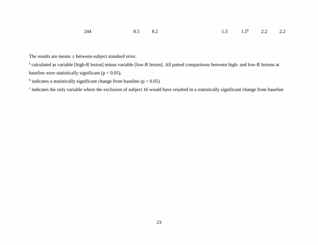
23
The results are means ± between-subject standard error. a calculated as variable [high-R lesion] minus variable [low-R lesion]. All paired comparisons between high- and low-R lesions at
baseline were statistically significant (p < 0.05). b indicates a statistically significant change from baseline (p < 0.05) c indicates the only variable where the exclusion of subject 16 would have resulted in a statistically significant change from baseline
244 8.5 8.2 1.3 1.5b 2.2 2.2

24
Table 2. Mean change in mineral loss (ΔΔZ; vol%min × µm) by subject, lesion type and treatment duration
high-R lesions low-R lesions Subject 3 wk 4 wk 3 wk 4 wk
1 600 270 n.s.a -225 2 540 740 -35 -255 3 540 120 -530 -1435 4 220 465 -140 15 5 340 195 -355 -1615 6 -110 140 -740 -725 7 655 475 -605 -650 8 370 990 165 205 9 810 1160 170 n.s. 10 535 975 -50 -1025 11 370 45 -200 -200 12 -160 270 -1335 -3915 13 435 710 -1060 -170 14 395 610 -830 -185 15 -1565 -3130 -2225 -3290 16 585 555 -670 -1345
a specimens did not yield sections suitable for TMR analysis

25
Figure Legends
Figure 1. Average mineral distributions for low- and high-R lesions at baseline and after the
four-week study duration.
Figure 2. Microradiographic image of a low-R caries lesion after the three-week study duration.
The original lesion (OL) and a clear lamination (Lam) are visible.


Recommended
![Fluoride toothpastes for preventing dental caries in ...neuron.mefst.hr/docs/katedre/znanstvena_metodologija/Fluoride... · [Intervention Review] Fluoride toothpastes for preventing](https://img.dokumen.tips/doc/110x75/5ac7a33f7f8b9aa3298b67ff/fluoride-toothpastes-for-preventing-dental-caries-in-intervention-review-fluoride.jpg)