Identification and functional characterisation of aquaporinsin the grapevine, Vitis vinifera
Megan C. SheldenA,B, Susan M. HowittC, Brent N. KaiserA and Stephen D. TyermanA,D
ASchool of Agriculture, Food andWine, University of Adelaide,Waite Campus, GlenOsmond, SA 5064, Australia.BPresent address: Australian Centre for Plant Functional Genomics, School of Botany, University of Melbourne,Parkville, Vic. 3010, Australia.
CBiochemistry and Molecular Biology, Research School of Biology, Australian National University, Canberra,ACT 0200, Australia.
DCorresponding author. Email: [email protected]
Abstract. Plant aquaporins belong to a large superfamily of conserved proteins called themajor intrinsic proteins (MIPs).There is limited information about the diversity of MIPs in grapevine, and their water transport capacity. The aim of thepresent study was to identify MIPs from grapevine and functionally characterise water transport of a subset of MIPs.Candidate genes were identified, by screening a Vitis vinifera L. (cv. Cabernet Sauvignon) cDNA library with gene specificprobes, for aquaporin cDNAs encoding members of the plasma membrane intrinsic protein (PIP) and tonoplast intrinsicprotein (TIP) subfamilies. The screen resulted in the identification of 11 full-length and two partial length aquaporin cDNAs.VvTIP2;1 isoforms had different 30 UTRs, immediately upstream of the poly(A) tail, suggesting the presence of multiplecleavage sites for polyadenylation. Using published genome sequences of grapevine, we conducted a phylogenetic analysisof the MIPs with previously characterised MIPs from Arabidopsis. We identified 23 full-length MIP genes from theV. viniferagenomesequenceof a nearhomozygous line (PN40024) that cluster into the fourmain subfamilies (andsubgroupswithin) identified in other species. However, based on the identification of PIP2 genes in Cabernet Sauvignon that were notpresent in the PN40024 genome, there are likely to be more than 23 MIP genes in other heterozygous grapevine cultivars.Water transport capacity was determined for several PIPs and TIPs, by expression in Xenopus oocytes. Only VvPIP2 andVvTIP proteins function as water channels with the exception of VvPIP2;5. VvPIP2;5 differs from the water conductingVvPIP2;1 by the substitution of two highly conserved amino acids in Loop B (G97S, G100W), which was shown byhomology modelling to likely form a hydrophobic block of the water pore.
Additional keywords: VvPIP1, VvPIP2, VvTIP, VvNIP, VvSIP, VvXIP.
Introduction
Grapevine is one of the worlds’ most economically importantfruit crops. It is the first fruit crop to have its genome fullysequenced (Jaillon et al. 2007; Velasco et al. 2007), and nowhas the potential to be a model organism for future studies offruit trees (Troggio et al. 2008). Although grapevines are ableto survive in a range of soil moisture conditions, their growthand yield is determined by their total water use (McCarthyet al. 2001), so it is important to identify and characterise themolecular components of water transport. The discovery andcharacterisation in plants of membrane intrinsic proteins (MIPs),some of which function as water permeable pores (aquaporins),has shown that aquaporins can have primary roles in regulatingplant water transport (Maurel et al. 2008). In grapevine, there arerelatively few studies characterising the MIPs in general, andidentifiying the roles of the MIPs in water transport (Baigeset al. 2001; Picaud et al. 2003; Vandeleur et al. 2009).
TheMIP superfamily can be divided into four subfamilies; theplasma membrane intrinsic proteins (PIPs), tonoplast intrinsicproteins (TIPs), nodulin-like intrinsic proteins (NIPs) and
the small basic intrinsic proteins (SIPs) (Johanson et al. 2001;Zardoya and Villalba 2001; Quigley et al. 2002; Forrest andBhave 2008). Two new subfamilies have recently been proposedin the moss Physcomitrella patens (Hedw.) B.S.G.; the GlypF-like intrinsic proteins (GIPs) (Gustavsson et al. 2005) and thehybrid intrinsic proteins (HIPs) (Danielson and Johanson2008). A third new subfamily called the x intrinsic proteins(XIPs) has recently been identified, with members in severaldicotyledonous plants including tomato (Lycopersiconesculentum L.; Sade et al. 2009) and grapevine (Danielson andJohanson 2008). The coding region of MIPs is typically between250 and 300 amino acid residues in length and they have amolecular mass between 26 and 34 kDa. Although the namingof the PIP and TIP groups imply a membrane location there arereports of their occurrence at different locations (Barkla et al.1999; Alexandersson et al. 2004; Whiteman et al. 2008).
The divergence of plant MIPs is believed to have occurredearly, with findings that MIPs in the moss, P. patens, fall into thesame subfamilies as in the angiosperms (Borstlap 2002). MIPgenes have been identified in over 30 plant species of both
CSIRO PUBLISHING
www.publish.csiro.au/journals/fpb Functional Plant Biology, 2009, 36, 1065–1078
� CSIRO 2009 10.1071/FP09117 1445-4408/09/121065

monocots and dicots in which they constitute a large gene family;35 members identified in Arabidopsis (Johanson et al. 2001), 31inmaize (ZeamaysL.; Chaumont et al. 2001), at least 35 inwheat(Triticum aestivum L.; Forrest and Bhave 2008) and 33 in rice(Oryza sativa L.; Sakurai et al. 2005). MIPs have been identifiedin woody plants such as grapevine (Baiges et al. 2001; Picaudet al. 2003; Fouquet et al. 2008; Vandeleur et al. 2009), walnut(Juglans regiaL.) (Sakr et al. 2003) and olive (Olea europaeaL.)(Secchi et al. 2007).
MIPs have a highly conserved structure, consisting of tandemrepeats of three membrane spanning a-helical domains. Eachtandem repeat has the highly conserved asparagine-proline-alanine (NPA) motif that forms the aqueous pore (Fu et al.2000). Loops B (cytoplasmic) and E (extracellular) form shorthydrophobic helices that correspond to two hemipores, dippinginto the membrane to form a single transmembrane aqueouspathway. Each monomer functions as a single water pore,although aquaporins are known to form both homotetramers(Engel et al. 2000) and heterotetramers in the membrane(Neely et al. 1999; Harvengt et al. 2000). The atomic structuresof several mammalian aquaporins selective for water have beendetermined, including AQP1 (Murata et al. 2000), AQP0(Gonen et al. 2004; Harries et al. 2004) and AQPZ fromEscherichia coli (Savage et al. 2003). A spinach plasmamembrane intrinsic protein, SoPIP2;1, has been crystallised inboth the open and closed conformation yielding new insightsinto the structural characteristics of plant aquaporins (Törnroth-Horsefield et al. 2006).
The transport selectivity of plant aquaporins has beencharacterised by expression in both Xenopus laevis oocytesand in yeast, and by reconstitution in proteoliposomes (Maurelet al. 2008).MIPs have a broad range of transport selectivity withvarious members permeable to water (Biela et al. 1999; Santoniet al. 2000; Tyerman et al. 2002), glycerol (Biela et al. 1999;Weig and Jakob 2000; Moshelion et al. 2002), urea (Gerbeauet al. 1999), CO2 (Uehlein et al. 2003), NH3 (Jahn et al. 2004;Loque et al. 2005), silicon (Ma et al. 2006) boron (Takano et al.2006; Tanaka et al. 2008) and arsenite (Kamiya et al. 2009). ThePIP2members have been shown to have highwater permeability,though there are exceptions (Zhou et al. 2007), and PIP1s havelow water permeability or no apparent permeability. In maize, allZmPIP2 proteins analysed show high water permeability andZmPIP1a and ZmPIP1b lowwater permeability (Chaumont et al.2000). The reason for this difference remains unclear, but theseinactive proteins were expressed in the plasma membrane ofoocytes, indicating correct targeting to the plasma membrane(Chaumont et al. 2000). Based on functional data in Xenopusoocytes, several authors have proposed that PIP1 and PIP2isoforms interact to form heterotetramers (Fetter et al. 2004;Mahdieh et al. 2008). Such an interaction has been observedwhen two grapevine isoforms, VvPIP1;1 and VvPIP2;2, areco-expressed in Xenopus oocytes (Vandeleur et al. 2009), butwas not observed for PIP1 PIP2 combinations from commonbean (Phaseolus vulgaris L.) (Zhou et al. 2007). Directinteraction of the two subgroups has been shown using FRETimaging in living maize cells overexpressing both these isoforms(Zelazny et al. 2007). High water channel activity has also beenobserved in somePIP1members includingNtAQP1 fromtobacco(Biela et al. 1999), BoPIP1b and BoPIP1;2 from Brassica
(Marin-Olivier et al. 2000) and the Arabidopsis aquaporins,AtPIP1a, AtPIP1b and AtPIP1c (Kammerloher et al. 1994).Two PIP1 aquaporins from grape berry show low waterpermeability but one (VvPIP1a) has been shown to transportglycerol (Picaud et al. 2003).
The aim of the present work was to identify and functionallycharacterise PIP and TIP aquaporins present in the grapevine,V. vinifera cv. Cabernet Sauvignon. During this study, two highquality draft genome sequences of grapevine were reported;a near-homozygous line PN40024, bred from V. vinifera cv.Pinot Noir (Jaillon et al. 2007), and a heterozygous grapevinevariety V. vinifera cv. Pinot Noir (clone ENTAV 115) (Velascoet al. 2007). Here, we present a complete analysis of the MIPsuperfamily in grapevine, based on sequences obtained from thesequencing of the grapevine genome, and cDNAs independentlyobtained in this study from Cabernet Sauvignon. The resultsshow a large diverse family of AQPs comprising four subgroups.Functional characterisation in Xenopus laevis oocytes of someof the aquaporins identified from Cabernet Sauvignon is alsopresented.
Materials and methodsPlant materialOne-year-old Vitis vinifera L. cv. Cabernet Sauvignon (cloneLC14) rootlings (Yalumba Nursery, Barossa Valley, SA,Australia), were planted in 12 inch pots containing a modifiedUC soil mix (61.5% (v/v) sand, 38.5% (v/v) peat moss, 0.50 gL–1
calcium hydroxide, 0.90 gL–1 calcium carbonate, 100 gL–1
Nitrophoska (12 : 5 : 14 N : P :K plus trace elements; IncitecPivot, Melbourne, Vic., Australia) at pH 6.8) and fertilised with0.08gL–1 per month of Osmocote Standard (Scotts Australia PtyLtd, Baulkham Hills, NSW, Australia). Plants were grown incontrolled temperature glasshouses maintained at 25�C day/20�Cnightwith extended light period provided bymercury halide lamps(14 h day/10 h night). Plants were watered to field capacity every2 days. Plant material was collected from the apical five nodesbetween 1000 and 1200 hours, frozen immediately in liquidnitrogen and stored at �80�C for RNA extraction.
Total and poly(A)+ RNA isolationTotal RNAwas extracted from200 to 500mgof young stem, leaf,tendril, petiole and root tissue using a 5M sodium perchlorateextraction buffer (5MNaCl04, 0.2MTris–HCl pH 8.3, 2% (w/v)SDS, 8.5% (w/v) polyvinylpolypyrrolidone (PVPP), 1% (v/v)b-mercaptoethanol). A modified protocol of the RNeasyExtraction Kit (Qiagen, Melbourne, Vic., Australia) was usedto purify total RNA (Franks et al. 2006). Poly(A)+ RNA waspurifiedusingOligo (dT)-cellulose spun columnchromatography(GE Healthcare, Piscataway, NJ, USA). The quality and purityof mRNA was checked on a denaturing agarose gel (1.2% (w/v)agarose, 1�MOPS, 8% (v/v) formaldehyde) before constructionof the cDNA library and by measuring the absorbance at260/280 nm.
Cloning of VvPIP2;1 and VvTIP1;1 by reversetranscriptase (RT)-PCR
One-step RT–PCR (Qiagen) was used to amplify VvPIP2;1 andVvTIP1;1 cDNAs from total RNA isolated from Cabernet
1066 Functional Plant Biology M. C. Shelden et al.

Sauvignon petiole tissue. RNA was treated with DNase 1(Ambion, Austin, TX, USA) before RT–PCR. PCR primersfor VitisPIP2;1 and VitisTIP3 (see Table S1 available as anAccessory Publication to this paper), were used to amplifyproducts using the following cycling conditions: 50�C for30min, 95�C for 15min, 35 cycles of 94�C for 30 s, 55�C for30 s, and 72�C for 1min, followed by cDNA extension at 72�Cfor 10min. PCR products were gel purified (QIAXII GelPurification Kit, Qiagen), cloned into pGEMT-easy (Promega,Alexandria, NSW, Australia) and transformed into E. coliXL1-B-cells. Transformants were selected using blue whitecolour selection and plasmid DNA purified (genelute plasmidpurification kit: Sigma Aldrich, Castle Hill, NSW, Australia).All cDNA inserts were sequenced using Dye Terminator 3(Applied Biosystems, Foster City, CA, USA) and analysed bythe Institute of Medical and Veterinary Sciences (Adelaide, SA,Australia).
Complementary DNA library construction
A grapevine cDNA library was constructed from a combinedmRNA pool consisting of individual mRNA extractions fromstems, leaves, tendrils, petioles and roots using the CloneminercDNA Library Construction Kit (Invitrogen, Melbourne, Vic.,Australia). The titre of the cDNA library inE.coliwas determinedto be 2.1� 107 colony forming units mL–1.
Macroarray synthesis
Individual E. coli transformants were spotted into 384 wellmicrotitre plates containing LB (supplemented with 7.5%glycerol, 50mgmL–1 kanamycin) using a VersArray ColonyPicker and Arrayer System (Biorad, Hercules, CA, USA).Microtitre plates were sealed in plastic bags, cells grown at37�C for between 20 and 24 h, covered with aluminium seal(AlumaSeal II, Excel Scientific, CA, USA) and stored at–80�C. Hybond N+ nylon membranes (GE Healthcare) wereplaced onto large plastic plates, spotted with six individual 384well plates containing cloned grapevine cDNAs. Membraneswere placed colony side up, onto large plates containingsolid LB media (supplemented with 50mgmL–1 kanamycin)and grown overnight at 37�C. Cells were lysed, DNAdenatured and fixed to the membrane following the protocolof Sambrook and Russel (2001). Membranes were air-driedfor 30min and DNA fixed using a UV transilluminator(GE Healthcare, Piscataway, NJ, USA).
Screening of cDNA library
Forward and reversePCRprimers (SigmaAldrich)weredesignedusingPrimer3 (http://frodo.wi.mit.edu/cgi-bin/primer3/primer3_www.cgi, accessed October 2003) to the 50 and 30 end of VitisRootstock Richter 110 aquaporin cDNAs and V. vinifera PinorNoir AQP cDNAs (Table S1).
Using the appropriate PCR primers, cDNA fragments wereamplified from purified plasmid cDNA library template with theDigoxygenin (DIG)-labelling PCR kit (Roche Diagnostics,Castle Hill, NSW, Australia) and TaqTi polymerase (FischerBiotech, Adelaide, SA, Australia). PCR cycling conditions wereas follows: 95�C for 15min for 1 cycle; 95�C for 30 s, 60�Cfor 30 s, 72�C for 1.5min for 35 cycles; extension at 72�C for
7min. DIG-labelled PCR products were separated by gelelectrophoresis, gel purified and cloned into pGEMT-Easy(Promega). Inserts were sequenced from both the 30 and 50 endto verify the amplified product. Equal amounts of eachDIG-labelled cDNA probe were combined and used togetherto screen the complete cDNA library under high stringencyconditions. Spotted cDNA membranes were prehybridised inDIG-Easy Hyb solution for 1 h at 42�C. The combined probeset was diluted in DIG-Easy Hyb solution, denatured at 100�Cfor 5min and then cooled quickly on ice. Denatured probewas added to the membranes and hybridised at 42�Covernight in a roller tube. Membranes were washed twice eachin the following solutions: low stringency wash buffer (2�SSC,0.1% SDS) at room temperature for 5min; high stringency washbuffer (0.1� SSC, 0.1% SDS) at 68�C for 15min; and 2�SSC.DIG-labelled DNA was detected by chemiluminescenceusing an anti DIG-alkaline phosphatase antibody and CDP star(Roche Diagnostics). Positive colonies were detected usingthe Molecular Imager ChemiDoc XRS System (Biorad) andImaGene 6.0 software (BioDiscovery, El Segundo, CA, USA)was used to locate and quantitate these signals on themacroarray.
Rapid amplification of cDNA endsRapid amplification of cDNA ends (RACE-PCR) was performedto amplify the 50 end of two incomplete aquaporin cDNAs.Primers were designed to amplify the 50 end of the partiallength aquaporin cDNAs, VvPIP2;3 and VvPIP1;4, using50 RACE-PCR (Table S1). The forward primer was designedin the multiple cloning site of pDONR222 (pDONR222F) andspecific reverse primerswere designed to the 50 end of each partiallength cDNA (VvPIP1;4R and VvPIP2;3R). 50 RACE-PCR wasperformed on the cDNA library template using high fidelityplatinum Taq polymerase (Invitrogen). PCR cycle for isolateVvPIP2;3 and VvPIP1;4 was as follows: 1 cycle of 95�C for2min; 35 cycles of 95�C for 30 s, 60.3�C for 30 s, 68�C for 1min;1 cycle of 68�C for 7 for min. PCR products were separated bygel electrophoresis, gel purified, cloned into pGEMT-Easy andtransformed intoE. coliXL1-B. Positives colonies were selected,plasmid DNA purified and then sequenced from both 50 and30 ends.
pGEMHE-DEST plasmid construction
The oocyte expression vector, pGEMHE (Liman et al. 1992)that contains the 50 and 30 b-globin untranslated regionsflanking the polylinker (Krieg and Melton 1984), was convertedto a Gateway destination vector using the gateway vectorconversion system (Invitrogen). pGEMHE was linearised withBamHI, end-filled using Klenow (Roche) and dephosphorylatedwith antartic phosphotase (NEB, Ipswich,MA,USA). The readingframe cassette A (RfA) was blunt end ligated to the linearisedpGEMHE vector and subsequently transformed into E. colistrain DB3.1. (Invitrogen). The reading frame cassette Acontains the chloramphenical resistance gene (CmR) and theccdB gene flanked by the attR1 and attR2 sites. Plasmidpreparations of putative pGEMHE/gateway transformantsresistant to antibiotic (chloramphenical, ampicillin) selectionwere digested with Bsr G1 (NEB) and sequenced with Gatewayforward and reverse primers to confirm conversion to a gateway
Characterisation of aquaporins in the grapevine Functional Plant Biology 1067

vector (Table S1). The resulting plasmid was designatedpGEMHE-DEST (Fig. S1 available as an Accessory Publicationto this paper).
Amplification of attB PCR products
AttB PCR primers were designed according to the GatewayTechnology Manual (Invitrogen) to amplify attB PCRproducts of VvPIP2;1 and VvTIP1;1 (Table S1). Pwo DNAPolymerase (Roche) was used to amplify attB/VvTIP1;1 byPCR from the cDNA library while AttB/VvPIP2;1 wasamplified from a previously cloned VvPIP2;1 cDNA. ThePCR cycling conditions were as follows: 94�C for 2min; 94�Cfor 15 s, 55�C for 30 s, 72�C for 1min cycled 10 times; 94�C for15 s, 55�C for 30 s, 72�C for 1min plus 5 s each cycle for15 cycles; and extension at 72�C for 7min. The attB PCRproducts were PEG purified (according to GatewayTechnology Manual) and recombined into pDONR222 usingBP Clonase (Invitrogen). The Gateway BP reaction facilitatedrecombination of the attB PCR products into pDONR222 tocreate attL entry clones of VvPIP2;1 and VvTIP1;1. LR Clonasewas used to recombine VvPIP2;1 and VvTIP1;1 into pGEMHE-DEST. Entry clones were digested and sequenced to confirmcorrect PCR product.
Cloning of AQP cDNAs into oocyte expression vector
Full-length V. vinifera aquaporin cDNAs identified in the libraryscreen were recombined into the pGEMHE-DEST vector usingthe LR recombination reaction (Invitrogen). The cDNA ofHsAQP1 from human red blood cells was obtained fromDaniel M. Roberts (University of Tennessee, Knoxville, TN,USA). HsAQP1 cDNA was cloned into the vector XbG-ev1between the 50 and 30 untranslated region (UTR) of the Xenopusb-globin cDNA (Preston et al. 1992). For cRNA synthesis,pGEMHE-DEST based vectors were digested with Nhe 1 at37�C overnight to linearise the plasmid, while HsAQP1XbG-ev1 plasmid was digested with Bam H1. ComplementaryRNA was transcribed using 1mg of linearised DNA with themMESSAGEmMachine Kit (Ambion) utilising the T7 promoterof pGEMHE, according to the manufacturers instructions.Phenol : chloroform : isoamyl alcohol (25 : 24 : 1 v/v) (SigmaAldrich) was used for extraction and isopropanol forprecipitation of the synthesised cRNA. cRNA was quantitatedand checked for purity using a spectrophotometer (Smartspec;Biorad) and by size separating on a denaturing RNA gel.
Harvesting oocytes
Oocytes were harvested from Xenopus laevis using the protocoldescribed by Hill et al. (2005). Following harvest, oocytes werewashed twice in cold Ca-free Ringers solution (96mM NaCl,2mM KCl, 5mM MgCl2; 5mM HEPES, pH 7.6), thendefolliculated in 1.7% (w/v) collagenase in Ca-free Ringerssolution for 90min with rotation. Oocytes were washed fivetimes in Ca-Ringers solution (96mM NaCl, 2mM KCl, 5mMMgCl2, 6mMCaCl2, 5mMHEPES, pH 7.6) to remove all tracesof collagenase. Oocytes were transferred to Ca-Ringers solutionsupplemented with antibiotics (100mgmL–1 tetracycline-HCl,100 unitsmL–1 penicillin, 100mgmL–1 streptomycin) and kept at18�C overnight. Defolliculated oocytes were microinjected with
the Nanoject II Auto-Nanolitre Injector (Drummond Scientific,Broomall, PA, USA) into the cytoplasm with 50 ng of cappedcRNA or with DEPC treated water (control) and incubatedfor three days at 18�C in Ca-Ringers solution supplementedwith antibiotics 100mgmL–1 tetracycline-HCl, 100 unitsmL–1
penicillin and 100mgmL–1 streptomycin.
Oocyte swelling and acidification assayOocyteswere preincubated at room temperature in an iso-osmoticsolution (Ca-Ringers solution) for 5min, 64–72 h after cRNAinjection, and then transferred to a hypoosmotic solution(Ca-Ringers solution diluted 5-fold with sterile water) at whichtime swellingwasmeasured for 2min. Oocyteswere viewedwitha Nikon SMZ800 light microscope with Vikam colour camera at2� magnification and imaged with Global Laboratory Image/2software (Data Translation, Marlboro, MA, USA). Images wereacquired every 4 s for 2min. Osmotic permeability (Pf) wascalculated for water injected and cRNA injected oocytes fromthe initial rate of change in relative volume (dVrel/dt)i calculatedfrom projected images assuming the oocytes were spherical:
Pf ¼ Vi � ðdV rel=dtÞiAi � Vw � DCo
; ð1Þ
whereVi andAi are the initial volume and area, respectively,Vw isthe partial molar volume of water; and DCo is the change inexternal osmolarity. At least two independent experiments with5–8 oocytes each were conducted for each gene. Osmolarity ofeach solutionwas determined using a vapour pressure osmometer5500 (Wescor, Logan, UT, USA). Cytosolic pH was loweredusing an external solution of 50mM Na-acetate at pH 5 inCa-Ringers solution (Tournaire-Roux et al. 2003). Controlscontained 50mM Na acetate at pH 7. Swelling assays wereconducted as above with oocytes incubated in 50mMNa-acetate pH 5 solution for 5min to allow acidification of theoocyte cytosol. Oocytes were then transferred to a hypotonicsolution (five times diluted with MilliQ water) and swellingimaged for 2min.
Modelling of AQP
Homology modelling was used to generate a predicted structurefor the VvPIP2;5 protein. The model was generated usingSWISS-MODEL (Schwede et al. 2003) with the structure ofthe open conformation of SoPIP2;1 (PDB number 285F) selectedas a template. Pictures were generated with Deep View-SwissPDB viewer.
Sequence analysis
Sequences identified in the library screenwere analysed using theBLASTn (nucleotide) and tBLASTn (translated nucleotide)algorithms at National Centre for Biotechnology Information(Altschul et al. 1997). MacVector 9.0 (Oxford Molecular Ltd,Oxford, UK) was used to translate sequences and ClustalW wasused to generate multiple sequence alignments using blosum30matrix and slow mode. The open gap penalty and extended gappenalty was set at 10.0 and 0.05, respectively. Phylogeneticanalysis was performed using a neighbour joining analysis ofthe deduced amino acid sequences (bootstrap 100 repetitions)usingMEGALIGN4.0.All novel aquaporin sequences identified
1068 Functional Plant Biology M. C. Shelden et al.
were submitted to GenBank (accession numbers shown inTable 1).
Results
Cloning of AQP cDNAs
A total of ~30 000 E. coli transformants containing grapevinecDNAs were screened under high stringency conditions for PIPand TIP cDNAs. Thirty colonies were identified based on initialDIG hybridisation with the combined DIG-labelled aquaporinprobe set. Each positive colony was regrown in liquid culture,plasmid purified and digested to release the cDNA insert.Plasmids containing cDNAs greater than ~300 bp weresequenced. Sixteen cDNAs were positively identified as beingmembers of the MIP super family by BLASTn searches usingGenBank. Of these eight were found to be full-length cDNAs andeightwere identified as partial length cDNAs. The cDNAs rangedin size from 278 bp to 1216 bp. The sequences were comparedwith previously identified MIP genes from both grapevineand Arabidopsis thaliana and were annotated according to thenomenclature set out by Johanson et al. (2001). Multiple cDNAisolates, encoding VvPIP1;2, VvPIP2;4 and VvTIP2;1, wereidentified in the library screen indicating degeneracy in thelibrary and potentially high expression of these genes withinthe plant. Of the cDNAs identified, eleven were found to benovel grapevine aquaporins. Two aquaporin cDNAs notisolated in the screen VvPIP1;1 and VvPIP1;3, were identifiedfrom sequencing of PCR generated probes. These have beenincluded in the bioinformatics analysis, but were not functionallycharacterised in Xenopus oocytes.
Full-length sequences were obtained for two partial cDNAsusing 50 RACE-PCR. Primers designed to the multiple cloningsite of pDONR222 and the 50 end of VvPIP2;3 and VvPIP1;4partial clones successfully amplified major products from thecDNA library at the expected size of 500 bp and 600 bp,respectively. These fragments were sequenced and aligned tothe corresponding partial cDNA, primers designed to the 50 and30 endof the full-length fragment and subsequently amplifiedwithPCR. The full-length cDNA of VvPIP2;3 is 1225 bp, encoding a
polypeptide of 280 amino acids, while the cDNA of VvPIP1;4 is1061 bp, encoding a PIP1 aquaporin, 287 amino acids in length.In total, 13 putative aquaporin PIP andTIP cDNAswere obtained(Table 1). Eleven cDNAs were submitted to GenBank anddesignated an accession number (Table 1).
Bioinformatics
The completion of two draft sequences of theV. vinifera genome,clone PN40024 (Jaillon et al. 2007) and Pinot Noir ENTAV 115(Velasco et al. 2007), has made it possible to identify putativeMIP genes in the grapevine genome. Currently, there are83 grapevine related MIPs annotated in GenBank, 73 from thewine grape V. vinifera (cvv. Syrah, Cabernet Sauvignon, PinotNoir and Nebbelio), eight from the rootstock Richter-110 (Vitisberlandieri�Vitis rupestris) and one from the Chinese wildgrape (Vitis pseudoreticulata). By using BLAST searches inGenBank, we identified 29 putative MIP genes in grapevine(clone PN40024): 23 of these encoded full-length proteins andsix encoded partial MIP proteins (Table 2). Nineteen putativeaquaporin genes (15 of these are full-length) were also identifiedin the heterozygous Pinot Noir genome (Table S2), including aputative gene encoding a XIP protein (Danielson and Johanson2008). Several genes encode partial proteins,missing one ormoretransmembrane domain, indicating these may be non-functionalpseudogenes, or misannotated from the genome sequencingproject.
All full-length PIP2 cDNAs identified in Cabernet Sauvignonare ~1200 bp in length and when compared at the nucleotidelevel show between 69 and 99% homology. We observedVvPIP2;4 and VvPIP2;5 are 99% homologous at the nucleotidelevel, and VvPIP2;1, VvPIP2;4 and VvPIP2;5 are 99%homologous at the amino acid level. VvPIP2;1 and VvPIP2;4differ by a conservative amino acid substitution at position193 from arginine to lysine. Similarly, VvPIP2;1 and VvPIP2;5were found to be highly similar with only two amino acidsubstitutions, Gly 97 Ser and Gly 100 Trp, both of whichare located in the highly conserved loop B. VvPIP2;3 andVvPIP2;2 are 77 and 80% homologous to VvPIP2;1,respectively. The PIP1 cDNAs are ~1100 bp in length andbetween 68 and 99% homologous. VvPIP1;2 and VvPIP1;4are 99% homologous at both the nucleotide and at the aminoacid level with only two amino acid differences in the codingregion, Ile 247 Val and Arg 250 Lys.
Five cDNA isolates from Cabernet Sauvignon were found tohave identical nucleotide sequences in their coding region. Thededuced amino acid sequence of these cDNAs encode theprotein, VvTIP2;1. Interestingly, the only difference observedbetween the isolates was in the 30 untranslated region. Twoisolates were both found to have a 15bp deletion in the 30 UTR,immediately upstream of the poly(A) tail, compared with theother three isolates. Analysis of other Vitis TIP2;1 homologueson the database, show that several these also have deletions in the30 UTR immediately upstream of the poly(A) tail suggesting thepresence of different cleavage sites for polyadenylation. VvTIP2;1is 72% identical to VvTIP1;1 at the nucleotide level and 72% atthe amino acid level. Multiple cDNA isolates of VvPIP1;2 andVvPIP2;4 were also identified in the library screen, however, nosequence differences were observed in their 30-UTR.
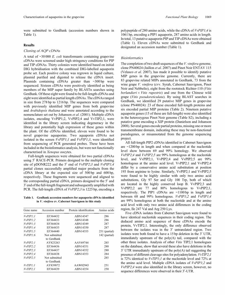
Table 1. GenBank accession numbers for aquaporin cDNAs identifiedin V. vinifera cv. Cabernet Sauvignon in this study
Gene name Accession number Protein identification Amino acids
VvPIP1;1 EF364432 ABN14347 286VvPIP1;2 EF364433 ABN14348 286VvPIP1;3 EF364434 ABN14349 287VvPIP1;4 EF364435 ABN14350 287VvPIP1;5 EF364440 ABN14355 251 (partial)VvPIP1;6 Not submitted
to GenBank142 (partial)
VvPIP2;1 AY823263 AAV69744 285VvPIP2;2 EF364436 ABN14351 280VvPIP2;3 EF364437 ABN14352 280VvPIP2;4 EF364438 ABN14353 284VvPIP2;5 Not submitted
to GenBank285
VvTIP1;1 AY839872 AAW02943 251VvTIP2;1 EF364439 ABN14354 250
Characterisation of aquaporins in the grapevine Functional Plant Biology 1069

To correctly annotate the grapevine MIPs, multiplealignments of translated sequences were generated withClustalW and compared with Arabidopsis MIPs. Evolutionaryhistory was inferred using the neighbour-joining method. Thesewere subsequently annotated according to the nomenclatureproposed by Johanson et al. (2001). The full-length grapevine
MIPs range in size from 236 to 354 amino acids (Table 2). Allgrapevine MIPs contain the conserved NPA motif in loops Band E, with the exception that in several of the NIPs, the alaninecan be replaced by a Ser or Val (Table S3). In VvSIP2 the alaninein loop B is replaced by leucine. The 23 grapevine MIPs clusterinto the four distinct MIP subfamilies: PIPs, TIPs, NIPs and SIPs
Table 2. Grapevine aquaporin genes identified in Vitis vinifera cv. Pinot Noir (clone PN40024)
Gene name Locus Chromosomenumber
Proteinaccession
Aminoacids
Locus tag Notes Comparison withFouquet et al. (2008)
PIPsVvPIP1;1 CU459265 13 CAO41326 286 GSVIVT00029248001VvPIP1;3 CU459322 2 CAO62835 287 GSVIVT00000433001VvPIP1;4 CU499257 15 CAO39626 286 GSVIVT00026881001 Annotated as
VvPIP1;2.VvPIP1;4 is onphylogenetic treebut not in the figurelegend
VvPIP1;5 CU459257 15 CAO39627 286 GSVIVT00026882001 Not on phylogenetictree but in figurelegend
VvPIP1 CU459376 12 CAO67902 250(partial)
GSVIVT00005839001 Truncated MIP.N-terminal protein ofunknown function
Missing from Fouquetanalysis
VvPIP2;2 CU459225 3 CAO47394 279 GSVIVT00036133001VvPIP2;3 CU459246 8 CAO18152 267 GSVIVT00023192001VvPIP2;4 CU459220 6 CAO21844 280 GSVIVT00024536001 Labelledon treebut not
in figure legend
TIPsVvTIP1;1 CU459234 13 CAO69259 251 GSVIVT00018548001VvTIP1;2 CU459323 8 CAO63006 251 GSVIVT00000605001VvTIP1;3 CU459242 6 CAO16745 251 GSVIVT00022146001VvTIP1;4 CU459220 6 CAO21720 252 GSVIVT00024394001VvTIP2;1 CU459224 9 CAO45860 249 GSVIVT00034350001VvTIP2;2 CU462185 Undetermined CAO23095 250 GSVIVT00012703001VvTIP2;3 CU460008 Undetermined CAO50033 126
(partial)GSVIVT00005226001 C-termini only Missing from Fouquet
analysisVvTIP3;1 CU459227 16 CAO62035 259 GSVIVT00013854001VvTIP4;1 CU459223 4 CAO44039 253 GSVIVT00032441001VvTIP5;1 CU459268 Undetermined CAO42713 249 GSVIVT00029946001VvTIP5;2 CU459236 15 CAO70596 262 GSVIVT00019170001
NIPsVvNIP1;1 CU459298 10 CAO48005 262 GSVIVT00035815001VvNIP2;1 CU461115 Undetermined CAO15462 201
(partial)GSVIVT00011149001 Missing first two TMD.
Starts in loop B.Misannotated asNIP4;1
VvNIP3;1 CU459243 14 CAO17108 270(partial)
GSVIVT00022377001 Missing Loop E and lastTMD.C-terminalTetrapyrole Methylase
VvNIP4;1 CU459401 Undetermined CAO70192 246 GSVIVT00007127001 Misannotated asVvNIP8;1
VvNIP4;2 CU459730 Undetermined CAO43338 217(partial)
GSVIVT00003903001 Missing the first two TMD Misannotated asVvNIP8;2
VvNIP5;1 CU459322 2 CAO62847 298 GSVIVT00000446001VvNIP6;1 CU459286 Undetermined CAO45476 354 GSVIVT00033750001VvNIP7;1 CU459219 5 CAO71103 293 GSVIVT00019910001
SIPsVvSIP1;1 CU459251 8 CAO23510 70
(partial)GSVIVT00025504001
VvSIP2;1 CU459251 8 CAO18284 236 GSVIVT00023346001
1070 Functional Plant Biology M. C. Shelden et al.

in a similar fashion to that of Arabidopsis (Figs 1, 2). The PIPmembers show divergence into the two subgroups, PIP1 andPIP2, and the grapevine TIPs cluster intofive groups, TIP1, TIP2,TIP3, TIP4 and TIP5 (Fig. 2). Several grapevine aquaporinscluster on one branch (VvPIP2;1, VvPIP2;4 and VvPIP2;5)separate to those identified in Arabidopsis which form separatebranches, respectively. This multiplicity of highly relatedaquaporins has been seen previously for both monocots(Chaumont et al. 2001; Sakurai et al. 2005) and dicots(Johanson et al. 2001).
Structural characteristics and conserved motifs
AllMIPmembers identified inV.vinifera cv.CabernetSauvignonshow characteristic sequence motifs found throughout the MIPsuperfamily.Multiple sequence alignments of the deduced amino
acid sequences, performed using ClustalW, show the highhomology between the PIP and TIP members identified in thisstudy (Fig. 3). The PIP1 members show an extended N-terminaltail of 14 amino acids compared with the PIP2 members.Conversely, the PIP2 members have an extended C-terminaltail comprising eight amino acids. TIP members have a muchshorter N-terminal tail than all the PIPs. Several residues arecompletely conserved in all grapevine PIP and TIP cDNAsidentified, with particularly high conservation found in thefunctional loops B and E. Several of these residues are alsoconserved in loops B and E of mammalian aquaporins (Murataet al. 2000). Hydropathy analysis of VvPIP1;1, VvPIP2;1 andVvTIP1;1 identified six transmembrane a spanning domainsthat are separated by hydrophilic loops (data not shown). TheC- andN termini were predicted to be cytoplasmic and the highlyconserved hydrophobic NPA motifs were in the loop betweenTMD1andTMD2, designated loopB, and loopEbetweenTMD5and TMD6.
Protein similarity betweenVitis species and cultivars is greaterthan 98% for all proteins identified (with the exception of thePIP2;3 homologue fromPN40024) indicating early divergenceofthe PIP and TIP genes within the genus Vitis. Closely relatedproteins (with homology >98%) have most likely arisen fromrecent gene duplications, as has been reported in other speciessuch as maize (Chaumont et al. 2001). The PIP2;3 homologuefrom PN40024 appears to be missing 20–30 amino acids andtherefore may be annotated incorrectly.
Functional analysis
The functionality of selected full-length V. viniferaAQP cDNAswas assessed by expression in Xenopus oocytes. Swelling wasmeasured in response to bathing in hypo-osmotic solution,~3 days after injection with cRNA. The water permeability(Pf) was calculated and compared with the water-injectedoocytes (control) (Fig. 4a). HsAQP1, from human red cells,was used as a positive control. The mean Pf value for AQP1was 22� 4� 10�3 cm s–1. The mean Pf of VvPIP2 proteinsranged from between 8.0 and 27.0� 10�3 cm s–1. The Pf valuefor VvPIP2;5 was 0.6� 10�3 cm s–1, higher than the waterinjected control 0.1� 10�3 cm s–1 (although not significantP> 0.05). PIP1 proteins (VvPIP1;2 and VvPIP1;4) alsoshowed low permeability with no significant differencecompared with the water injected controls, 0.5� 10�3 cm s–1.The Pf value for VvTIP2;1 was 9.0� 5.0� 10�3 cm s–1, butVvTIP1;1 was significantly lower at 5.0� 2.0� 10�3 cm s–1.
A highly conserved His residue in loop D (His197) has beenimplicated in channel gating under anoxic conditions in anArabidopsis PIP2 aquaporin (Tournaire-Roux et al. 2003). Thecorresponding His residue (His196) is conserved in all VvPIPaquaporins identified. In order to check the effect of acidiccytosolic pH on regulation of selected Vitis aquaporins,oocytes expressing HsAQP1, VvPIP2;1, VvTIP1;1 and H2Oinjected controls, were acid stressed with a weak acid at lowpH. A significant decrease in Pf from 9.0� 1.8� 10�3 to2.0� 1.0� 10�3 was observed for VvPIP2;1 when exposed toNa-acetate pH5, comparedwithNa-acetate pH7 (P < 0.001). ThePf value of VvTIP1;1 was unaltered. HsAQP1 showed a 2-foldincrease in Pf when exposed to Na-acetate pH 5 solution,compared with the pH 7 control solution (P < 0.001).
VvPIP1;4
VvPIP2;3
VvTIP1;2
VvNIP4;1
VvTIP4;1
VvPIP1;3VvPIP
1;1
VvPIP1;2
VvNIP5;1
VvNIP6;1
VvNIP7;1
VvNIP1;1
VvTIP5;1
VvTIP2;2
VvTIP5;2
VvPIP2;
4
VvPIP2;2
VvTIP3;1
VvTIP2;1
PIP
VvTIP1;3
VvTIP1;1
VvTIP1;4
NIP
TIP
SIPVvSIP2;10.1
VvPIP1;4
VvPIP2;3
VvTIP1;2
VvNIP4;1
VvTIP4;1
VvPIP1;3VvPIP
1;1
VvPIP1;2
VvNIP5;1
VvNIP6;1
VvNIP7;1
VvNIP1;1
VvTIP5;1
VvTIP2;2
VvTIP5;2
VvPIP2;
4
VvPIP2;2
VvTIP3;1
VvTIP2;1
PIP
VvTIP1;3
VvTIP1;1
VvTIP1;4
NIP
TIP
SIPVvSIP2;1
Fig. 1. Phylogenetic analysis of putative full-length MIPs identified fromhomozygous PN40024 genotype of Vitis Vinifera L. cv. Pinot noir (Jaillonet al. 2007). ClustalW (MacVector 9.0) was used to perform multiplesequence alignments of the deduced amino acid sequences. Phylogeneticanalyses were conducted in MEGA4 (Tamura Dudley et al. 2007) where theevolutionary history was inferred using the Neighbour-Joining method(Saitou and Nei 1987). The evolutionary distances were computedusing the JTT matrix-based method (Jones Taylor et al. 1992) and are inthe units of the number of amino acid substitutions per site. All positionscontaining alignment gaps and missing data were eliminated only in pairwisesequence comparisons (pairwise deletion option). Accessions numbersare as follows: VvPIP1;1 (CAO41326), VvPIP1;3 (CAO62835), VvPIP1;4(CAO39626), VvPIP1;5 (CAO39627), VvPIP2;2 (CAO47394),VvPIP2;3 (CAO18152), VvPIP2;4 (CAO21844), VvTIP1;1 (CAO69259),VvTIP1;2 (CAO63006), VvTIP1;3 (CAO16745), VvTIP1;4(CAO21720), VvTIP2;1 (CAO45860), VvTIP2;2 (CAO23095), VvTIP3;1(CAO62035), VvTIP4;1 (CAO44039), VvTIP5;1 (CAO42713),VvTIP5;2 (CAO70596), VvNIP1;1 (CAO48005), VvNIP4;1(CAO70192), VvNIP5;1 (CAO62847), VvNIP6;1 (CAO45476), VvNIP7;1(CAO71103) and VvSIP2;1 (CAO71103).
Characterisation of aquaporins in the grapevine Functional Plant Biology 1071

Discussion
Identification and phylogenetic analysis of grapevineaquaporins
The screening of a V. vinifera cv. Cabernet Sauvignon cDNAlibrary for PIP and TIP cDNAs has resulted in the identificationof 13 cDNAs, 11 of these encode full-length proteins and twoencode partial proteins. These were identified as members of theMIP superfamily based on homology to other plant aquaporins.
Of these cDNAs, five are PIP2 aquaporins, six are PIP1 and twoare TIP aquaporins. Using the high quality draft genomicsequence of the homozygous V. vinifera Pinot Noir clonePN40024 (Jaillon et al. 2007), we identified 23 full-lengthgenes encoding MIP proteins and have designated these intotheir respective subgroups: the PIPs, TIPs, NIPs and SIPs. Sixpartial sequences were also identified in the grapevine genome,one PIP, one TIP, three NIPs and one SIP. Based on ourbioinformatics and phylogenetic analysis, we have proposed
Fig. 2. Phylogenetic analysis of the PIP and TIP subfamilies from grapevine andArabidopsis. ClustalW (MacVector 9.0) was usedto perform multiple sequence alignments of the deduced amino acid sequences and the phylogenetic tree was generated using theneighbour joiningmethod (see Fig. 1). HsAQP1was used as the outgroup. TheMIP cDNAs identified in this study are in bold. Onlyamino acid sequences of full-length cDNAswere included in the phylogenetic analysis. Accession numbers are shown in brackets forprotein sequences obtained for clone PN24004.
1072 Functional Plant Biology M. C. Shelden et al.

different nomenclature for several the PN40024 genes(Table 2) compared with a previous study (Fouquet et al.2008). Furthermore, Danielson and Johanson (2008) recentlyreported a gene encoding aXIPprotein in the genome sequence ofPinot Noir (ENTAV 115). No members of the XIP family havebeen found in Arabidopsis (Danielson and Johanson 2008), or inthe grapevine genome of PN40024.
Using the genomic sequence as a reference we believe wehave isolated the majority of PIP genes found in CabernetSauvignon. The PIPs identified in this study are highlyhomologous at both the nucleotide and amino acid level. Thisis characteristic of PIP aquaporins with very high homologiesfound in other species such as Arabidopsis (Johanson et al. 2001;Quigley et al. 2002),Brassica (Marin-Olivier et al. 2000), walnut(Sakr et al. 2003), maize (Chaumont et al. 2001), rice (Sakuraiet al. 2005) and wheat (Forrest and Bhave 2008).
A phylogenetic analysis of the PIPs from the monocot maizeand the dicot Arabidopsis, indicate that the divergence of theseaquaporin genes occurred after the monocot – dicot divergence(Chaumont et al. 2001).Comparison of theV. vinifera aquaporinswith Arabidopsis aquaporins clearly shows that the divergence
of plant aquaporins into the four subfamilies occurred earlyin evolution (Danielson and Johanson 2008). The grapevineaquaporins segregate onto distinct branches within eachsubfamily indicating that highly homologous genes most likelyarose from recent gene duplications. An example of this is thehigh homology that exists between the cDNAs for VvPIP2;1,VvPIP2;4 and VvPIP2;5. Note that VvPIP2;1 and VvPIP2;5are missing from the PN40024 sequence (Fig. 1) but VvPIP2;4is present and may perform the same function as the missinggenes. This high nucleotide homology has been seen foraquaporins in other species such as maize, where ZmPIP1–3and ZmPIP1–4 have 98% nucleotide homology and encodeidentical proteins. In maize, these genes were also shown tohave a similar transcript distribution pattern, and the authorshypothesised that the role of having two genes encodingidentical proteins may be to increase the expression levels ofthe duplicated genes (Chaumont et al. 2001). VvPIP1;2 andVvPIP1;4 are 99% identical at the nucleotide level and 100%similar at the amino acid level. The transcript abundance andtissue location in grapevine of each of these isoforms is currentlyunknown.
Fig. 3. Alignment of the deduced amino acid sequences of the 11 full-length putative V. vinifera aquaporin cDNAs identified from Cabernet Sauvignon.ClustalW (MacVector 9.0) was used to generate multiple sequence alignments using blosum30matrix with an open gap penalty of 10 and extended gap penaltyof 0.05. The black bars indicate the position of the six transmembrane spanning domains. The dark shaded boxes show identical residues and the light shadedboxes show similar residues in over 50% of sequences. The consensus sequence is shown below the cDNAs. The black box highlights the NPAmotif conservedin all sequences.
Characterisation of aquaporins in the grapevine Functional Plant Biology 1073

The V. vinifera TIP aquaporins have lower homologies thanobserved for the PIP aquaporins. This has been seen previouslyin both Arabidopsis (Johanson et al. 2001) and maize TIPs(Chaumont et al. 2001). The full-length cDNA of VvTIP2;1was independently identified in the library screen five times,indicating that this gene is most likely highly expressedthroughout the plant. The Arabidopsis homologue d-TIP(renamed AtTIP2;1), has been shown to be highly expressedin the leaves (Alexandersson et al. 2005). Further, two of theVvTIP2;1 cDNAs identified had a 15-bp deletion at the 30 end ofthe cDNA, immediately before the poly(A) tail. Differences inboth the 30 non-coding region and the length of the poly(A)tail have been reported previously for Vitis PIP1b cDNAs(Picaud et al. 2003). Plant polyadenylation cleavage sites havea consensus signal which comprises a pyrimidine/adenosinedinucleotide (YA) within a U rich region (Hunt 1994). Thepresence of multiple cleavage sites in the 30 UTR can result inthe addition of the poly(A) tail at different positions. This mayaffect the stability of the transcript, translation of the protein and
trafficking of the transcript to the cytoplasm, and ultimately maylead to changes in protein activity.
Five full-length grapevine NIPs were identified in thegrapevine genome of V. vinifera clone PN40024. Phylogeneticanalyses show the NIPs are more divergent than both thegrapevine PIP and TIP aquaporins as has been reportedpreviously for other plants (Wallace et al. 2006). The NIPscan be divided into two subgroups: group I (NIP1, NIP2, NIP3and NIP4) and group II (NIP5, NIP6 and NIP7) based on solutepermeability and the structure of selectivity-determining ar/Rfilter (Wallace et al. 2006). We propose that NIP4;1, NIP8;1 andNIP8;2, previously reported by Fouquet et al. (2008) should berenamed as VvNIP2;1, VvNIP4;1 and VvNIP4;2, respectively.Functional characterisation of plant NIPs have shown theyare permeable to a broad range of small solutes (Wallace et al.2006), however, to date, none of the grapevine NIPs have beenfunctionally characterised.
The SIPs comprise the smallest subgroup and are moredivergent,than the other MIPs (Chaumont et al. 2001;Johanson et al. 2001) in particular the NPA motif in loop B isnot conserved and instead consists of the motif asparagine-proline-threonine (NPT) or asparagine-proline-leucine (NPL)(Chaumont et al. 2001). The SIP identified from grapevine,designated as a VvSIP2, has the motif NPL in loop Bconsistent with SIPs identified in other higher plants.
Water channel activity
Nine of the full-length AQP cDNAs from the grapevineV. vinifera cv. Cabernet Sauvignon have been assessed forfunctionality by expression in Xenopus laevis oocytes. AllVvPIP2 AQPs examined (with the exception of VvPIP2;5)function as water channels with high water permeability, ashas been shown previously for other plant PIP2 aquaporinmembers (reviewed by Tyerman et al. 2002). Waterpermeability varied slightly between the different V. viniferaPIP2 isoforms with VvPIP2;1 and VvPIP2;4 having thehighest Pf values. These two genes are very closely related(99% homologous) with only one conservative amino acidsubstitution at position 209 from Arg to Lys. VvPIP2;3 andVvPIP2;2 are between 74% and 82% homologous to the otherV. vinifera PIP2 aquaporins and have slightly lower Pf values.Both the VvPIP1 AQPs examined here showed low or nowater transport activity, as shown previously for VvPIP1;1(Vandeleur et al. 2009). VvPIP1;2 and VvPIP1;4 are 89%identical to the previously characterised VvPIP1b, and 98 and99% identical to PIP1a, respectively (Picaud et al. 2003). Bothof these PIP1 AQPs had no permeability for water, but PIP1afacilitated glycerol uptake (Picaud et al. 2003). Given their highhomology to VvPIP1a and VvPIP1b, it is likely that VvPIP1;2and VvPIP1;4 may also be permeable to glycerol. The inactivityof VvPIP1 aquaporins when expressed in Xenopus oocytes mayalso be a result of incorrect trafficking and/or insertion into theoocyte membrane. Although inactive maize PIP1 aquaporinswere shown to be trafficked correctly to the plasma membrane(Chaumont et al. 2000), this would need to be confirmed forVvPIP1 members. It has also been postulated that the inactivityof PIP1 aquaporins in oocytes, may be because they require apositive regulator for functionality that is not present in oocytes
0.00
0.015
a
a
b
b
pH 7
pH 5
0.010
0.005
0.000
HsAQP
HsAQP1
VvPIP
2;1
VvTIP
1;1
VvPIP
1;2
VvPIP
1;4
VvPIP
2;1
VvPIP
2;2
VvPIP
2;3
VvPIP
2;4
VvPIP
2;5
VvTIP
1;1
VvTIP
2;1
H 2O
H 2O
0.01Pf (
cm s
–1)
Pf (
cm s
–1)
0.02
0.03(a)
(b)
Fig. 4. Water permeability ofXenopus oocytes expressingVitis aquaporins.(a) Pf values were calculated for water injected and cRNA (50 ng) injectedoocytes from the increase in volume with time (mean� s.e., of at least fivebiological replicates). At least two independent experiments were conductedfor each gene. (b) Pf values of oocytes expressing Vitis aquaporins and theeffect of cytosolic pH.Cytosolic pHwas lowered using an external solution ofCa-Ringerswith the addition ofNa-acetate, pH5. Control solutions containedCa-Ringers with 50mM Na-acetate at pH 7. Data represents the mean� s.e.of at least five biological replicates. For each gene, different letters indicatevalues that are significantly different (P < 0.001).
1074 Functional Plant Biology M. C. Shelden et al.

(Fetter et al. 2004). Using affinity chromatography, Fetter et al.(2004) showed that co-expression of PIP1 and PIP2 aquaporinsresulted in both being targeted to the oocyte membrane, possiblyforming heterotetramers. A role in planta for the formation ofheterotetramers has yet to be demonstrated. Mammalian AQP1was used as a positive control for the Vitis aquaporins. SimilarPf values for AQP1 were observed to those shown previously(Pf ~20� 10�3 cm s–1) (Preston et al. 1992).
Until recently, the molecular basis of aquaporin gating in bothplants and mammals remained unknown. However, it is knownthat plant aquaporins are gated by pH (Tournaire-Roux et al.2003; Fischer and Kaldenhoff 2008), calcium (Alleva et al.2006) and phosphorylation (Johansson et al. 1996; Johanssonet al. 1998; Prak et al. 2008; Endler et al. 2009). The mammalianaquaporin,AQP0, has been shown to be regulated by external pH,when expressed in Xenopus oocytes (Nemeth-Cahalan and Hall2000; Nemeth-Cahalan et al. 2004; Hedfalk et al. 2006). Theeffect of cytosolic pH on gating of human AQP1, VvPIP2;1 andVvTIP1;1 was examined by expression in Xenopus oocytes(Fig. 4b). Our results show a 2-fold increase in Pf of AQP1 atpH 5. Human AQP1 has previously been shown to be insensitiveto external pH in the range 5.5 to 8.0, although both acidand alkaline pH sensitivity was induced by the addition ofhistidines in loops A and C (Nemeth-Cahalan et al. 2004).Although mammalian AQP1 does not contain a His residue inloop D, there is a stretch of highly charged residues in this regionwhich may respond to the presence of cytosolic protons andresult in the increase in Pf. There is also a His in the extracellularpart of loop E (Nemeth-Cahalan and Hall 2000) that mayaccount for the response at the lower external pH used in ourexperiments.
Tournaire-Roux et al. (2003) showed plant PIPs to be regulatedby cytoplasmic pH.Mutation of a conservedHis residue (His 197)in the intracellular loop D of the Arabidopsis plasma membraneaquaporin, AtPIP2, showed reduced effects in water permeabilityto a reduction in cytosolic pH. The corresponding His residueis conserved throughout the PIP family (but not the TIPs) and isfound in all V. vinifera PIP members identified in this study.Reduction of cytosolic pH with Na-acetate pH 5, resulted in asignificant decrease in water permeability of VvPIP2;1 notobserved for VvTIP1;1. The X-ray structure of SoPIP2;1 in theclosed confirmation supports the proposed mechanism of pHregulated gating (Törnroth-Horsefield et al. 2006). Conservationof this His residue (His 196) in VvPIP2;1, combined with thedecrease in Pf observed by acidifying the cytosol, is evidencethat His 196 is most likely protonated as is the case for SoPIP2;1and AtPIP2.
Structural characteristics of grapevine aquaporins
Loop D in the PIP aquaporin members differs from other plantaquaporins, in that there are usually four to seven additionalamino acid residues. Several these residues in loopDof SoPIP2;1have been identified as being involved in gating of the channel(Törnroth-Horsefield et al. 2006). The amino acid alignment ofthe V. vinifera aquaporins shows that all the PIPs identifiedhave four additional amino acid residues in loop D, not foundin the TIP aquaporins. In SoPIP2;1, Leu 197 was identified as akey residue, thought to effectively form a lid to cap the channel,
and in combinationwith other conserved residuesHis 99,Val 104and Leu 108, contribute to the hydrophobic barrier that blocksthe pore region. Leu 200 in VvPIP2;1, corresponding to Leu 197in SoPIP2;1, is also fully conserved in all the PIP members, asare His 102, Val 108 and Leu 111. In the closed conformationloop D occludes the pore from the cytosol.
VvPIP2;5, identified in the cDNA library screen of CabernetSauvignon, has two amino acid substitutions, Gly 97 issubstituted for Ser, and Gly 100 is substituted for Trp, in thehighly conserved region of loop B. Both Gly residues arecompletely conserved in all plant PIP members. Gly 100 ishighly conserved in the MIP superfamily and it is alsoconserved in mammalian aquaporins. The structural model ofAQP1 shows this glycine faces the inside of the aqueous pore(Murata et al. 2000). This, combinedwith the conservation of thisresidue throughout the aquaporin family, indicates it most likelyplays a crucial role in water transport through the channel. Ser 99is conserved in all identified PIP, TIP and NIP aquaporins fromV. vinifera and the corresponding serine residue in AQP1 formsa hydrogen bond with Tyr 137 which helps to stabilise loop B(Mitsuoka et al. 1999). The substitution of the Gly residue by aTrp residue at position 100 may also affect the stability of thisinteraction. Therefore, it is expected that the addition of the Trp100 will significantly affect the structure and/or functionality ofthe protein. It is interesting to note this gene is not present in eitherof the V. vinifera genome sequences which may indicate a novelfeature of the Cabernet Sauvignon genome. SemiquantitativePCR showed very low levels of expression of this gene in thevegetative tissues (results not shown). EST database searcheshave revealed the presence of a Trp amino acid at the sameposition in a PIP1 cDNA (AI443117) identified from the roottissue of soybean (Glycine max). Comon bean PvPIP2;2 also hasamino acid substitutions in the highly conserved Loop B and it isalso non-functional in Xenopus oocytes (Zhou et al. 2007). Thefunctional role of these genes in planta are unknown.
An alignment of loop B with VvPIP2;1 and SoPIP2;1 isshown with the two residues highlighted (Fig. 5). A homologymodel of the VvPIP2;5 structure was generated using the openstructure of SoPIP2;1 as a template (Fig. 5). As both Ser and Trpare larger than the Gly residues they replace, the pore mouthbecomes more crowded in VvPIP2;5 and both residues makecontact with other parts of the protein that are not present in theSoPIP2 structure. In particular, because Trp 100 is large andhydrophobic, it may block the pore. This could explain the verylow water permeability observed for VvPIP2;5 when expressedin Xenopus oocytes. A point mutation of the correspondingresidue in AQP4 (Gly 72 Trp) showed the abolition of waterpermeability and a decrease in trafficking of the protein tothe oocyte plasma membrane (Shi and Verkman 1996). It istherefore possible that the substitution of Gly 100 Trp in VvPIP2could interfere in correct protein folding and/or trafficking tothe plasma membrane.
In summary, we have isolated grapevine PIPs and TIPs,and have shown several of these are permeable to water whenexpressed in Xenopus oocytes. Some have novel features thatrequire further study. From in silico analysis of the grapevinegenome sequence of a near homozygous line (PN40024), wepresent an alternative classification of grapevine MIPs from thatalready published (Fouquet et al. 2008) We identified 29 MIP
Characterisation of aquaporins in the grapevine Functional Plant Biology 1075

genes belonging to four subfamilies, 23 of which encode full-length proteins. Further examination of grapevine MIPs isrequired to elucidate their functional roles in planta.
Acknowledgements
We thank Wendy Sullivan for expert technical assistance, Christa Niemietzfor assistance with Xenopus oocytes and Lars Bredmose for assistance withthe macroarray. This research was supported by the Australian ResearchCouncil, and the Grape and Wine Research and Development Corporation.
References
Alexandersson E, Saalbach G, Larsson C, Kjellbom P (2004) Arabidopsisplasma membrane proteomics identifies components of transport, signaltransduction and membrane trafficking. Plant & Cell Physiology 45,1543–1556. doi: 10.1093/pcp/pch209
Alexandersson E, Fraysse L, Sjovall-Larsen S, Gustavsson S, Fellert M,Karlsson M, Johanson U, Kjellbom P (2005) Whole gene familyexpression and drought stress regulation of aquaporins. PlantMolecular Biology 59, 469–484. doi: 10.1007/s11103-005-0352-1
AllevaK,Niemietz CM,Maurel C, ParisiM, TyermanSD,AmodeoG (2006)Plasma membrane of Beta vulgaris storage root shows high waterchannel activity regulated by cytoplasmic pH and a dual range ofcalcium concentrations. Journal of Experimental Botany 57, 609–621.doi: 10.1093/jxb/erj046
Altschul SF, Madden TL, Schaffer AA, Zhang JH, Zhang Z, Miller W,Lipman DJ (1997) Gapped BLAST and PSI-BLAST: a new generationof protein database search programs. Nucleic Acids Research 25,3389–3402. doi: 10.1093/nar/25.17.3389
Baiges I, Schaffner AR, Mas A (2001) Eight cDNA encoding putativeaquaporins in Vitis hybrid Richter-110 and their differential expression.Journal of Experimental Botany 52, 1949–1951. doi: 10.1093/jexbot/52.362.1949
Barkla BJ, Vera-Estrella R, Pantoja O, Kirch HH, Bohnert HJ (1999)Aquaporin localization – how valid are the TIP and PIP labels? Trendsin Plant Science 4, 86–88. doi: 10.1016/S1360-1385(99)01388-6
Biela A, Grote K, Otto B, Hoth S, Hedrich R, Kaldenhoff R (1999) TheNicotiana tabacum plasma membrane aquaporin NtAQP1 is mercury-insensitive and permeable for glycerol. The Plant Journal 18, 565–570.doi: 10.1046/j.1365-313X.1999.00474.x
Borstlap AC (2002) Early diversification of plant aquaporins. Trends inPlant Science 7, 529–530. doi: 10.1016/S1360-1385(02)02365-8
Chaumont F, Barrieu F, Jung R, Chrispeels MJ (2000) Plasma membraneintrinsic proteins from maize cluster in two sequence subgroupswith differential aquaporin activity. Plant Physiology 122, 1025–1034.doi: 10.1104/pp.122.4.1025
Chaumont F, Barrieu F,Wojcik E, ChrispeelsMJ, Jung R (2001) Aquaporinsconstitute a large and highly divergent protein family in maize. PlantPhysiology 125, 1206–1215. doi: 10.1104/pp.125.3.1206
Danielson JAH, Johanson U (2008) Unexpected complexity of the aquaporingene family in themossPhyscomitrella patens. BMCPlant Biology 8, 45.doi: 10.1186/1471-2229-8-45
Endler A, Reiland S, Gerrits B, Schmidt UG, Baginsky S,Martinoia E (2009)In vivo phosphorylation sites of barley tonoplast proteins identified bya phosphoproteomic approach. Proteomics 9, 310–321. doi: 10.1002/pmic.200800323
Engel A, Fijiyoshi Y, Agre P (2000) The importance of aquaporin waterchannel protein structures. EMBO Journal 19, 800–806. doi: 10.1093/emboj/19.5.800
(a)
(b) (c)
90VvPIP2;5VvPIP2;1SoPIP2;1
100 110
Fig. 5. (a) Sequence alignment of the conserved loopBofVvPIP2;5,VvPIP2;1 andSoPIP2;1.The twodifferent residues inVvPIP2;5,G97S andG100Ware highlighted. (b) The open conformation of SoPIP2;1 (PDBnumber 285F) looking down the pore from the extracellular side.Gly97 andGly100 are shown in yellow. (c) The same view of the modelled structure of VvPIP2;5 with Ser97 and Trp100 shown in yellow.
1076 Functional Plant Biology M. C. Shelden et al.

Fetter K, Van Wilder V, Moshelion M, Chaumont F (2004) Interactionsbetween plasma membrane aquaporins modulate their water channelactivity. The Plant Cell 16, 215–228. doi: 10.1105/tpc.017194
Fischer M, Kaldenhoff R (2008) On the pH regulation of plant aquaporins.The Journal of Biological Chemistry 283, 33 889–33 892. doi: 10.1074/jbc.M803865200
ForrestKL,BhaveM(2008)ThePIP andTIPaquaporins inwheat forma largeand diverse familywith unique gene structures and functionally importantfeatures. Functional & Integrative Genomics 8, 115–133. doi: 10.1007/s10142-007-0065-4
Fouquet R, Leon C, Ollat N, Barrieu F (2008) Identification of grapevineaquaporins and expression analysis in developing berries. Plant CellReports 27, 1541–1550. doi: 10.1007/s00299-008-0566-1
Franks TK, Powell KS, Choimes S, Marsh E, Iocco P, Sinclair BJ, Ford CM,van Heeswijck R (2006) Consequences of transferring three sorghumgenes for secondary metabolite (cyanogenic glucoside) biosynthesis tograpevine hairy roots. Transgenic Research 15, 181–195. doi: 10.1007/s11248-005-3737-7
Fu DX, Libson A, Miercke LJW, Weitzman C, Nollert P, Krucinski J,Stroud RM (2000) Structure of a glycerol-conducting channel and thebasis for its selectivity. Science 290, 481–486. doi: 10.1126/science.290.5491.481
Gerbeau P, Guclu J, Ripoche P, Maurel C (1999) Aquaporin Nt-TIPa canaccount for the high permeability of tobacco cell vacuolar membraneto small neutral solutes. The Plant Journal 18, 577–587. doi: 10.1046/j.1365-313x.1999.00481.x
Gonen T, Sliz P, Kistler J, ChengYF,Walz T (2004) Aquaporin-0membranejunctions reveal the structure of a closed water pore. Nature 429,193–197. doi: 10.1038/nature02503
Gustavsson S, Lebrun AS, Norden K, Chaumont F, Johanson U (2005)A novel plant major intrinsic protein in Physcomitrella patens mostsimilar to bacterial glycerol channels. Plant Physiology 139, 287–295.doi: 10.1104/pp.105.063198
Harries WEC, Akhavan D, Miercke LJW, Khademi S, Stroud RM (2004)The channel architecture of aquaporin 0 at a 2.2-angstrom resolution.Proceedings of the National Academy of Sciences of the United States ofAmerica 101, 14 045–14 050. doi: 10.1073/pnas.0405274101
Harvengt P, VlerickA, Fuks B,Wattiez R, Ruysschaert JM,Homble F (2000)Lentil seed aquaporins form a hetero-oligomer which is phosphorylatedby a Mg2+-dependent and Ca2+-regulated kinase. The BiochemicalJournal 352, 183–190. doi: 10.1042/0264-6021:3520183
Hedfalk K, Tornroth-Horsefield S, Nyblom M, Johanson U, Kjellbom P,Neutze R (2006) Aquaporin gating. Current Opinion in StructuralBiology 16, 447–456. doi: 10.1016/j.sbi.2006.06.009
Hill WG, Southern NM, MacIver B, Potter E, Apodaca G, Smith CP,Zeidel ML (2005) Isolation and characterization of the Xenopus oocyteplasma membrane: a new method for studying activity of water andsolute transporters. American Journal of Physiology. Renal Physiology289, F217–F224. doi: 10.1152/ajprenal.00022.2005
Hunt AG (1994) Messenger-RNA 30 end formation in plants. Annual Reviewof Plant Physiology and Plant Molecular Biology 45, 47–60.
Jahn TP, Moller ALB, Zeuthen T, Holm LM, Klaerke DA, Mohsin B,Kuhlbrandt W, Schjoerring JK (2004) Aquaporin homologues inplants and mammals transport ammonia. FEBS Letters 574, 31–36.doi: 10.1016/j.febslet.2004.08.004
Jaillon O, Aury JM, Noel B, Policriti A, Clepet C, et al. (2007) The grapevinegenome sequence suggests ancestral hexaploidization in majorangiosperm phyla. Nature 449, 463–467. doi: 10.1038/nature06148
Johanson U, Karlsson M, Johansson I, Gustavsson S, Sjovall S, Fraysse L,Weig AR, Kjellbom P (2001) The complete set of genes encoding majorintrinsic proteins in Arabidopsis provides a framework for a newnomenclature for major intrinsic proteins in plants. Plant Physiology126, 1358–1369. doi: 10.1104/pp.126.4.1358
Johansson I, Larsson C, Ek B, Kjellbom P (1996) The major integral proteinsof spinach leaf plasma membranes are putative aquaporins and arephosphorylated in response to Ca2+ and apoplastic water potential. ThePlant Cell 8, 1181–1191.
Johansson I, Karlsson M, Shukla VK, Chrispeels MJ, Larsson C, Kjellbom P(1998) Water transport activity of the plasma membrane aquaporinPM28A is regulated by phosphorylation. The Plant Cell 10, 451–459.
Jones DT, TaylorWR, Thornton JM (1992) The rapid generation of mutationdata matrices from protein sequences. Computer Applications in theBiosciences 8, 275–282.
Kamiya T, Tanaka M, Mitani N, Ma JF, Maeshima M, Fujiwara T (2009)NIP1;1, an aquaporin homolog, determines the arsenite sensitivity ofArabidopsis thaliana. Journal of Biological Chemistry 284, 2114–2120.doi: 10.1074/jbc.M806881200
Kammerloher W, Fischer U, Piechottka GP, Schaffner AR (1994) Waterchannels in the plant plasma-membrane cloned by immunoselection froma mammalian expression system. The Plant Journal 6, 187–199.doi: 10.1046/j.1365-313X.1994.6020187.x
Krieg PA, Melton DA (1984) Functional messenger-RNAs are producedby SP6 invitro transcription of cloned cDNAs. Nucleic Acids Research12, 7057–7070. doi: 10.1093/nar/12.18.7057
Liman ER, Tytgat J, Hess P (1992) Subunit stoichiometry of a mammalianK+ channel determined by construction of multimeric cDNAs. Neuron9, 861–871. doi: 10.1016/0896-6273(92)90239-A
Loque D, Ludewig U, Yuan LX, von Wiren N (2005) Tonoplastintrinsic proteins AtTIP2;1 and AtTIP2;3 facilitate NH3 transport intothe vacuole. Plant Physiology 137, 671–680. doi: 10.1104/pp.104.051268
Ma JF, Tamai K, Yamaji N, Mitani N, Konishi S, Katsuhara M, Ishiguro M,Murata Y, Yano M (2006) A silicon transporter in rice. Nature 440,688–691. doi: 10.1038/nature04590
MahdiehM,MostajeranA,Horie T,KatsuharaM (2008)Drought stress alterswater relations and expression of PIP-type aquaporin genes in Nicotianatabacum plants. Plant & Cell Physiology 49, 801–813. doi: 10.1093/pcp/pcn054
Marin-Olivier M, Chevalier T, Fobis-Loisy I, Dumas C, Gaude T (2000)Aquaporin PIP genes are not expressed in the stigma papillae in Brassicaoleracea. The Plant Journal 24, 231–240. doi: 10.1046/j.1365-313x.2000.00874.x
Maurel C, Verdoucq L, Luu DT, Santoni V (2008) Plant aquaporins:membrane channels with multiple integrated functions. Annual Reviewof Plant Biology 59, 595–624. doi: 10.1146/annurev.arplant.59.032607.092734
McCarthy MG, Jones ID, Due G (2001) Irrigation – principles and practice.In ‘Viticulture 2 Practices’. (Eds BG Coombe, PR Dry) pp. 104–128.(Winetitles: Adelaide)
Mitsuoka K, Murata K, Walz T, Hirai T, Agre P, Heymann JB, Engel A,Fujiyoshi Y (1999) The structure of aquaporin-1 at 4.5-angstromresolution reveals short alpha-helices in the center of the monomer.Journal of Structural Biology 128, 34–43. doi: 10.1006/jsbi.1999.4177
Moshelion M, Becker D, Biela A, Uehlein N, Hedrich R, Otto B, Levi H,Moran N, Kaldenhoff R (2002) Plasma membrane aquaporins in themotor cells of Samanea saman: diurnal and circadian regulation.The Plant Cell 14, 727–739. doi: 10.1105/tpc.010351
Murata K, Mitsuoka K, Hirai T, Walz T, Agre P, Heymann JB, Engel A,Fujiyoshi Y (2000) Structural determinants of water permeation throughaquaporin-1. Nature 407, 599–605. doi: 10.1038/35036519
Neely JD, Christensen BM, Nielsen S, Agre P (1999) Heterotetramericcomposition of aquaporin-4 water channels. Biochemistry 38,11 156–11 163. doi: 10.1021/bi990941s
Nemeth-Cahalan KL, Hall JE (2000) pH and calcium regulate the waterpermeability of aquaporin 0. Journal of Biological Chemistry 275,6777–6782. doi: 10.1074/jbc.275.10.6777
Characterisation of aquaporins in the grapevine Functional Plant Biology 1077

Nemeth-Cahalan KL, Kalman K, Hall JE (2004) Molecular basis of pH andCa2+ regulation of aquaporin water permeability. Journal of GeneralPhysiology 123, 573–580. doi: 10.1085/jgp.200308990
Picaud S, Becq F, Dedaldechamp F, Ageorges A, Delrot S (2003) Cloningand expression of two plasma membrane aquaporins expressed duringthe ripening of grape berry. Functional Plant Biology 30, 621–630.doi: 10.1071/FP02116
Prak S, Hem S, Boudet J, Viennois G, Sommerer N, Rossignol M, Maurel C,SantoniV (2008)Multiple phosphorylations in theC-terminal tail of plantplasma membrane aquaporins. Molecular & Cellular Proteomics 7,1019–1030. doi: 10.1074/mcp.M700566-MCP200
Preston GM, Carroll TP, Guggino WB, Agre P (1992) Appearance of waterchannels in Xenopus oocytes expressing red-cell Chip28 protein. Science256, 385–387. doi: 10.1126/science.256.5055.385
Quigley F, Rosenberg JM, Shachar-Hill Y, Bohnert HJ (2002) Fromgenome to function: the Arabidopsis aquaporins. Genome Biology 3,research0001.1–research0001.17. doi: 10.1186/gb-2001-3-1-research0001
Sade N, Vinocur BJ, Diber A, Shatil A, Ronen G, Nissan H, Wallach R,Karchi H, Moshelion M (2009) Improving plant stress tolerance andyield production: is the tonoplast aquaporin SlTIP2;2 a key to isohydricto anisohydric conversion? New Phytologist 181, 651–661.doi: 10.1111/j.1469-8137.2008.02689.x
Saitou N, Nei M (1987) The neighbor-joining method – a new method forreconstructing phylogenetic trees. Molecular Biology and Evolution 4,406–425.
Sakr S, Alves G, Morillon RL, Maurel K, Decourteix M, Guilliot A,Fleurat-Lessard P, Julien JL, Chrispeels MJ (2003) Plasma membraneaquaporins are involved in winter embolism recovery in walnut tree.Plant Physiology 133, 630–641. doi: 10.1104/pp.103.027797
Sakurai J, Ishikawa F, Yamaguchi T, Uemura M, Maeshima M (2005)Identification of 33 rice aquaporin genes and analysis of theirexpression and function. Plant & Cell Physiology 46, 1568–1577.doi: 10.1093/pcp/pci172
Sambrook J, Russel DW (2001) ‘Molecular cloning: a laboratory manual.’(Cold Spring Harbor Laboratory: Cold Spring Harbor, NY)
Santoni V, Gerbeau P, Javot H, Maurel C (2000) The high diversity ofaquaporins reveals novel facets of plant membrane functions. CurrentOpinion in Plant Biology 3, 476–481. doi: 10.1016/S1369-5266(00)00116-3
Savage DF, Egea PF, Robles-Colmenares Y, O’Connell JD, Stroud RM(2003) Architecture and selectivity in aquaporins: 2.5 Å X-ray structureof aquaporin Z. PLoS Biology 1, 334–340. doi: 10.1371/journal.pbio.0000072
Schwede T, Kopp J, Guex N, Peitsch MC (2003) SWISS-MODEL: anautomated protein homology-modeling server. Nucleic Acids Research31, 3381–3385. doi: 10.1093/nar/gkg520
Secchi F, Lovisolo C, Uehlein N, Kaldenhoff R, Schubert A (2007) Isolationand functional characterization of three aquaporins from olive (Oleaeuropaea L.). Planta 225, 381–392. doi: 10.1007/s00425-006-0365-2
Shi LB, Verkman AS (1996) Selected cysteine point mutations confermercurial sensitivity to the mercurial-insensitive water channel MIWC/AQP-4. Biochemistry 35, 538–544. doi: 10.1021/bi9520038
Takano J, Wada M, Ludewig U, Schaaf G, von Wiren N, Fujiwara T (2006)The Arabidopsis major intrinsic protein NIP5;1 is essential for efficientboron uptake and plant development under boron limitation. The PlantCell 18, 1498–1509. doi: 10.1105/tpc.106.041640
TamuraK,Dudley J,NeiM,KumarS (2007)MEGA4:Molecular evolutionarygenetics analysis (MEGA) software version 4.0. Molecular Biology andEvolution 24, 1596–1599. doi: 10.1093/molbev/msm092
TanakaM,Wallace IS, Takano J, Roberts DM, Fujiwara T (2008) NIP6;1 is aboric acid channel for preferential transport of boron to growing shoottissues in Arabidopsis. The Plant Cell 20, 2860–2875. doi: 10.1105/tpc.108.058628
Törnroth-Horsefield S, Wang Y, Hedfalk K, Johanson U, Karlsson M,Tajkhorshid E, Neutze R, Kjellbom P (2006) Structural mechanism ofplant aquaporin gating. Nature 439, 688–694. doi: 10.1038/nature04316
Tournaire-Roux C, SutkaM, Javot H, Gout E, Gerbeau P, Luu DT, Bligny R,Maurel C (2003) Cytosolic pH regulates root water transport duringanoxic stress through gating of aquaporins. Nature 425, 393–397.doi: 10.1038/nature01853
Troggio M, Vezzulli S, Pindo M, Malacarne G, Fontana P, Moreira FM,Costantini L,GrandoMS,ViolaR,VelascoR (2008)Beyond the genome,opportunities for a modern viticulture: a research overview. AmericanJournal of Enology and Viticulture 59, 117–127.
Tyerman SD, Niemietz CM, Bramley H (2002) Plant aquaporins:multifunctional water and solute channels with expanding roles.Plant, Cell & Environment 25, 173–194. doi: 10.1046/j.0016-8025.2001.00791.x
Uehlein N, Lovisolo C, Siefritz F, Kaldenhoff R (2003) The tobaccoaquaporin NtAQP1 is a membrane CO2 pore with physiologicalfunctions. Nature 425, 734–737. doi: 10.1038/nature02027
Vandeleur RK, Mayo G, Shelden MC, GillihamM, Kaiser BN, Tyerman SD(2009) The role of plasmamembrane intrinsic protein aquaporins inwatertransport through roots: diurnal and drought stress responses revealdifferent strategies between isohydric and anisohydric cultivars ofgrapevine. Plant Physiology 149, 445–460. doi: 10.1104/pp.108.128645
Velasco R, Zharkikh A, Troggio M, Cartwright DA, Cestaro A, et al. (2007)A high quality draft consensus sequence of the genome of a heterozygousgrapevine variety. PLoS ONE 2, e1326.
Wallace IS, Choi WG, Roberts DM (2006) The structure, functionand regulation of the nodulin 26-like intrinsic protein family ofplant aquaglyceroporins. Biochimica et Biophysica Acta (BBA) –
Biomembranes 1758, 1165–1175. doi: 10.1016/j.bbamem.2006.03.024Weig AR, Jakob C (2000) Functional identification of the glycerol
permease activity of Arabidopsis thaliana NLM1 and NLM2 proteinsby heterologous expression in Saccharomyces cerevisiae. FEBS Letters481, 293–298. doi: 10.1016/S0014-5793(00)02027-5
Whiteman SA, Nuhse TS, Ashford DA, Sanders D, Maathuis FJM (2008)A proteomic and phosphoproteomic analysis of Oryza sativa plasmamembrane and vacuolar membrane. The Plant Journal 56, 146–156.doi: 10.1111/j.1365-313X.2008.03578.x
Zardoya R, Villalba S (2001) A phylogenetic framework for the aquaporinfamily in eukaryotes. Journal of Molecular Evolution 52, 391–404.
Zelazny E, Borst JW, Muylaert M, Batoko H, Hemminga MA, Chaumont F(2007) FRET imaging in livingmaize cells reveals that plasmamembraneaquaporins interact to regulate their subcellular localization. Proceedingsof the National Academy of Sciences of the United States of America 104,12 359–12 364. doi: 10.1073/pnas.0701180104
Zhou Y, Setz N, Niemietz C, Qu H, Offler CE, Tyerman SD, Patrick JW(2007) Aquaporins and unloading of phloem-imported water in coats ofdeveloping bean seeds. Plant, Cell & Environment 30, 1566–1577.doi: 10.1111/j.1365-3040.2007.01732.x
Manuscript received 21 May 2009, accepted 28 July 2009
1078 Functional Plant Biology M. C. Shelden et al.
http://www.publish.csiro.au/journals/fpb
Recommended