Digestive System

Bellwork
• Define (use the glossary in the back of
small books)
• Peristalsis
• Chyme
• Mastication
Anatomy & Physiology
State Standard
• 44) Trace food from the time it enters the
body until it is released, outlining the major
organs involved and the digestive processes
that occur.

Anatomy & Physiology
Objectives
• Students will be able to trace food as it
enters the body until it is released outlining
all of the major digestive organs.
• Students will be able to describe the
function of each digestive organ.

Diagnostic Medicine
State Standard
• 12) Outline the in-depth normal structure and function of the
musculoskeletal, digestive, and cardio-respiratory systems, specifically
as they relate to diagnostic medical imaging.
• Review directions, planes, and sections of the body in order to perform
diagnostic imaging procedures.
• Summarize appropriate medical text(s) in order to list signs and
symptoms of common diseases and disorders associated with each
system.

Objectives
• Outline the normal structure and function of
the digestive system and the appropriate
section each organ resides in through a
group lab activity.

Structures of the Digestive
System
Digestive system
– Also known as:» Alimentary Canal
» Digestive Tract
» Gastrointestinal Tract
• Upper GI (esophagus, stomach, small intestine)
• Lower GI (large intestine, rectum)
• Approximately 30’ in length from mouth to anus
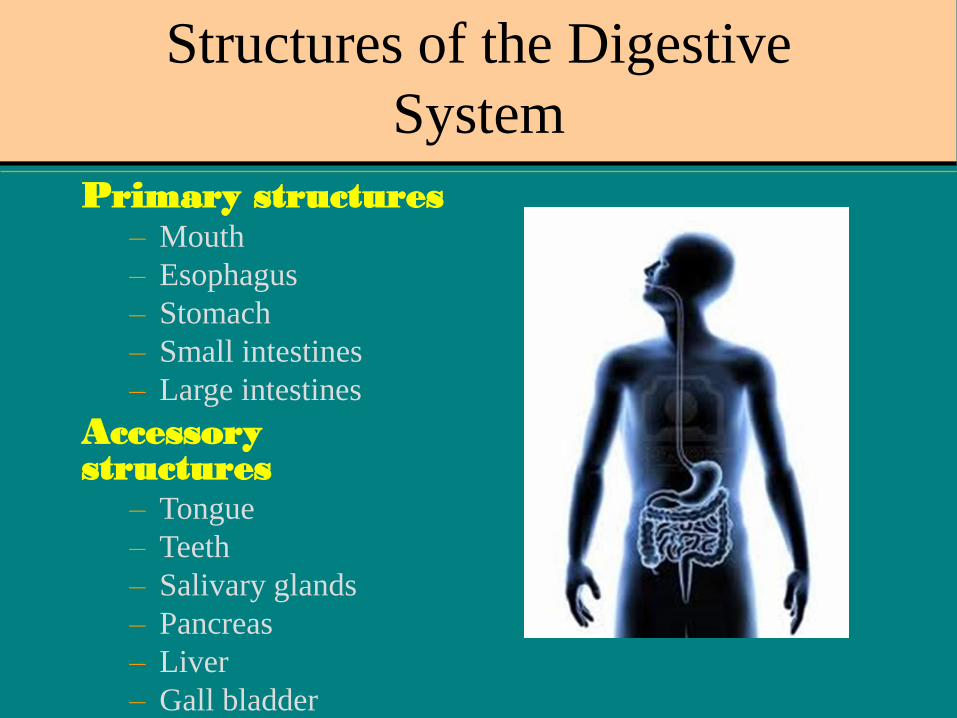
Structures of the Digestive
System
Primary structures– Mouth
– Esophagus
– Stomach
– Small intestines
– Large intestines
Accessory structures
– Tongue
– Teeth
– Salivary glands
– Pancreas
– Liver
– Gall bladder
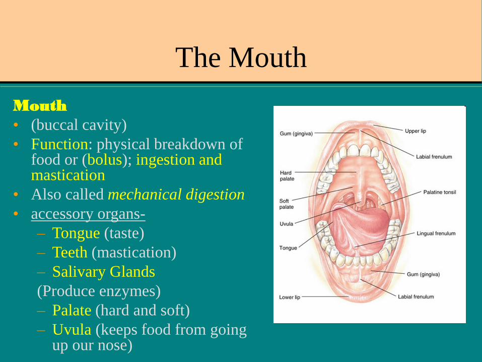
The Mouth
Mouth
• (buccal cavity)
• Function: physical breakdown of food or (bolus); ingestion and mastication
• Also called mechanical digestion
• accessory organs-
– Tongue (taste)
– Teeth (mastication)
– Salivary Glands
(Produce enzymes)
– Palate (hard and soft)
– Uvula (keeps food from going up our nose)
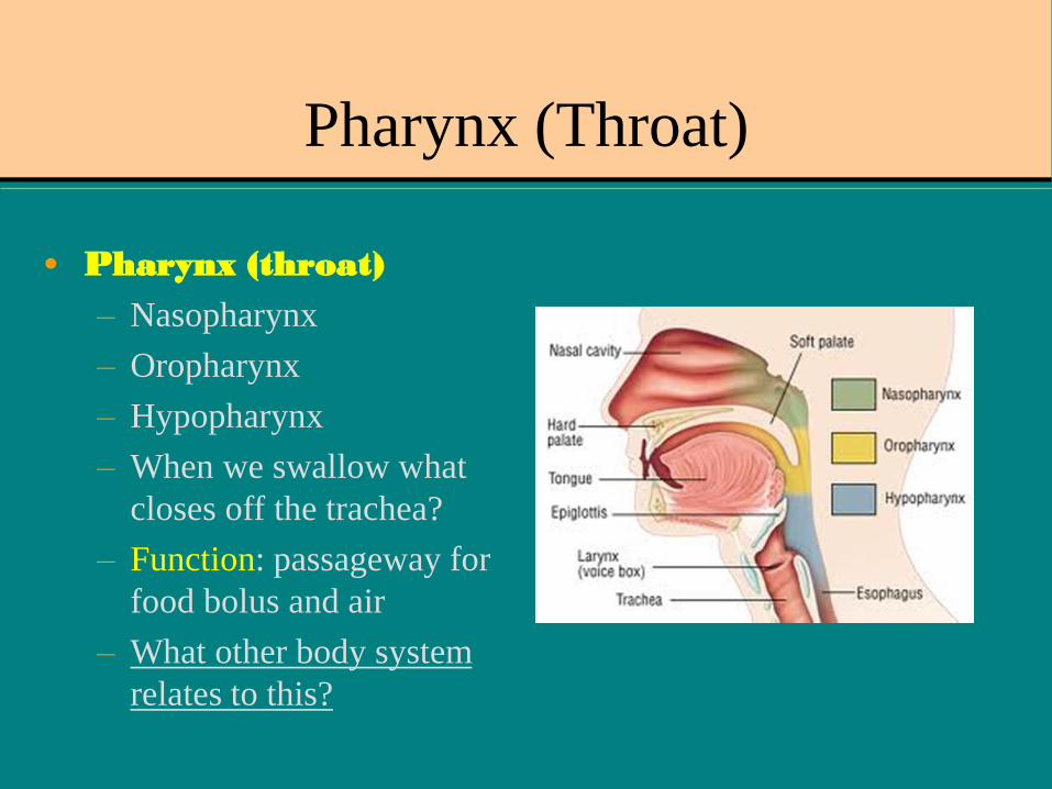
Pharynx (Throat)
• Pharynx (throat)
– Nasopharynx
– Oropharynx
– Hypopharynx
– When we swallow what
closes off the trachea?
– Function: passageway for
food bolus and air
– What other body system
relates to this?

Esophagus
• Esophagus– Muscular tube, 10”
long
– Connects the pharynx and stomach
– Peristalsis occurs here
• Esophageal wall layers
– Mucosa
– Submucosa
– Muscular
– External serous Is the esophagus anterior or
posterior to the trachea?

Stomach
• Stomach» Upper left quadrant
of the of the abdominal cavity
» Fundus
» Body
» Pylorus
– Cardiac Sphincter (b/t esophagus & stomach)
– Pyloric Sphincter (b/t stomach & small intestine)
– Rugae (folds which expand when stomach
fills)
Function:
Chemical digestion of food to
the end products of fat,
carbohydrates, and protein
Which is the distal sphincter?
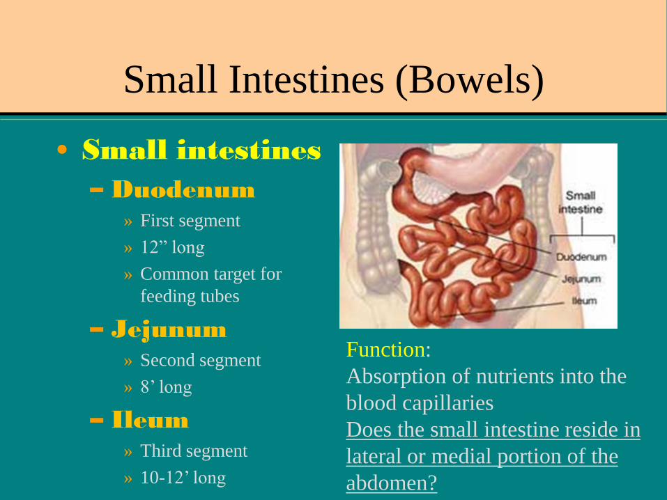
Small Intestines (Bowels)
• Small intestines
– Duodenum» First segment
» 12” long
» Common target for
feeding tubes
– Jejunum» Second segment
» 8’ long
– Ileum» Third segment
» 10-12’ long
Function:
Absorption of nutrients into the
blood capillaries
Does the small intestine reside in
lateral or medial portion of the
abdomen?

Large Intestines (Colon)
• Large intestines– Approximately 2” in diameter &
5’ long
– Cecum
– Ascending, Transverse,
Descending Colon
– Sigmoid and Rectum
– Hepatic and Splenic flexures
– Function: eliminates
waste products through
excretion and
defecation Label the parts of the colon listed to the left.

Group Activity
• Click on the
Digestive System
lab activities,
• Work with your
small group to
complete Station 2.
• You will be
provided with
string and tape to
complete this
activity.

Fecal Transplant for
Clostridium Difficile (C. Diff.)
• Bacteria found
all around us.
• Remember
normal flora?
• Triggered by
abundant use of
antibiotics.
• Good bacteria
killed off.

EXIT TICKET
• NAME EACH PART OF THE DIGESTIVE
SYSTEM AS A BOLUS PASSES
THROUGH IT (BEGINNING TO END)!!

Day 2 Bell Work
Draw the schematic diagram to the right of the
image.

Objectives Day 2
• Students will be able to label the accessory
organs of the digestive system and describe
their functions.
• Students will investigate further the phases,
processes, and possible complications of the
digestive system through a group research
project.

Accessory Organs:
Liver, Pancreas, Gallbladder
Based on previous lessons, what is the function of the ducts shown?
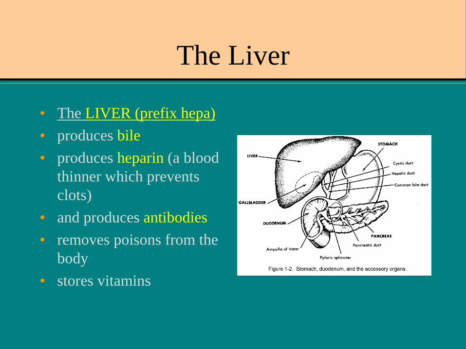
The Liver
• The LIVER (prefix hepa)
• produces bile
• produces heparin (a blood
thinner which prevents
clots)
• and produces antibodies
• removes poisons from the
body
• stores vitamins

Cirrhosis
• Chronic progressive
disease of the liver
• As the liver is damaged it
tries to heal itself, scarring
and turning into fibrous
tissue
• Bleed and bruise easily,
itchy skin, jaundice,
nausea, and fluid build up
• 75% caused by excessive
alcohol consumption
-Condition associated with Hepatitis B and C
-Also occurs in those with fatty liver disease
**Treated through weight loss, medications,
paracentesis, alcohol cessation.
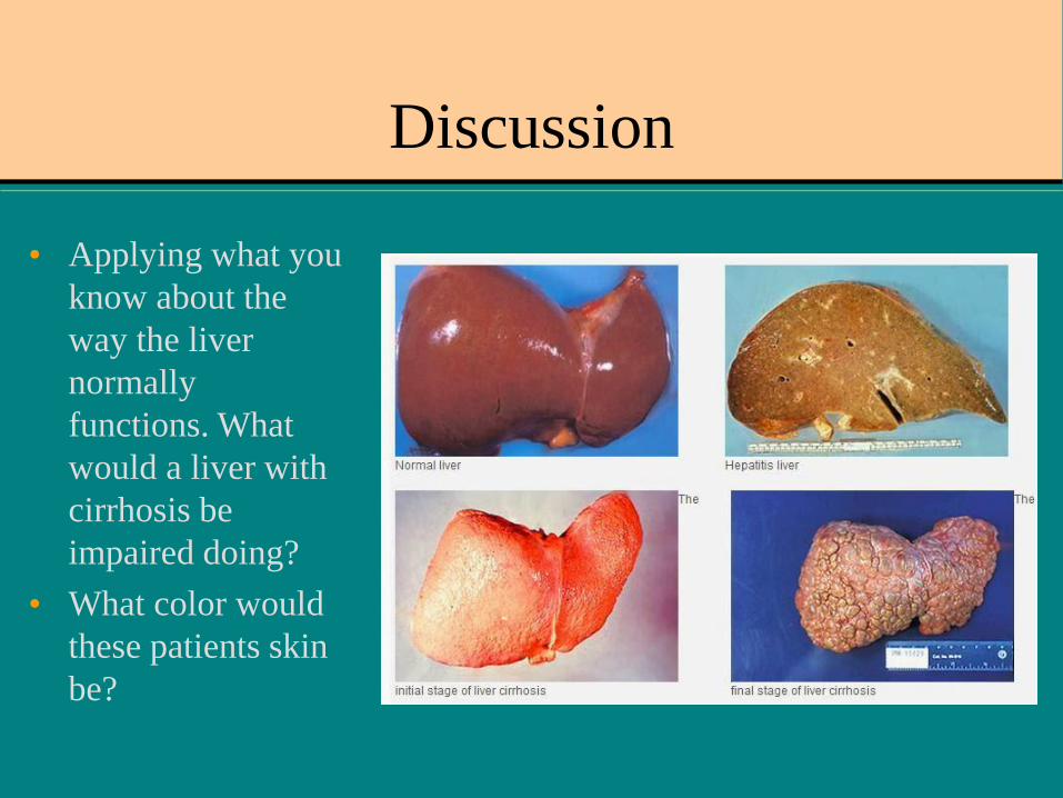
Discussion
• Applying what you
know about the
way the liver
normally
functions. What
would a liver with
cirrhosis be
impaired doing?
• What color would
these patients skin
be?
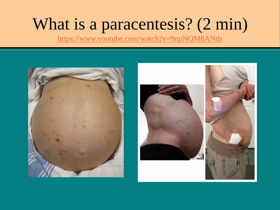
What is a paracentesis? (2 min)https://www.youtube.com/watch?v=9npNQM8ANds

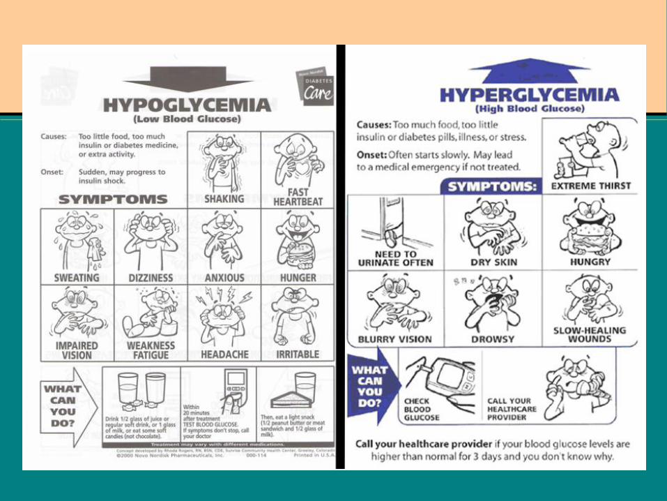
The Pancreas
• The PANCREAS
• helps chemically
break down food
• manufactures
insulin which
regulates the
amount of sugar or
glucose used by the
body
What is the sphincter of Oddi?
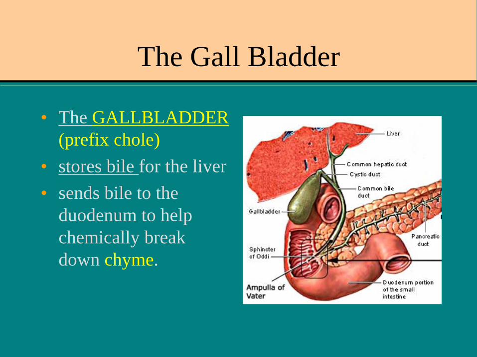
The Gall Bladder
• The GALLBLADDER
(prefix chole)
• stores bile for the liver
• sends bile to the
duodenum to help
chemically break
down chyme.
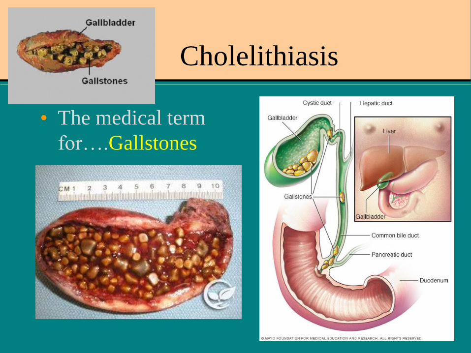
Cholelithiasis
• The medical term
for….Gallstones

WHAT ABOUT THE APPENDIX??
• A sac attached to the cecum and has no known function.
• Appendix can become infected by trapped fecal matter
• If it ruptures, bacteria from appendix can spread to peritoneal cavity.
• Symptoms- RLQ pain, rebound tenderness, fever, nausea, and vomiting
• RX – surgical appendectomy-What does that mean?

Possible theory for its purpose…
• Click on the following link…
• https://www.webmd.com/digestive-
disorders/news/20071012/appendix-may-
have-purpose#1
• What is suggested as a possible purpose and
function for the appendix?
• Consider past lessons on normal flora? Do
you agree with the possibility?
Group Activity:
Create a Digital Presentation
• Briefly describe the
following
diseases/disorders related
to the digestive system:
• Stomach Ulcer
• Diverticulitis
• Colon Cancer
• Chron Disease
• Hepatitis (A, B, C)
• Diabetes (Type I and II)
• Describe/Explain in
each:
• Signs/symptoms
• Diagnostic tools
• Possible treatments
• Prevention methods
• Include appropriate
pictures
Additional Activities
• Research the following feeding tubes.
• DOBHOFF
• PEG tube
• G-tube
• GJ-tube
• What are the differences?
• Where are they placed?
• What are some reasons a patient would need a
feeding tube?
Individual Activity
• Go to the class website and choose the link:
Evaluation of Dysphagia Directed Reading.
• 1st read the article and answer the directed
reading questions.
• 2nd go to the class website again and choose
the extended learning for directed reading
tab. Complete the extended learning
assignments for Dysphagia.

Exit Ticket
• Where does peristalsis begin in the digestive
system?
• Which is larger in diameter? Large or small
intestine?
• Which organ absorbs nutrients?
• Which organ produces bile, heparin, and
antibodies?
• Which organ manufactures insulin and regulates
blood sugar?
Recommended