Comparison of Caudal and Antero-Posterior Glide Mobilisation … - Sarkari, Dhakshinamoorthy & Multani
59
Comparison of Caudal and Antero-Posterior Glide Mobilisation for
the Improvement of Abduction Range of Motion
Sarkari, E.*, Dhakshinamoorthy, P.
** and Multani, N. K.
***
*Lecturer M.M.I.P.R. Mullana (Ambala) **SBSPGI, Balawala, Dehradun ***Principal M.M.I.P.R. Mullana (Ambala)
Abstract
The study was conducted on twenty patients of age between 40-65 year of adhesive capsulitis to
compare caudal and antero-posterior glide mobilisation technique for the improvement of
abduction range of motion, pain relief, and improvement in ADL‟S. Significant improvement in
abduction active ROM & passive ROM as well as alleviation in pain and disability was observed,
when end range mobilisation was administered for three weeks. It was further observed that caudal
glide was more effective than the antero-posterior glide.
Key Words: Caudal Glide, Antero-posterior Glide, End Range Mobilisation
Introduction
Adhesive Capsulitis is one of the
most common and disabling Orthopaedic
disorder characterized by painful restriction of shoulder motion for which
patients seeks treatment (Codman, 1934)
Adhesive Capsulitis is characterized by an insidious and progressive loss of active
and passive mobility in glenohumeral
joint presumably due to capsular
contracture. Despite research in the last century its etiology and pathology
remains enigmatic (Howel et al., 1988).
This painful, debilitating disorder reportedly affects 2-5% of the general
adult population (Neer et al., 1989) and 10-20% of people with diabetes (Kevin et
al., 1997). Incidence is slightly higher in
women than in men and is somewhat more common in the non dominant arm
(Dan et al., 1987). This condition most
frequently affects persons aged 40-60 years (Uitvlugt et al., 1993).
Primary frozen shoulder is classically described as having three
stages, “Freezing”, “Frozen” and
“Thawing” (Richard et al., 1986). Pain
particularly in the 1st phase often keeps
patients from performing activities of
daily living (ADL). In the second phase pain appears to be less pronounced but the
restriction in active motion appears to
limit the patient in personal care, ADL, and occupational activities. In the third
stage there is increase in mobility, which
leads to full or almost full recovery
(Richard et al., 1986).
Inspite of various approaches there remains a lack of evidence that
treatment speeds up recovery Joint
mobilisation has become a widely
employed physical therapy procedure for treating patients with joint hypomobility
(Maitland, 1983). It is accomplished by
performing gliding movements in the direction of limited joint glide (Henricus
& Obesmann, 2000). Antero-posterior
Glide and Caudal Glide mobilisations are frequently employed by physical
therapists to mobilise the shoulder joint to
decrease pain, improve mobility and

Journal of Exercise Science and Physiotherapy, Vol. 2: 59-65, 2006
60
regain normal joint function (Goldstein,
2004).
This study is conducted to
investigate if Antero-posterior Glide mobilisation is effective in increasing
abduction range of motion when given at
the end of available range of motion. It is also to compare Antero-posterior Glide
with Caudal Glide so as to analyse which
of the two is more effective. These glides
are given along with lateral distraction, capsular stretching, hot fomentation and
exercises. This study done on patients of
Adhesive Capsulitis is also to know about the effect of treatment on pain and
functional recovery by evaluating through
Shoulder Pain and Disability Index.
Materials and Methods
Subjects: Patients of Adhesive Capsulitis (n=20) in between the age of 40-65 years
were included in the study. They were taken from out patient department of
Prayas Physiotherapy Center, Dehradun
and SBSPGI, Balawala. The sample studied includes 11 males and 9 female
subjects with a mean age of 57.2. Thirteen
subjects had left arm involvement and 7
had right arm involvement. They were instructed not to do any other exercises.
Inclusion Criteria
1. Case of pure Adhesive Capsulitis
2. Painful restriction of more then 50%
of active & passive range of motion of the shoulder.
3. Capsular pattern of motion restriction.
4. An absence of radiological evidence of glenohumeral joint arthritis.
5. Symptoms present for at least 3
months.
Exclusion Criteria
Local corticosteroid injection to the
affected shoulder within the last 3
months or current corticosteroid
therapy.
Neuromuscular diseases.
Shoulder symptoms due to other
causes
Pregnancy
History of metastatic cancer or
diagnosis of cancer within 12 months.
Unstable angina
Insulin dependent diabetes
Prior shoulder surgery
Arthritis of shoulder
Variables of the Study
Independent Variable
1. Joint Mobilisation (Caudal glide,
Antero-posterior glide & Lateral
distraction) 2. Hot pack
3. Capsular stretching (Posterior &
Anterior) 4. Codman‟s exercise
Dependent Variable
1. Abduction range of Motion (Active
and Passive) 2. Pain and functional ability through
Shoulder Pain and Disability index
(Warren et al., 1984).
Study Protocol
Group A (n=10) (Caudal Glide + Lateral Distraction + Conventional treatment)
Lateral distraction was given
with Shoulder in neutral position followed by caudal glide, given at the shoulder
joint line after the end of available
abduction range was achieved. Grade 3 and 4 of Maitland Mobilisation was given
for 10-15 repetitions for 5-6 times. Total
duration lasted for 20 minutes. This was
followed by conventional treatment.
Group B (n=10) (Antero-Posterior Glide
+ Lateral Distraction + Conventional
Treatment)

Comparison of Caudal and Antero-Posterior Glide Mobilisation … - Sarkari, Dhakshinamoorthy & Multani
61
Lateral distraction was given with shoulder in neutral position followed by
Antero-posterior glide given at the
shoulder joint line after the end of available abduction range was achieved.
Grade 3 & 4 of Maitland mobilisation was
given for 10-15 repetitions for 5-6 times for 20 minutes. This was followed by
conventional treatment.
Procedure
With the initiations of each treatment session the subjects‟ abduction
range of motion was measured actively
and passively using standard goniometer according to the method as described by
Lippman (1943) and Norkin and White
(1995). The level of pain and disability was measured with the help of Shoulder
Pain and Disability Index. Once
measurements were recorded the patients
were then treated according to the assigned groups. Intervention started with
hot fomentation for 10 minutes followed
by few minutes of warm up consisting of rhythmic mid range mobilisation. This
was done with patient in supine position;
the joint was taken through full range of
available range of motion 3 times. After the patient were given capsular stretching
for the posterior and anterior part of the
capsule with 20 seconds hold in order to maintain the stretched position. This was
repeated for 4 times. Thereafter the joint
mobilisation was given according to the group the patients were assigned to. For
all the glides 10-15 repetitions were made
of grade 3 and 4 for Maitland
Mobilisation technique, which were performed at the end of available range.
Intermittently the shoulder was moved
once or twice through full range of available ROM to obtain muscle
relaxation. Total duration of end range
mobilisation technique lasted for 30 minutes.
Codman‟s Exercise was first
started with 10-15 repetitions without any weights. All other exercises were also
performed for 10-15 repetitions in a
particular session. All these exercises were first demonstrated to the patient and
then were asked to repeat the same. They
were instructed that while performing
these exercises at home, they should avoid causing pain of greater than 5 out of
10 on pain scale (10 being the worst).
After the completion of intervention the measurement of
abduction range of motion and values for pain and disability through SPADI was
again taken. This protocol was given for
total 9 sessions, which was completed in duration of 3 weeks.
Data Analysis
Unrelated and Paired t test was used to
compare AROM, PROM, pain and disability. The significance (probability –
P) has been selected as 0.05.
Results
Ten subjects were taken in each group
with the mean age of 56.1 4.95 and
58.3 4.37 respectively (table 1)
Table 1. Subject Information
Serial No. Group N Age
Mean + S.D.
1 A 10 56.1 + 4.95
2 B 10 58.3 + 4.37
Student t test was used for the comparison of mean of AROM between
group A & B. At zero session calculated t
value was 1.36 which is less than the
tabulated value at significance level 0.05. This indicates that there was no much
disparity amongst the subject of the two

Journal of Exercise Science and Physiotherapy, Vol. 2: 59-65, 2006
62
groups, before starting the intervention.
The mean value of AROM was
43.7 10.44, 52.6 15.77 and that of
PROM was 53.4 0.16, 62 17.08 respectively of the groups. (Tables 2 & 3).
Table 2. Comparison of mean of AROM
between Group A &Group B
Sessio
ns
Group A
X + S.D.
Group B
X + S.D. t
S/
Ns
0 43.7 + 10.44 52.6+15.77 1.36 NS
3 67.3 +11.31 66.7 + 19.8 0.1 NS
6 99.7 +7.16 88 + 17.37 1.89 NS
9 126.5 + 10.9 104.3 +17.63 3.58 S
P < 0.05
Table 3. Comparison of mean of PROM between
Group A & Group B
Sessions Group A
X + S.D.
Group B
X + S.D. t S/Ns
0 53.4 + 0.16 62 + 17.08 1.61 NS
3 77.5 + 9.4 77.2 + 20.5 0.04 NS
6 110.8+ 7.08 98.1+ 17.47 2.27 S
9 137.7+ 9.28 115.6+15.99 4.02 S
P<0.05
Student t test was done to compare the means of AROM group A &
B at 0, 3rd
, 6th and 9
th sessions. The result
of 0, 3rd
and 6th session were found to be
insignificant whereas that of 9th session
were significant with the means of
AROM being 126.5 10.9, 104.3 17.63 respectively of the groups (Table 2).
While comparing the means of PROM between groups A & B at 0, 3
rd,
6th & 9
thsessions the results of 3rd session
were insignificant .The 6th and 9
th sessions
showed significant improvement with the
t values of 2.27 and 4.02 respectively of
the sessions (table 3).
Paired t test was used to compare AROM within the group A between 0 and
9th sessions. The mean difference of the
session was 82.8 and the t value was
43.13 thus showing a significant
improvement. Similarly of the group B
the mean difference was 51.7 and the t value was 36.35 which was also
significant (table 4).
Table 4. Comparison of Improvement in Mean of
ROM within Group A&B between 0-9 sessions.
G SESSION 0 SESSION 9 Mean
Difference t
S/N
S
A 43.7+10.44 126.5+10.90 82.8 43.12 S
B 52.6 +15.77 104.3+17.63 51.7 36.35 S
G stands for Group
When comparison of PROM was done within group A between 0 and 9
th
sessions the mean difference was found to
be 84.3 and the t value was 67.6 which
was significant .Similarly of group B the mean difference was 53.6 and the t value
was 37.06 which was significant (Table 5).
Table 5. Comparison of Improvement in Mean of
PROM within Group A & B between 0-9 sessions.
G SESSION 0 SESSION 9 Mean
Difference t
S/N
S
A 53.4 +10.16 137.7 +9.28 84.3 67.6 S
B 62.0 +17.08 115.6+15.99 53.6 37.06 S
G stands for Group, P<0.05
Evaluation of pain and disability was
carried out through SPADI and student t
test was used to compare the means of 0, 3
rd, 6
th and 9
th sessions. At 0 session, the t
value was significant whereas at 3rd
, 6th
and 9th sessions it was found to be
insignificant (Table 6).
Table 6: Comparison of mean SPADI between
groups A & B
Sessions Group A
X + S.D.
Group B
X + S.D. t S/Ns
0 80.98+6.78 69.77+ 14.4 2.39 S
3 73.83+ 6.61 64.27+ 14.35 2.03 NS
6 65.13+ 6.67 58.97+14.78 1.28 NS
9 58.04+ 6.78 52.96+ 14.5 1.06 NS
Comparison of Caudal and Antero-Posterior Glide Mobilisation … - Sarkari, Dhakshinamoorthy & Multani
63
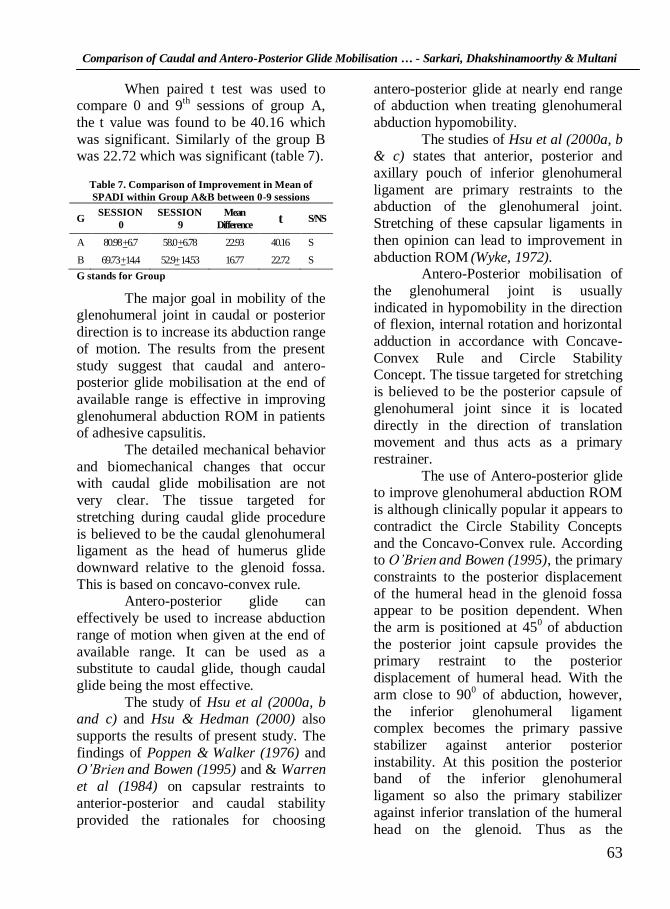
When paired t test was used to compare 0 and 9
th sessions of group A,
the t value was found to be 40.16 which
was significant. Similarly of the group B was 22.72 which was significant (table 7).
Table 7. Comparison of Improvement in Mean of
SPADI within Group A&B between 0-9 sessions
G SESSION
0
SESSION
9
Mean
Difference t S/NS
A 80.98 +6.7 58.0 +6.78 22.93 40.16 S
B 69.73 +14.4 52.9+ 14.53 16.77 22.72 S
G stands for Group
The major goal in mobility of the glenohumeral joint in caudal or posterior
direction is to increase its abduction range
of motion. The results from the present
study suggest that caudal and antero-posterior glide mobilisation at the end of
available range is effective in improving
glenohumeral abduction ROM in patients of adhesive capsulitis.
The detailed mechanical behavior
and biomechanical changes that occur with caudal glide mobilisation are not
very clear. The tissue targeted for
stretching during caudal glide procedure
is believed to be the caudal glenohumeral ligament as the head of humerus glide
downward relative to the glenoid fossa.
This is based on concavo-convex rule. Antero-posterior glide can
effectively be used to increase abduction
range of motion when given at the end of
available range. It can be used as a substitute to caudal glide, though caudal
glide being the most effective.
The study of Hsu et al (2000a, b and c) and Hsu & Hedman (2000) also
supports the results of present study. The
findings of Poppen & Walker (1976) and O’Brien
and Bowen (1995) and & Warren
et al (1984) on capsular restraints to
anterior-posterior and caudal stability
provided the rationales for choosing
antero-posterior glide at nearly end range of abduction when treating glenohumeral
abduction hypomobility.
The studies of Hsu et al (2000a, b & c) states that anterior, posterior and
axillary pouch of inferior glenohumeral
ligament are primary restraints to the abduction of the glenohumeral joint.
Stretching of these capsular ligaments in
then opinion can lead to improvement in
abduction ROM (Wyke, 1972).
Antero-Posterior mobilisation of
the glenohumeral joint is usually
indicated in hypomobility in the direction of flexion, internal rotation and horizontal
adduction in accordance with Concave-
Convex Rule and Circle Stability Concept. The tissue targeted for stretching
is believed to be the posterior capsule of
glenohumeral joint since it is located
directly in the direction of translation movement and thus acts as a primary
restrainer.
The use of Antero-posterior glide to improve glenohumeral abduction ROM
is although clinically popular it appears to
contradict the Circle Stability Concepts
and the Concavo-Convex rule. According to O’Brien
and Bowen (1995), the primary
constraints to the posterior displacement
of the humeral head in the glenoid fossa appear to be position dependent. When
the arm is positioned at 450 of abduction
the posterior joint capsule provides the primary restraint to the posterior
displacement of humeral head. With the
arm close to 900 of abduction, however,
the inferior glenohumeral ligament complex becomes the primary passive
stabilizer against anterior posterior
instability. At this position the posterior band of the inferior glenohumeral
ligament so also the primary stabilizer
against inferior translation of the humeral
head on the glenoid. Thus as the

Journal of Exercise Science and Physiotherapy, Vol. 2: 59-65, 2006
64
glenohumeral joint approaches end range
of abduction the posterior band of the inferior glenohumeral ligament becomes
the primary structure against inferior
gliding of humeral head on the glenoid
fossa. In this position Antero-Posterior glide will most effectively stretch the
posterior band of the inferior
glenohumeral ligament releasing the tightened posterior band and allowing
more inferior glide of the humeral head to
occur during abduction. Selecting an appropriate joint
position could be a very important factor
in the success of the joint mobilisation
procedure. Several authors (Goldstein 2004) have advocated that resting position
is not the most effective position for
increasing ROM of the joint treated. Results of various studies
Hsu et al
(2000a, b and c) showed that mobilisation
of glenohumeral joint at its resting position is less effective than the end
range position in improving abduction
ROM. This may be because the
periarticular tissue that limits joint ROM is most stretched when the joint is
positioned close to the restricted range.
The SPADI is a shoulder region functional status measure that is
responsive to clinical change. There is
remarkable increase in functional status of
the patient following mobilisation procedure. There was a marked decrease
in pain and disability following treatment
session especially in group A followed by group B .There is no previous study as per
the supporting article of similar study.
As the increase in abduction ROM was seen there was remarkable
decrease in pain and disability in the
similar fashion.
Limitation of the Study 1. Number of subjects was less.
2. No control group was taken.
3. No groups had similar patients with
the same degree of involvement. 4. Age variation was there from 40-
65years.
5. Patients built was variable
6. Adhesive capsulitis is a self-limiting disease so the actual improvement
through the treatment cannot be
evaluated. 7. Photographic method for
measurement of abduction ROM was
not used. 8. Marked amount of tissue resistance is
experienced while applying the glide
which has not been taken into
consideration 9. Proper strengthening program was not
followed after mobilisation sessions
due to lack of time.
Conclusions
This study provides preliminary
evidence that antero-posterior glide is also effective in improving glenohumeral
abduction ROM when given at the end of
available range. However, it is less
effective than the traditional caudal glide mobilisation.
Clinical Implication
This study provides some evidence for the use of both Antero
posterior and inferior glide performed
close to the end range of abduction to
increase the abduction ROM. This study also states that inferior glide is most
effective in increasing abduction ROM.
The significant increase in abduction range seen after Antero posterior glide
procedure performed at a joint angle close
to its end might be a good alternative for treating abduction hypomobility. This is
because inferior glenohumeral ligament is
preferentially stressed in this position.
Patient‟s functional recovery was evaluated using SPADI. A significant

Comparison of Caudal and Antero-Posterior Glide Mobilisation … - Sarkari, Dhakshinamoorthy & Multani
65
improvement in functional activities was in the same pattern as for the
improvement in the abduction ROM. This
provides an evidence for the functional rehabilitation of the patient.
References
Codman E.A. 1934. In: The Shoulder, Ed. Thomas Todd. Boston.
Dan, L., Riddle, J. and M. Rothstein 1987. Goniometric Reliability in Clinical Setting Shoulder Measurements. Physical Therapy. 67(5): 668-73.
Goldstein, B. 2004. Shoulder Anatomy and Biomechanics. Physical Medicine Rehabilitation Clinics of North America. 15: 313-49.
Henricus, M. V. and Obesmann, W. R. 2000. End Range Mobilisation Technique in Adhesive Capsulitis of the Shoulder Joint: A Multiple Subject Case Report. Physical Therapy, 80(12): 1204-12.
Howel, S.M., Galinat, B.J., Renzi, A. J. and Marone, P.J. 1988. Normal and Abnormal Mechanics of Glenohumeral Joint in Horizontal Plane. Journal of Bone Joint and Surgery (Am). 70: 227-32.
Hsu, A. T. and Hedman, T. 2000. Change in Abduction and Rotational Range of Motion in Responses to Simulated Dorsal and Ventral Translational Mobilisation of Glenohumeral Joint. Physical Therapy, 82(6). 544-56
Hsu, A. T., Ho, C., Jia., C., Chih, H. 2000a. Determining the Resting Position of Glenohumeral Joint: A Cadaver Study. Journal of Orthopaedic and Sports Physical Therapy, 32: 605-12.
Hsu, A. T., Ho, L. S., Ho, T. and Hedman 2000b. Immediate Response of Glenohumeral Abductor Range of Motion to a Caudally
Directed Translational Mobilisation: A Fresh Cadaver Simulation. Archives Physical Medicine Rehabilitation, 81: Nov. 1516-16
Hsu, A. T., Ho, L. S., Ho, T. and Hedman 2000c. Joint Position during Anterior-Posterior Glide Mobilisation: It‟s Effect on Glenohumeral Abduction Range of Motion. Archives Physical Medicine Rehabilitation, 81: Feb, 210-40
Kevin E W, Christopher, A. A., James, and Andrews, R. 1997 Current Concept: The Stabilizing Structure of the Glenohumeral Joint. Journal of Orthopedics and Sports Physical Therapy, 26(6): 364-79
Lippman, R. K. 1943. Frozen Shoulder Periarthritis Bicipital Tenosynovitis. Archives Surgery. 47: 283.
Maitland, G.D. 1983. Treatment of Glenohumeral Joint by Passive Movement. Physiotherapy, 69(1): Jan 3-7.
Neer, C.S., Satterlee, C.C. and Dalsey, R.M. 1989. On the Value of Coracohumeral Ligament Release. Orthopedic Trans., 13: 235.
Norkin, C.C. and White, D. J. 1995. Measurement of Joint Motion. In: A Guide to Goniometry. 2nd Ed., Davis Philadelphia.
O‟Brien, S.J. and Schwartz, H.S. 1995. Capsule Restraint to Anterior Posterior Motion of Abducted Shoulder Biomechanical Study. Journal of Shoulder and Elbow Surgery. 4(2): 298-05.
Poppen, N.K. and Walker, P.S. 1976. Normal and Abnormal Motion of Shoulder. Journal of Bone Joint Surgery (Am). 58, 195-201.
Richard, W., Bowling, A. P. and Rockar, J.R. 1986. Examination of Shoulder Complex. Physical Therapy, 66(12): 1866-77.
Uitvlugt G, Detrisae, D.A. and Johson, L.L. 1993. Arthroscopic Observation Before and After Manipulation of Frozen Shoulder. Arthroscopy, 9: 181
Warren RF, Karnblatt, I.B. and Morehand, R.1984. Static Factors Affecting Posterior Shoulder Stability. Orthopedic Trans., 8: 89-93
Wyke, B. 1972. Articular Neurology: A Review, Physiotherapy, 58: 94
Recommended