© WINFOCUS’ CRITICAL CARE ECHOCARDIOGRAPHY
WINFOCUS BASIC ECHO (WBE)!
Ritesh Dhar, MD Director, Echocardiography Lab
and Staff Cardiologist Intermountain Medical Center
Murray, Utah
Basic Assessment of Left Ventricular Systolic Function
© WINFOCUS’ CRITICAL CARE ECHOCARDIOGRAPHY
!
Outline
LV GLOBAL SYSTOLIC FUNCTION
• Fractional Shortening (FS)
• Fractional Area Change (FAC)
• Volumetric Ejection Fraction (EF)
• Long Axis Function (MAPSE)

© WINFOCUS’ CRITICAL CARE ECHOCARDIOGRAPHY
!
Outline
LV GLOBAL SYSTOLIC FUNCTION
• Fractional Shortening (FS)
• Fractional Area Change (FAC)
• Volumetric Ejection Fraction (EF)
• Long Axis Function (MAPSE)
© WINFOCUS’ CRITICAL CARE ECHOCARDIOGRAPHY
!Systolic Heart Function
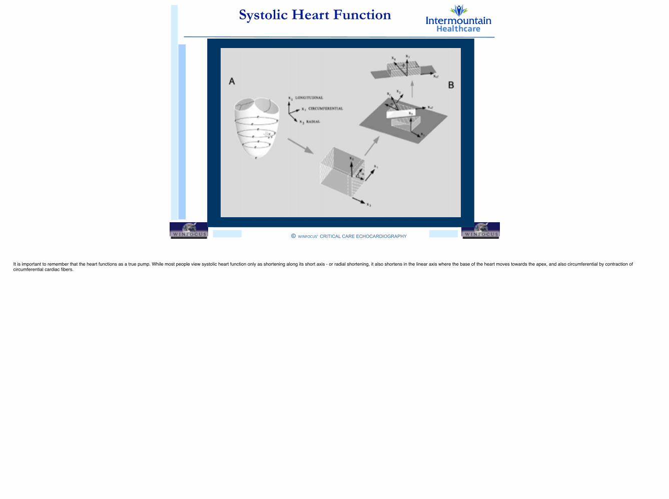
It is important to remember that the heart functions as a true pump. While most people view systolic heart function only as shortening along its short axis - or radial shortening, it also shortens in the linear axis where the base of the heart moves towards the apex, and also circumferential by contraction of circumferential cardiac fibers.
© WINFOCUS’ CRITICAL CARE ECHOCARDIOGRAPHY
!
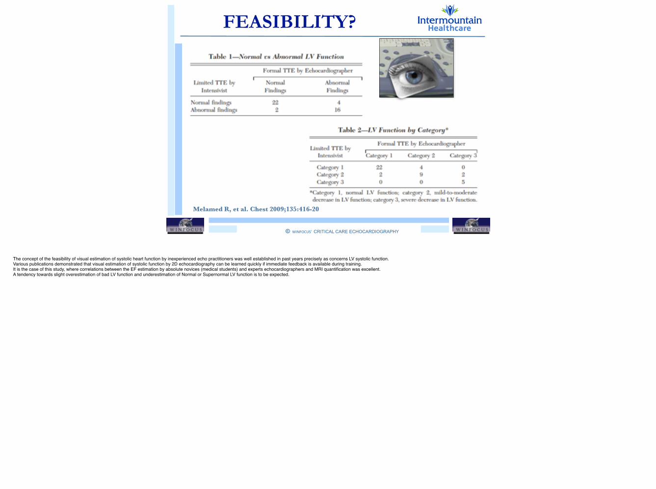
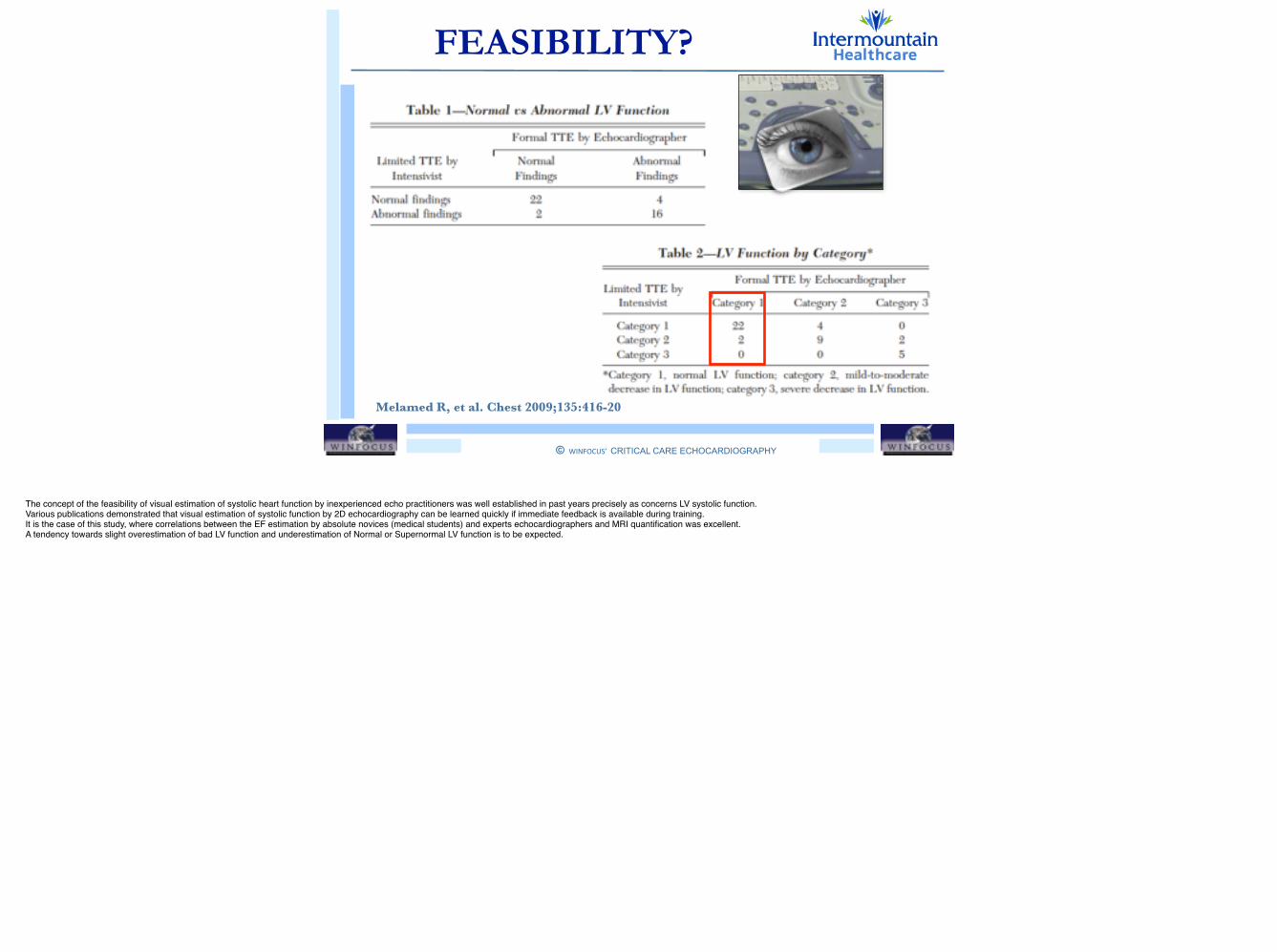
FEASIBILITY?
Melamed R, et al. Chest 2009;135:416-20
The concept of the feasibility of visual estimation of systolic heart function by inexperienced echo practitioners was well established in past years precisely as concerns LV systolic function.!Various publications demonstrated that visual estimation of systolic function by 2D echocardiography can be learned quickly if immediate feedback is available during training. !It is the case of this study, where correlations between the EF estimation by absolute novices (medical students) and experts echocardiographers and MRI quantification was excellent.!A tendency towards slight overestimation of bad LV function and underestimation of Normal or Supernormal LV function is to be expected.!
© WINFOCUS’ CRITICAL CARE ECHOCARDIOGRAPHY
!
FEASIBILITY?
Melamed R, et al. Chest 2009;135:416-20
The concept of the feasibility of visual estimation of systolic heart function by inexperienced echo practitioners was well established in past years precisely as concerns LV systolic function.!Various publications demonstrated that visual estimation of systolic function by 2D echocardiography can be learned quickly if immediate feedback is available during training. !It is the case of this study, where correlations between the EF estimation by absolute novices (medical students) and experts echocardiographers and MRI quantification was excellent.!A tendency towards slight overestimation of bad LV function and underestimation of Normal or Supernormal LV function is to be expected.!

© WINFOCUS’ CRITICAL CARE ECHOCARDIOGRAPHY
!
FEASIBILITY?
Melamed R, et al. Chest 2009;135:416-20
YES !
The concept of the feasibility of visual estimation of systolic heart function by inexperienced echo practitioners was well established in past years precisely as concerns LV systolic function.!Various publications demonstrated that visual estimation of systolic function by 2D echocardiography can be learned quickly if immediate feedback is available during training. !It is the case of this study, where correlations between the EF estimation by absolute novices (medical students) and experts echocardiographers and MRI quantification was excellent.!A tendency towards slight overestimation of bad LV function and underestimation of Normal or Supernormal LV function is to be expected.!
© WINFOCUS’ CRITICAL CARE ECHOCARDIOGRAPHY
!
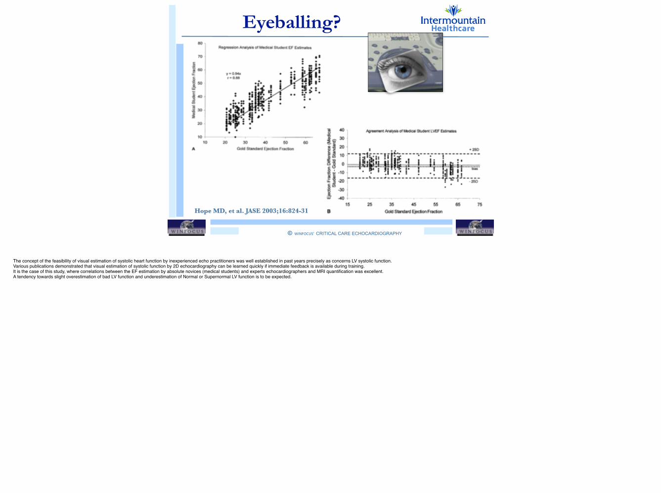
Eyeballing?
Hope MD, et al. JASE 2003;16:824-31
The concept of the feasibility of visual estimation of systolic heart function by inexperienced echo practitioners was well established in past years precisely as concerns LV systolic function.!Various publications demonstrated that visual estimation of systolic function by 2D echocardiography can be learned quickly if immediate feedback is available during training. !It is the case of this study, where correlations between the EF estimation by absolute novices (medical students) and experts echocardiographers and MRI quantification was excellent.!A tendency towards slight overestimation of bad LV function and underestimation of Normal or Supernormal LV function is to be expected.!
© WINFOCUS’ CRITICAL CARE ECHOCARDIOGRAPHY
!
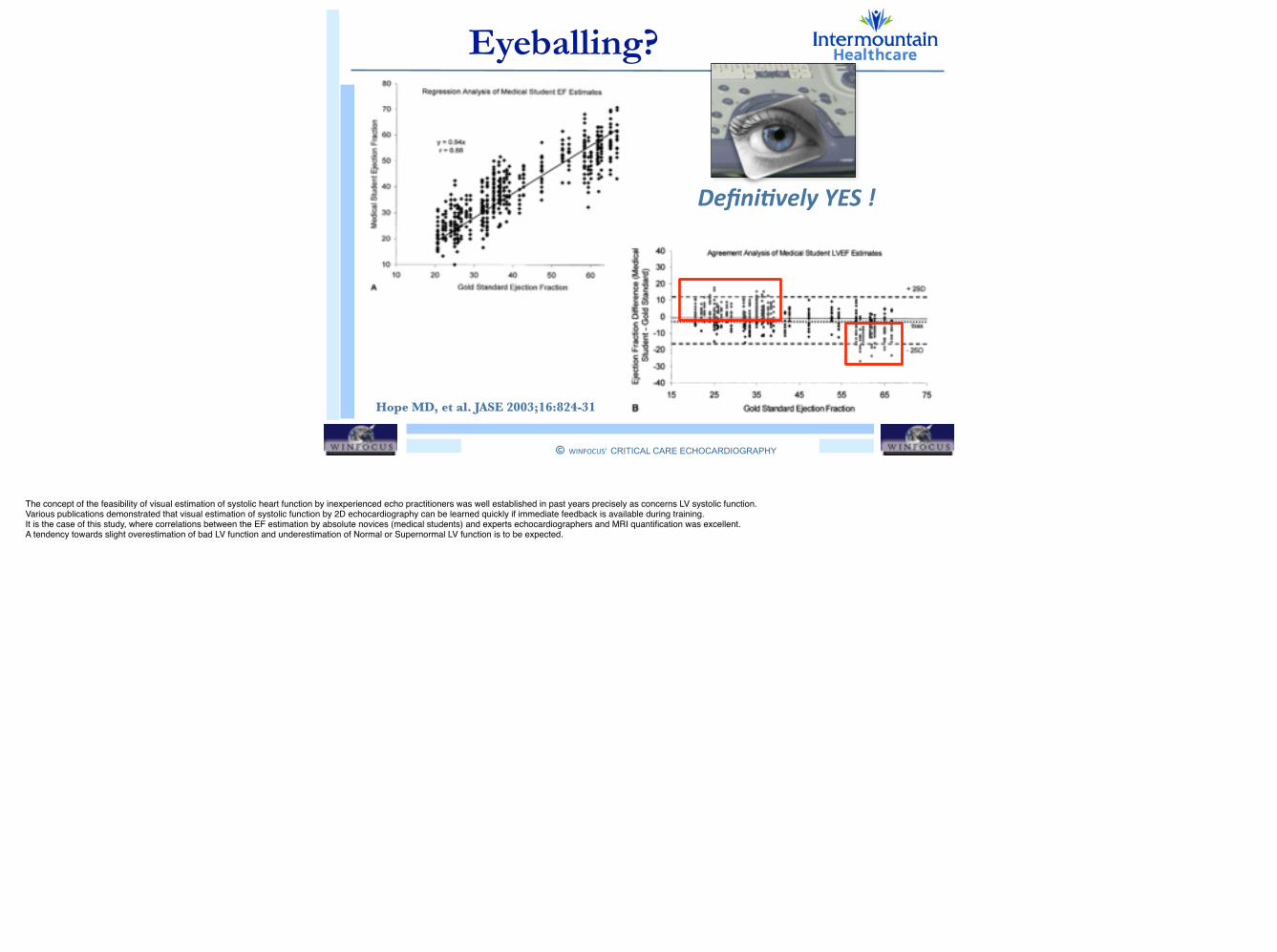
Eyeballing?
Hope MD, et al. JASE 2003;16:824-31
Defini+vely YES !
The concept of the feasibility of visual estimation of systolic heart function by inexperienced echo practitioners was well established in past years precisely as concerns LV systolic function.!Various publications demonstrated that visual estimation of systolic function by 2D echocardiography can be learned quickly if immediate feedback is available during training. !It is the case of this study, where correlations between the EF estimation by absolute novices (medical students) and experts echocardiographers and MRI quantification was excellent.!A tendency towards slight overestimation of bad LV function and underestimation of Normal or Supernormal LV function is to be expected.!
© WINFOCUS’ CRITICAL CARE ECHOCARDIOGRAPHY
!
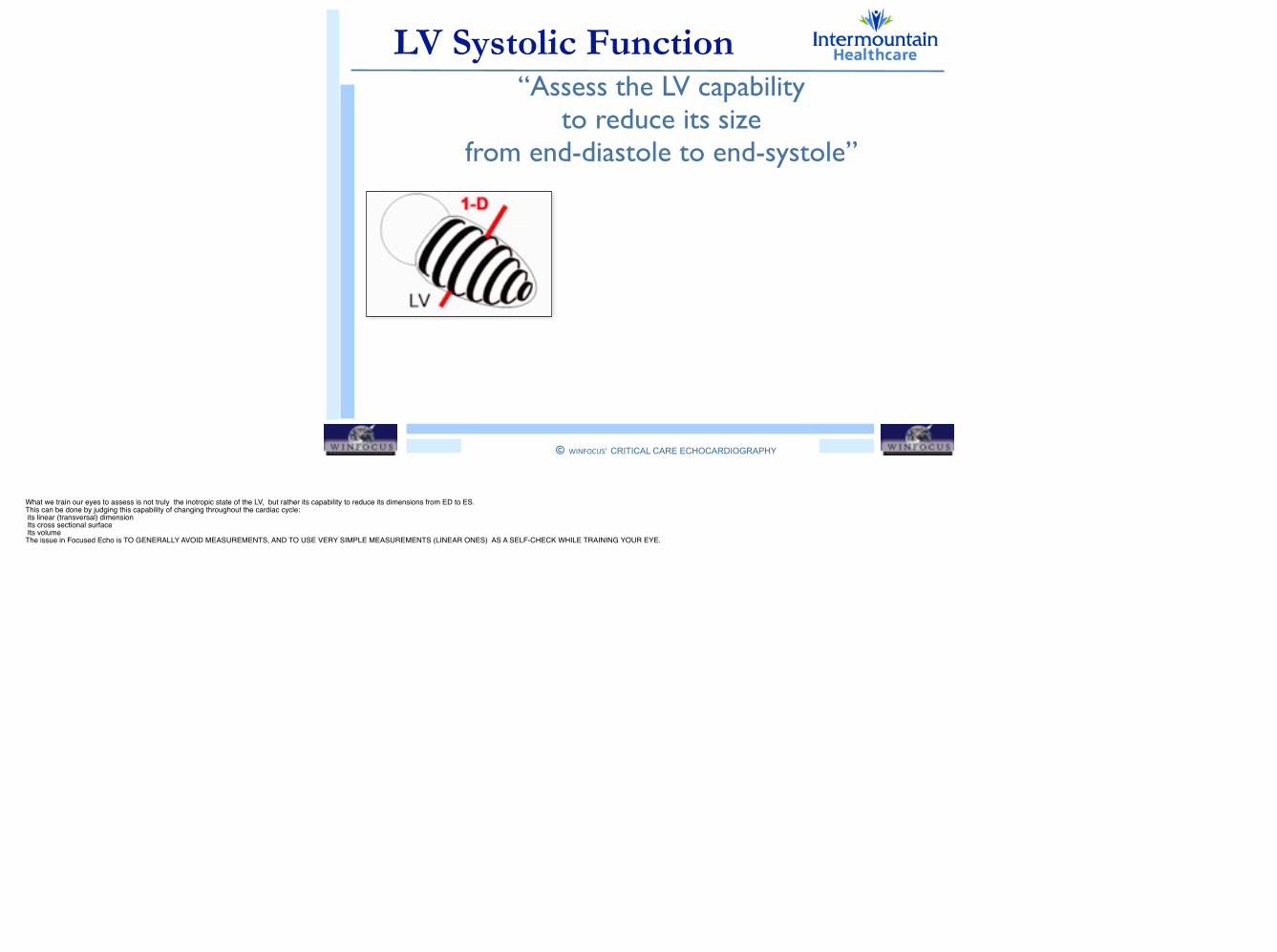
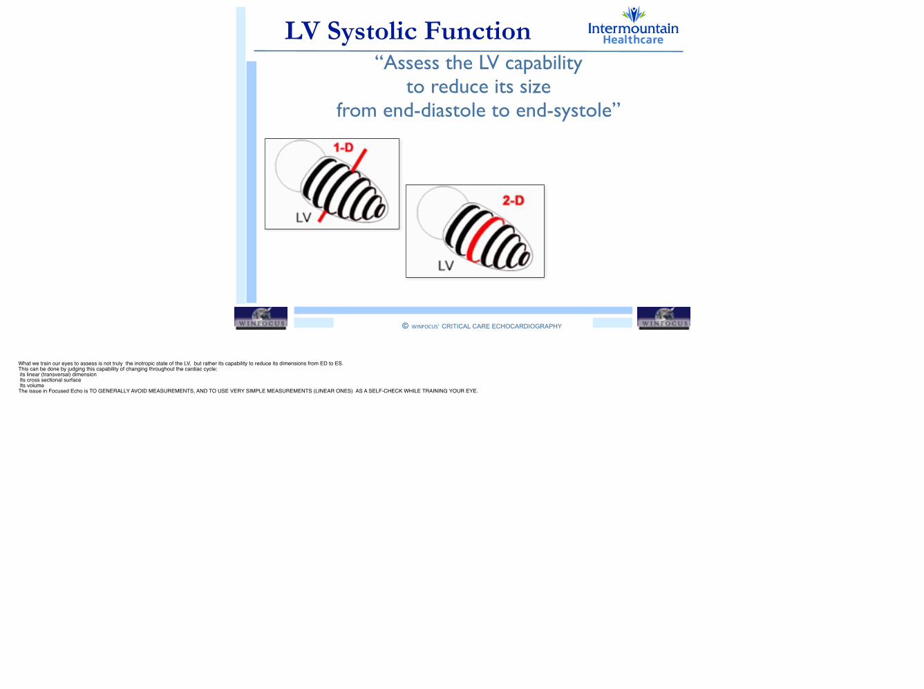
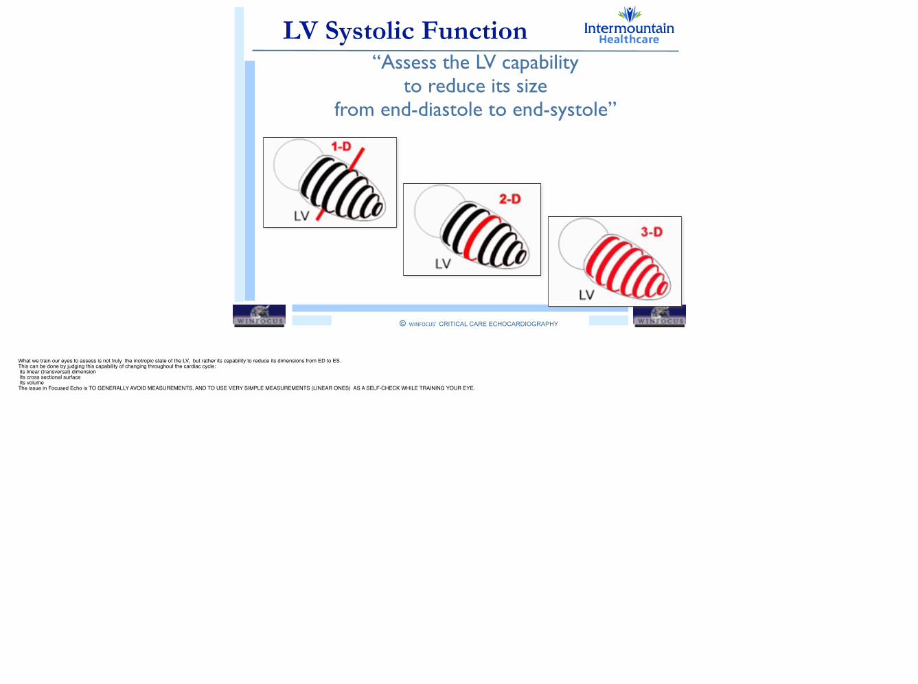
LV Systolic Function“Assess the LV capability
to reduce its size from end-diastole to end-systole”
What we train our eyes to assess is not truly the inotropic state of the LV, but rather its capability to reduce its dimensions from ED to ES.!This can be done by judging this capability of changing throughout the cardiac cycle:! its linear (transversal) dimension ! Its cross sectional surface! Its volume!The issue in Focused Echo is TO GENERALLY AVOID MEASUREMENTS, AND TO USE VERY SIMPLE MEASUREMENTS (LINEAR ONES) AS A SELF-CHECK WHILE TRAINING YOUR EYE.
© WINFOCUS’ CRITICAL CARE ECHOCARDIOGRAPHY
!
Outline
LV GLOBAL SYSTOLIC FUNCTION
• Fractional Shortening (FS)
• Fractional Area Change (FAC)
• Volumetric Ejection Fraction (EF)
• Long Axis Function (MAPSE)

© WINFOCUS’ CRITICAL CARE ECHOCARDIOGRAPHY
!
Outline
LV GLOBAL SYSTOLIC FUNCTION
• Fractional Shortening (FS)
• Fractional Area Change (FAC)
• Volumetric Ejection Fraction (EF)
• Long Axis Function (MAPSE)
© WINFOCUS’ CRITICAL CARE ECHOCARDIOGRAPHY
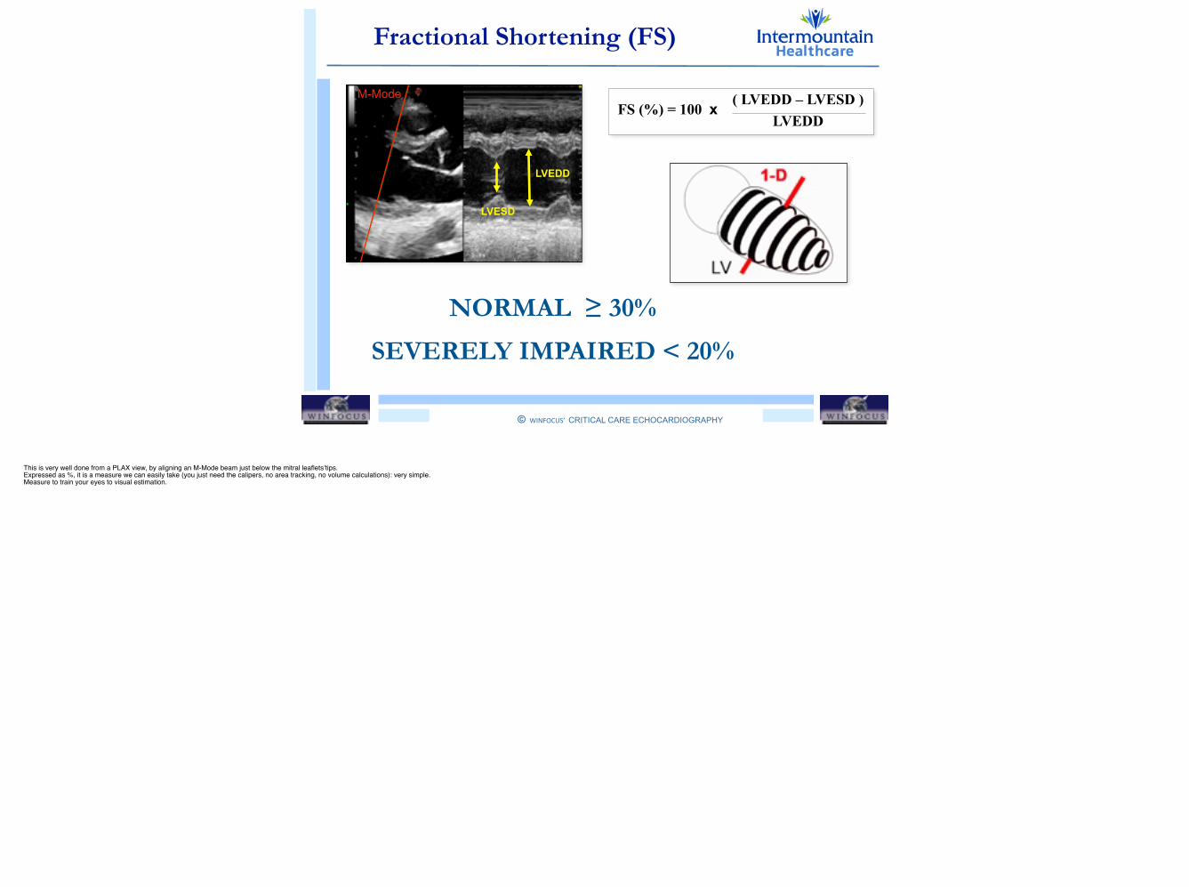
!Fractional Shortening (FS)
NORMAL ≥ 30%
SEVERELY IMPAIRED < 20%
FS (%) = 100 x ( LVEDD – LVESD ) LVEDD
LVEDD
LVESD
M-Mode
This is very well done from a PLAX view, by aligning an M-Mode beam just below the mitral leaflets’tips.!Expressed as %, it is a measure we can easily take (you just need the calipers, no area tracking, no volume calculations): very simple. !Measure to train your eyes to visual estimation.
© WINFOCUS’ CRITICAL CARE ECHOCARDIOGRAPHY
!
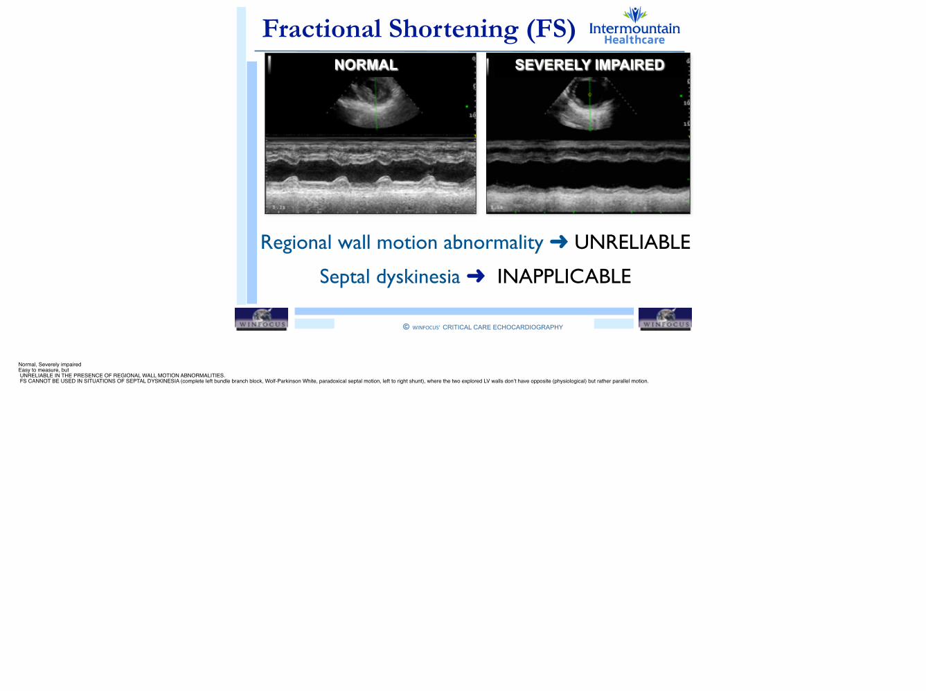
Fractional Shortening (FS)SEVERELY IMPAIRED
Regional wall motion abnormality ➜ UNRELIABLE
Septal dyskinesia ➜ INAPPLICABLE
NORMAL
Normal, Severely impaired!Easy to measure, but ! UNRELIABLE IN THE PRESENCE OF REGIONAL WALL MOTION ABNORMALITIES.! FS CANNOT BE USED IN SITUATIONS OF SEPTAL DYSKINESIA (complete left bundle branch block, Wolf-Parkinson White, paradoxical septal motion, left to right shunt), where the two explored LV walls don’t have opposite (physiological) but rather parallel motion.
© WINFOCUS’ CRITICAL CARE ECHOCARDIOGRAPHY
!
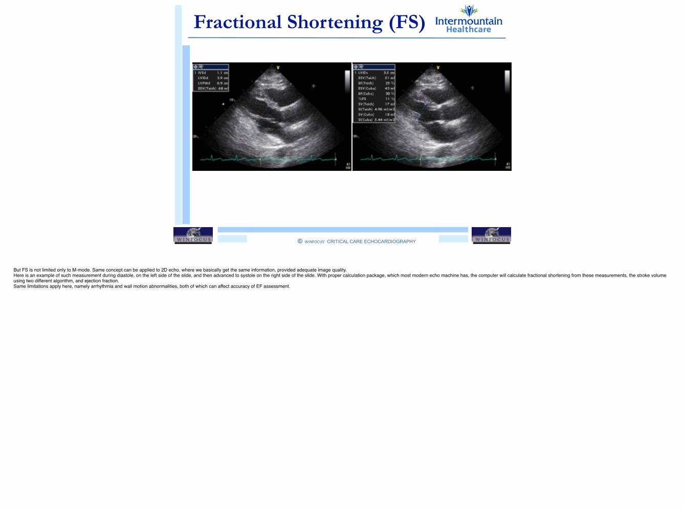
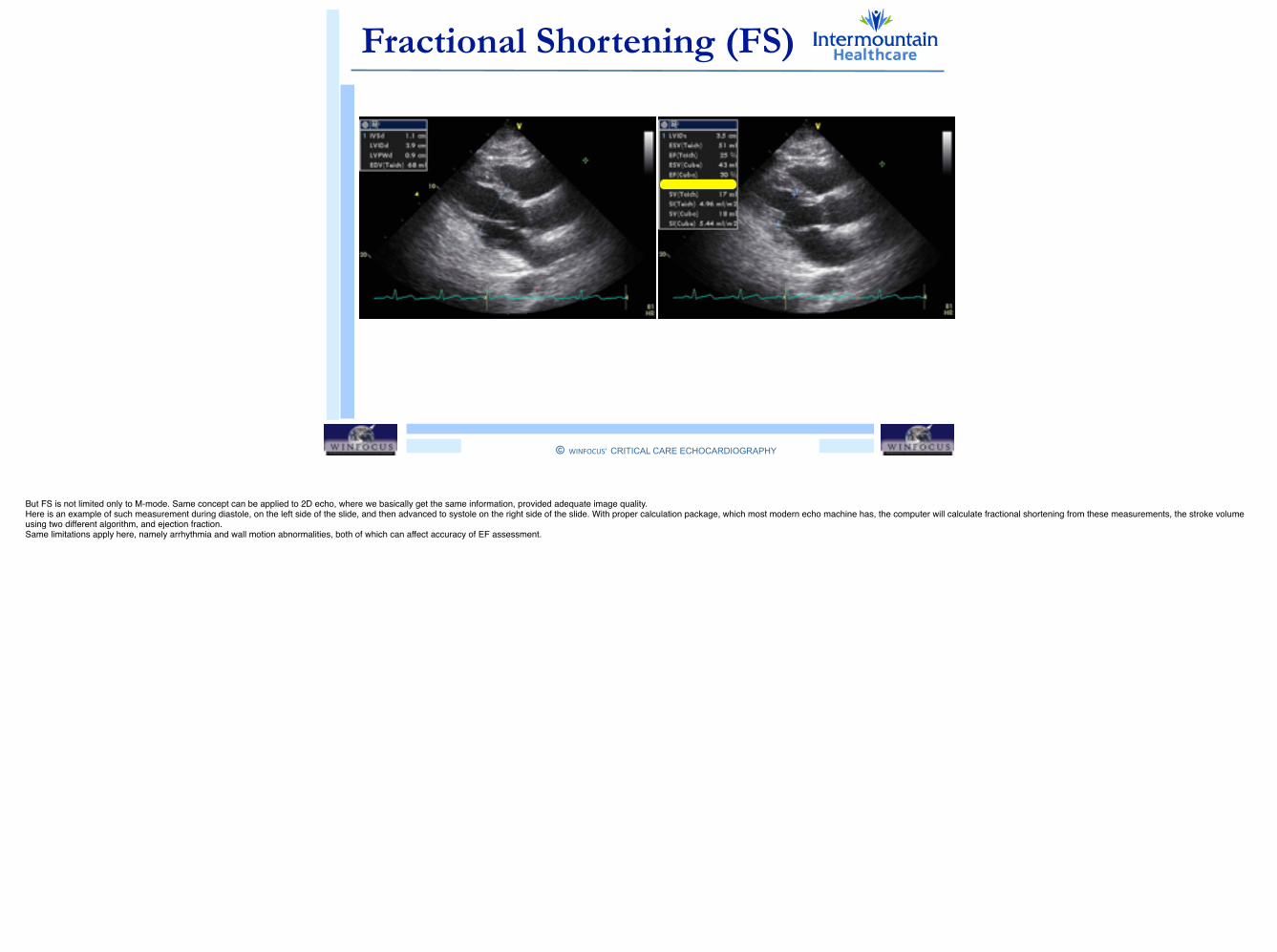
Fractional Shortening (FS)
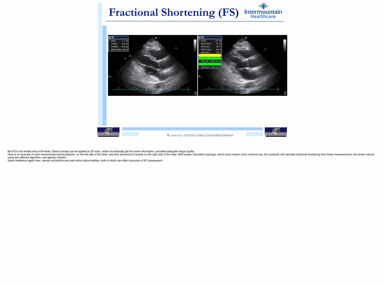
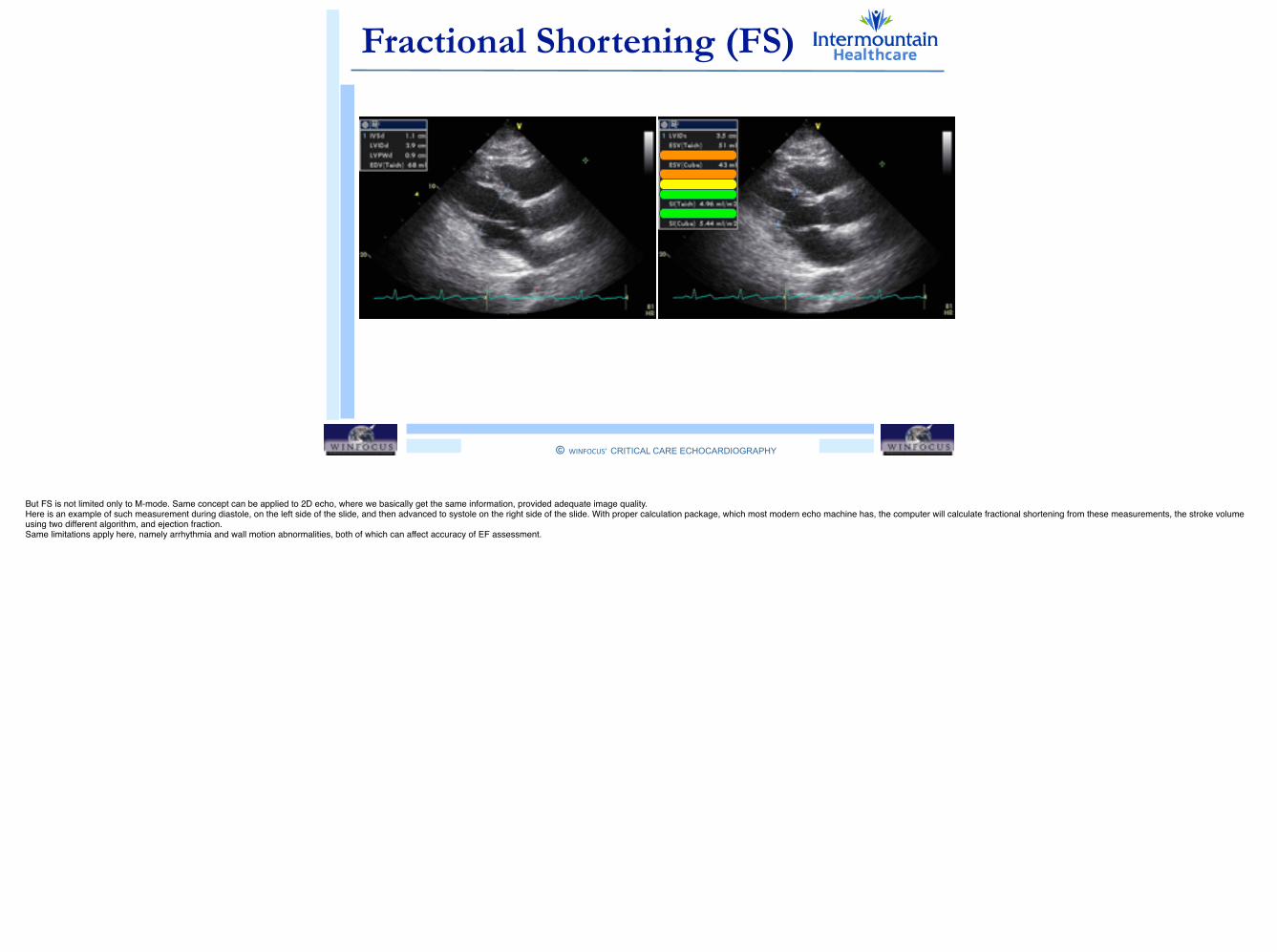
But FS is not limited only to M-mode. Same concept can be applied to 2D echo, where we basically get the same information, provided adequate image quality. !Here is an example of such measurement during diastole, on the left side of the slide, and then advanced to systole on the right side of the slide. With proper calculation package, which most modern echo machine has, the computer will calculate fractional shortening from these measurements, the stroke volume using two different algorithm, and ejection fraction. !Same limitations apply here, namely arrhythmia and wall motion abnormalities, both of which can affect accuracy of EF assessment.
© WINFOCUS’ CRITICAL CARE ECHOCARDIOGRAPHY
!
Fractional Shortening (FS)
But FS is not limited only to M-mode. Same concept can be applied to 2D echo, where we basically get the same information, provided adequate image quality. !Here is an example of such measurement during diastole, on the left side of the slide, and then advanced to systole on the right side of the slide. With proper calculation package, which most modern echo machine has, the computer will calculate fractional shortening from these measurements, the stroke volume using two different algorithm, and ejection fraction. !Same limitations apply here, namely arrhythmia and wall motion abnormalities, both of which can affect accuracy of EF assessment.
© WINFOCUS’ CRITICAL CARE ECHOCARDIOGRAPHY
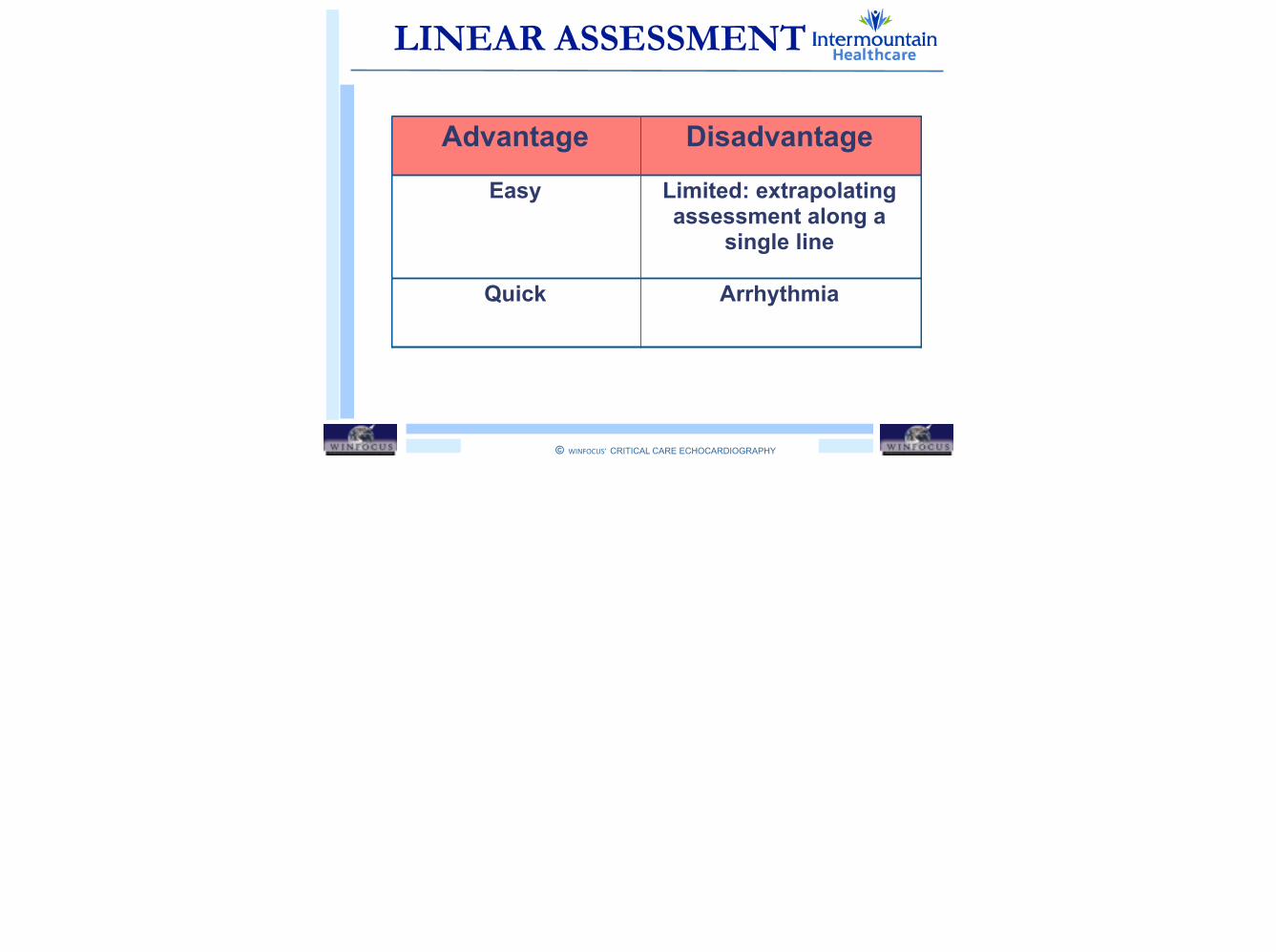
!LINEAR ASSESSMENT
Advantage Disadvantage
Easy Limited: extrapolating assessment along a
single line
Quick Arrhythmia

© WINFOCUS’ CRITICAL CARE ECHOCARDIOGRAPHY
!
Outline
LV GLOBAL SYSTOLIC FUNCTION
• Fractional Shortening (FS)
• Fractional Area Change (FAC)
• Volumetric Ejection Fraction (EF)
• Long Axis Function (MAPSE)
© WINFOCUS’ CRITICAL CARE ECHOCARDIOGRAPHY
!
Outline
LV GLOBAL SYSTOLIC FUNCTION
• Fractional Shortening (FS)
• Fractional Area Change (FAC)
• Volumetric Ejection Fraction (EF)
• Long Axis Function (MAPSE)
© WINFOCUS’ CRITICAL CARE ECHOCARDIOGRAPHY
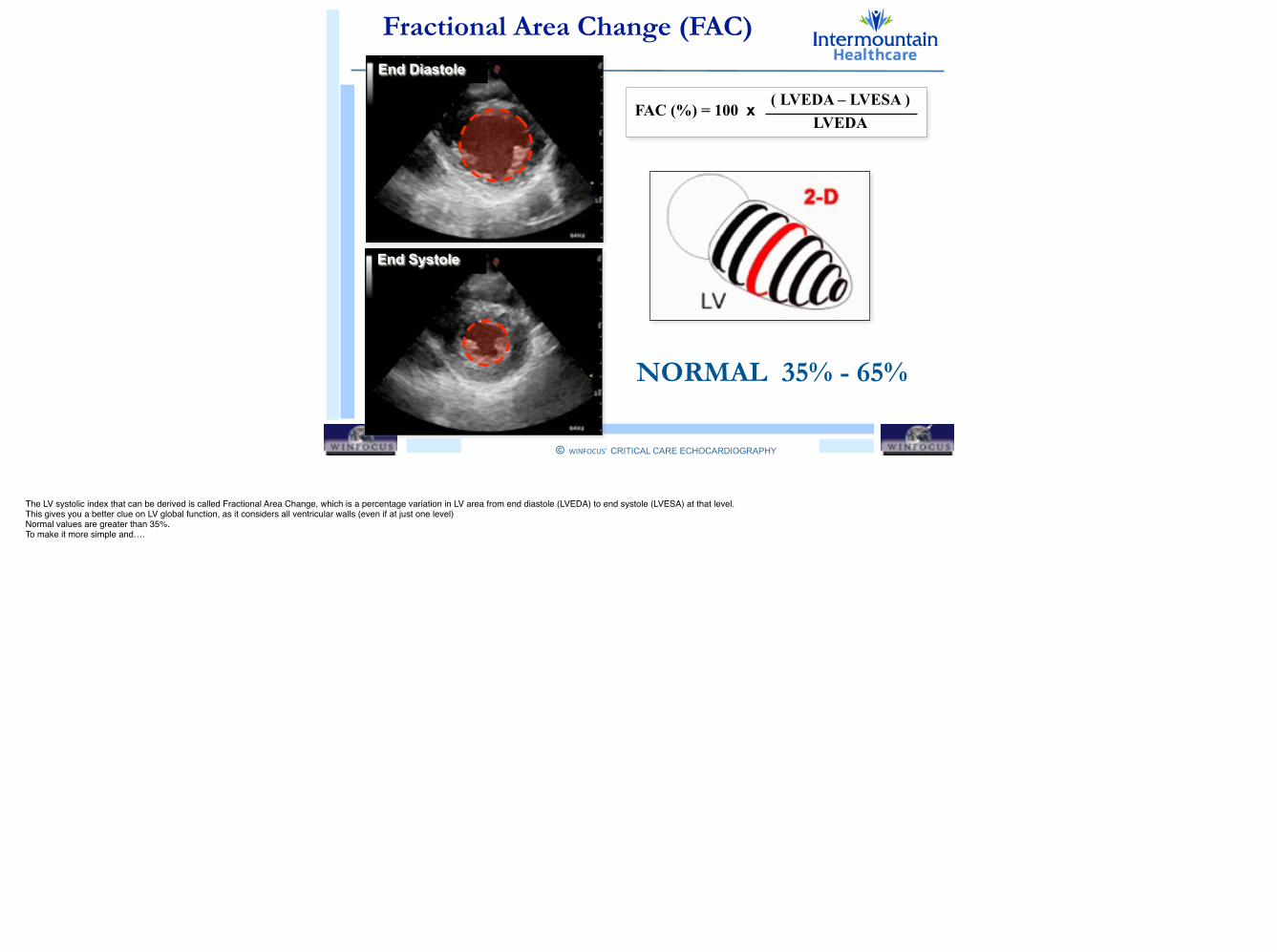
!Fractional Area Change (FAC)
NORMAL 35% - 65%
FAC (%) = 100 x ( LVEDA – LVESA ) LVEDA
End Diastole
End Systole
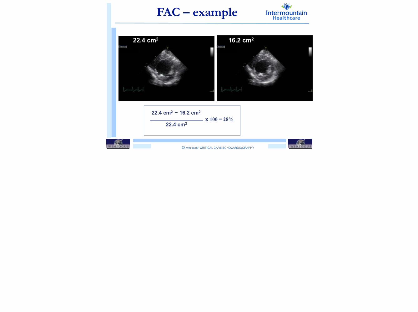
The LV systolic index that can be derived is called Fractional Area Change, which is a percentage variation in LV area from end diastole (LVEDA) to end systole (LVESA) at that level. !This gives you a better clue on LV global function, as it considers all ventricular walls (even if at just one level)!Normal values are greater than 35%.!To make it more simple and….

© WINFOCUS’ CRITICAL CARE ECHOCARDIOGRAPHY
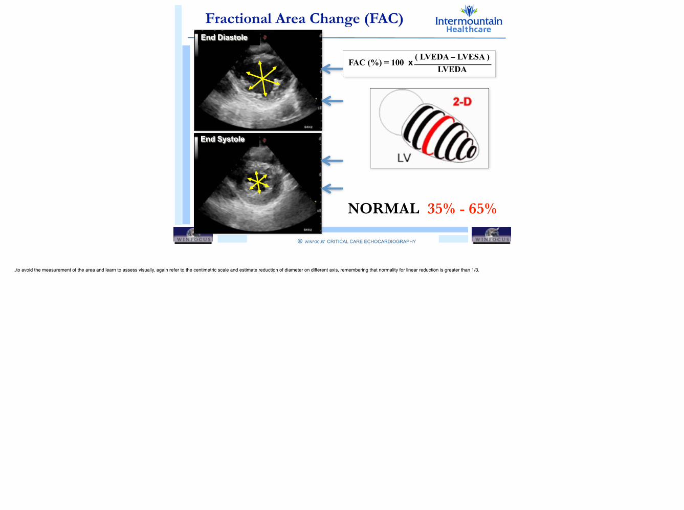
!Fractional Area Change (FAC)
NORMAL 35% - 65%
End Diastole
End Systole
FAC (%) = 100 x ( LVEDA – LVESA ) LVEDA
..to avoid the measurement of the area and learn to assess visually, again refer to the centimetric scale and estimate reduction of diameter on different axis, remembering that normality for linear reduction is greater than 1/3.
© WINFOCUS’ CRITICAL CARE ECHOCARDIOGRAPHY
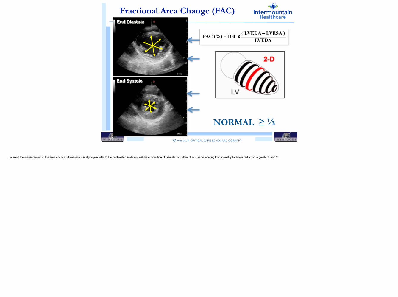
!Fractional Area Change (FAC)
NORMAL 35% - 65%NORMAL ≥ ⅓
End Diastole
End Systole
FAC (%) = 100 x ( LVEDA – LVESA ) LVEDA
..to avoid the measurement of the area and learn to assess visually, again refer to the centimetric scale and estimate reduction of diameter on different axis, remembering that normality for linear reduction is greater than 1/3.
© WINFOCUS’ CRITICAL CARE ECHOCARDIOGRAPHY
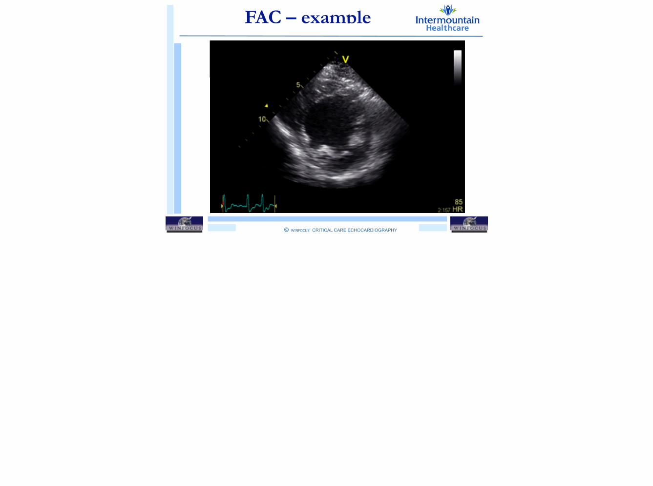
!FAC – example

© WINFOCUS’ CRITICAL CARE ECHOCARDIOGRAPHY
!FAC – example
© WINFOCUS’ CRITICAL CARE ECHOCARDIOGRAPHY
!FAC – example
22.4 cm2 16.2 cm2
22.4 cm2 − 16.2 cm2
22.4 cm2x 100 = 28%
© WINFOCUS’ CRITICAL CARE ECHOCARDIOGRAPHY
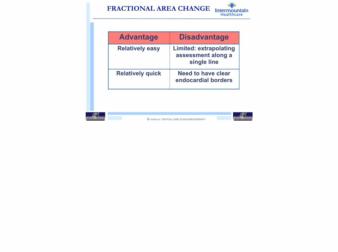
!FRACTIONAL AREA CHANGE
Advantage DisadvantageRelatively easy Limited: extrapolating
assessment along a single line
Relatively quick Need to have clear endocardial borders
© WINFOCUS’ CRITICAL CARE ECHOCARDIOGRAPHY
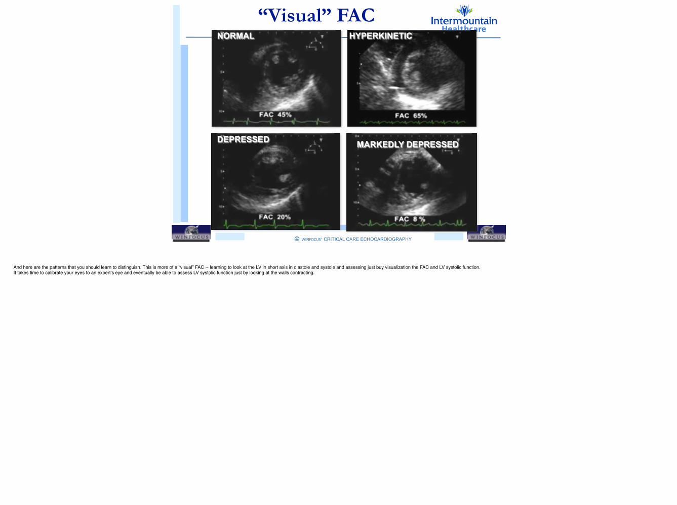
!“Visual” FACNORMAL HYPERKINETIC
DEPRESSED MARKEDLY DEPRESSED
And here are the patterns that you should learn to distinguish. This is more of a “visual” FAC -- learning to look at the LV in short axis in diastole and systole and assessing just buy visualization the FAC and LV systolic function. It takes time to calibrate your eyes to an expert’s eye and eventually be able to assess LV systolic function just by looking at the walls contracting. !

© WINFOCUS’ CRITICAL CARE ECHOCARDIOGRAPHY
!“Visual” FACNORMAL HYPERKINETIC
DEPRESSED MARKEDLY DEPRESSED
And here are the patterns that you should learn to distinguish. This is more of a “visual” FAC -- learning to look at the LV in short axis in diastole and systole and assessing just buy visualization the FAC and LV systolic function. It takes time to calibrate your eyes to an expert’s eye and eventually be able to assess LV systolic function just by looking at the walls contracting. !
© WINFOCUS’ CRITICAL CARE ECHOCARDIOGRAPHY
!
Outline
• LV GLOBAL SYSTOLIC FUNCTION
• Fractional Shortening (FS)
• Fractional Area Change (FAC)
• Volumetric Ejection Fraction (EF)
• Long Axis Function (MAPSE)

© WINFOCUS’ CRITICAL CARE ECHOCARDIOGRAPHY
!
Outline
• LV GLOBAL SYSTOLIC FUNCTION
• Fractional Shortening (FS)
• Fractional Area Change (FAC)
• Volumetric Ejection Fraction (EF)
• Long Axis Function (MAPSE)
© WINFOCUS’ CRITICAL CARE ECHOCARDIOGRAPHY
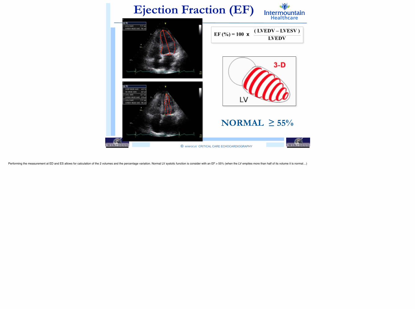
!Ejection Fraction (EF)
EF (%) = 100 x ( LVEDV – LVESV ) LVEDV
NORMAL ≥ 55%
Performing the measurement at ED and ES allows for calculation of the 2 volumes and the percentage variation. Normal LV systolic function is consider with an EF > 55% (when the LV empties more than half of its volume it is normal…)
© WINFOCUS’ CRITICAL CARE ECHOCARDIOGRAPHY
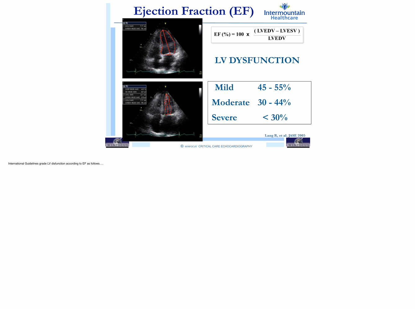
!Ejection Fraction (EF)
Mild 45 - 55%
Moderate 30 - 44%
Severe < 30%
LV DYSFUNCTION
Lang R, et al. JASE 2005
EF (%) = 100 x ( LVEDV – LVESV ) LVEDV
International Guidelines grade LV disfunction according to EF as follows….
!
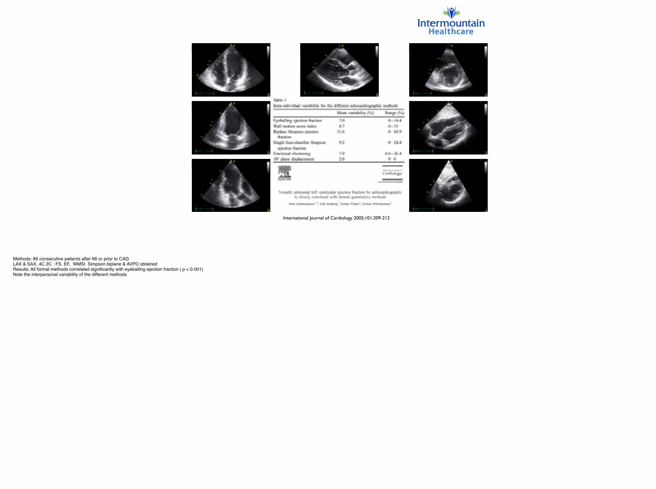
International Journal of Cardiology 2005;101:209-212
Methods: 89 consecutive patients after MI or prior to CAG!LAX & SAX, 4C 2C : FS, EF, WMSI Simpson biplane & AVPD obtained!Results: All formal methods correlated significantly with eyeballing ejection fraction ( p < 0.001)!Note the interpersonal variability of the different methods

!
International Journal of Cardiology 2005;101:209-212
Methods: 89 consecutive patients after MI or prior to CAG!LAX & SAX, 4C 2C : FS, EF, WMSI Simpson biplane & AVPD obtained!Results: All formal methods correlated significantly with eyeballing ejection fraction ( p < 0.001)!Note the interpersonal variability of the different methods
© WINFOCUS’ CRITICAL CARE ECHOCARDIOGRAPHY
!
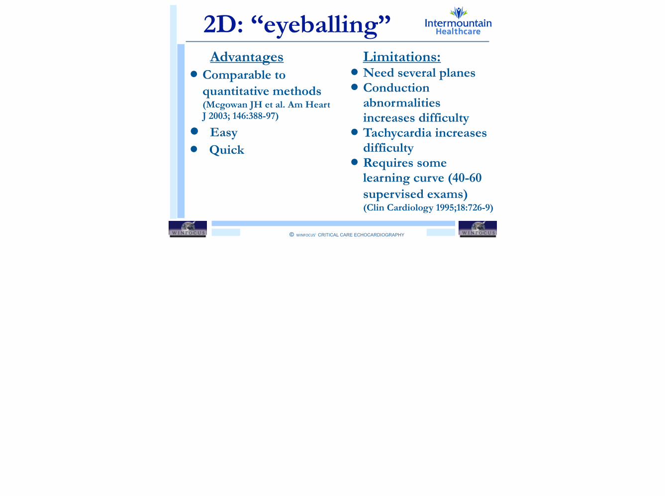
2D: “eyeballing” Advantages
• Comparable to quantitative methods (Mcgowan JH et al. Am Heart J 2003; 146:388-97)
• Easy
• Quick !!!
Limitations: • Need several planes • Conduction
abnormalities increases difficulty
• Tachycardia increases difficulty
• Requires some learning curve (40-60 supervised exams) (Clin Cardiology 1995;18:726-9)
© WINFOCUS’ CRITICAL CARE ECHOCARDIOGRAPHY
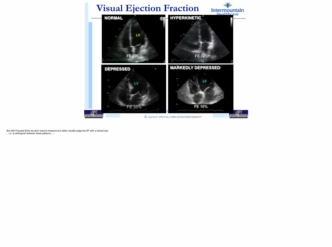
!Visual Ejection Fraction NORMAL HYPERKINETIC
DEPRESSED MARKEDLY DEPRESSED
But with Focused Echo we don’t want to measure but rather visually judge the EF with a trained eye.!…i.e. to distinguish between these patterns:…..

© WINFOCUS’ CRITICAL CARE ECHOCARDIOGRAPHY
!Visual Ejection Fraction NORMAL HYPERKINETIC
DEPRESSED MARKEDLY DEPRESSED
But with Focused Echo we don’t want to measure but rather visually judge the EF with a trained eye.!…i.e. to distinguish between these patterns:…..
© WINFOCUS’ CRITICAL CARE ECHOCARDIOGRAPHY
!
Outline
LV GLOBAL SYSTOLIC FUNCTION
• Fractional Shortening (FS)
• Fractional Area Change (FAC)
• Volumetric Ejection Fraction (EF)
• Long Axis Function (MAPSE)

© WINFOCUS’ CRITICAL CARE ECHOCARDIOGRAPHY
!
Outline
LV GLOBAL SYSTOLIC FUNCTION
• Fractional Shortening (FS)
• Fractional Area Change (FAC)
• Volumetric Ejection Fraction (EF)
• Long Axis Function (MAPSE)
© WINFOCUS’ CRITICAL CARE ECHOCARDIOGRAPHY
!
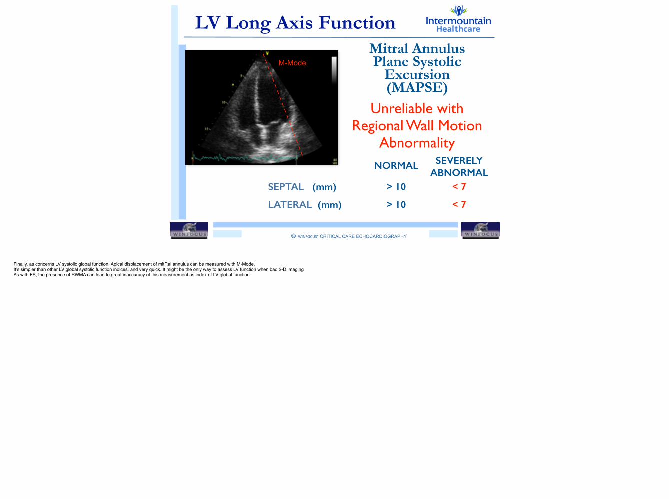
LV Long Axis Function
M-ModeMitral Annulus Plane Systolic
Excursion (MAPSE)
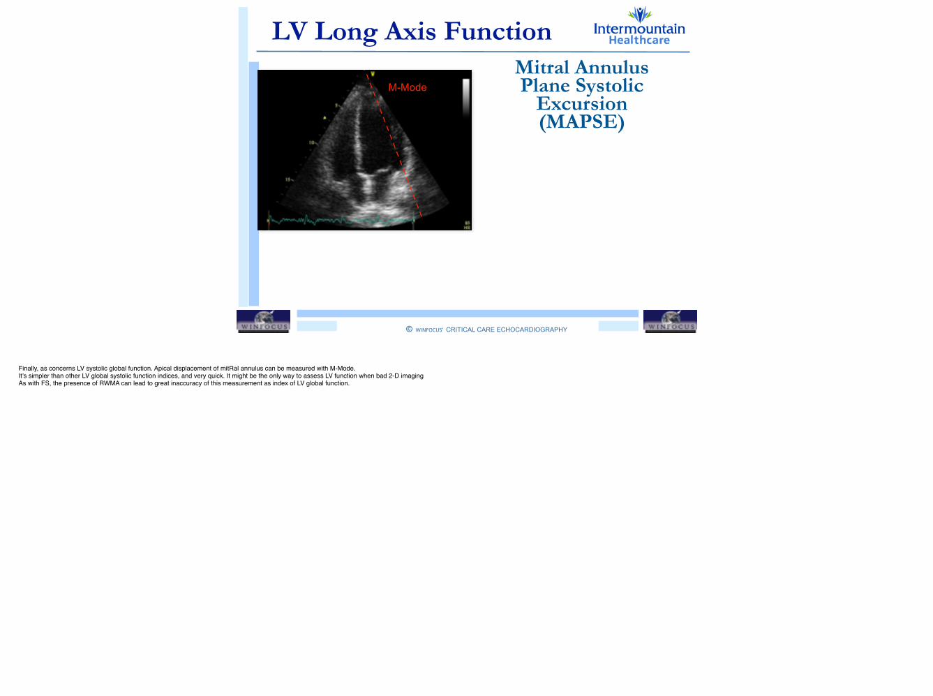
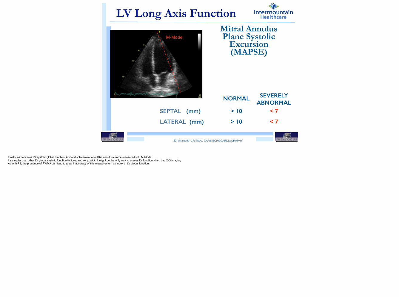
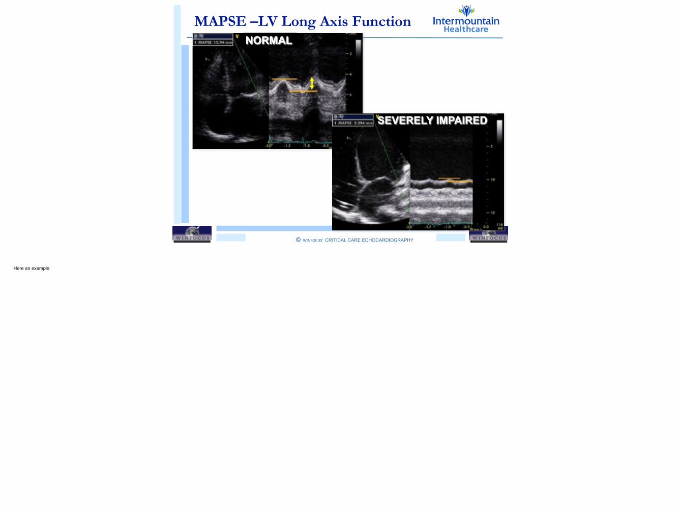
Finally, as concerns LV systolic global function. Apical displacement of mitRal annulus can be measured with M-Mode.!It’s simpler than other LV global systolic function indices, and very quick. It might be the only way to assess LV function when bad 2-D imaging!As with FS, the presence of RWMA can lead to great inaccuracy of this measurement as index of LV global function.
© WINFOCUS’ CRITICAL CARE ECHOCARDIOGRAPHY
!
LV Long Axis Function
M-ModeMitral Annulus Plane Systolic
Excursion (MAPSE)
NORMAL SEVERELY ABNORMAL
SEPTAL (mm) > 10 < 7
LATERAL (mm) > 10 < 7
Unreliable with Regional Wall Motion
Abnormality
Finally, as concerns LV systolic global function. Apical displacement of mitRal annulus can be measured with M-Mode.!It’s simpler than other LV global systolic function indices, and very quick. It might be the only way to assess LV function when bad 2-D imaging!As with FS, the presence of RWMA can lead to great inaccuracy of this measurement as index of LV global function.
© WINFOCUS’ CRITICAL CARE ECHOCARDIOGRAPHY
!
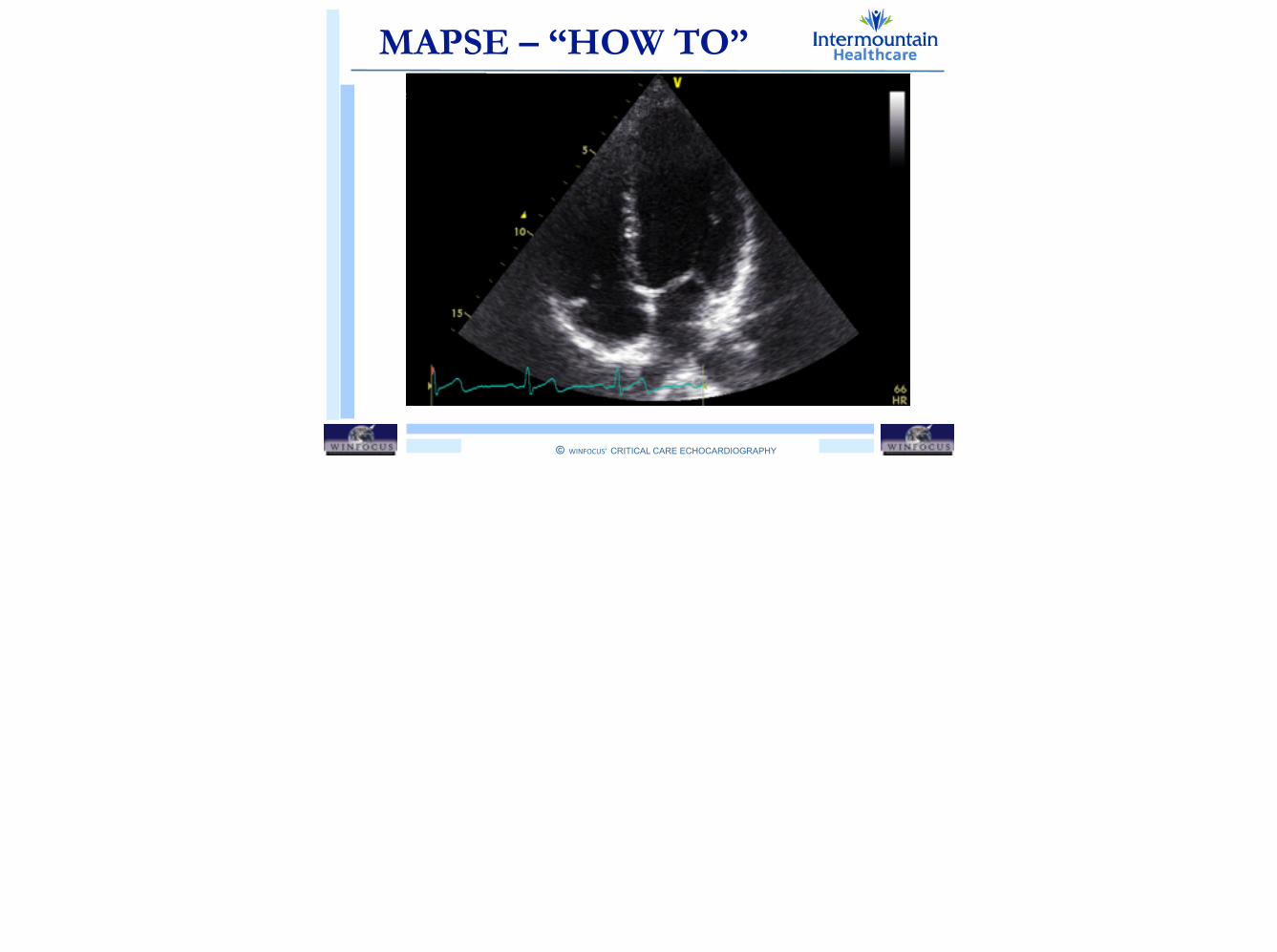
MAPSE – “HOW TO”

© WINFOCUS’ CRITICAL CARE ECHOCARDIOGRAPHY
!
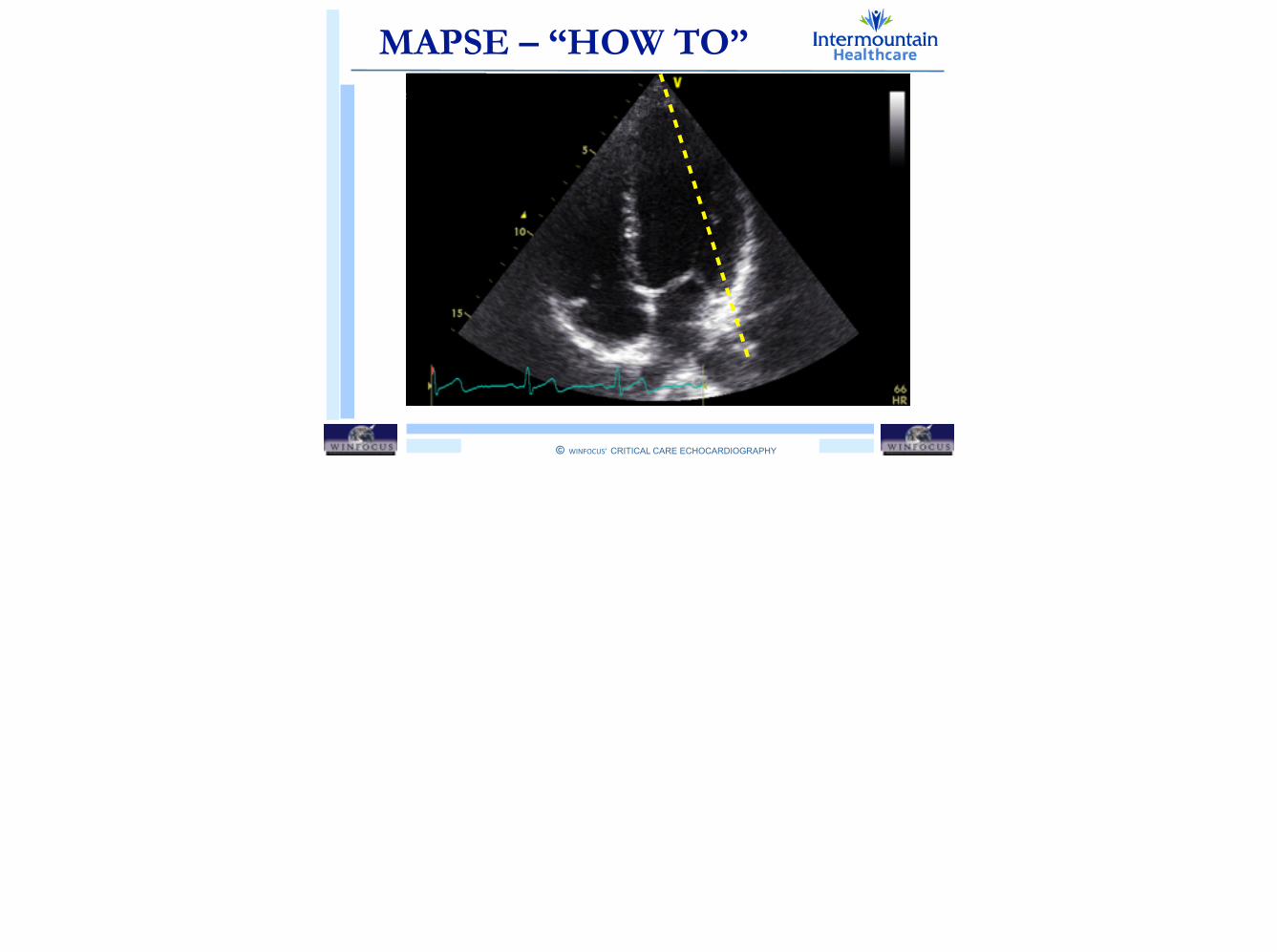
MAPSE – “HOW TO”
© WINFOCUS’ CRITICAL CARE ECHOCARDIOGRAPHY
!
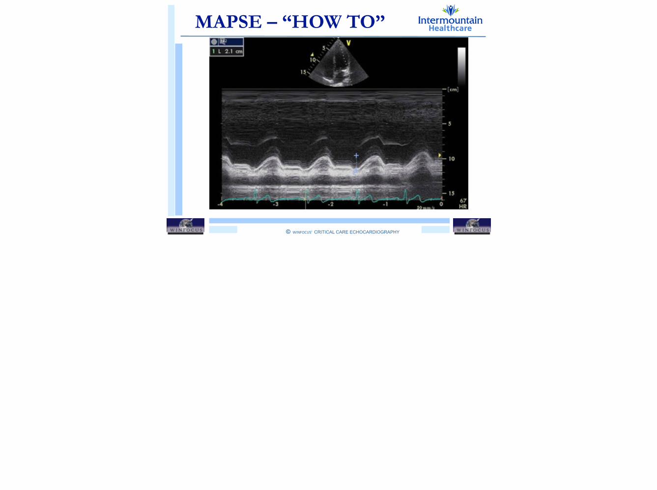
MAPSE – “HOW TO”
© WINFOCUS’ CRITICAL CARE ECHOCARDIOGRAPHY
!
MAPSE – “HOW TO”
© WINFOCUS’ CRITICAL CARE ECHOCARDIOGRAPHY
!
NORMALMAPSE –LV Long Axis Function
SEVERELY IMPAIRED
Here an example

© WINFOCUS’ CRITICAL CARE ECHOCARDIOGRAPHY
!
Outline
LV GLOBAL SYSTOLIC FUNCTION
• Fractional Shortening (FS)
• Fractional Area Change (FAC)
• Volumetric Ejection Fraction (EF)
• Long Axis Function (MAPSE)
© WINFOCUS’ CRITICAL CARE ECHOCARDIOGRAPHY
!
Outline
LV GLOBAL SYSTOLIC FUNCTION
• Fractional Shortening (FS)
• Fractional Area Change (FAC)
• Volumetric Ejection Fraction (EF)
• Long Axis Function (MAPSE)
Recommended