AD_________________
AWARD NUMBER: W81XWH-07-1-0292 TITLE: Mumps Parotitis and Ovarian Cancer: Modern Significance of an Historic Association PRINCIPAL INVESTIGATOR: Daniel W. Cramer, M.D.
CONTRACTING ORGANIZATION: Brigham and Women’s Hospital Boston, MA 02115
REPORT DATE: October 2009 TYPE OF REPORT: Final PREPARED FOR: U.S. Army Medical Research and Materiel Command Fort Detrick, Maryland 21702-5012 DISTRIBUTION STATEMENT: Approved for Public Release; Distribution Unlimited The views, opinions and/or findings contained in this report are those of the author(s) and should not be construed as an official Department of the Army position, policy or decision unless so designated by other documentation.
REPORT DOCUMENTATION PAGE Form Approved
OMB No. 0704-0188 Public reporting burden for this collection of information is estimated to average 1 hour per response, including the time for reviewing instructions, searching existing data sources, gathering and maintaining the data needed, and completing and reviewing this collection of information. Send comments regarding this burden estimate or any other aspect of this collection of information, including suggestions for reducing this burden to Department of Defense, Washington Headquarters Services, Directorate for Information Operations and Reports (0704-0188), 1215 Jefferson Davis Highway, Suite 1204, Arlington, VA 22202-4302. Respondents should be aware that notwithstanding any other provision of law, no person shall be subject to any penalty for failing to comply with a collection of information if it does not display a currently valid OMB control number. PLEASE DO NOT RETURN YOUR FORM TO THE ABOVE ADDRESS. 1. REPORT DATE 1 October 2009
2. REPORT TYPEFinal
3. DATES COVERED 1 Nov 2008 – 30 Sep 2009
4. TITLE AND SUBTITLE
5a. CONTRACT NUMBER
Mumps Parotitis and Ovarian Cancer: Modern Significance of an Historic Association 5b. GRANT NUMBER W81XWH-07-1-0292
5c. PROGRAM ELEMENT NUMBER
6. AUTHOR(S)
5d. PROJECT NUMBER
Daniel W. Cramer, M.D. 5e. TASK NUMBER
E-Mail: [email protected] 5f. WORK UNIT NUMBER7. PERFORMING ORGANIZATION NAME(S) AND ADDRESS(ES) Brigham and Women’s Hospital Boston, MA 02115
8. PERFORMING ORGANIZATION REPORT NUMBER
9. SPONSORING / MONITORING AGENCY NAME(S) AND ADDRESS(ES) 10. SPONSOR/MONITOR’S ACRONYM(S)U.S. Army Medical Research and Materiel Command Fort Detrick, Maryland 21702-5012 11. SPONSOR/MONITOR’S REPORT NUMBER(S)
12. DISTRIBUTION / AVAILABILITY STATEMENT Approved for Public Release; Distribution Unlimited 13. SUPPLEMENTARY NOTES
14. ABSTRACT Epidemiologic studies found childhood mumps might protect against ovarian cancer. We investigated whether mumps might engender immunity against a tumor-like form of the glycoprotein mucin 1 (MUC1) to explain this association. Through various health agencies, we obtained sera that had been saved from 161 individuals with mumps parotitis. Sera from 194 individuals without mumps were assembled from the health agencies, blood bank donors, or university volunteers. We used an ELISA to measure anti-MUC1 antibodies and electro-chemiluminescence assays to measure MUC1 and CA 125. Log-transformed measurements were analyzed by t tests, generalized linear models, and Pearson or Spearman correlations. We also conducted a meta-analysis of published studies regarding mumps and ovarian cancer. From the meta-analysis, the pooled odds ratio estimate (and 95% CL) for the mumps and ovarian cancer association was 0.66 (0.47 to 0.91) (p = 0.01). Adjusting for assay batch, age, and sex, the level of anti-MUC1 antibodies was significantly higher in mumps cases compared to controls (p =0.002). In a subset of cases with sufficient sera remaining, CA 125, but not MUC1, levels were higher in cases. Mumps parotitis may lead to immune recognition of a tumor-like form of MUC1 and create effective immunosurveillance of ovarian cancer cells that express this form of MUC1.
15. SUBJECT TERMS MUC1, Mumps, Ovarian cancer 16. SECURITY CLASSIFICATION OF:
17. LIMITATION OF ABSTRACT
18. NUMBER OF PAGES
19a. NAME OF RESPONSIBLE PERSONUSAMRMC
a. REPORT U
b. ABSTRACT U
c. THIS PAGEU UU 27
19b. TELEPHONE NUMBER (include area code)
Standard Form 298 (Rev. 8-98)Prescribed by ANSI Std. Z39.18
Table of Contents
Page
Introduction…………………………………………………………….………..….. 4
Body………………………………………………………………………………….. 4
Accomplishments…………………………………………………………….…….. 12
Reportable Outcomes……………………………………………………………… 12
Conclusion…………………………………………………………………………… 13
References……………………………………………………………………………. 13
Appendices…………………………………………………………………………… 15
4
Introduction
In one of the earliest case-control studies of ovarian cancer, West observed that women with the disease were less likely to report having had mumps compared to women with benign ovarian cysts(1), suggesting that childhood mumps might protect against the subsequent development of ovarian cancer. Since then, eight additional observational studies addressing mumps and ovarian cancer have been published(2-9); and, in all but two(2, 9), controls were more likely to report a history of mumps than cases, suggesting that mumps might be associated with lower ovarian cancer risk. However, no credible biologic explanation was offered; and, as time passed and with the introduction of mumps vaccination in the late 1960’s, the association between mumps and ovarian cancer was rendered seemingly irrelevant and largely forgotten.
Recently, we have proposed and tested a new hypothesis which can unite many apparently unrelated ovarian cancer risk factors(10). Protective risk factors may work through events that may raise immunity against normal cell molecules that may be abnormally expressed on cells and tissues and also later on nascent ovarian cancer cells making them targets of effective immunosurveillance. As an example of such molecules, we have studied the cell surface glycoprotein and tumor associated antigen, mucin 1 (MUC1). MUC1 (CA 15.3) is a product of the mucin family of genes that also includes MUC16 (CA 125). MUC1 is normally expressed at low levels and in an extensively glycosylated form on epithelia of the genito-urinary, respiratory, and digestive tracts as well as breast ducts. It is over-expressed in a hypoglycosylated (immunogenic) form in most adenocarcinomas, including breast and ovarian tumors. We previously published that (acute) inflammatory events that affect tissues that normally express MUC1, like a tubal ligation or a breast mastitis, found to protect against ovarian cancer, might do so by causing overexpression of the hypoglycosylated form of MUC1 leading to an immune response and immune memory for the type of MUC1 later found on ovarian cancer cells(10). Just as the presence of anti-MUC1 antibodies in cancer patients at diagnosis may be associated with a more favorable disease prognosis(11), pre-existing antibodies might prevent disease in the first place.
Salivary glands also express a low level of the fully glycosylated form of MUC1(12) on the apical ductal surfaces. We reasoned that abnormal expression of MUC1 during a mumps infection might induce anti-MUC1 immunity (measured by anti-MUC1 antibodies) and immune memory, giving a mechanistic explanation to the largely historic and phenomenological association between mumps and ovarian cancer. In the current study, we sought to test this hypothesis by comparing levels of anti- MUC1 antibodies (as well as MUC1 and CA 125 antigens) in samples obtained from patients diagnosed with mumps versus healthy controls. BODY Methods Cases
We surveyed several Regional and State Health Departments about the availability of specimens for research purposes from patients with a mumps infection. Our final sample included a total of 161 patients with clinically diagnosed mumps parotitis from four different agencies, three contributed 149 paired samples (during the acute and convalescent phase of the infection) and one contributed single samples. Thirty-nine paired samples were donated from patients with clinically diagnosed mumps parotitis who reported to the Health Protection Agency Centre for Infections (London, UK). Fifty-two paired samples were obtained from the Specialist Virology Centre Edinburgh collected between fall 2004 and mid 2005. These patients were predominantly young adults and older teenagers born before routine mumps immunization was introduced in the region. Additionally, 58 paired serum samples collected from patients with confirmed or probable mumps infection during a 2007 outbreak were received from The Queen Elizabeth II Health Sciences Centre (Nova Scotia, Canada). These included 35 paired sera from laboratory confirmed cases of mumps and 23 paired sera from patients with parotitis but low mumps titer. Of these 23 samples, 18 had an epidemiologic link to a laboratory confirmed case. Finally, 12 one-time samples obtained at an unknown
5
point of the mumps infection and collected between 2000 and 2008 were provided by the North Dakota Department of Public Health. All samples were shipped to our laboratory on dry-ice via overnight courier. IRBs associated with the local Health departments or hospitals approved release of specimens without identifying information. Only age, sex, and mumps viral titer (where available) were linked to the specimens; and they were therefore considered anonymized and exempt by the IRB at the Brigham and Women’s Hospital and University of Pittsburgh. Table 1 summarizes details regarding these cases. Controls
A total of 194 controls were obtained from four sites. Twelve samples were received from the Department of Immunology at the University of Pittsburgh. These were college-aged students who had been enrolled as controls in various studies in which response to a MUC1 peptide vaccine was being assessed. A total of 67 single serum samples from participants of the health screening program (who did not have mumps parotitis) were obtained from the Queen Elizabeth II Health Sciences Centre (Nova Scotia, Canada). In the course of surveying State Laboratories, the Illinois Department of public health indicated they could not provide mumps specimens but could supply 39 specimens from individuals who were healthy but sought to assess the status of their immunity to mumps. Finally, 76 samples were collected from healthy volunteers in Boston from Research Blood Components, a commercial blood bank in Boston, MA where all blood donors must have been free of symptoms suggestive of a viral illness. As with the mumps cases, all specimens were de-identified and linked only to age at collection and sex. Assays for anti-MUC1 antibodies.
Anti-MUC1 antibodies were measured against a synthetic 100-mer MUC1 peptide corresponding to five tandem repeats of the MUC1 polypeptide core repeat region(10). Briefly, MUC1-coated Immulon wells (Dynax, Chantilly, VA) and peptide-negative plates were incubated overnight and washed three times with PBS before addition of 100 μl of 2.5% bovine serum albumin in PBS. Serially diluted plasma (1:40 to 1:80 in PBS) were added to MUC1-coated plates and incubated at room temperature. Plates were washed 5x with 100 μl PBS and 0.1% Tween 20 detergent. Alkaline phosphatase-labeled goat anti-human polyvalent IgM, IgG, IgA (50 μl) (Sigma-Aldrich, St. Louis, MO) diluted 1:1,000 was added before plates were again washed 5x with PBS-Tween. Alkaline phosphatase substrate pNPP (100 μl) (Sigma-Aldrich) was added. Plates were incubated before the stop solution (0.5 mol/L NaOH) was added. We used the MRX Revelation plate reader (Thermo Labsystems, Chantilly, VA) to read absorbance values at 405 to 410 nm, which were subtracted from absorbance values obtained from antigen-negative plates to account for nonspecific binding.
Laboratory personnel were blinded to case/control status of the samples. Blood specimens from cases and controls as well as paired samples from the same participants were assayed on the same plate, which also included masked quality control samples (3 to 5 replicates per plate). Samples were assayed in two batches. Batch 1 included samples from London and controls from the University of Pittsburg, which were assayed in triplicate and in four dilutions (1:40, 1:80, 1:160, and 1:320). Average values across triplicates were reported for each sample. In order to increase the number of specimens per assay plate (and thus decrease inter-plate variability, samples in batch 2 (which included all the remaining study specimens) were assayed only in two dilutions (1:40 and 1:80). Triplicates of each sample were assayed on different plates and average values of triplicates were reported for each sample. The coefficients of variation for the batch 1 and 2 positive controls were 18% and 4%, respectively. The Spearman rank correlation between the 1:40 and 1:80 dilutions was 0.95. Readings at 1:40 were used for the current analysis. Assays for CA 125 and MUC1
Serum levels of CA 125 and MUC1 were measured using electro-chemiluminescence (ECL) assays and Imager 2400 (Meso Scale Discovery, Gaithersburg, MD, USA). The ECL platform allows assays using very small volumes and has been validated against traditional ELISA (13). The linearity ranges were 1.2 to 5000 U/ml for the CA 125 and 0.98 to 4000 mU/ml for the MUC1 assay. The serum samples were tested undiluted for the CA 125 and diluted 1:200 for the MUC1 assay. A positive quality control (QC) sample was run on each plate in duplicate. The QC sample had a mean CA 125 concentration of 450.47 U/ml and a mean
6
MUC1 concentration of 63.73 U/ml. The coefficient of variation was calculated as 100*(SD/average) for each assay plate and between plates. The intra-plate CV% for CA 125 varied from 2.2% to 13.1% with inter-plate CV=9.1%. The intra-plate CV% for MUC1 varied from 0% to 4.7% with inter-plate CV=10.97%. Meta-analysis
We conducted a systematic literature search of published studies reporting on the association between history of mumps parotitis and subsequent development of ovarian cancer. On June of 2009, we searched MEDLINE (via PubMed), Web of Science and EMBASE using the search terms mumps, parotitis, ovarian neoplasms, ovarian cancer. No limits on publication dates or on language were posed. This search yielded a total of 34 references, nine of which referred to original contributions. Of these, one(1) did not provide estimates for the association between mumps parotitis and ovarian cancer; and, therefore, was not included in the meta-analysis, allowing 8 studies to be included in the analysis. We estimated a summary odds ratio of ovarian cancer and the associated 95% confidence interval across studies using a random effect model, which assumes that the true effects are normally distributed.(14) Statistical heterogeneity was assessed using the I2 statistic. All p-values are two-sided with significance levels set at less than 0.05. Statistical Analyses
The anti-MUC1 antibody, MUC1, and CA 125 distributions were skewed right, so we first log transformed the values to normalize their distributions. After verifying that there were no significant differences, acute and convalescent values were averaged for sites where paired samples were available (London, Edinburgh, Nova Scotia). We examined geometric mean levels by case-control status and used t-tests to assess differences by case-control status stratified by lab batch number, site, age and sex. We used linear regression to examine the mean difference in anti-MUC1 antibody or MUC1 and CA 125 antigen levels, adjusted for age, sex and lab batch. For sites with continuous mumps titer data available (London and Edinburgh), we used Spearman rank correlations to assess the relationship between titer levels and anti-MUC1 antibody levels. Pearson correlations were used to examine the relationship between anti-MUC1 antibody levels and MUC1 antigen levels. The SAS version 9.1 statistical package (SAS Institute, Cary, NC) was used for all analyses. Results
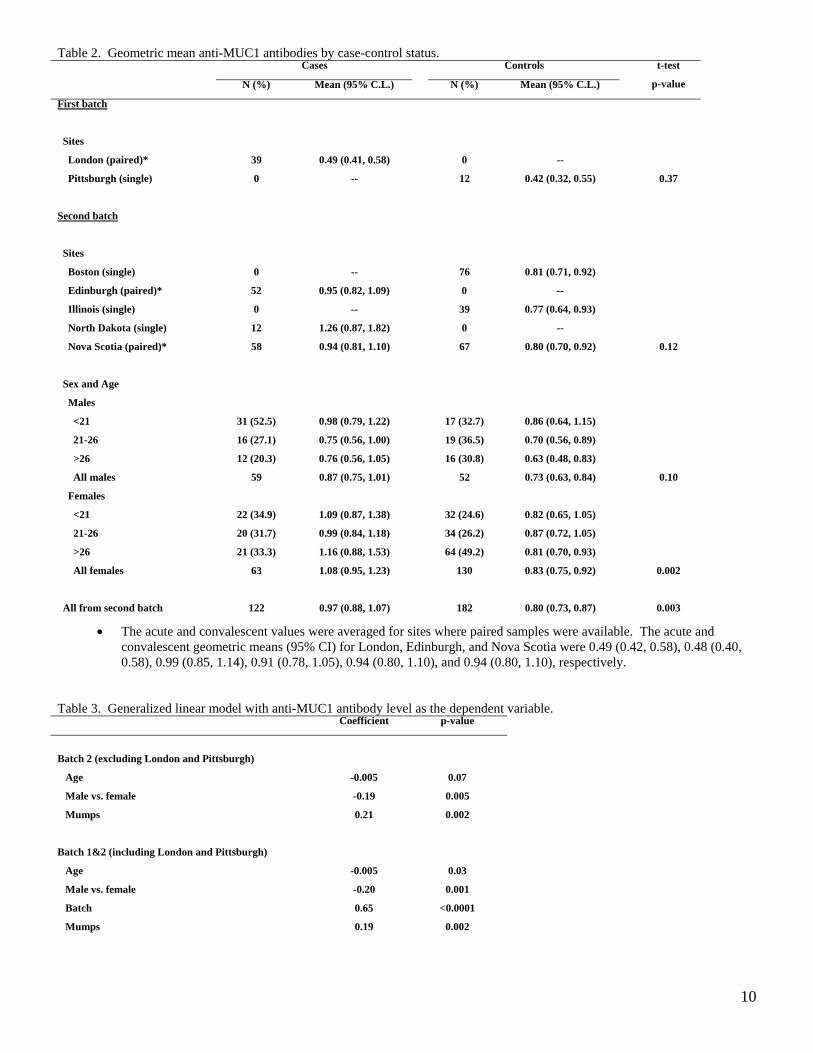
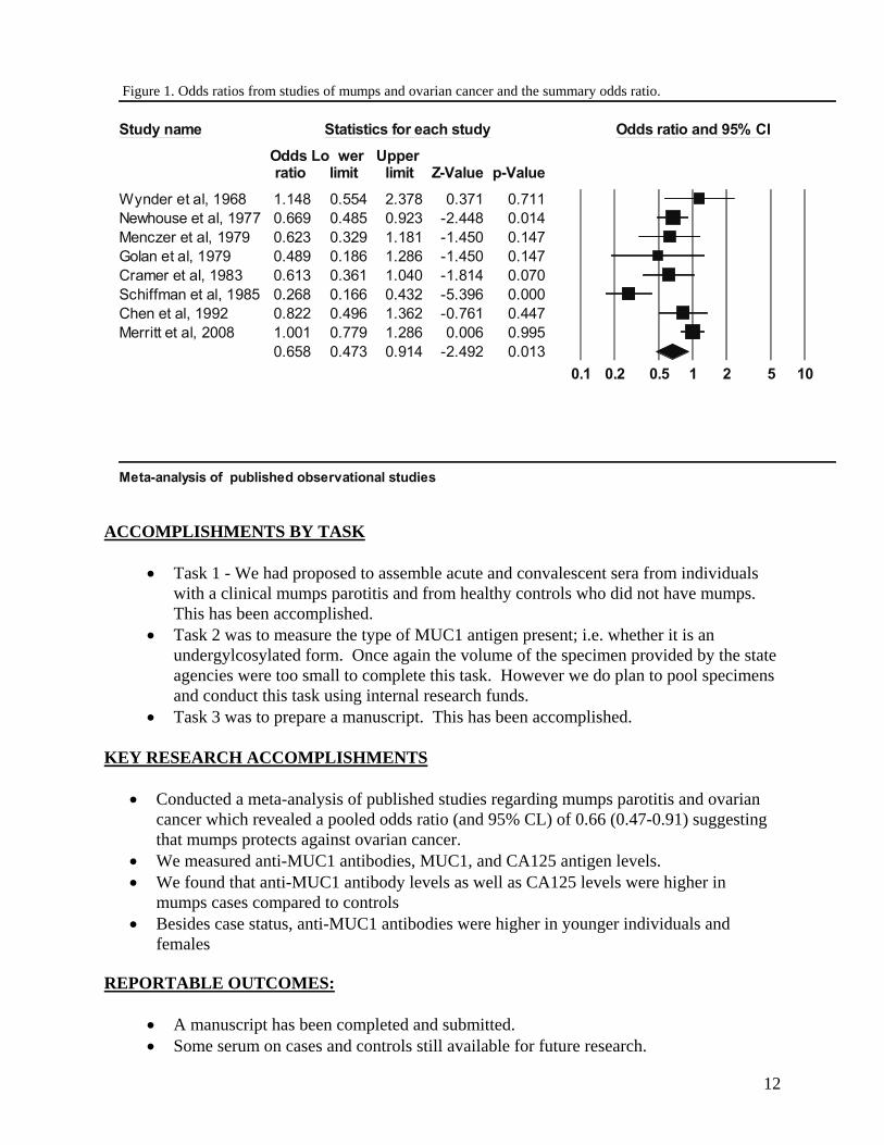
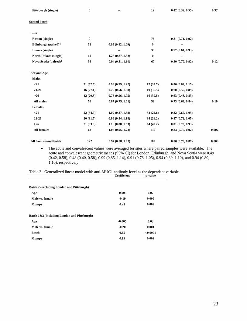
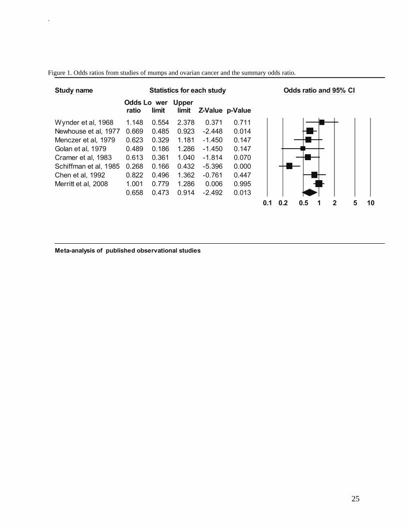
We identified eight epidemiologic studies that provided odds ratios for the association between mumps parotitis and ovarian cancer(2-8). In all but two of these studies (2), controls were more likely to report a history of mumps than cases. From these studies, using a random effects model, we estimated that the overall risk for ovarian cancer associated with history of mumps parotitis was 0.66 with 95% confidence interval of 0.47-0.91 (p = 0.01), suggesting that mumps is significantly and inversely associated with ovarian cancer risk (Figure 1). No significant differences in anti-MUC1 antibody levels were observed between acute and convalescent specimens from those sites providing paired samples (see footnote to Table 2). Therefore, the values were averaged for each subject. Geometric mean anti-MUC1 antibody levels are described in Table 2 by batch and mumps status. There was a clear batch effect for the mumps samples run first with London cases and Pittsburgh controls; both groups having lower mean levels compared to the larger number of specimens run in the second batch. Some variation was noted in anti-MUC1 antibody levels in mumps cases by the source of the specimens contributed to batch 2, but this reflected differences in the composition of the samples by age and sex further illustrated in Table 2. In general, males had lower levels of anti-MUC1 antibodies than females and antibody levels appeared to decline with age in the male cases and controls. In all age and sex categories from batch 2, the levels of anti-MUC1 antibodies were higher in the mumps cases compared to controls (p = 0.003). As illustrated in Table 3, mumps was a significant predictor of higher anti-MUC1 antibody levels after adjustment for age and sex in generalized linear models either restricted to batch 2 data (p = 0.002) or in a second model which included batch 1 data, as well as a variable for batch in the model (p = 0.002). The models confirmed that age and sex were also significant predictors with higher anti-MUC1 antibody levels in younger individuals and women.
7
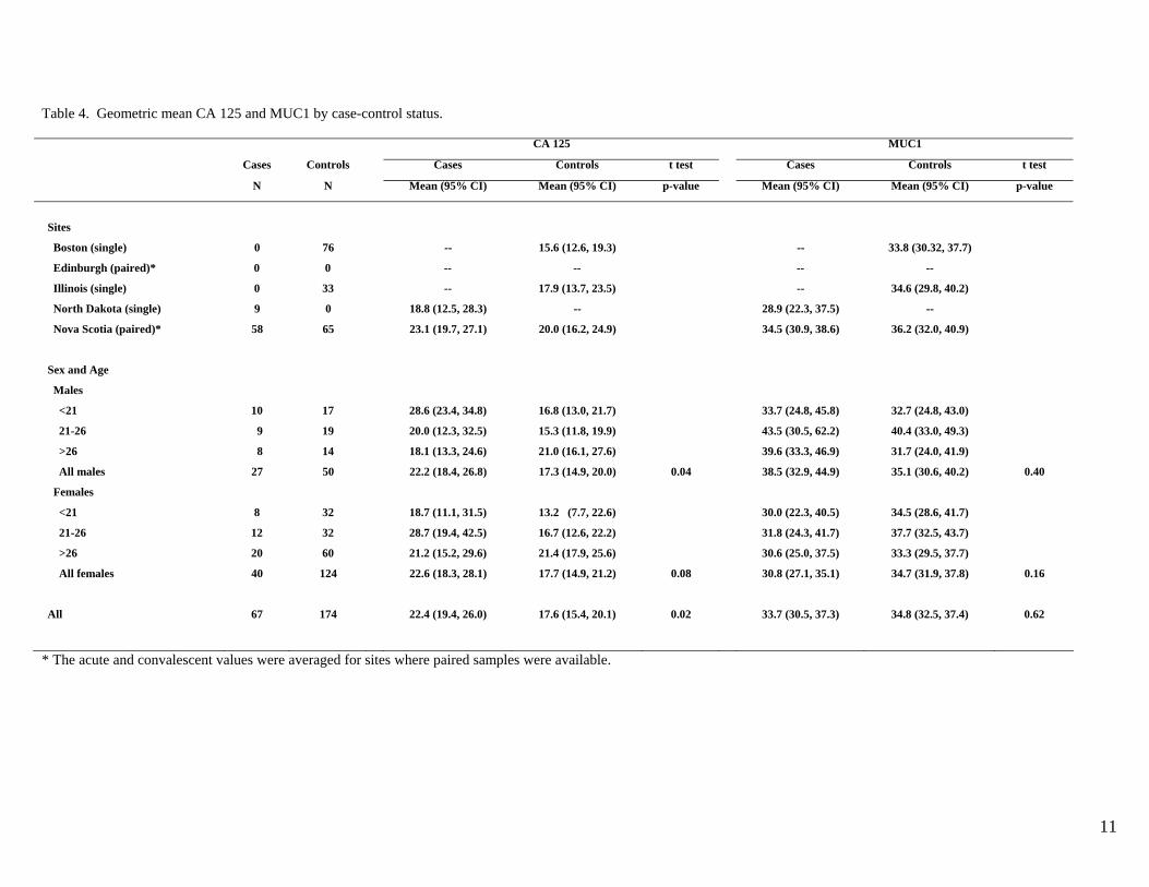
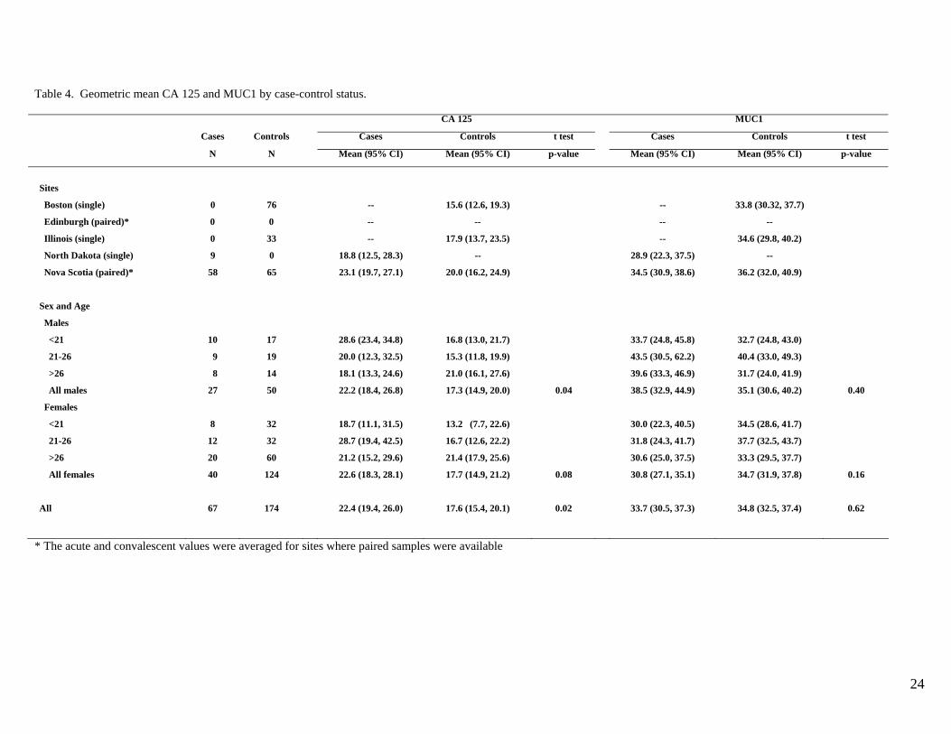
We also examined whether CA 125 or MUC1 antigen levels differed between mumps cases and controls. For many of the samples, including all of those from London and Edinburgh, there was insufficient volume remaining after the anti-MUC1 antibody measurement for the CA 125 and MUC1 measurements. No significant differences were observed for MUC1 levels but, as shown in Table 4, mumps cases had higher CA 125 levels than controls (p = 0.02). Neither age nor sex influenced MUC1 or CA 125 levels in this small series. No significant correlations were observed between mumps viral titers supplied for the London and Edinburgh cases and anti-MUC1 antibody levels (data not shown). These were the same cases with insufficient volume to measure MUC1 and CA 125, so the correlations of the antigens with viral titers could not be examined. We examined the correlation between anti-MUC1 antibody levels and MUC1 antigen levels. There was a weak but significant inverse correlation between antigen and antibody levels for mumps cases (r = -0.24, p = 0.05), but not controls (r = -0.09, p = 0.21).
Discussion
In this study, we found that sera from individuals during (or just after) symptomatic mumps parotitis have a significantly higher level of anti-MUC1 antibodies than sera from controls without active parotitis. Showing the relevance of this observation to ovarian cancer clearly requires background data that would link mumps, ovarian cancer, and anti-MUC1 antibodies. In the introduction we briefly reviewed the epidemiologic data that history of childhood mumps parotitis was inversely associated with risk for ovarian cancer. As part of this study we performed a meta-analysis of published studies to show the consistency of this observation and obtain an overall estimate of the effect. In eight observational studies addressing the association, the summary odds ratio was 0.66 with 95% confidence limits of 0.47-0.91 (p = 0.01) suggesting a 34% decrease in risk for ovarian cancer associated with history of mumps parotitis.
Despite epidemiologic evidence that mumps reduced the risk for ovarian cancer, this association has largely been forgotten, probably due to the lack of a plausible biologic explanation. We have presented data here in support of an immunological basis for the association. A protective effect of mumps parotitis on ovarian cancer risk can be explained under a model related to immunity against the surface glycoprotein, MUC1. Events affecting tissues that normally express MUC1, like a tubal ligation or a breast mastitis which have been shown to protect against ovarian cancer(10), might confer protection because of injury to the tissue causing expression and presentation of a tumor-like (less glycosylated) form of MUC1 to the immune system. This would allow immune recognition of the protein core of MUC1 and generation of an immune response.(15, 16) Since MUC1 is expressed in normal salivary glands, acute inflammation of this tissue with mumps would be expected to induce similar changes in MUC1 levels and glycosylation leading to an antibody response. Inflammation-induced overexpression and hypoglycosylation of MUC1 has already been reported(17-21). Thus, our observation that anti-MUC1 antibodies are elevated in individuals with mumps is consistent with the interpretation that mumps infection causes similar changes that elicit an immune response that could later protect against ovarian cancer. We began this study with the expectation that, in mumps cases with paired sera, we would find higher MUC1 levels in the acute specimen and higher anti-MUC1 antibody levels in the convalescent specimen. Neither was observed. The fact that anti-MUC1 antibody levels were similar (but higher than controls) in both the acute and convalescent sera could be explained if this were not the first time that the mumps cases had seen an inflammatory type of MUC1. In this circumstance, a much more rapid immune response would be expected and might have occurred because of prior inflammatory conditions involving the genito-urinary, respiratory, digestive tract, or breast ducts—tissues which all express MUC1. MUC1 antigen itself was not elevated in cases compared to controls in either the acute or convalescent specimens. It is possible that blood obtained at even earlier stages of the disease might have been revealing or that saliva would have been a better body fluid to look for MUC1. It is also possible that the presence of anti-MUC1 antibodies interfered with measurement of MUC1 since immune complexes involving MUC1 and antibodies against it have been described(22). Supporting this possibility was an inverse correlation between anti-MUC1 antibody levels and MUC1 antigen levels in cases noted in our presentation of the results of Table 4. The low volume of mumps specimens precluded measurement of immune complexes. Interestingly, CA 125 was significantly elevated in sera from the
8
mumps cases compared to controls. CA 125 is expressed in salivary gland tumors(23); but we could find no reports of its expression in normal salivary glands. There are many limitations of this study, not the least of which was the difficulty of obtaining specimens from individuals with mumps parotitis. The samples we obtained were collected between 2000 and 2008 and were stored under variable conditions. Titer data were available from only two sites (London and Edinburgh). Although age and sex of the cases was known, information on precisely when during the course of the infection the samples were collected was quite limited.
Regarding controls, we are certain they did not have an active mumps infection; but it was a diverse group which included university students for the London cases in batch 1 and, in batch 2, individuals without mumps in Illinois who were tested for immunity and blood bank controls from Boston. Community matched controls were only available for the Nova Scotia subjects in batch 2. Despite this, the levels of anti-MUC1 antibodies in all control groups from batch 2 were very similar and nearly identical to the Nova Scotia controls. Another limitation was variability in the assay from batch 1 to batch 2. To decrease inter-plate variability after running batch 1, we increased the number of unique specimens per plate for batch 2, which may have contributed to the batch differences. However despite batch variation, both batches suggested higher antibody levels in mumps cases compared to controls. Moreover, analyses including and excluding batch 1 yielded a similar result.
The epidemiology of mumps parotitis has obviously changed dramatically in the last 40 years. Mumps parotitis was a very common illness in infants and children prior to 1970. With now close to universal vaccination except in the third world, mumps has become a disease of adults who were either born too early for routine vaccination or who have lost immunity after vaccination. For this reason, inferences about the consequences of parotitis on MUC1 immunity based on observations from the specimens tested here may not be generalizable to what might have occurred with childhood infection before vaccination programs began. We point out, however, that the anti-MUC1 antibody response was most robust in younger women suggesting that childhood infection, as would have occurred in the past, might have been the optimum time for engendering immunity against ovarian cancer.
If it is true that symptomatic mumps protected against ovarian cancer through an immune reaction, a logical consequence is that we might expect an increased incidence of ovarian cancer as symptomatic mumps parotitis infections have decreased through vaccination. In a paper examining incidence patterns for ovarian cancer from 1978 to 1998, rates of invasive serous, endometrioid, and clear cell tumor increased over this time period among white females (24). Endometrioid and clear cell cancers are the types of ovarian cancer that we found were most strongly linked to the conditions we proposed might be mediated through anti-MUC1 antibodies (10). However, these incidence data are confounded by trends towards more precise pathology coding that occurred over the same period. A re-examination of these trends with more recent data and a focus on the birth cohorts most likely to been vaccinated is underway.
Readers will need to judge whether or not we have made a convincing case that the protective association between mumps and ovarian cancer is real and that an immunologic connection with MUC1 (or other mucins) explains it. The interpretation we do not wish readers to take from this report is that we are suggesting childhood vaccination for mumps should be abandoned. Prior to vaccination, mumps was generally a mild illness but could have serious sequelae including orchitis and sterility, meningitis and deafness, and pancreatitis. Nevertheless, our study suggests there could also have been unanticipated long term benefits of a mumps infection, such as we have described in this paper. Understanding the scope of and basis for the potential benefits of childhood infections may allow immunologists to duplicate the beneficial effects at the same time as providing the means for avoiding a natural infection and its possible adverse consequences. Further study of individuals going through a mumps infection, especially with a focus on mucin immunity, may provide clues to pathways for duplicating the beneficial effects of mumps parotitis suggested by this study.
9
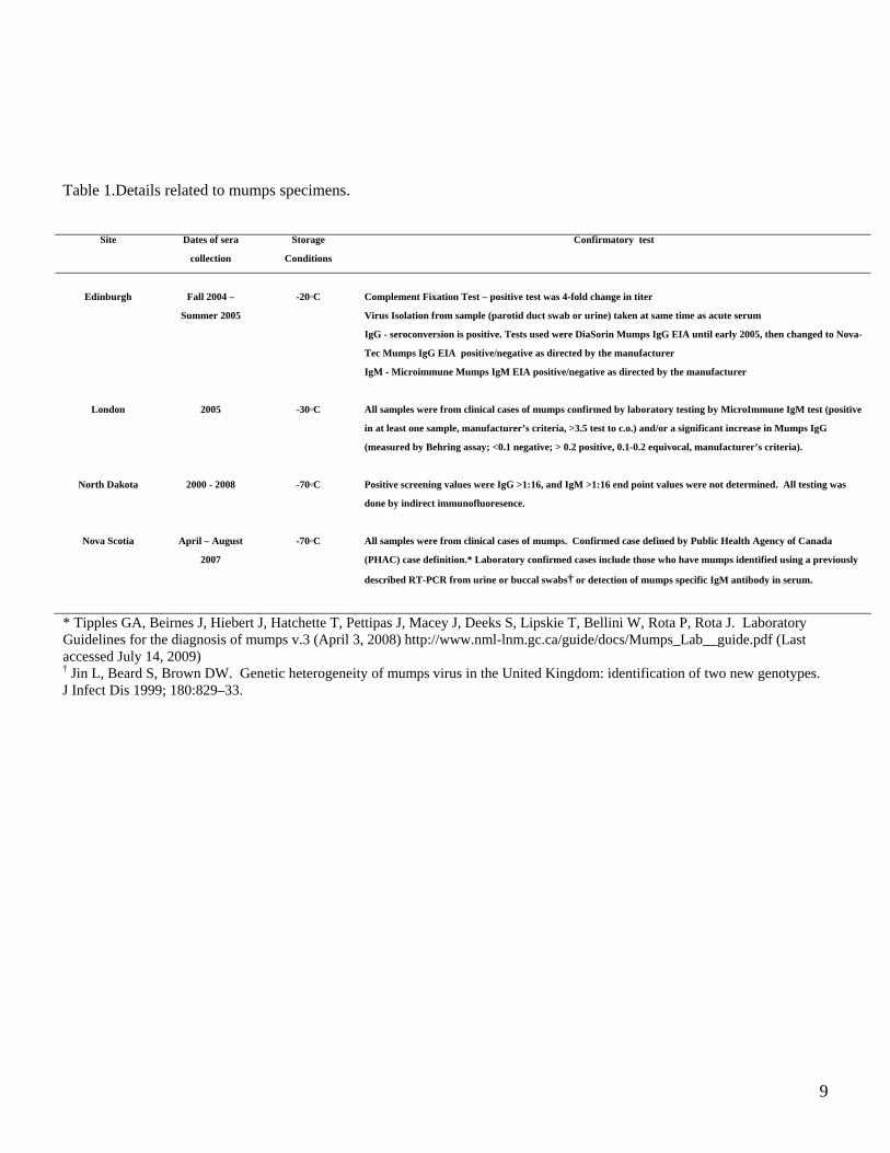
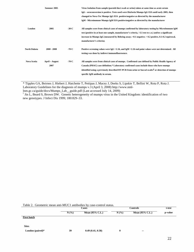
Table 1.Details related to mumps specimens.
Site Dates of sera
collection
Storage
Conditions
Confirmatory test
Edinburgh Fall 2004 –
Summer 2005
-20◦C Complement Fixation Test – positive test was 4-fold change in titer
Virus Isolation from sample (parotid duct swab or urine) taken at same time as acute serum
IgG - seroconversion is positive. Tests used were DiaSorin Mumps IgG EIA until early 2005, then changed to Nova-
Tec Mumps IgG EIA positive/negative as directed by the manufacturer
IgM - Microimmune Mumps IgM EIA positive/negative as directed by the manufacturer
London 2005 -30◦C All samples were from clinical cases of mumps confirmed by laboratory testing by MicroImmune IgM test (positive
in at least one sample, manufacturer’s criteria, >3.5 test to c.o.) and/or a significant increase in Mumps IgG
(measured by Behring assay; <0.1 negative; > 0.2 positive, 0.1-0.2 equivocal, manufacturer’s criteria).
North Dakota 2000 - 2008 -70◦C Positive screening values were IgG >1:16, and IgM >1:16 end point values were not determined. All testing was
done by indirect immunofluoresence.
Nova Scotia April – August
2007
-70◦C All samples were from clinical cases of mumps. Confirmed case defined by Public Health Agency of Canada
(PHAC) case definition.* Laboratory confirmed cases include those who have mumps identified using a previously
described RT-PCR from urine or buccal swabs† or detection of mumps specific IgM antibody in serum.
* Tipples GA, Beirnes J, Hiebert J, Hatchette T, Pettipas J, Macey J, Deeks S, Lipskie T, Bellini W, Rota P, Rota J. Laboratory Guidelines for the diagnosis of mumps v.3 (April 3, 2008) http://www.nml-lnm.gc.ca/guide/docs/Mumps_Lab__guide.pdf (Last accessed July 14, 2009) † Jin L, Beard S, Brown DW. Genetic heterogeneity of mumps virus in the United Kingdom: identification of two new genotypes. J Infect Dis 1999; 180:829–33.
10
Table 2. Geometric mean anti-MUC1 antibodies by case-control status. Cases Controls
N (%) Mean (95% C.L.) N (%) Mean (95% C.L.)
t-test
p-value
First batch
Sites
London (paired)* 39 0.49 (0.41, 0.58) 0 --
Pittsburgh (single) 0 -- 12 0.42 (0.32, 0.55) 0.37
Second batch
Sites
Boston (single) 0 -- 76 0.81 (0.71, 0.92)
Edinburgh (paired)* 52 0.95 (0.82, 1.09) 0 --
Illinois (single) 0 -- 39 0.77 (0.64, 0.93)
North Dakota (single) 12 1.26 (0.87, 1.82) 0 --
Nova Scotia (paired)* 58 0.94 (0.81, 1.10) 67 0.80 (0.70, 0.92) 0.12
Sex and Age
Males
<21 31 (52.5) 0.98 (0.79, 1.22) 17 (32.7) 0.86 (0.64, 1.15)
21-26 16 (27.1) 0.75 (0.56, 1.00) 19 (36.5) 0.70 (0.56, 0.89)
>26 12 (20.3) 0.76 (0.56, 1.05) 16 (30.8) 0.63 (0.48, 0.83)
All males 59 0.87 (0.75, 1.01) 52 0.73 (0.63, 0.84) 0.10
Females
<21 22 (34.9) 1.09 (0.87, 1.38) 32 (24.6) 0.82 (0.65, 1.05)
21-26 20 (31.7) 0.99 (0.84, 1.18) 34 (26.2) 0.87 (0.72, 1.05)
>26 21 (33.3) 1.16 (0.88, 1.53) 64 (49.2) 0.81 (0.70, 0.93)
All females 63 1.08 (0.95, 1.23) 130 0.83 (0.75, 0.92) 0.002
All from second batch 122 0.97 (0.88, 1.07) 182 0.80 (0.73, 0.87) 0.003
The acute and convalescent values were averaged for sites where paired samples were available. The acute and convalescent geometric means (95% CI) for London, Edinburgh, and Nova Scotia were 0.49 (0.42, 0.58), 0.48 (0.40, 0.58), 0.99 (0.85, 1.14), 0.91 (0.78, 1.05), 0.94 (0.80, 1.10), and 0.94 (0.80, 1.10), respectively.
Table 3. Generalized linear model with anti-MUC1 antibody level as the dependent variable. Coefficient p-value
Batch 2 (excluding London and Pittsburgh)
Age -0.005 0.07
Male vs. female -0.19 0.005
Mumps 0.21 0.002
Batch 1&2 (including London and Pittsburgh)
Age -0.005 0.03
Male vs. female -0.20 0.001
Batch 0.65 <0.0001
Mumps 0.19 0.002
11
Table 4. Geometric mean CA 125 and MUC1 by case-control status. CA 125 MUC1
Cases Controls Cases Controls t test Cases Controls t test
N N Mean (95% CI) Mean (95% CI) p-value Mean (95% CI) Mean (95% CI) p-value
Sites
Boston (single) 0 76 -- 15.6 (12.6, 19.3) -- 33.8 (30.32, 37.7)
Edinburgh (paired)* 0 0 -- -- -- --
Illinois (single) 0 33 -- 17.9 (13.7, 23.5) -- 34.6 (29.8, 40.2)
North Dakota (single) 9 0 18.8 (12.5, 28.3) -- 28.9 (22.3, 37.5) --
Nova Scotia (paired)* 58 65 23.1 (19.7, 27.1) 20.0 (16.2, 24.9) 34.5 (30.9, 38.6) 36.2 (32.0, 40.9)
Sex and Age
Males
<21 10 17 28.6 (23.4, 34.8) 16.8 (13.0, 21.7) 33.7 (24.8, 45.8) 32.7 (24.8, 43.0)
21-26 9 19 20.0 (12.3, 32.5) 15.3 (11.8, 19.9) 43.5 (30.5, 62.2) 40.4 (33.0, 49.3)
>26 8 14 18.1 (13.3, 24.6) 21.0 (16.1, 27.6) 39.6 (33.3, 46.9) 31.7 (24.0, 41.9)
All males 27 50 22.2 (18.4, 26.8) 17.3 (14.9, 20.0) 0.04 38.5 (32.9, 44.9) 35.1 (30.6, 40.2) 0.40
Females
<21 8 32 18.7 (11.1, 31.5) 13.2 (7.7, 22.6) 30.0 (22.3, 40.5) 34.5 (28.6, 41.7)
21-26 12 32 28.7 (19.4, 42.5) 16.7 (12.6, 22.2) 31.8 (24.3, 41.7) 37.7 (32.5, 43.7)
>26 20 60 21.2 (15.2, 29.6) 21.4 (17.9, 25.6) 30.6 (25.0, 37.5) 33.3 (29.5, 37.7)
All females 40 124 22.6 (18.3, 28.1) 17.7 (14.9, 21.2) 0.08 30.8 (27.1, 35.1) 34.7 (31.9, 37.8) 0.16
All 67 174 22.4 (19.4, 26.0) 17.6 (15.4, 20.1) 0.02 33.7 (30.5, 37.3) 34.8 (32.5, 37.4) 0.62
* The acute and convalescent values were averaged for sites where paired samples were available.
12
Figure 1. Odds ratios from studies of mumps and ovarian cancer and the summary odds ratio.
Study name Statistics for each study Odds ratio and 95% CI
Odds Lo wer Upper ratio limit limit Z-Value p-Value
Wynder et al, 1968 1.148 0.554 2.378 0.371 0.711Newhouse et al, 1977 0.669 0.485 0.923 -2.448 0.014Menczer et al, 1979 0.623 0.329 1.181 -1.450 0.147Golan et al, 1979 0.489 0.186 1.286 -1.450 0.147Cramer et al, 1983 0.613 0.361 1.040 -1.814 0.070Schiffman et al, 1985 0.268 0.166 0.432 -5.396 0.000Chen et al, 1992 0.822 0.496 1.362 -0.761 0.447Merritt et al, 2008 1.001 0.779 1.286 0.006 0.995
0.658 0.473 0.914 -2.492 0.0130.1 0.2 0.5 1 2 5 10
Meta-analysis of published observational studies
ACCOMPLISHMENTS BY TASK
Task 1 - We had proposed to assemble acute and convalescent sera from individuals with a clinical mumps parotitis and from healthy controls who did not have mumps. This has been accomplished.
Task 2 was to measure the type of MUC1 antigen present; i.e. whether it is an undergylcosylated form. Once again the volume of the specimen provided by the state agencies were too small to complete this task. However we do plan to pool specimens and conduct this task using internal research funds.
Task 3 was to prepare a manuscript. This has been accomplished.
KEY RESEARCH ACCOMPLISHMENTS
Conducted a meta-analysis of published studies regarding mumps parotitis and ovarian cancer which revealed a pooled odds ratio (and 95% CL) of 0.66 (0.47-0.91) suggesting that mumps protects against ovarian cancer.
We measured anti-MUC1 antibodies, MUC1, and CA125 antigen levels. We found that anti-MUC1 antibody levels as well as CA125 levels were higher in
mumps cases compared to controls Besides case status, anti-MUC1 antibodies were higher in younger individuals and
females REPORTABLE OUTCOMES:
A manuscript has been completed and submitted. Some serum on cases and controls still available for future research.
13
CONCLUSIONS:
Mumps parotitis may lead to immune recognition of a tumor-like form of MUC1 and create effective immunosurveillance of ovarian cancer cells that express this form of MUC1.
Documentation of the precise immune changes that occur during mumps parotitis may provide a better basis for understanding (and perhaps duplicating) this possible beneficial effect of a natural infection,
The above research may be very important since mumps parotitis has been virtually eliminated in the Western World through vaccination. Increases in ovarian cancer (and possibly other MUC1 related cancers) might be a consequence without a means to duplicate the protective effect of an natural infection.
REFERENCES:
1. West RO (1966) Epidemiology study of malignancies of the ovaries. Cancer 19: 1001-1007.
2. Wynder EL, Dodo H, Barber HR (1969) Epidemiology of cancer of the ovary. Cancer 23: 352-70.
3. Newhouse ML, Pearson RM, Fullerton JM, Boesen EA, Shannon HS (1977) A case control study of carcinoma of the ovary. Br J Prev Soc Med 31: 148-53.
4. Menczer J, Modan M, Ranon L, Golan A (1979) Possible role of mumps virus in the etiology of ovarian cancer. Cancer 43: 1375-9.
5. Golan A, Joosting AC, Orchard ME (1979) Mumps virus and ovarian cancer. S Afr Med J 56: 18-20.
6. Cramer DW, Welch WR, Cassells S, Scully RE (1983) Mumps, menarche, menopause, and ovarian cancer. Am J Obstet Gynecol 147: 1-6.
7. Schiffman MH, Hartge P, Lesher LP, McGowan L (1985) Mumps and postmenopausal ovarian cancer. Am J Obstet Gynecol 152: 116-8.
8. Chen Y, Wu PC, Lang JH, Ge WJ, Hartge P, Brinton LA (1992) Risk factors for epithelial ovarian cancer in Beijing, China. Int J Epidemiol 21: 23-9.
9. Merritt MA, Green AC, Nagle CM, Webb PM (2008) Talcum powder, chronic pelvic inflammation and NSAIDs in relation to risk of epithelial ovarian cancer. Int J Cancer 122: 170-6.
10. Cramer DW, Titus-Ernstoff L, McKolanis JR, Welch WR, Vitonis AF, Berkowitz RS, et al. (2005) Conditions associated with antibodies against the tumor-associated antigen MUC1 and their relationship to risk for ovarian cancer. Cancer Epidemiol Biomarkers Prev 14: 1125-31.
11. Hamanaka Y, Suehiro Y, Fukui M, Shikichi K, Imai K, Hinoda Y (2003) Circulating anti-MUC1 IgG antibodies as a favorable prognostic factor for pancreatic cancer. Int J Cancer 103: 97-100.
12. Alos L, Lujan B, Castillo M, Nadal A, Carreras M, Caballero M, et al. (2005) Expression of membrane-bound mucins (MUC1 and MUC4) and secreted mucins (MUC2, MUC5AC, MUC5B, MUC6 and MUC7) in mucoepidermoid carcinomas of salivary glands. Am J Surg Pathol 29: 806-13.
13. Fichorova RN, Richardson-Harman N, Alfano M, Belec L, Carbonneil C, Chen S, et al. (2008) Biological and technical variables affecting immunoassay recovery of cytokines from human serum and simulated vaginal fluid: a multicenter study. Anal Chem 80: 4741-51.
14
14. Egger M, Smith G, Altman D. Systematic Reviews in Health Care: Meta-Analysis in Context. 2nd Edition ed. Tavistock Square, London: BMJ Publishing Group; 2001.
15. Baldus SE, Engelmann K, Hanisch FG (2004) MUC1 and the MUCs: a family of human mucins with impact in cancer biology. Crit Rev Clin Lab Sci 41: 189-231.
16. Vlad AM, Finn OJ (2004) Glycoprotein tumor antigens for immunotherapy of breast cancer. Breast Dis 20: 73-9.
17. Campbell BJ, Yu LG, Rhodes JM (2001) Altered glycosylation in inflammatory bowel disease: a possible role in cancer development. Glycoconj J 18: 851-8.
18. Jerome KR, Kirk AD, Pecher G, Ferguson WW, Finn OJ (1997) A survivor of breast cancer with immunity to MUC-1 mucin, and lactational mastitis. Cancer Immunol Immunother 43: 355-60.
19. Nakajima M, Manabe T, Niki Y, Matsushima T (1998) Serum KL-6 level as a monitoring marker in a patient with pulmonary alveolar proteinosis. Thorax 53: 809-11.
20. Takaishi H, Ohara S, Hotta K, Yajima T, Kanai T, Inoue N, et al. (2000) Circulating autoantibodies against purified colonic mucin in ulcerative colitis. J Gastroenterol 35: 20-7.
21. Kohno N (1999) Serum marker KL-6/MUC1 for the diagnosis and management of interstitial pneumonitis. J Med Invest 46: 151-158.
22. Vlad AM, Muller S, Cudic M, Paulsen H, Otvos L, Jr., Hanisch FG, et al. (2002) Complex carbohydrates are not removed during processing of glycoproteins by dendritic cells: processing of tumor antigen MUC1 glycopeptides for presentation to major histocompatibility complex class II-restricted T cells. J Exp Med 196: 1435-46.
23. Okamura K, Kiyoshima T, Shima K, Kobayashi I, Matsuo K, Ishibashi H, et al. (2002) Immunohistochemical expression of CA19-9 and CA125 in mucoepidermoid and adenoid cystic carcinomas of the salivary gland. Oral Oncol 38: 244-50.
24. Mink PJ, Sherman ME, Devesa SS (2002) Incidence patterns of invasive and borderline ovarian tumors among white women and black women in the United States. Results from the SEER Program, 1978-1998. Cancer 95: 2380-9.
15
Appendices :
Mumps and Ovarian Cancer: Modern Interpretation of an Historic Association
Daniel W. Cramer1 Allison F. Vitonis1
Simone P. Pinheiro2 John McKolanis3 Raina Fichorova4 Kevin Brown5 Todd F. Hatchette6,7 Olivera J. Finn3 1Obstetrics and Gynecology Epidemiology Center, Brigham and Women's Hospital (BWH), Harvard Medical School, Boston, MA 2Channing Laboratory, Department of Medicine, Brigham and Women’s Hospital and Harvard Medical School, Boston, MA 3Department of Immunology, University of Pittsburgh School of Medicine, Pittsburgh, PA 4Laboratory of Genital Tract Biology, Department of Obstetrics, Gynecology and Reproductive Biology, Brigham and Women's Hospital and Harvard Medical School, Boston, MA 5Centre for Infections, Health Protection Agency, London 6Division of Microbiology, Department of Pathology and Laboratory Medicine, Queen Elizabeth II Health Sciences Centre 7Dalhousie University, Department of Pathology Acknowledgements We thank Claudia Krause, Bernard Johnson, and Michael Trythall for helping us assemble the specimen sets and for their thoughtful comments on the manuscript and Huaiping Yan and Vanessa Tang-Fernandez for lab assistance. Grants: U.S. Department of Defense #W81XWH-07-1-0292 and NIH Grant R01CA123170
16
Abstract Background: Epidemiologic studies found childhood mumps might protect against ovarian cancer. We investigated whether mumps might engender immunity against a tumor-like form of the glycoprotein mucin 1 (MUC1) to explain this association. Methods: Through various health agencies, we obtained sera that had been saved from 161 individuals with mumps parotitis. Sera from 194 individuals without mumps were assembled from the health agencies, blood bank donors, or university volunteers. We used an ELISA to measure anti-MUC1 antibodies and electro-chemiluminescence assays to measure MUC1 and CA 125. Log-transformed measurements were analyzed by t tests, generalized linear models, and Pearson or Spearman correlations. We also conducted a meta-analysis of published studies regarding mumps and ovarian cancer. Results: From the meta-analysis, the pooled odds ratio estimate (and 95% CL) for the mumps and ovarian cancer association was 0.66 (0.47 to 0.91) (p = 0.01). Adjusting for assay batch, age, and sex, the level of anti-MUC1 antibodies was significantly higher in mumps cases compared to controls (p =0.002). In a subset of cases with sufficient sera remaining, CA 125, but not MUC1, levels were higher in cases. Conclusion: Mumps parotitis may lead to immune recognition of a tumor-like form of MUC1 and create effective immunosurveillance of ovarian cancer cells that express this form of MUC1. Introduction
In one of the earliest case-control studies of ovarian cancer, West observed that women with the disease were less likely to report having had mumps compared to women with benign ovarian cysts(1), suggesting that childhood mumps might protect against the subsequent development of ovarian cancer. Since then, eight additional observational studies addressing mumps and ovarian cancer have been published(2-9); and, in all but two(2, 9), controls were more likely to report a history of mumps than cases, suggesting that mumps might be associated with lower ovarian cancer risk. However, no credible biologic explanation was offered; and, as time passed and with the introduction of mumps vaccination in the late 1960’s, the association between mumps and ovarian cancer was rendered seemingly irrelevant and largely forgotten.
Recently, we have proposed and tested a new hypothesis which can unite many apparently unrelated ovarian cancer risk factors(10). Protective risk factors may work through events that may raise immunity against normal cell molecules that may be abnormally expressed on cells and tissues and also later on nascent ovarian cancer cells making them targets of effective immunosurveillance. As an example of such molecules, we have studied the cell surface glycoprotein and tumor associated antigen, mucin 1 (MUC1). MUC1 (CA 15.3) is a product of the mucin family of genes that also includes MUC16 (CA 125). MUC1 is normally expressed at low levels and in an extensively glycosylated form on epithelia of the genito-urinary, respiratory, and digestive tracts as well as breast ducts. It is over-expressed in a hypoglycosylated (immunogenic) form in most adenocarcinomas, including breast and ovarian tumors. We previously published that (acute) inflammatory events that affect tissues that normally express MUC1, like a tubal ligation or a breast mastitis, found to protect against ovarian cancer, might do so by causing overexpression of the hypoglycosylated form of MUC1 leading to an immune response and immune memory for the type of MUC1 later found on ovarian cancer cells(10). Just as the presence of anti-MUC1 antibodies in cancer patients at diagnosis may be associated with a more favorable disease prognosis(11), pre-existing antibodies might prevent disease in the first place.
Salivary glands also express a low level of the fully glycosylated form of MUC1(12) on the apical ductal surfaces. We reasoned that abnormal expression of MUC1 during a mumps
17
infection might induce anti-MUC1 immunity (measured by anti-MUC1 antibodies) and immune memory, giving a mechanistic explanation to the largely historic and phenomenological association between mumps and ovarian cancer. In the current study, we sought to test this hypothesis by comparing levels of anti- MUC1 antibodies (as well as MUC1 and CA 125 antigens) in samples obtained from patients diagnosed with mumps versus healthy controls. Methods Cases
We surveyed several Regional and State Health Departments about the availability of specimens for research purposes from patients with a mumps infection. Our final sample included a total of 161 patients with clinically diagnosed mumps parotitis from four different agencies, three contributed 149 paired samples (during the acute and convalescent phase of the infection) and one contributed single samples. Thirty-nine paired samples were donated from patients with clinically diagnosed mumps parotitis who reported to the Health Protection Agency Centre for Infections (London, UK). Fifty-two paired samples were obtained from the Specialist Virology Centre Edinburgh collected between fall 2004 and mid 2005. These patients were predominantly young adults and older teenagers born before routine mumps immunization was introduced in the region. Additionally, 58 paired serum samples collected from patients with confirmed or probable mumps infection during a 2007 outbreak were received from The Queen Elizabeth II Health Sciences Centre (Nova Scotia, Canada). These included 35 paired sera from laboratory confirmed cases of mumps and 23 paired sera from patients with parotitis but low mumps titer. Of these 23 samples, 18 had an epidemiologic link to a laboratory confirmed case. Finally, 12 one-time samples obtained at an unknown point of the mumps infection and collected between 2000 and 2008 were provided by the North Dakota Department of Public Health. All samples were shipped to our laboratory on dry-ice via overnight courier. IRBs associated with the local Health departments or hospitals approved release of specimens without identifying information. Only age, sex, and mumps viral titer (where available) were linked to the specimens; and they were therefore considered anonymized and exempt by the IRB at the Brigham and Women’s Hospital and University of Pittsburgh. Table 1 summarizes details regarding these cases. Controls
A total of 194 controls were obtained from four sites. Twelve samples were received from the Department of Immunology at the University of Pittsburgh. These were college-aged students who had been enrolled as controls in various studies in which response to a MUC1 peptide vaccine was being assessed. A total of 67 single serum samples from participants of the health screening program (who did not have mumps parotitis) were obtained from the Queen Elizabeth II Health Sciences Centre (Nova Scotia, Canada). In the course of surveying State Laboratories, the Illinois Department of public health indicated they could not provide mumps specimens but could supply 39 specimens from individuals who were healthy but sought to assess the status of their immunity to mumps. Finally, 76 samples were collected from healthy volunteers in Boston from Research Blood Components, a commercial blood bank in Boston, MA where all blood donors must have been free of symptoms suggestive of a viral illness. As with the mumps cases, all specimens were de-identified and linked only to age at collection and sex. Assays for anti-MUC1 antibodies.
Anti-MUC1 antibodies were measured against a synthetic 100-mer MUC1 peptide corresponding to five tandem repeats of the MUC1 polypeptide core repeat region(10). Briefly, MUC1-coated Immulon wells (Dynax, Chantilly, VA) and peptide-negative plates were incubated overnight and washed three times with PBS before addition of 100 μl of 2.5% bovine serum albumin in PBS. Serially diluted plasma (1:40 to 1:80 in PBS) were added to MUC1-coated plates and incubated at room temperature. Plates were washed 5x with 100 μl PBS and
18
0.1% Tween 20 detergent. Alkaline phosphatase-labeled goat anti-human polyvalent IgM, IgG, IgA (50 μl) (Sigma-Aldrich, St. Louis, MO) diluted 1:1,000 was added before plates were again washed 5x with PBS-Tween. Alkaline phosphatase substrate pNPP (100 μl) (Sigma-Aldrich) was added. Plates were incubated before the stop solution (0.5 mol/L NaOH) was added. We used the MRX Revelation plate reader (Thermo Labsystems, Chantilly, VA) to read absorbance values at 405 to 410 nm, which were subtracted from absorbance values obtained from antigen-negative plates to account for nonspecific binding.
Laboratory personnel were blinded to case/control status of the samples. Blood specimens from cases and controls as well as paired samples from the same participants were assayed on the same plate, which also included masked quality control samples (3 to 5 replicates per plate). Samples were assayed in two batches. Batch 1 included samples from London and controls from the University of Pittsburg, which were assayed in triplicate and in four dilutions (1:40, 1:80, 1:160, and 1:320). Average values across triplicates were reported for each sample. In order to increase the number of specimens per assay plate (and thus decrease inter-plate variability, samples in batch 2 (which included all the remaining study specimens) were assayed only in two dilutions (1:40 and 1:80). Triplicates of each sample were assayed on different plates and average values of triplicates were reported for each sample. The coefficients of variation for the batch 1 and 2 positive controls were 18% and 4%, respectively. The Spearman rank correlation between the 1:40 and 1:80 dilutions was 0.95. Readings at 1:40 were used for the current analysis. Assays for CA 125 and MUC1
Serum levels of CA 125 and MUC1 were measured using electro-chemiluminescence (ECL) assays and Imager 2400 (Meso Scale Discovery, Gaithersburg, MD, USA). The ECL platform allows assays using very small volumes and has been validated against traditional ELISA (13). The linearity ranges were 1.2 to 5000 U/ml for the CA 125 and 0.98 to 4000 mU/ml for the MUC1 assay. The serum samples were tested undiluted for the CA 125 and diluted 1:200 for the MUC1 assay. A positive quality control (QC) sample was run on each plate in duplicate. The QC sample had a mean CA 125 concentration of 450.47 U/ml and a mean MUC1 concentration of 63.73 U/ml. The coefficient of variation was calculated as 100*(SD/average) for each assay plate and between plates. The intra-plate CV% for CA 125 varied from 2.2% to 13.1% with inter-plate CV=9.1%. The intra-plate CV% for MUC1 varied from 0% to 4.7% with inter-plate CV=10.97%. Meta-analysis
We conducted a systematic literature search of published studies reporting on the association between history of mumps parotitis and subsequent development of ovarian cancer. On June of 2009, we searched MEDLINE (via PubMed), Web of Science and EMBASE using the search terms mumps, parotitis, ovarian neoplasms, ovarian cancer. No limits on publication dates or on language were posed. This search yielded a total of 34 references, nine of which referred to original contributions. Of these, one(1) did not provide estimates for the association between mumps parotitis and ovarian cancer; and, therefore, was not included in the meta-analysis, allowing 8 studies to be included in the analysis. We estimated a summary odds ratio of ovarian cancer and the associated 95% confidence interval across studies using a random effect model, which assumes that the true effects are normally distributed.(14) Statistical heterogeneity was assessed using the I2 statistic. All p-values are two-sided with significance levels set at less than 0.05. Statistical Analyses The anti-MUC1 antibody, MUC1, and CA 125 distributions were skewed right, so we first log transformed the values to normalize their distributions. After verifying that there were no significant differences, acute and convalescent values were averaged for sites where paired samples were available (London, Edinburgh, Nova Scotia). We examined geometric mean levels by case-control status and used t-tests to assess differences by case-control status stratified by
19
lab batch number, site, age and sex. We used linear regression to examine the mean difference in anti-MUC1 antibody or MUC1 and CA 125 antigen levels, adjusted for age, sex and lab batch. For sites with continuous mumps titer data available (London and Edinburgh), we used Spearman rank correlations to assess the relationship between titer levels and anti-MUC1 antibody levels. Pearson correlations were used to examine the relationship between anti-MUC1 antibody levels and MUC1 antigen levels. The SAS version 9.1 statistical package (SAS Institute, Cary, NC) was used for all analyses. Results
We identified eight epidemiologic studies that provided odds ratios for the association between mumps parotitis and ovarian cancer(2-8). In all but two of these studies (2), controls were more likely to report a history of mumps than cases. From these studies, using a random effects model, we estimated that the overall risk for ovarian cancer associated with history of mumps parotitis was 0.66 with 95% confidence interval of 0.47-0.91 (p = 0.01), suggesting that mumps is significantly and inversely associated with ovarian cancer risk (Figure 1).
No significant differences in anti-MUC1 antibody levels were observed between acute and convalescent specimens from those sites providing paired samples (see footnote to Table 2). Therefore, the values were averaged for each subject. Geometric mean anti-MUC1 antibody levels are described in Table 2 by batch and mumps status. There was a clear batch effect for the mumps samples run first with London cases and Pittsburgh controls; both groups having lower mean levels compared to the larger number of specimens run in the second batch. Some variation was noted in anti-MUC1 antibody levels in mumps cases by the source of the specimens contributed to batch 2, but this reflected differences in the composition of the samples by age and sex further illustrated in Table 2. In general, males had lower levels of anti-MUC1 antibodies than females and antibody levels appeared to decline with age in the male cases and controls. In all age and sex categories from batch 2, the levels of anti-MUC1 antibodies were higher in the mumps cases compared to controls (p = 0.003). As illustrated in Table 3, mumps was a significant predictor of higher anti-MUC1 antibody levels after adjustment for age and sex in generalized linear models either restricted to batch 2 data (p = 0.002) or in a second model which included batch 1 data, as well as a variable for batch in the model (p = 0.002). The models confirmed that age and sex were also significant predictors with higher anti-MUC1 antibody levels in younger individuals and women.
We also examined whether CA 125 or MUC1 antigen levels differed between mumps cases and controls. For many of the samples, including all of those from London and Edinburgh, there was insufficient volume remaining after the anti-MUC1 antibody measurement for the CA 125 and MUC1 measurements. No significant differences were observed for MUC1 levels but, as shown in Table 4, mumps cases had higher CA 125 levels than controls (p = 0.02). Neither age nor sex influenced MUC1 or CA 125 levels in this small series. No significant correlations were observed between mumps viral titers supplied for the London and Edinburgh cases and anti-MUC1 antibody levels (data not shown). These were the same cases with insufficient volume to measure MUC1 and CA 125, so the correlations of the antigens with viral titers could not be examined. We examined the correlation between anti-MUC1 antibody levels and MUC1 antigen levels. There was a weak but significant inverse correlation between antigen and antibody levels for mumps cases (r = -0.24, p = 0.05), but not controls (r = -0.09, p = 0.21).
Discussion In this study, we found that sera from individuals during (or just after) symptomatic
mumps

20
parotitis have a significantly higher level of anti-MUC1 antibodies than sera from controls without active parotitis. Showing the relevance of this observation to ovarian cancer clearly requires background data that would link mumps, ovarian cancer, and anti-MUC1 antibodies. In the introduction we briefly reviewed the epidemiologic data that history of childhood mumps parotitis was inversely associated with risk for ovarian cancer. As part of this study we performed a meta-analysis of published studies to show the consistency of this observation and obtain an overall estimate of the effect. In eight observational studies addressing the association, the summary odds ratio was 0.66 with 95% confidence limits of 0.47-0.91 (p = 0.01) suggesting a 34% decrease in risk for ovarian cancer associated with history of mumps parotitis.
Despite epidemiologic evidence that mumps reduced the risk for ovarian cancer, this association has largely been forgotten, probably due to the lack of a plausible biologic explanation. We have presented data here in support of an immunological basis for the association. A protective effect of mumps parotitis on ovarian cancer risk can be explained under a model related to immunity against the surface glycoprotein, MUC1. Events affecting tissues that normally express MUC1, like a tubal ligation or a breast mastitis which have been shown to protect against ovarian cancer(10), might confer protection because of injury to the tissue causing expression and presentation of a tumor-like (less glycosylated) form of MUC1 to the immune system. This would allow immune recognition of the protein core of MUC1 and generation of an immune response.(15, 16) Since MUC1 is expressed in normal salivary glands, acute inflammation of this tissue with mumps would be expected to induce similar changes in MUC1 levels and glycosylation leading to an antibody response. Inflammation-induced overexpression and hypoglycosylation of MUC1 has already been reported(17-21). Thus, our observation that anti-MUC1 antibodies are elevated in individuals with mumps is consistent with the interpretation that mumps infection causes similar changes that elicit an immune response that could later protect against ovarian cancer. We began this study with the expectation that, in mumps cases with paired sera, we would find higher MUC1 levels in the acute specimen and higher anti-MUC1 antibody levels in the convalescent specimen. Neither was observed. The fact that anti-MUC1 antibody levels were similar (but higher than controls) in both the acute and convalescent sera could be explained if this were not the first time that the mumps cases had seen an inflammatory type of MUC1. In this circumstance, a much more rapid immune response would be expected and might have occurred because of prior inflammatory conditions involving the genito-urinary, respiratory, digestive tract, or breast ducts—tissues which all express MUC1. MUC1 antigen itself was not elevated in cases compared to controls in either the acute or convalescent specimens. It is possible that blood obtained at even earlier stages of the disease might have been revealing or that saliva would have been a better body fluid to look for MUC1. It is also possible that the presence of anti-MUC1 antibodies interfered with measurement of MUC1 since immune complexes involving MUC1 and antibodies against it have been described(22). Supporting this possibility was an inverse correlation between anti-MUC1 antibody levels and MUC1 antigen levels in cases noted in our presentation of the results of Table 4. The low volume of mumps specimens precluded measurement of immune complexes. Interestingly, CA 125 was significantly elevated in sera from the mumps cases compared to controls. CA 125 is expressed in salivary gland tumors(23); but we could find no reports of its expression in normal salivary glands. There are many limitations of this study, not the least of which was the difficulty of obtaining specimens from individuals with mumps parotitis. The samples we obtained were collected between 2000 and 2008 and were stored under variable conditions. Titer data were available from only two sites (London and Edinburgh). Although age and sex of the cases was known, information on precisely when during the course of the infection the samples were collected was quite limited.
Regarding controls, we are certain they did not have an active mumps infection; but it was a diverse group which included university students for the London cases in batch 1 and, in batch 2, individuals without mumps in Illinois who were tested for immunity and blood bank
21
controls from Boston. Community matched controls were only available for the Nova Scotia subjects in batch 2. Despite this, the levels of anti-MUC1 antibodies in all control groups from batch 2 were very similar and nearly identical to the Nova Scotia controls. Another limitation was variability in the assay from batch 1 to batch 2. To decrease inter-plate variability after running batch 1, we increased the number of unique specimens per plate for batch 2, which may have contributed to the batch differences. However despite batch variation, both batches suggested higher antibody levels in mumps cases compared to controls. Moreover, analyses including and excluding batch 1 yielded a similar result.
The epidemiology of mumps parotitis has obviously changed dramatically in the last 40 years. Mumps parotitis was a very common illness in infants and children prior to 1970. With now close to universal vaccination except in the third world, mumps has become a disease of adults who were either born too early for routine vaccination or who have lost immunity after vaccination. For this reason, inferences about the consequences of parotitis on MUC1 immunity based on observations from the specimens tested here may not be generalizable to what might have occurred with childhood infection before vaccination programs began. We point out, however, that the anti-MUC1 antibody response was most robust in younger women suggesting that childhood infection, as would have occurred in the past, might have been the optimum time for engendering immunity against ovarian cancer.
If it is true that symptomatic mumps protected against ovarian cancer through an immune reaction, a logical consequence is that we might expect an increased incidence of ovarian cancer as symptomatic mumps parotitis infections have decreased through vaccination. In a paper examining incidence patterns for ovarian cancer from 1978 to 1998, rates of invasive serous, endometrioid, and clear cell tumor increased over this time period among white females (24). Endometrioid and clear cell cancers are the types of ovarian cancer that we found were most strongly linked to the conditions we proposed might be mediated through anti-MUC1 antibodies (10). However, these incidence data are confounded by trends towards more precise pathology coding that occurred over the same period. A re-examination of these trends with more recent data and a focus on the birth cohorts most likely to been vaccinated is underway.
Readers will need to judge whether or not we have made a convincing case that the protective association between mumps and ovarian cancer is real and that an immunologic connection with MUC1 (or other mucins) explains it. The interpretation we do not wish readers to take from this report is that we are suggesting childhood vaccination for mumps should be abandoned. Prior to vaccination, mumps was generally a mild illness but could have serious sequelae including orchitis and sterility, meningitis and deafness, and pancreatitis. Nevertheless, our study suggests there could also have been unanticipated long term benefits of a mumps infection, such as we have described in this paper. Understanding the scope of and basis for the potential benefits of childhood infections may allow immunologists to duplicate the beneficial effects at the same time as providing the means for avoiding a natural infection and its possible adverse consequences. Further study of individuals going through a mumps infection, especially with a focus on mucin immunity, may provide clues to pathways for duplicating the beneficial effects of mumps parotitis suggested by this study.
Table 1.Details related to mumps specimens.
Site Dates of sera
collection
Storage
Conditions
Confirmatory test
Edinburgh Fall 2004 – -20◦C Complement Fixation Test – positive test was 4-fold change in titer
22
Summer 2005 Virus Isolation from sample (parotid duct swab or urine) taken at same time as acute serum
IgG - seroconversion is positive. Tests used were DiaSorin Mumps IgG EIA until early 2005, then
changed to Nova-Tec Mumps IgG EIA positive/negative as directed by the manufacturer
IgM - Microimmune Mumps IgM EIA positive/negative as directed by the manufacturer
London 2005 -30◦C All samples were from clinical cases of mumps confirmed by laboratory testing by MicroImmune IgM
test (positive in at least one sample, manufacturer’s criteria, >3.5 test to c.o.) and/or a significant
increase in Mumps IgG (measured by Behring assay; <0.1 negative; > 0.2 positive, 0.1-0.2 equivocal,
manufacturer’s criteria).
North Dakota 2000 - 2008 -70◦C Positive screening values were IgG >1:16, and IgM >1:16 end point values were not determined. All
testing was done by indirect immunofluoresence.
Nova Scotia April – August
2007
-70◦C All samples were from clinical cases of mumps. Confirmed case defined by Public Health Agency of
Canada (PHAC) case definition.* Laboratory confirmed cases include those who have mumps
identified using a previously described RT-PCR from urine or buccal swabs† or detection of mumps
specific IgM antibody in serum.
* Tipples GA, Beirnes J, Hiebert J, Hatchette T, Pettipas J, Macey J, Deeks S, Lipskie T, Bellini W, Rota P, Rota J. Laboratory Guidelines for the diagnosis of mumps v.3 (April 3, 2008) http://www.nml-lnm.gc.ca/guide/docs/Mumps_Lab__guide.pdf (Last accessed July 14, 2009) † Jin L, Beard S, Brown DW. Genetic heterogeneity of mumps virus in the United Kingdom: identification of two new genotypes. J Infect Dis 1999; 180:829–33. Table 2. Geometric mean anti-MUC1 antibodies by case-control status. Cases Controls
N (%) Mean (95% C.L.) N (%) Mean (95% C.L.)
t-test
p-value
First batch
Sites
London (paired)* 39 0.49 (0.41, 0.58) 0 --
23
Pittsburgh (single) 0 -- 12 0.42 (0.32, 0.55) 0.37
Second batch
Sites
Boston (single) 0 -- 76 0.81 (0.71, 0.92)
Edinburgh (paired)* 52 0.95 (0.82, 1.09) 0 --
Illinois (single) 0 -- 39 0.77 (0.64, 0.93)
North Dakota (single) 12 1.26 (0.87, 1.82) 0 --
Nova Scotia (paired)* 58 0.94 (0.81, 1.10) 67 0.80 (0.70, 0.92) 0.12
Sex and Age
Males
<21 31 (52.5) 0.98 (0.79, 1.22) 17 (32.7) 0.86 (0.64, 1.15)
21-26 16 (27.1) 0.75 (0.56, 1.00) 19 (36.5) 0.70 (0.56, 0.89)
>26 12 (20.3) 0.76 (0.56, 1.05) 16 (30.8) 0.63 (0.48, 0.83)
All males 59 0.87 (0.75, 1.01) 52 0.73 (0.63, 0.84) 0.10
Females
<21 22 (34.9) 1.09 (0.87, 1.38) 32 (24.6) 0.82 (0.65, 1.05)
21-26 20 (31.7) 0.99 (0.84, 1.18) 34 (26.2) 0.87 (0.72, 1.05)
>26 21 (33.3) 1.16 (0.88, 1.53) 64 (49.2) 0.81 (0.70, 0.93)
All females 63 1.08 (0.95, 1.23) 130 0.83 (0.75, 0.92) 0.002
All from second batch 122 0.97 (0.88, 1.07) 182 0.80 (0.73, 0.87) 0.003
The acute and convalescent values were averaged for sites where paired samples were available. The acute and convalescent geometric means (95% CI) for London, Edinburgh, and Nova Scotia were 0.49 (0.42, 0.58), 0.48 (0.40, 0.58), 0.99 (0.85, 1.14), 0.91 (0.78, 1.05), 0.94 (0.80, 1.10), and 0.94 (0.80, 1.10), respectively.
Table 3. Generalized linear model with anti-MUC1 antibody level as the dependent variable. Coefficient p-value
Batch 2 (excluding London and Pittsburgh)
Age -0.005 0.07
Male vs. female -0.19 0.005
Mumps 0.21 0.002
Batch 1&2 (including London and Pittsburgh)
Age -0.005 0.03
Male vs. female -0.20 0.001
Batch 0.65 <0.0001
Mumps 0.19 0.002
24
Table 4. Geometric mean CA 125 and MUC1 by case-control status. CA 125 MUC1
Cases Controls Cases Controls t test Cases Controls t test
N N Mean (95% CI) Mean (95% CI) p-value Mean (95% CI) Mean (95% CI) p-value
Sites
Boston (single) 0 76 -- 15.6 (12.6, 19.3) -- 33.8 (30.32, 37.7)
Edinburgh (paired)* 0 0 -- -- -- --
Illinois (single) 0 33 -- 17.9 (13.7, 23.5) -- 34.6 (29.8, 40.2)
North Dakota (single) 9 0 18.8 (12.5, 28.3) -- 28.9 (22.3, 37.5) --
Nova Scotia (paired)* 58 65 23.1 (19.7, 27.1) 20.0 (16.2, 24.9) 34.5 (30.9, 38.6) 36.2 (32.0, 40.9)
Sex and Age
Males
<21 10 17 28.6 (23.4, 34.8) 16.8 (13.0, 21.7) 33.7 (24.8, 45.8) 32.7 (24.8, 43.0)
21-26 9 19 20.0 (12.3, 32.5) 15.3 (11.8, 19.9) 43.5 (30.5, 62.2) 40.4 (33.0, 49.3)
>26 8 14 18.1 (13.3, 24.6) 21.0 (16.1, 27.6) 39.6 (33.3, 46.9) 31.7 (24.0, 41.9)
All males 27 50 22.2 (18.4, 26.8) 17.3 (14.9, 20.0) 0.04 38.5 (32.9, 44.9) 35.1 (30.6, 40.2) 0.40
Females
<21 8 32 18.7 (11.1, 31.5) 13.2 (7.7, 22.6) 30.0 (22.3, 40.5) 34.5 (28.6, 41.7)
21-26 12 32 28.7 (19.4, 42.5) 16.7 (12.6, 22.2) 31.8 (24.3, 41.7) 37.7 (32.5, 43.7)
>26 20 60 21.2 (15.2, 29.6) 21.4 (17.9, 25.6) 30.6 (25.0, 37.5) 33.3 (29.5, 37.7)
All females 40 124 22.6 (18.3, 28.1) 17.7 (14.9, 21.2) 0.08 30.8 (27.1, 35.1) 34.7 (31.9, 37.8) 0.16
All 67 174 22.4 (19.4, 26.0) 17.6 (15.4, 20.1) 0.02 33.7 (30.5, 37.3) 34.8 (32.5, 37.4) 0.62
* The acute and convalescent values were averaged for sites where paired samples were available
25
. Figure 1. Odds ratios from studies of mumps and ovarian cancer and the summary odds ratio.
Study name Statistics for each study Odds ratio and 95% CI
Odds Lo wer Upper ratio limit limit Z-Value p-Value
Wynder et al, 1968 1.148 0.554 2.378 0.371 0.711Newhouse et al, 1977 0.669 0.485 0.923 -2.448 0.014Menczer et al, 1979 0.623 0.329 1.181 -1.450 0.147Golan et al, 1979 0.489 0.186 1.286 -1.450 0.147Cramer et al, 1983 0.613 0.361 1.040 -1.814 0.070Schiffman et al, 1985 0.268 0.166 0.432 -5.396 0.000Chen et al, 1992 0.822 0.496 1.362 -0.761 0.447Merritt et al, 2008 1.001 0.779 1.286 0.006 0.995
0.658 0.473 0.914 -2.492 0.0130.1 0.2 0.5 1 2 5 10
Meta-analysis of published observational studies
26
References 1. West RO (1966) Epidemiology study of malignancies of the ovaries. Cancer 19: 1001-
1007. 2. Wynder EL, Dodo H, Barber HR (1969) Epidemiology of cancer of the ovary. Cancer 23:
352-70. 3. Newhouse ML, Pearson RM, Fullerton JM, Boesen EA, Shannon HS (1977) A case
control study of carcinoma of the ovary. Br J Prev Soc Med 31: 148-53. 4. Menczer J, Modan M, Ranon L, Golan A (1979) Possible role of mumps virus in the
etiology of ovarian cancer. Cancer 43: 1375-9. 5. Golan A, Joosting AC, Orchard ME (1979) Mumps virus and ovarian cancer. S Afr Med
J 56: 18-20. 6. Cramer DW, Welch WR, Cassells S, Scully RE (1983) Mumps, menarche, menopause,
and ovarian cancer. Am J Obstet Gynecol 147: 1-6. 7. Schiffman MH, Hartge P, Lesher LP, McGowan L (1985) Mumps and postmenopausal
ovarian cancer. Am J Obstet Gynecol 152: 116-8. 8. Chen Y, Wu PC, Lang JH, Ge WJ, Hartge P, Brinton LA (1992) Risk factors for
epithelial ovarian cancer in Beijing, China. Int J Epidemiol 21: 23-9. 9. Merritt MA, Green AC, Nagle CM, Webb PM (2008) Talcum powder, chronic pelvic
inflammation and NSAIDs in relation to risk of epithelial ovarian cancer. Int J Cancer 122: 170-6.
10. Cramer DW, Titus-Ernstoff L, McKolanis JR, Welch WR, Vitonis AF, Berkowitz RS, et al. (2005) Conditions associated with antibodies against the tumor-associated antigen MUC1 and their relationship to risk for ovarian cancer. Cancer Epidemiol Biomarkers Prev 14: 1125-31.
11. Hamanaka Y, Suehiro Y, Fukui M, Shikichi K, Imai K, Hinoda Y (2003) Circulating anti-MUC1 IgG antibodies as a favorable prognostic factor for pancreatic cancer. Int J Cancer 103: 97-100.
12. Alos L, Lujan B, Castillo M, Nadal A, Carreras M, Caballero M, et al. (2005) Expression of membrane-bound mucins (MUC1 and MUC4) and secreted mucins (MUC2, MUC5AC, MUC5B, MUC6 and MUC7) in mucoepidermoid carcinomas of salivary glands. Am J Surg Pathol 29: 806-13.
13. Fichorova RN, Richardson-Harman N, Alfano M, Belec L, Carbonneil C, Chen S, et al. (2008) Biological and technical variables affecting immunoassay recovery of cytokines from human serum and simulated vaginal fluid: a multicenter study. Anal Chem 80: 4741-51.
14. Egger M, Smith G, Altman D. Systematic Reviews in Health Care: Meta-Analysis in Context. 2nd Edition ed. Tavistock Square, London: BMJ Publishing Group; 2001.
15. Baldus SE, Engelmann K, Hanisch FG (2004) MUC1 and the MUCs: a family of human mucins with impact in cancer biology. Crit Rev Clin Lab Sci 41: 189-231.
16. Vlad AM, Finn OJ (2004) Glycoprotein tumor antigens for immunotherapy of breast cancer. Breast Dis 20: 73-9.
17. Campbell BJ, Yu LG, Rhodes JM (2001) Altered glycosylation in inflammatory bowel disease: a possible role in cancer development. Glycoconj J 18: 851-8.
18. Jerome KR, Kirk AD, Pecher G, Ferguson WW, Finn OJ (1997) A survivor of breast cancer with immunity to MUC-1 mucin, and lactational mastitis. Cancer Immunol Immunother 43: 355-60.
19. Nakajima M, Manabe T, Niki Y, Matsushima T (1998) Serum KL-6 level as a monitoring marker in a patient with pulmonary alveolar proteinosis. Thorax 53: 809-11.
27
0. Takaishi H, Ohara S, Hotta K, Yajima T, Kanai T, Inoue N, et al. (2000) Circulating
autoantibodies against purified colonic mucin in ulcerative colitis. J Gastroenterol 35: 20-7.
21. Kohno N (1999) Serum marker KL-6/MUC1 for the diagnosis and management of interstitial pneumonitis. J Med Invest 46: 151-158.
22. Vlad AM, Muller S, Cudic M, Paulsen H, Otvos L, Jr., Hanisch FG, et al. (2002) Complex carbohydrates are not removed during processing of glycoproteins by dendritic cells: processing of tumor antigen MUC1 glycopeptides for presentation to major histocompatibility complex class II-restricted T cells. J Exp Med 196: 1435-46.
23. Okamura K, Kiyoshima T, Shima K, Kobayashi I, Matsuo K, Ishibashi H, et al. (2002) Immunohistochemical expression of CA19-9 and CA125 in mucoepidermoid and adenoid cystic carcinomas of the salivary gland. Oral Oncol 38: 244-50.
24. Mink PJ, Sherman ME, Devesa SS (2002) Incidence patterns of invasive and borderline ovarian tumors among white women and black women in the United States. Results from the SEER Program, 1978-1998. Cancer 95: 2380-9.
Supporting data: Tables and Figures from manuscript
Recommended


![gunderson (3).ppt [Read-Only] - ndsu.edu · Mumps Pertussis Polio (paralytic) ... • Parotitis rare • Deafness rare ... • 12 children with development delays, 8 with autism](https://img.dokumen.tips/doc/110x75/5b8875267f8b9a5b688b8d9a/gunderson-3ppt-read-only-ndsuedu-mumps-pertussis-polio-paralytic-.jpg)