Wissenschaftliche Leitung:
Prof. Dr. H. Messmann, AugsburgProf. Dr. H.-D. Allescher, Garmisch-Partenkirchen
28. – 29. November 2014
Kongress am Park Augsburg
www.endoupdate.de

P. Siersema, Utrecht/NiederlandeDiagnostische Endoskopie –
Die 5 wichtigsten Publikationen 2014
D. Hartmann, BerlinERCP – Die 5 wichtigsten Publikationen 2014
J. Hochberger, Strasbourg/FrankreichTechnische Innovationen und neue Produkte
in der Endoskopie
C. Meyenberger, St. Gallen/SchweizSicherheit in der Endoskopie
S. Faiss, HamburgEUS – Die 5 wichtigsten Publikationen 2014
P. N. Meier, HannoverProktologie – Die 5 wichtigsten Publikationen 2014
I. Steinbrück, HamburgEnteroskopie und Kapselendoskopie –
Die 5 wichtigsten Publikationen 2014

P. Siersema
Diagnos�sche Endoskopie Die 5 wich�gsten Publika�onen 2014
Prof. Dr. Peter Siersema Department of Gastroenterology and Hepatology
University Medical Center Utrecht Utrecht/Niederlande

����
Diagnostische�Endoskopie��Die�5�wichtigsten�Publikationen�2014�
�
Peter�D.�Siersema,�MD�
Professor�and�Head�
Dept.�of�Gastroenterology�and�Hepatology�
Diagnostic�Endoscopy�What�was�hot�in�2014?�
• Esophagus�
• Stomach�
• HPB�
• Colon�

Background Published prevalence of Barrett’s esophagus (BE) varies from 0.9% to 25%, in part because of differences in the endoscopic interpretation of the disease
Objective What is the accuracy of diagnosis in 130 patients previously labeled as having BE?
Intervention All patients underwent endoscopy+biopsy by 1 of 3 endoscopists; the video tapes/photos were also reviewed by the other 2 endoscopists
Overdiagnosis of BE in clinical practice
Barrett’s esophagus is frequently overdiagnosed in clinical practice: results of the Barrett’s Esophagus Endoscopic Revision (BEER) study
Robert A. Ganz, John I. Allen, Sam Leon, Kenneth P. Batts Gastrointest Endosc 2014;79:565-73
Results
Previous endoscopy results Barrett’s esophagus length, cm Mean 1,82 (IQR 0-2) Hiatal herna size, cm Median 1
Total revised and nonrevised BE diagnosis Revised 42/130 (32%)*# Nonrevised 88/310 (68%) *on 38/42 revised diagnoses all three endoscopists agreed
# 41/42 revised diagnoses included SSBE
Overdiagnosis of BE in clinical practice

Results
Overdiagnosis of BE in clinical practice
Results
Visible columnar-lined esophagus in 42 revised cases No. of cases Visible CE Intestinal Metaplasis 5 - + (cardia) 18 - - 19 + -
Predictors of a revised dBE diagnosis
- Practice setting - Shorter BE length - Younger age - Shorther length/ no HH - Female sex
Overdiagnosis of BE in clinical practice

Conclusion • BE is overdiagnosed in clinical practice leading to increased costs, insurance issues and psychological stress
• The true BE cancer risk may also be underestimated TAKE HOME MESSAGE
This study suggest that a diagnosis of BE can only be made with
an optimal endoscopic technique (no overinsufflation!), taking into
account the anatomical landmarks around the GEJ
Overdiagnosis of BE in clinical practice
Kim et al. Clin Gastroenterol Hepatol 2012; 10: 988-96
Eosinophilic esophagitis Endoscopic findings

S
E
F
E
R
Eosinophilic esophagitis Endoscopic classification and grading
Hirano et al. Gut 2013; 62: 489-95
Eosinophilic esophagitis Evaluation of EREFS system
Objective
To evaluate the Endoscopic Reference Score for EoE in clinical practice
Methods
• Endoscopic images from 30 EoE patients (6 in remission) • Scored using the EREFS by 4 expert and 4 trainee endoscopists • After 4 weeks, images rescored for intraobserver agreement
Evaluating the Endoscopic Reference Score for eosinophilic esophagitis: moderate to substantial intra- and interobserver reliability van Rhijn BD, Warners MJ, Curvers WL, van Lent AU, Bekkali NL, Takkenberg RB, Kloek
JJ, Bergman JJ, Fockens P, Bredenoord AJ Endoscopy 2014 Sep 10. (Epub)

Van Rhijn et al. Endoscopy 2014 (Epub)
Eosinophilic esophagitis Evaluation of EREFS system
Conclusion
The Endoscopic Reference Score (EREFS) system provides a quantifiable way to assess the presence/absence of EoE
→ comparable with Prague classification for Barrett’s esophagus
TAKE HOME MESSAGE
Intra- and interobserver agreement for a diagnosis of EoE has
improved with the EREFS System, but histological confirmation
of EoE is still required
Eosinophilic esophagitis Evaluation of EREFS system

EUS for remnant stones in symptomatic patients after cholecystectomy
Background
Stones in the cystic duct stump (CDS) or gallbladder remnant after cholecystectomy are difficult to identify
Objective To evaluate EUS in the diagnosis of stones in the CDS or gallbladder remnant in patients with postcholecystectomy syndrome
Utility of endoscopic ultrasound to diagnose remnant stones in symptomatic patients after cholecystectomy
Mehdi Mohamadnejad, Sayed Jalal Hashemi, Farhad Zamani, Massoud Baghai-Wadji, Reza Malekzadeh, Mohamad A. Eloubeidi
Endoscopy 2014; 46: 650-5
EUS for remnant stones in symptomatic patients after cholecystectomy
Methods
• Consecutive patients with pancreaticobiliary-type pain or acute pancreatitis (n=112) following cholecystectomy
• Diagnostic modalities including EUS were used to diagnose the cause of postcholecystectomy syndrome
• A final diagnosis was based on the results of clinical findings, CT scan, MRCP, EUS, liver biochemical tests, pathology results, and patient follow-up
• A tentative diagnosis of SOD was made if the patient had biliary-type abdominal pain, dilated CBD on EUS with no CBD stone

EUS for remnant stones in symptomatic patients after cholecystectomy
Results
Conclusion EUS should be considered in the study of patients with symptoms after cholecystectomy, as the diagnosis or residual stones in the CDS or gallbladder remnant is frequently missed by other imaging modalities
TAKE HOME MESSAGE
Stones in the CDS or gallbladder remnant are a frequently
missed cause of PCS and should be considered in patients with
normal liver enzyme levels and a recent open cholecystectomy
EUS for remnant stones in symptomatic patients after cholecystectomy

Reassessment of the Forrest classification peptic ulcer rebleeding
Background
• Rebleeding is a frequently observed complication of peptic ulcer bleeds • Potential occurrence of rebleeding often prevents early discharge from the hospital
Objective • To reassess whether the Forrest classification is still useful for the prediction of rebleeding and mortality in peptic ulcer bleedings
• Based on this, whether the classification could be simplified
Reassessment of the predictive value of the Forrest classification for peptic ulcer rebleeding and mortality: can classification be simplified?
Nicolette L. de Groot, Martijn G. H. van Oijen, Koen Kessels, et al. Endoscopy 2014; 46: 46-52
Forrest Ia: spurting hemorrhage
Forrest IIa: visible vessel
Forrest Ib: oozing hemorrhage
Forrest IIb: Adherent clot
Forrest IIc: hematin on ulcer base
Forrest III: clean ulcer base
Reassessment of the Forrest classification peptic ulcer rebleeding
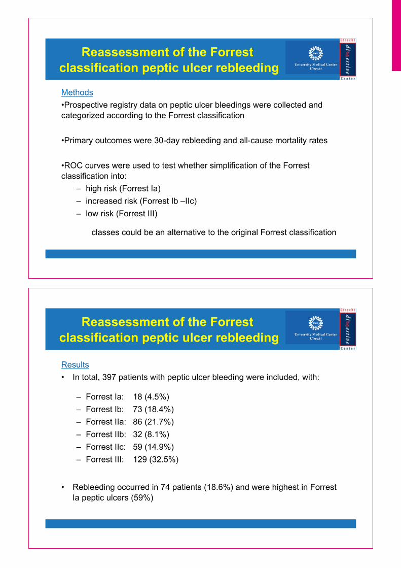
Methods
• Prospective registry data on peptic ulcer bleedings were collected and categorized according to the Forrest classification
• Primary outcomes were 30-day rebleeding and all-cause mortality rates
• ROC curves were used to test whether simplification of the Forrest classification into:
– high risk (Forrest Ia) – increased risk (Forrest Ib –IIc) – low risk (Forrest III)
classes could be an alternative to the original Forrest classification
Reassessment of the Forrest classification peptic ulcer rebleeding
Results
• In total, 397 patients with peptic ulcer bleeding were included, with:
– Forrest Ia: 18 (4.5%) – Forrest Ib: 73 (18.4%) – Forrest IIa: 86 (21.7%) – Forrest IIb: 32 (8.1%) – Forrest IIc: 59 (14.9%) – Forrest III: 129 (32.5%)
• Rebleeding occurred in 74 patients (18.6%) and were highest in Forrest Ia peptic ulcers (59%)
Reassessment of the Forrest classification peptic ulcer rebleeding

Results
Reassessment of the Forrest classification peptic ulcer rebleeding
Results
Reassessment of the Forrest classification peptic ulcer rebleeding

Results
Reassessment of the Forrest classification peptic ulcer rebleeding
The simplified Forrest classification had similar test characteristics to the original Forrest classification
CONCLUSION • The Forrest classification is still clinically useful to identify patients at an increased risk of rebleeding, with the highest prognostic significance for gastric ulcers.
• Reclassifying and simplifying the Forrest classification into low risk (Forrest III), increased risk (Forrest Ib –Iic) and high risk (Forrest Ia ulcers) of rebleeding is possible
TAKE HOME MESSAGE
Reassessment of the Forrest classification peptic ulcer rebleeding
Peptic ulcer bleeds should be classified according the (simplified)
Forrest classification as it helps in predicting which patients can
be discharged early from the hospital

ADR and risk of CRC and death
N= 314,872 colonoscopies by 136 colonoscopists
Kaiser Permanente Northern California database (3.3 million people) 712 interval cancers: – 255 advanced-stage cancers – 147 deaths from interval CRCs
ADR quintiles – Q1: 16.56% (7.35-19.05%) – Q2: 21.50% (19.06-23.85% – Q3: 25.70% (23.86-28.40%) – Q4: 30.96% (28.41-33.50%) – Q5: 38.86% (33.51-52.51%)
Corley et al. New Engl J Med 2013
ADR and risk of CRC and death
Corley et al. New Engl J Med 2013

EndoCuff
New developments
G-Eye
New developments

EndoRings�
New�developments�
Full�Spectrum�Endoscopy�FUSE�colonoscopy�
New�developments�

31�
Lancet Oncol 2014;�15:�353–60�
Study�Aims�
1. Primary:�Adenoma�Miss�Rates�
2. Secondary:�• Polyp�Miss�Rates�• Advanced�Adenoma�Miss�Rates�• Time�to�Cecal�Intubation�• Colonoscope�Withdrawal�Time�• Adverse�Events�
32�

Study�Design�
Randomized�(concealed�allocation)�• Tandem�colonoscopy�design�• Same�day,�back-to-back,�by�the�same�endoscopist�• 170o�SFV�vs.�Fuse�330o��
All�polyps�removed�when�identified�• Except�hyperplastic�rectal�polyps�(1mm-2mm)�• All�adenomas�and�cancers�confirmed�by�pathology�
Multicenter�• Israel�(3)�Netherlands�(1)�USA�(2)�• February�1,�2012�–�March�31,�2013�
33�
Subject�Demographics�
• Mean�age�55.8�±�9.7�years�
• 101�female�(54.6%)�/�84�male�(45.4%)�
• Baseline�characteristics�• Age,�gender,�reason�for�colonoscopy�were�similar�
• Indications�for�colonoscopy�• CRC�screening�=�103�(55.7%)�• Polyp�surveillance�=�36�(19.5%)�• Diagnostic�evaluation�=�46�(24.8%)�
34�

Time�to�Cecum�(median�time)
Withdrawal�Time��
(median�time)
SFV��Colonoscopy 5.1�minutes 5.6�minutes
Fuse™�Colonoscopy
4.8�minutes 6.2�minutes
Procedure�Times�Comparable�
p<0.0001�p=NS�35�
Fuse™�Study�Conclusions�
FUSE�colonoscopy�
• found�an�additional�69%�more�adenomas�after�SFV�
• had�a�significantly�lower�adenoma�miss�rate�(7%)�������������������
compared�to�SFV�(41%)��
• Fuse�had�no�negative�colonoscopies;�whereas,�6%�of�patients�with�
SFV�first�had�a�false�negative�colonoscopy.��
• Fuse�shortened�the�interval�recommendations�in�53%�of�the�exams�
where�SFV�colonoscopy�failed�to�identify�all�polyps�present�in�9%�of�
total�patient�population.��
�36�

Diagnostic Endoscopy 2014 Conclusions
Diagnostic Endoscopy 2014 Conclusions

D. Hartmann
ERCP Die 5 wich�gsten Publika�onen 2014
PD Dr. Dirk Hartmann Klinik für Innere Medizin I Sana Klinikum Lichtenberg
Berlin

Klinik für Innere Medizin I Fanningerstraße 32 | 10365 Berlin
Tel. 030 5518-2210 | Fax 030 5518-2250 [email protected] | www.sana-kl.de
Sana Kliniken Berlin-Brandenburg GmbH Sana Klinikum Lichtenberg
ERCP: Die 5 wichtigsten Publikationen 2014
D. Hartmann
RFA VS. PDT

CHOLANGIOZELLULÄRES KARZINOM RFA VS. PDT
Ortner et al., Gastroenterology 2003 Zöpf et al., Am J Gastroenterol 2005
1 Ø 2 randomisierte Studien zur photodynamischen Therapie (PDT)
bei nicht resezierbarem CCC
Ø Vergleich PDT plus Stenting versus Stenting alleine
Autor n Sensitizer Überleben p-Wert
PDT + Stenting Stenting
Ortner 2003
39 Photofrin 16,5 Monate 3,5 Monate <0,0001
Zöpf 2005
32 Photosan-3 21 Monate 7 Monate <0,01
1 CHOLANGIOZELLULÄRES KARZINOM RFA VS. PDT
Nachteile der PDT
ü Vermeidung von Sonnenlicht für 4-6 Wochen
ü sehr aufwendige Endoskopie ü Applikation des Sensitizers 48h vorher notwendig
ü Hohe Kosten für den Photosensitizer und das Equipment
8 F Katheter Vorteile RFA
ü einfache Handhabung ü Einbringen über Draht ü Betrieb mit konventionellem HF-Generator

1 CHOLANGIOZELLULÄRES KARZINOM RFA VS. PDT
Ø retrospektive Kohorten Studie Ø keine Randomisierung Ø vergleichbare Gruppen
Strand et al., Gastrointest Endosc 2014
1
Strand et al., Gastrointest Endosc 2014
CHOLANGIOZELLULÄRES KARZINOM RFA VS. PDT

1
Strand et al., Gastrointest Endosc 2014
CHOLANGIOZELLULÄRES KARZINOM RFA VS. PDT
Gruppe RFA: 13/16 (81%) mit Metastasen Gruppe PDT: 18/32 (56%) mit Metastasen
1
Strand et al., Gastrointest Endosc 2014
CHOLANGIOZELLULÄRES KARZINOM RFA VS. PDT
PDT-Gruppe 7,5 Monate
RFA-Gruppe 9,6 Monate
Überleben

1
RFA scheint vergleichbar mit PDT in Bezug auf das Überleben !
RFA bei CCC ist technisch machbar (einfache Handhabung) !
Aktuell keine randomisierten Studien !
CHOLANGIOZELLULÄRES KARZINOM RFA VS. PDT
BENIGNE STRIKTUREN

BENIGNE�GALLENGANGSTENOSEN�FULLY�COVERED�SEMS�2�
Benigne�Gallengangstenosen:��ü Chronsiche�Pankreatitis:� �10-30%�ü Lebertransplantation:� �4-9%�ü Postoperativ:� ��0,3�bis�0,4%�
Endoskopische�Therapie:��ü Plastikstents�in�aufsteigender�Anzahl�ü Wechsel�alle�3�Monate�ü Therapie�über�1�Jahr��
Therapiealternativen:��ü voll�gecoverter�SEMS�ü bisher�nur�wenige�Daten�
Deviere�et�al.,�Gastroenterology�2014�
BENIGNE�GALLENGANGSTENOSEN�FULLY�COVERED�SEMS�2�
ü Prospektve�Studie�ü 13�Institutionen�aus�11�Ländern�ü Entfernung�FCSEMS�nach�10-12�Monate�bei�chronischer�Pankreatitis�bzw.�postoperativ�
ü Entfernung�FCSEMS�nach�4-6�Monate�bei�Patienten�nach�OLT��
Deviere et al., Gastroenterology 2014
BENIGNE GALLENGANGSTENOSEN FULLY COVERED SEMS 2
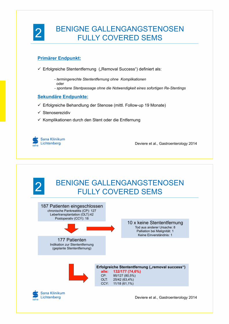
Primärer Endpunkt: ü Erfolgreiche Stententfernung („Removal Success“) definiert als:
- termingerechte Stententfernung ohne Komplikationen oder - spontane Stentpassage ohne die Notwendigkeit eines sofortigen Re-Stentings
Sekundäre Endpunkte:
ü Erfolgreiche Behandlung der Stenose (mittl. Follow-up 19 Monate) ü Stenoserezidiv ü Komplikationen durch den Stent oder die Entfernung
BENIGNE GALLENGANGSTENOSEN FULLY COVERED SEMS 2
187 Patienten eingeschlossen chronische Pankreatitis (CP): 127 Lebertransplantation (OLT):42 Postoperativ (CCY): 18
177 Patienten Indikation zur Stententfernung (geplante Stententfernung)
10 x keine Stententfernung Tod aus anderer Ursache: 8 Palliation bei Malignität: 1 Keine Einverständnis: 1
Erfolgreiche Stententfernung („removal success“) alle: 132/177 (74,6%) CP: 95/127 (80,5%) OLT: 25/42 (63,4%) CCY: 11/18 (61,1%)
Deviere et al., Gastroenterology 2014

Und die anderen 45/177 Patienten…………??
ü Frühe endoskopische Entfernung wegen Komplikationen: n=25 Cholangitits: 14 Cholestase: 3 Cholecysitits: 1 Andere: 7
ü Lost of Follow-up: n=5
ü Komplette Stentmigration: n=16
ohne Notwendigkeit des Re-Stentings: 8 („removal success“) mit Notwendigkeit des Re-Stentings: 8
ü SAE nach Stententfernung: n=7
Cholangitits: 5 Blutung: 1 Pankreatitis: 1
BENIGNE GALLENGANGSTENOSEN FULLY COVERED SEMS 2
Deviere et al., Gastroenterology 2014
Stentmigration
- Gesamt: 55 Patienten - komplett: 16 Patienten - partiell distal: 20 Patienten - partiell proximal: 19 Patienten
Stententfernung
- beim ersten Versuch: 149/155 Patienten - beim zweiten oder dritten Versuch: 6/155 Patienten Stent in Stent Technik: 3/6 Patienten
Deviere et al., Gastroenterology 2014
BENIGNE GALLENGANGSTENOSEN FULLY COVERED SEMS 2
Bei allen Patienten konnte der Stent wieder entfernt werden, überwiegend problemlos und einfach.

BENIGNE GALLENGANGSTENOSEN FULLY COVERED SEMS 2
Deviere et al., Gastroenterology 2014
ü Cholangitis ist die häufigste Komplikation (13,9%)
ü Pankreatitis und Cholecystitis selten (2,7% und 3,0%)
Deviere et al., Gastroenterology 2014
BENIGNE GALLENGANGSTENOSEN FULLY COVERED SEMS 2
Erfolgreiche Therapie mit FCSEMS

Die Entfernung des Stents ist quasi immer möglich. !
FCSEMS bei benignen Stenosen sind sicher und effektiv. !
BENIGNE GALLENGANGSTENOSEN FULLY COVERED SEMS 2
Bei chronischer Pankreatitis scheinen FCSEMS dem Multistenting überlegen. !
POST-ERCP PANKREATITIS
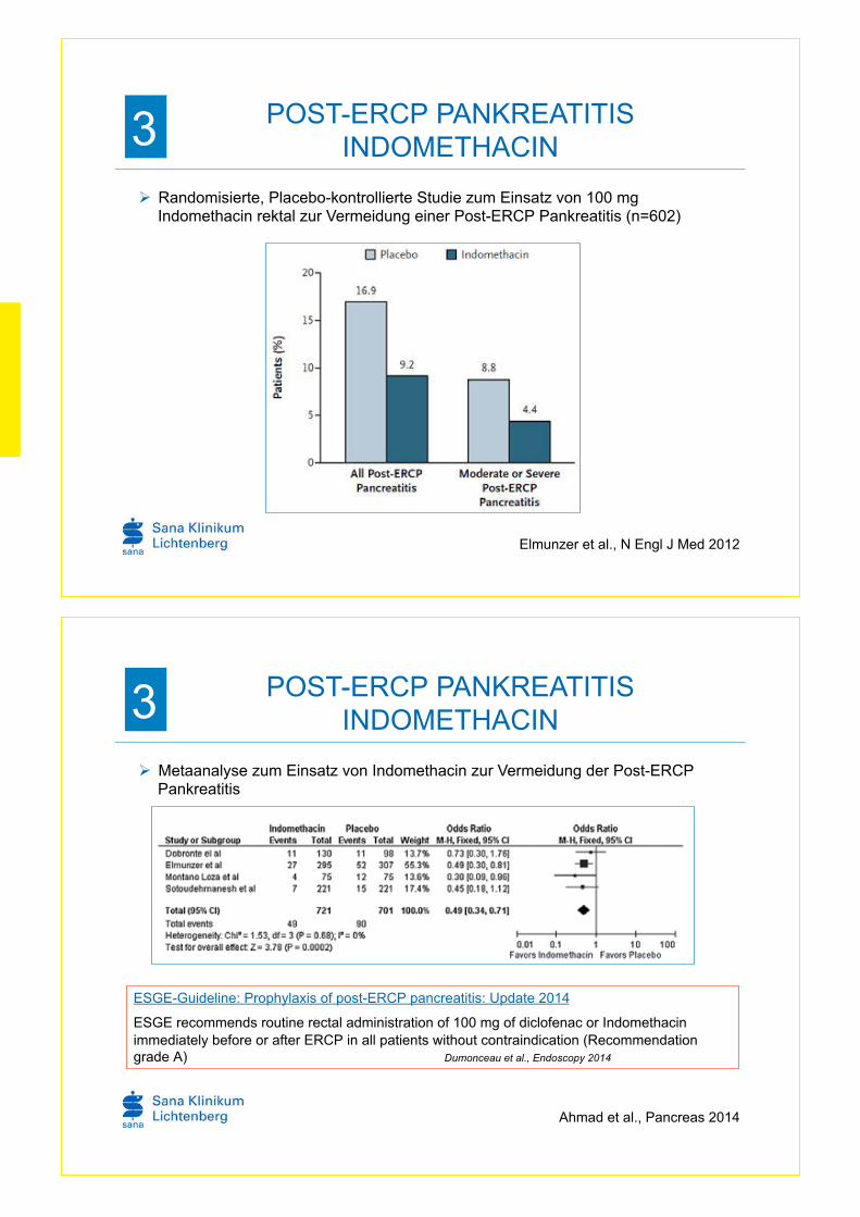
POST-ERCP PANKREATITIS INDOMETHACIN 3
Ahmad et al., Pancreas 2014
Ø Metaanalyse zum Einsatz von Indomethacin zur Vermeidung der Post-ERCP Pankreatitis
ESGE-Guideline: Prophylaxis of post-ERCP pancreatitis: Update 2014
ESGE recommends routine rectal administration of 100 mg of diclofenac or Indomethacin immediately before or after ERCP in all patients without contraindication (Recommendation grade A) Dumonceau et al., Endoscopy 2014
POST-ERCP PANKREATITIS INDOMETHACIN 3
Elmunzer et al., N Engl J Med 2012
Ø Randomisierte, Placebo-kontrollierte Studie zum Einsatz von 100 mg Indomethacin rektal zur Vermeidung einer Post-ERCP Pankreatitis (n=602)

POST-ERCP PANKREATITIS NITRATE 3
Ø Metaanalyse zum Einsatz von Nitroglycerin zur Vermeidung der Post-ERCP Pankreatitis (1662 Patienten)
Bang et al., Aliment Pharmacol Ther 2009
Vorteil nur bei sublingualer nicht bei transdermaler Applikation
ESGE-Guideline: Prophylaxis of post-ERCP pancreatitis: Update 2014
Nitroglycerin may be effective in preventing PEP when administered sublingually. ESGE does not recommend the routine use of GTN for PEP prophylaxis.
Dumonceau et al., Endoscopy 2014
POST-ERCP PANKREATITIS INDOMETHACIN PLUS NITRAT 3
ü 300 Patienten (150 Patienten in jeder Gruppe) ü Gruppe A: 100 mg Indomethacin rektal plus 5 mg Isosobiddinitrat sublingual
ü Gruppe B: 100 mg Indomethacin plus Placebo ü Applikation 5 Minuten vor der ERCP
Sotoudehmanesh et al., Am J Gastroenterol 2014

POST-ERCP PANKREATITIS INDOMETHACIN PLUS NITRAT 3
Indomethacin plus Nitrat
Indomethacin alleine
6,7% 15,3%
Sotoudehmanesh et al., Am J Gastroenterol 2014
Nitrate wirksam, jedoch nicht generell empfohlen (ESGE Guideline 2014) !
Indomethacin oder Diclofenac sind der medikamentöse Standard zur Prophylaxe der Post-ERCP Pankreatitis !
Kombination scheint der alleinigen Gabe von Indomethacin überlegen. Muticenterstudien erwartet. !
POST-ERCP PANKREATITIS INDOMETHACIN PLUS NITRAT 3
POST-ERCP PANKREATITIS FRÜHE ERCP PLUS PROTHESE 4
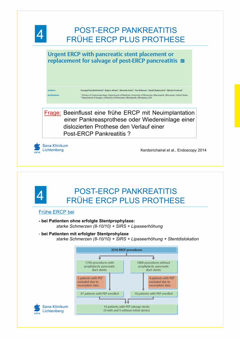
Frage: Beeinflusst eine frühe ERCP mit Neuimplantation einer Pankreasprothese oder Wiedereinlage einer dislozierten Prothese den Verlauf einer Post-ERCP Pankreatitis ?
Kerdsirichairat et al., Endoscopy 2014
POST-ERCP PANKREATITIS FRÜHE ERCP PLUS PROTHESE 4
Frühe ERCP bei
- bei Patienten ohne erfolgte Stentprophylaxe: starke Schmerzen (8-10/10) + SIRS + Lipaseerhöhung
- bei Patienten mit erfolgter Stentprohylaxe starke Schmerzen (8-10/10) + SIRS + Lipaseerhöhung + Stentdislokation

POST-ERCP PANKREATITIS FRÜHE ERCP PLUS PROTHESE 4
Problem der Studie: Natürlicher Verlauf unklar. Keine Vergleichsgruppe. !
Frühe ERCP bei PEP: Neue Option, keine Empfehlung !
Was passiert, wenn Zugang zum Pankreasgang nicht gelingt? Verschlechterung der Situation? !
POST-ERCP PANKREATITIS FRÜHE ERCP PLUS PROTHESE 4
Schmerzen Lipase
Kerdsirichairat et al., Endoscopy 2014
ü SIRS innerhalb von 24 Stunden nicht mehr nachweisbar ü Mittl. KH-Aufenthalt von 2 Tagen (1-4,75 Tage) ü 5/14 Patienten konnten nach 24 h entlassen werden
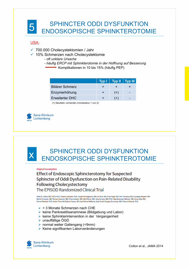
SPHINCTER ODDI DYSFUNKTION ENDOSKOPISCHE SPHINKTEROTOMIE 5
USA:
ü 700.000 Cholecystektomien / Jahr ü 10% Schmerzen nach Cholecystektomie
- oft unklare Ursache - häufig ERCP mit Sphinkterotomie in der Hoffnung auf Besserung Komplikationen in 10 bis 15% (häufig PEP)
Typ I Typ II Typ III
Biliärer Schmerz + + +
Enzymerhöhung + (+) -
Erweiterter DHC + (+) - (+) fakultativ vorhanden (mindestens 1 von 2)
SPHINCTER ODDI DYSFUNKTION ENDOSKOPISCHE SPHINKTEROTOMIE x
Cotton et al., JAMA 2014
Ø > 3 Monate Schmerzen nach CHE Ø keine Pankreatitisanamnese (Bildgebung und Labor) Ø keine Sphinkterintervention in der Vergangenheit Ø unauffällige ÖGD Ø normal weiter Gallengang (<9mm) Ø Keine signifikanten Laborveränderungen

SPHINCTER ODDI DYSFUNKTION ENDOSKOPISCHE SPHINKTEROTOMIE 5
1584 Patienten gescreent
Sphinkterotomie, n=141 99 mit pathol. Manometrie 42 normale Manometrie
214 randomisiert
1298 ausgeschlossen path. Bildgebung oder Endoskopie atypische Schmerzen tgl. Schmerzmittel abnorme Laborwerte
Sham, n=73 48 mit pathol. Manometrie 25 normale Manometrie
Cotton et al., JAMA 2014
2 : 1
SPHINCTER ODDI DYSFUNKTION ENDOSKOPISCHE SPHINKTEROTOMIE 5
Definition Therapieerfolg (primärer Endpunkt) ü RAPID Score < 6 Tage Verlust der Produktivität in Monat 9 und 12 ü Keine weitere Intervention ü Keine Schmerzmittel zu Monat 10,11 und 12
RAPID Score:
Recurrent abdominal pain intensity and disability

SPHINCTER ODDI DYSFUNKTION ENDOSKOPISCHE SPHINKTEROTOMIE 5
Cotton et al., JAMA 2014
SPHINCTER ODDI DYSFUNKTION ENDOSKOPISCHE SPHINKTEROTOMIE 5 Therapie n Therapieerfolg
Sham 73 27 (37%)
Sphinkterotomie 141 32 (23%)
Manometrie Gallengang
Manometrie Pankreas
Sphinkterotomie Therapieerfolg
Sham Therapieerfolg
pathol. pathol. 27% (14/52) 38% (8/21)
normal pathol. 21% (9/42) 36% (8/22)
pathol. normal 18% (3/16) 11% (1/9)
normal normal 19 %(6/31) 48% (10/21)
Cotton et al., JAMA 2014

SPHINCTER ODDI DYSFUNKTION ENDOSKOPISCHE SPHINKTEROTOMIE 5
Manometrie hilft nicht um Subgruppen zu identifizieren, die ggf. von einer EST profitieren. !
Kein Nutzen der Sphinkterotomie bei Patienten mit SOD Typ III im Vergleich zur Sham-Gruppe !
“These findings do not support ERCP and sphincterotomy for these patients” !
VIELEN DANK

J. Hochberger
Technische Innova�onen und neue Produkte in der Endoskopie
Prof. Dr. Jürgen Hochberger Chairman of Gastroenterology and GI Endoscopy
Strasbourg University Hospitals – Nouvel Hôpital Civil and IHU Strasbourg/Frankreich

Page 1 of 4
Technical Innovations and Treatment Modalities in EndoscopyJuergen Hochberger, M.D. PhD, Univ. Hospital Strasbourg – NHC and IHU, Strasbourg, France
In the talk new different devices and techniques will be highlighted. Among other devices and
techniques foci will be put on endoscopic treatment of morbid obesity and new resection and ablation
devices.
Endoscopic Treatment of Morbid ObesityMorbid obesity represents an increasing problem in the Western World. Obesity is classified according
to the body mass index weight (kg) / height (m2) into the following categories: Acceptable Range 18.5–
24.9 kg/m2; Overweight 25–29.9 kg/m2; Obese 30–34.9 kg/m2; Severe Obesity 35–39.9 kg/m2; Morbid
Obesity 40–49.9 kg/m2; Super-Morbid Obesity 50 +++ kg/m2. In 2002 34% of the 15 year old boys and
20 % of the 15 year old girls had overweight according to the American Center of Disease Control
(CDC). The percentage of obese people with an BMI of greater than 30 ranges in Europe between 15-
28% and is suspected to raise to 19-35% in 2020 and to 23-43% in 2030.
Concerning treatment of morbid obesity ‘conservative measures’ show only a limited effect. Diet plus
exercise lead to weight loss of 5-7%. Adding behavioral therapy may achieve a weight loss of 8-10%.
Pharmacotherapy such as inhibitors of intestinal fat absorption, appetite suppressants and agonists of
the endo-cannabinoid system may add another 5-8%. However, the mid- and longterm effect of these
measures is often very limited. ‘Bariatric’ surgery as specific term for surgery in morbid obesity is
currently the only therapeutic measure with a proven profund longterm effect. Restrictive surgical
procedures include: ‘Vertical Banded Gastroplasty’ (VBG), ‘Adjustable Gastric Band’ (AGB) and
‘Sleeve Gastrectomy’ (SG). Restrictive surgery for obesity leading to malabsorption include ‘Bilio-
pancreatic Diversion’ (BPD), ‚Duodenal Switch' (DS) and ‚Roux-en-Y Gastric Bypass’ (RYGB).
Longterm studies up to 15 years could prove a reduction in weight loss of 15-30% for gastric banding
procedures or gastric bypass compared to control subjects. A second positive effect of bariatric
surgery is a significant improvement in a diabetes type 2 often associated with morbid obesity within a
metabolic syndrome.
Within the last 10 years different endoscopic procedures have tried to mimic these effects. They
include balloons, barriers, sleeves and restrictive endoscopic interventional procedures. Some of
those tools for a primary intervention include intragastric balloons (Allergan, Spatz-FGIA Inc,
Helioscopie), the RESTORe Suturing System (C.R. Bard Inc), the TOGa Stapler device (Satiety Inc)
the TERIS system (Barosense) or the Incisionless Operating Platform (USGI Medical Inc) to be used

Hochberger J: Technical innovations and treatment modalities in end. - EndoUpdate Augsburg, Nov. 29th, 2014
Page 2 of 4
to mimic e.g. a vertical band gastroplasty. Bypass Liners (GI Dynamics, ValenTx) cover part of the
proximal small intestine ± stomach with an impermeable flouropolymer sheath to induce
malabsorption. The AspireAssist system (Aspire Bariatrics, Inc., King of Prussia, USA) works by
aspirating predigested food from the stomach.
Of the different techniques applied and devices used only limited clinical series have been published
and longterm outcomes are often lacking so far. Different of the devices and procedures such as the
TOGA system have disappeared already from the market in the meantime. While a initial echoes
where rather enthusiastic midterm or longterm results were rather disappointing. Suturing devices
have been used to reduce the lumen and e.g. simulate a vertical band gastroplasty. New devices such
as the G-prox or the Overstich device were used to achieve transmural suturing. However, those
stiches could often not resists mechanical forces due to increased food intake when patients fell back
into former eating behaviors. This is the reason why current endoscopic attempts often try to induce
an initial clinical weight loss in combination with behavioral therapy in order to induce a first step
toward a change in the life habits. Especially for super-obese patients endoscopic ‘first-line’ treatment
may reduce the operative risk of a secondary more definitive surgical procedure. Some easy to apply
systems not changing the GI anatomy include balloons, the AspireAssist and the EndoBarrier Liner.
New concepts include the Factyl system for superficial coagulation of the duodenal and upper jejunal
mucosa to induce malabsorption and intestinal endocrine changes.
A first generation of balloons like the ‘Garren-Edwards Gastric Bubble (GEGB)’ dates back to more
than 15 years. However, technical problems included a rupture of the balloon and secondary intestinal
obstruction but especially a rapid regain in weight after removal of the balloon. 10 years later a second
generation like the BioEnterics Intragastric Balloon (BIB; Allergan, Irvine, CA, USA) show a clearly
reduced risk profile and studies have demonstrated a significant weight loss in the short-term and mid-
term ranges. However, long-term weight loss following balloon removal, has again been equivocal.
The ‘AspireAssist’ is a new gastric aspiration tube. A type of large gastric (PEG) feeding tube is placed
under endoscopic control and aseptical conditions. In the following the patient is taught to meticulously
chew all food until it becomes an almost liquid mash. This way the patient spends as a first effect more
time for the meal, eats more consciously and has an earlier saturation effect. Directly after finishing
the meal the patient aspirates the food through the AspireAssist tube from the stomach in order to
reduce the quantity of food being digested. Limited patient series e.g. from the Czech Republic are
promising.
The duodeno-jejunal Bypass-Liner (DJBS; Endobarrier; GI Dynamics, Watertown, MA, USA) uses a

Hochberger J: Technical innovations and treatment modalities in end. - EndoUpdate Augsburg, Nov. 29th, 2014
Page 3 of 4
malabsorption principle of the upper small intestine which leads secondarily to important GI peptide
changes such as a reduced level of hormones like ‘ghrelin’ which play an important metabolic role in
morbid obesity. A nitinol anchor placed at the level of the duodenal bulb holds a 60 cm long, thin
fluoropolymer sleeve impermeable for nutritional content. As with the other procedures described a
positive effect on weight loss can be observed in the order of 15-25% within the first 6 month in
patients tolerating the devices (80-90%). However, this reduction in weight is reversible in a major part
of patients on the longterm run. However, a secondary important effect on a type2-diabetes often
insulin-resistant can often be observed already the first days after implantation of the devices /
beginning of treatment.
To summarize first results of these new endoscopic treatment modalities are promising. However,
their place in the multi-disciplinary treatment of morbid obesity is still not clear yet. An important role
may be a reduction of the pre-operative risk of super-obese or multi-morbid obese patients. This
includes the improvement of an often treatment-resistant accompanying diabetes. Further comparative
multicentric studies have to be awaited. The different systems and working mechanisms will be
presented.
New resection and ablation techniquesDifferent devices and products have recently been presented. Full thickness resection is now possible
using the new FTRD device (Ovesco Tübingen). An over-the-scope-clip (OTSC) is used in
combination with an embedded snare. First a full-thickness capture of the intestinal wall is induced by
the OTSC and secondarily an artificial ‘mushroom’ is created by preferably pulling the future resection
specimen into the distal transparent cylinder by a grasper or anchor. Preliminary results by Caca and
Bauernfeind are promising. The device could be a perspective for colonic recurrences after piecemeal
resection on a scar or full thickness resection of small submucosal tumors in the colo-rectum such as
hind gut carcinoids. Limitations may be the current restriction to the lower GI tract and the impossibility
of passage to cranial lesions in case of a narrow sigmoid.
The combination of a central high-pressure capillary for submucosal saline injection and a
conventional argon plasma coagulation probe have recently been presented by Pech, Manner, Ell and
co-workers and will be shortly described.
New applications for fluid supported resection devices include POEM or small intestinal ESD.
Dissectors, graspers vices or resection forceps have recently been propagated for applications in
different areas of the GI tract. Clinical examples in the esophagus and colo-rectum using e.g. the

Hochberger J: Technical innovations and treatment modalities in end. - EndoUpdate Augsburg, Nov. 29th, 2014
Page 4 of 4
Clutch Cutter (Fujifilm, Düsseldorf) are shown.
PerspectivesFinally new perspectives in flexible endoscopy such as new imaging and recognition systems for prior
lesions or biopsies as well as perspectives on new manipulators allowing surgery-like traction for
endoscopic resections in the rectum such as the Anubis System (Storz, Tuttlingen) and a tele-robotic
version of this endoscope will be presented.
Contact:
Juergen Hochberger, M.D. PhD
Professor of Medicine
Chairman of Gastroenterology and GI Endoscopy
Strasbourg University Hospitals – Nouvel Hôpital Civil and IHU
1, Place de l’Hôpital (BP 426)
F-67091 STRASBOURG, FRANCE
Secr. +33-3695-50313 resp. +33-3695-50313-51589
Fax: +33-3695-51857
E-mail: [email protected] or [email protected]
E-mail secretariat: [email protected]


C. Meyenberger
Sicherheit in der Endoskopie
Prof. Dr. Christa Meyenberger Gastroenterologie/Hepatologie
Kantonsspital St. Gallen St. Gallen/Schweiz

Klinik für Gastroenterologie / Hepatologie Sicherheit in der Endoskopie
Sicherheit in der Endoskopie
Prof. Dr. Christa Meyenberger Chefärztin Gastroenterologie / Hepatologie, St. Gallen, Schweiz Dr. med. Nobert Rose Leiter Abteilung Qualitätsmanagement, St. Gallen, Schweiz
www.meldeportal.ch
Klinik für Gastroenterologie / Hepatologie Sicherheit in der Endoskopie 2

Klinik für Gastroenterologie / Hepatologie Sicherheit in der Endoskopie
St.Gallen
Bodensee
Appenzellerland
Bodensee
Klinik für Gastroenterologie / Hepatologie Sicherheit in der Endoskopie
St.Gallen Rorschach Flawil
900 Betten 40`000 stationäre Patienten 4300 Mitarbeitende 760 Mio CHF Ertrag
Kantonsspital St.Gallen – ein Unternehmen, drei Spitäler
4

Klinik für Gastroenterologie / Hepatologie Sicherheit in der Endoskopie
Krankenhaus
Kliniken Institute Support bereiche
Ärzte Pflege Administration
Behandlungskette / Behandlungsprozess
Infrastruktur
Qualitätsmanagement: Risikomanagement
Klinik für Gastroenterologie / Hepatologie Sicherheit in der Endoskopie
Prozesse und Verhaltensweisen, die darauf ausgerichtet sind, eine Organisation bezüglich medizinisch/pflegerische Patienten-Risiken zu steuern.
Klinisches Risikomanagement
Risikokultur / Sicherheitskultur
Umsetzung des Risikomanagement

Klinik�für�Gastroenterologie�/�Hepatologie � �Sicherheit�in�der�Endoskopie�
Auf�der�Basis�ISO�31000�Risikomanagement��
Meldesysteme�sind�der�Einstiegspunkt�für�ein�umfassendes�Risikomanagement�
RM�
RM-Prozess�
IKS�
Melde-systeme�
Schaden-analyse�
Schaden-abwicklung�
Krisen-management�
Risiko-politik�
Risiko-beurteilung�
Dr.�med.�Norbert�Rose
Klinik�für�Gastroenterologie�/�Hepatologie � �Sicherheit�in�der�Endoskopie�
Fehler-�abwehr-systeme�
Fehlerablauf�
Schaden�
J.�Reason�1991�
Schweizer�Käsemodell�der�Systemfehler�

Klinik�für�Gastroenterologie�/�Hepatologie � �Sicherheit�in�der�Endoskopie�
Komplikationen:�
Prävention�
Register�Behandlungspfade�
Guidelines�
Zertifizierung�
Risikoanalysen�Risikoanalysen
MoMo-Konferenzen�
Schulung�
Training�
Standards�
Fehlerkultur�
Team-Time-Out�
Melde�
systeme�
Klinik�für�Gastroenterologie�/�Hepatologie � �Sicherheit�in�der�Endoskopie�
St.Galler�CIRS:��Critical�Incident�Reporting�System�Critical�Incident�=�Kritischer�Zwischenfall�
Definition:�Ein�kritischer�Zwischenfall�ist�ein�Ereignis,�das�den�Patienten�gefährden�kann,�aber�nicht�schädigt.�
„Near�misses“�
Institute�of�Medicine,�Linda�T�Kohn�et�al.:�To�err�is�human,�2000,�p87�
Ein�kritischer�Zwischenfall�ist�ein�vermeidbares�Ereignis�
Konsequent�keine�Schäden,�sondern�nur�Gefährdungen�melden�

Klinik�für�Gastroenterologie�/�Hepatologie � �Sicherheit�in�der�Endoskopie�
Keine�Schäden�melden� Anonymität�wahren�
Interprofessionalität�Interdisziplinarität�
fördern�
Kurzes�CIRS-Meldeformular�
CIRS-Verantwortliche�einsetzen�
Leadership��in�Sicherheitskultur�
CIRS-Fall-Besprechungen�durchführen�
Daten-und��Informationspolitik�
festlegen�
Meldekreise��einführen�
Thieme�Fachzeitschrift:�Gesundheitsökonomie�&�Qualitätsmanagement,�Gesundh�ökon�Qual�manag�2005;�10: 83-89;�Rose, N.;�Germann, D.:�Resultate�eines�krankenhausweiten�Critical�Incident�Reporting�System�(CIRS)�
St.�Galler�CIRS:�9�Kernmerkmale�
Klinik�für�Gastroenterologie�/�Hepatologie � �Sicherheit�in�der�Endoskopie�
CIRS�
Sicherheitskultur�Organisations-�u.�Prozessentwicklung�
Ergebnis�eines�CIRS�

Klinik für Gastroenterologie / Hepatologie Sicherheit in der Endoskopie
www.meldeportal.ch
Meldesystem in einem Portal: web basiert
Klinik für Gastroenterologie / Hepatologie Sicherheit in der Endoskopie 14
Einschätzung Schweregrad (I-III) Menschliche Fehler Organisation / Kommunikation
Beschreibung Ereignis
Mögliche Massnahmen
Kurzes 3 Minuten Melde-formular

Klinik für Gastroenterologie / Hepatologie Sicherheit in der Endoskopie
Kurzes 3 Minuten Melde-formular
15
Meldung anonymisieren, Original löschen
Klinik für Gastroenterologie / Hepatologie Sicherheit in der Endoskopie
596
874 962
1200
1424
1852
1520
1676
1849
1722 1725
83 120 149 123 102 107 100 45 37 49 71
0
200
400
600
800
1000
1200
1400
1600
1800
2000
2003 2004 2005 2006 2007 2008 2009 2010 2011 2012 2013
CIRS Massnahmen
CIRS-Jahresstatistik Kantonsspital SG

Klinik�für�Gastroenterologie�/�Hepatologie � �Sicherheit�in�der�Endoskopie�
2009-2013�(n�=�448)��(n�=�88�/�Jahr)��
Gastroenterologie:�CIRS-Schweregrad�
Klinik�für�Gastroenterologie�/�Hepatologie � �Sicherheit�in�der�Endoskopie�
• Administration�und�Organisation�• Präinterventionelle�Massnahmen�• Aufklärung�• Sedation�• Behandlung�• Postinterventionelle�Nachsorge�• Material,�Medikamente�und�Infrastruktur�
CIRS�in�der�Endoskopie�

Klinik für Gastroenterologie / Hepatologie Sicherheit in der Endoskopie
Datenqualität • Stammdaten im KIS falsch ausgewählt. • Verwechslungen: Patientendaten, Akten, Zuweiser (Spitäler, Ärzte) Ein normaler Bürger hat Arztbericht erhalten! • Auf Überwachungsblatt fehlt Patientenetikette • Laboranalysen für falschen Patienten verordnet (von Hand) • Krankengeschichte und Pflegedokumentation auf falsches Patientenbett deponiert • Hausarzt fordert Labor an. Werte werden an eine Papeterie gefaxt. • Akten von verschiedenen Patienten verhaken sich in Dossiers mit Büroklammern • Verstorbener Patient wird aufgeboten, da Aufgebot in falsche Akte abgelegt wurde
CIRS: Administration / Organisation
Klinik für Gastroenterologie / Hepatologie Sicherheit in der Endoskopie
Ø Einführung «Patientenarmbänder»: Alle Patienten erhalten ein Armband mit allen erforderlichen Patientendaten.
Ø Einführung «Team-Time-Out»: Patientenidentifikation mit „double-check“: Name, Geburtsdatum,
Adresse, Adressat und Befragung des Patienten.
Ø Patientenidentifikation (Formulare / Probenmaterial): Doppelvisum Arzt / Pflege
Ø Laborverordnungen elektronisch Ø Akten: Büroklammern werden verboten und Akten werden Geheftet und / oder in Mappen deponiert.
Ø Todesfälle werden spitalweit gemeldet und Akten gekennzeichnet
Massnahmen

Klinik für Gastroenterologie / Hepatologie Sicherheit in der Endoskopie
• Patient musste im Gang auf Endoskopie warten. Hatte keine Glocke um sich für einen Toilettengang zu melden. • Flächendeckendes Alarmsystem im Wartebereich fehlt (keine «Holding Area)
• Patient wartet im Gang auf auswärtigen Transport. Er fällt von der Liege und verletzt sich. • Patient wird nach ÖGD abgeholt. 60mg Disoprivan erhalten, hustend… Keine Schwester auffindbar, kein Rapport. BD 77/45 Sättigung 87%. • Patient kommt nicht nüchtern zur PEG-Einlage und erbricht schwallartig! Weisungen für Vorbereitung missachtet • PEG-Einlage bei Pat. mit HNO-Tumor: akute Dyspnoe. Fehleinschätzung der Risikosituation
CIRS: Patientenbetreuung
Klinik für Gastroenterologie / Hepatologie Sicherheit in der Endoskopie
Ø Alarmanlage in allen Untersuchungsräumen und Wartezonen (mobiler Alarm). Schulung aller Personen.
Ø Alle Betten und Liegen werden vor dem Transport in die Endoskopie mit Bettgittern ausgerüstet. Reservegitter sind in der Endoskopie gelagert.
Ø Seitenstützen an der Untersuchungsliege werden nach jedem Eingriff durch den Arzt hochgeklappt.
Ø PEG bei HNO- und ALS-Patienten: Interdisziplinärer Standard / Anästhesie für Risikopatienten
Ø Aspirationsgefahr (Stenosen etc.): Kopftieflage Ø Patientenübergabe: durch diplomierte Pflege oder Arzt Ø Vier-Augen-Prinzip Ø Regelmässiges REA-Training / Materialkunde Ø Regelmässiger Check der Basisinfrastruktur in der Endoskopie
Massnahmen

Klinik für Gastroenterologie / Hepatologie Sicherheit in der Endoskopie
Team Time Out
• Überprüfung der Patientenidentität • Bewertung vorliegender Befunde, weitere Diagnostik • Einbeziehung Angehörige (bei Kindern und nicht urteilsfähigen Patienten)
• Nach Abwaschen und Abdecken des OP-Feldes: Time-Out vor Schnitt • Kontrolle nach Checkliste: • 1. Patientenidentität • 2. Einriffsart und Lokalisation
• 3. Probleme Anästhesie
• 4. präop. Medikation (z.B. Antibiotika)
• Besprechen von Befund, OP-Indikation und Alternativen • Dokumentation (Aufklärung) und Visum Patient und Arzt • Markierung Lokalisation • Stationär und Ambulant: Gespräch Anästhesie, Aufklärung Anästhesie mit Visum Patient und Arzt, weitere Diagnostik?
• Überprüfung der Patientenidentität • Kontrolle der vorliegenden Befunde und ergänzende Anordnungen Anästhesie • Bestätigung der OP-Indikation und Markierung der Lokalisation • Malignität? Histologie? • Vorhandesein OP-und Narkoseeinwilligung
• Überprüfung der Patientenidentität • Kontrolle der vorliegenden Befunde/Bilder und ergänzende Anordnungen Anästhesie • Blutpräparate, Antibiotika vorhanden? • Kontrolle der OP-Lokalisation, Markierung • Vorhandensein Op-Und Anästhesieeinwilligung
3. Zuweisung OP-Saal
4. Team Time Out
2. Markierung Eingriffsort
1. Identifikation Patient
Klinik für Gastroenterologie / Hepatologie Sicherheit in der Endoskopie
Team Time Out Einführung und Umsetzung in allen operativen und interventionellen Bereichen:Gastro, Kardio, Pneumo, Radiolgie
Haynes A.B.et al.: A Surgical Safety Checklist Reduce Morbidity and Mortality in a Global Population. NEJM 360:491, January 29, 2009

Klinik für Gastroenterologie / Hepatologie Sicherheit in der Endoskopie
• Pat. nach Sättigungsabfall beatmet und REA-Alarm ausgelöst.
Klingelalarm in der Endo 3 betätigt. Es kommt niemand.
Alarmton zu leise!
• Patient erbricht: Absaugsystem nicht komplett, Absaugkatheter fehlt,
Sättigungsabfall
• Transportable Sauerstoffflasche leer
• Stichverletzung: Entsorgungsbox zu voll
• Material für Blutstillung (Histoacryl) fehlend
CIRS: Materialprobleme
Klinik für Gastroenterologie / Hepatologie Sicherheit in der Endoskopie
Ø Systematischer Check aller Einrichtungen vor jeder Untersuchung und am Abend (Endoskopieräume / Notfallwagen)
Ø Stichverletzungen: Grosse Entsorgungsboxen, regelmässiger Check
Ø Spezialbehälter für jede Notfall-Intervention (Sklerosierung, Ligatur, Histoacryl, Hemospray etc.) mit Handlungsanweisung
klar im Lager gekennzeichnet
Ø Einführung neuer Mitarbeiter (Ärzte / Pflege) und Refresher
Massnahmen

Klinik für Gastroenterologie / Hepatologie Sicherheit in der Endoskopie
• Patient mit Infusion samt Zusätzen zur Behandlung gebracht
• Prämedikation vergessen
• Adrenalin-Verordnung 1:100 für Polypektomie statt dessen 1:10
verabreicht.
• In Schachtel Indigocarmin war «Tusche» gelagert
• Propofol nach Endoskopie in Infusionsschlauch belassen • Patient nach Endoskopie mit diskonnektiertem Port-a-cath verlegt
• Patient mit Port-a-cath ohne Verschlusskappen verlegt
• Falsches Medikament verordnet
CIRS: Infusionen und Medikamente
Klinik für Gastroenterologie / Hepatologie Sicherheit in der Endoskopie
Ø Infusion ohne Zusätze zur Behandlung bringen
Ø «Team-Time-Out» für alle Vorbereitungsmassnahmen
Ø Eingriffe: Standards für Ärzte / Pflege für Material / Medikamente
Vier-Augenprinzip
Ø Propofol nach Endoskopie: Aspiration und Spülung der Infusion
Ø Genereller «Infusions-Check» nach Endoskopie
Massnahmen

Klinik für Gastroenterologie / Hepatologie Sicherheit in der Endoskopie
Richtiges Arzneimittel Verordnung richtig lesen
Wenn möglich Doppelkontrolle
Richtige Dosierung Verordnung immer inkl. Dosierung mit entsprechend
dosierter Tabletten, Ampullen etc.
Richtige Darreichungsform Sicherstellung der richtigen Applikationsart Patientengerechte Arzneiform
Richtiger Zeitpunkt prä- und postprandiale Einnahme beachten
Gleichmässige Verteilung auf 24h einhalten
Chronopharmakologie beachten
Richtiger Patient Arzneimittel einzeln pro Patient bereitstellen
Korrekte Beschriftung der bereitgestellten Arzneimittel
Abgabe der Tagesdosis nur bei selbständigen Patienten
Richtige Information Stete Information zwischen Ärzten und Pflegenden
Richtige Aufklärung «Patientengerechte» Aufklärung
Richtige Dokumentation Aller Verordnungen, Verabreichungen, Änderungen
Medikamente: Massnahmen «8-er-Regel»
Klinik für Gastroenterologie / Hepatologie Sicherheit in der Endoskopie
Meldesysteme führen zu
Sicherheitskultur Verbesserungen in: Organisation Kommunikation Prozessen Material usw....
Ergebnisse von Meldesystemen

Klinik für Gastroenterologie / Hepatologie Sicherheit in der Endoskopie
Persönliche Fehler Fehler entstehen durch einzelne Personen
Persönliche Sanktionen verhindern Fehler
Systemfehler Fehler entstehen, weil das System es zulässt
Fehler werden durch Fehlerabwehrsysteme verhindert
Standpunkt der Fehlerweise ändern
Klinik für Gastroenterologie / Hepatologie Sicherheit in der Endoskopie
Systematische Fehler erkennen
Fehler systematisch lösen
Ergebnisse von Meldesystemen
Ø Patientenidentifikation Ø Team-Time-Out für gesamten Behandlungsprozess Ø Checklisten Ø Vier-Augen-Prinzip / Double-check mit Visum Ø Standards (Material, Medikamente, Eingriff) Ø Schulung interprofessionell (Ärzte / Pflege) Ø Schnittstellen minimieren

Klinik für Gastroenterologie / Hepatologie Sicherheit in der Endoskopie
Schulung interprofessionell Standards interprofessionell
Klinik für Gastroenterologie / Hepatologie Sicherheit in der Endoskopie
Danke für Ihre Aufmerksamkeit

S. Faiss
EUS
Die 5 wich�gsten Publika�onen 2014
PD Dr. Siegbert Faiss III. Medizinische Klinik
Asklepios Klinik Barmbek Hamburg

Endo Update 2014: Endosonographie
Die 5 wichtigsten Publikationen 2014
PD Dr. med. S. Faiss Chefarzt Gastroenterologie & Interventionelle Endoskopie Asklepios Klinik Barmbek Rübenkamp 220 22291 Hamburg
23. November 2014 Endo Update 2014: Endosonographie – Die 5 wichtigsten Publikationen 2014 2
Themenauswahl
1.EUS Staging: T1a vs. T1b (Ösophagus)
2.CH-EUS Pankreas
3.EUS-FNA vs. EUS-FNB
4. IPMN: Detektion Adenokarzinom

23. November 2014 3
EUS Staging: T1a vs. T1b (Ösophagus)
Endo Update 2014: Endosonographie – Die 5 wichtigsten Publikationen 2014
23. November 2014 4
EUS Staging: T1a vs. T1b (Ösophagus)
Endo Update 2014: Endosonographie – Die 5 wichtigsten Publikationen 2014
He et al. World J Gastroenterol 2014; 20:1340-1347

23. November 2014 5
EUS Staging: T1a vs. T1b (Ösophagus)
Endo Update 2014: Endosonographie – Die 5 wichtigsten Publikationen 2014
He et al. World J Gastroenterol 2014; 20:1340-1347
23. November 2014 6
EUS Staging: T1a vs. T1b (Ösophagus)
Endo Update 2014: Endosonographie – Die 5 wichtigsten Publikationen 2014
Thosani et al. GI Endoscopy 2012; 75:242-253

23. November 2014 7
CH-EUS Pankreas
Endo Update 2014: Endosonographie – Die 5 wichtigsten Publikationen 2014
23. November 2014 8
CH-EUS Pankreas
Endo Update 2014: Endosonographie – Die 5 wichtigsten Publikationen 2014
Gincul et al. Endoscopy 2014; 46:373-379

23. November 2014 9
CH-EUS Pankreas
Endo Update 2014: Endosonographie – Die 5 wichtigsten Publikationen 2014
Gincul et al. Endoscopy 2014; 46:373-379
23. November 2014 10
CH-EUS Pankreas
Endo Update 2014: Endosonographie – Die 5 wichtigsten Publikationen 2014
Gincul et al. Endoscopy 2014; 46:373-379

23. November 2014 11
CH-EUS Pankreas
Endo Update 2014: Endosonographie – Die 5 wichtigsten Publikationen 2014
Editorial: Fusaroli et al. Endoscopy 2014; 46:380-381
23. November 2014 12
EUS-FNA vs. EUS-FNB
Endo Update 2014: Endosonographie – Die 5 wichtigsten Publikationen 2014
Endoscopy 2014 online first

23. November 2014 13
EUS-FNA vs. EUS-FNB
Endo Update 2014: Endosonographie – Die 5 wichtigsten Publikationen 2014
Endscopy 2014 online first
23. November 2014 14
EUS-FNA vs. EUS-FNB
Endo Update 2014: Endosonographie – Die 5 wichtigsten Publikationen 2014
Lee et al. Endoscopy 2014 online first

23. November 2014 15
EUS-FNA vs. EUS-FNB
Endo Update 2014: Endosonographie – Die 5 wichtigsten Publikationen 2014
Lee et al. Endoscopy 2014 online first
23. November 2014 16
EUS-FNA vs. EUS-FNB
Endo Update 2014: Endosonographie – Die 5 wichtigsten Publikationen 2014
Endoscopy 2014 online first

23. November 2014 17
EUS-FNA vs. EUS-FNB
Endo Update 2014: Endosonographie – Die 5 wichtigsten Publikationen 2014
23. November 2014 18
EUS-FNA vs. EUS-FNB
Endo Update 2014: Endosonographie – Die 5 wichtigsten Publikationen 2014
Vanbiervliet et al. Endoscopy 2014 online first

23. November 2014 19
EUS-FNA vs. EUS-FNB
Endo Update 2014: Endosonographie – Die 5 wichtigsten Publikationen 2014
Vanbiervliet et al. Endoscopy 2014 online first
23. November 2014 20
IPMN: Detektion Adenokarzinom
Endo Update 2014: Endosonographie – Die 5 wichtigsten Publikationen 2014

23. November 2014 21
IPMN: Detektion Adenokarzinom
Endo Update 2014: Endosonographie – Die 5 wichtigsten Publikationen 2014
23. November 2014 22
IPMN: Detektion Adenokarzinom
Endo Update 2014: Endosonographie – Die 5 wichtigsten Publikationen 2014
Kamata et al. Endoscopy 2014; 46: 22-29

23. November 2014 23
IPMN: Detektion Adenokarzinom
Endo Update 2014: Endosonographie – Die 5 wichtigsten Publikationen 2014
Kamata et al. Endoscopy 2014; 46: 22-29
23. November 2014 Endo Update 2014: Endosonographie – Die 5 wichtigsten Publikationen 2014 24
Themenauswahl
1.EUS Staging: T1a vs. T1b (Ösophagus)
2.CH-EUS Pankreas
3.EUS-FNA vs. EUS-FNB
4. IPMN: Detektion Adenokarzinom

Vielen Dank für Ihre Aufmerksamkeit
PD Dr. S. Faiss Tel. 040/18 18-82 38 10 Fax 040/18 18-82 38 09 [email protected]

P. N. Meier
Proktologie Die 5 wich�gsten Publika�onen 2014
Dr. Peter N. Meier Medizinische Klinik II Henrie�ens��ung
Hannover

1. Hemorrhoids N Engl J Med 371;10, 944-‐951
Jacobs D.
2. Sexually transmi�ed infec�ons of the anus and rectum Assi R., Hashim PW., Reddy VB., Einarsdo�r H., Longo WE.
World J Gastroenterol 20(41), 15262-‐15268
3. Hidradeni�s Suppura�va and Pruritus Ani Asgeirsson T., Nunoo R., Luch�eld MA.
Clin Colon Rectal Surg 24:71-‐80
4. Posterior �bial nerve s�mula�on for fecal incon�nence: where are we? George AT., Maitra RK., Maxwell-‐Armstrong C.
World J Gastroenterol 19(48), 9139-‐9145
5. Medical student recogni�on of benign anorectal condi�ons: the effect of a�ending the outpa�ent colorectal clinic Spanos CP, Tsapas A., Abatzis-‐Papadopoulos M., Theodorakou E., Marakis GN.
BMC Surgery 14:95

Sexually transmi�ed infec�ons of the anus and rectum
Assi R, Hashim PW, Reddy VB, Einarsdo�r H, Longo WE. Abstract Sexually transmi�ed infec�ons (STIs) represent a significant public health concern. Several STIs, once thought to be on the verge of ex�nc�on, have recently reemerged. This change is thought to be par�ally related to an increase in STIs of the anus and rectum. Importantly, the global human immunodeficiency virus and acquired immunodeficiency syndrome (HIV/AIDS) epidemic has contributed to the emergence of par�cular anorectal lesions that require specialized approaches. In this report, we review common anorectal STIs that are frequently referred to colorectal surgeons in the United States. Epidemiology, clinical presenta�on, and management are summarized, including the latest treatment recommenda�ons. The par�cularity of anorectal diseases in HIV/AIDS is addressed, along with recent trends in anal cytology and human papillomavirus vaccina�on. World J Gastroenterol. 2014 Nov 7;20(41):15262-‐15268.

Hidradeni�s Suppura�va and Pruritus Ani Asgeirsson T, Nunoo R, Luchtefeld MA.
Abstract Hidradeni�s suppura�va (HS) is a chronic debilita�ng disorder that can affect any areas bearing apocrine glands. Perineal HS is associated with high morbidity compared with other anatomic regions. Early-‐stage disease may mimic various other forms of cutaneous disorders, but as HS progresses pathognomonic skin changes occur. Clinical stage can guide the therapeu�c approach, but the lowest recurrence rate is obtained by removing all involved skin and subcutaneous fat. Pruritus ani is a complex disease with a mul�tude of e�ologies. Its management can be frustra�ng and disappoin�ng for the pa�ent and doctor alike. The key is to start with simple treatment op�ons focusing on perianal hygiene and avoidance of the most common offending foods and beverages. If these measures fail, topical medica�ons should be a�empted before gradua�ng to perianal injec�ons of methylene blue as a last resort Clin Colon Rectal Surg. 2011 Mar;24(1):71-‐80. doi: 10.1055/s-‐0031-‐1272826

Posterior �bial nerve s�mula�on for fecal incon�nence: where are we? George AT, Maitra RK, Maxwell-‐Armstrong C. Abstract Neuros�mula�on remains the mainstay of treatment for pa�ents with faecal incon�nence who fails to respond to available conserva�ve measures. Sacral nerve s�mula�on (SNS) is the main form of neuros�mula�on that is in use today. Posterior �bial nerve s�mula�on (PTNS)-‐-‐both the percutaneous and the transcutaneous routes-‐-‐remains a rela�vely new entry in neuros�mula�on. Though in its infancy, PTNS holds promise to be an effec�ve, pa�ent friendly, safe and cheap treatment. However, presently PTNS only appears to have a minor role with SNS having the limelight in trea�ng pa�ents with faecal incon�nence. This seems to have arisen as the strong, uniform and evidence based data on SNS remains to have been unchallenged yet by the weak, disjointed and unsupported evidence for both percutaneous and transcutaneous PTNS. The use of PTNS is slowly gaining acceptance. However, several ques�ons remain unanswered in the delivery of PTNS. These have raised dilemmas which as long as they remain unsolved can considerably weaken the argument that PTNS could offer a viable alterna�ve to SNS. This paper reviews available informa�on on PTNS and focuses on these dilemmas in the light of exis�ng evidence. World J Gastroenterol. 2013 Dec 28;19(48):9139-‐45. doi: 10.3748/wjg.v19.i48.9139.

Medical student recogni�on of benign anorectal condi�ons: the effect of a�ending the outpa�ent colorectal clinic Spanos CP, Tsapas A, Abatzis-‐Papadopoulos M, Theodorakou E, Marakis GN. Abstract BACKGROUND: Benign anorectal condi�ons are fairly common. Physicians of various special�es usually see pa�ents with these condi�ons before being referred to colorectal specialists, frequently with an incorrect diagnosis. We sought to evaluate the effect of a�ending an outpa�ent colorectal clinic by medical students on the diagnos�c accuracy of these condi�ons. METHODS: Over a 1-‐year period, medical students were randomized into a group that a�ended the clinic, and one that did not. Both groups were shown images of six common benign anorectal condi�ons. The overall diagnos�c accuracy as well as the diagnos�c accuracy for each one of these condi�ons was prospec�vely evaluated for both groups. RESULTS: Nineteen students a�ended clinic and 17 did not. Overall diagnos�c accuracy was 80.6% for students a�ending clinic and 43.1% for non-‐a�ending students (p < 0.05). In the a�ending group, diagnos�c accuracy was significantly greater for prolapsed internal hemorrhoids (73.6% versus 35.2%, p < 0.05), thrombosed external hemorrhoid, (73.6% versus 17.6%, p < 0.05) fissure (100% versus 47%, p < 0.05), and anal tags (68.4% versus 11.7%, p < 0.05%). CONCLUSION: Exposure to these condi�ons during surgical clerkships in medical school may help future specialists provide be�er care for pa�ents with benign anorectal disorders BMC Surg. 2014 Nov 19;14(1):95

I. Steinbrück
Enteroskopie und Kapselendoskopie Die 5 wich�gsten Publika�onen 2014
Dr. Ingo Steinbrück I. Medizinische Abteilung Asklepios Klinik Altona
Hamburg


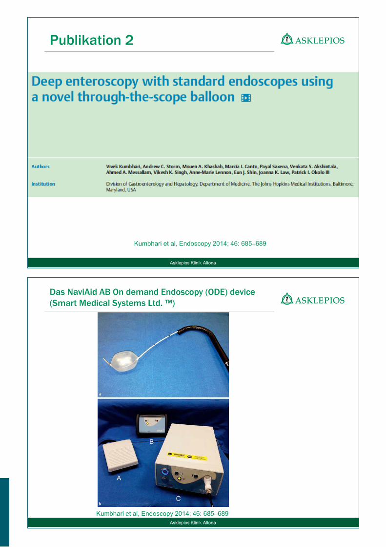


Recommended