2033-1
Joint ICTP/IAEA Advanced School on Dosimetry in DiagnosticRadiology and its Clinical Implementation
Claire-Louise Chapple
11 - 15 May 2009
Freeman HospitalNewcastle
UK
Introduction to Radiological Dosimetry

IAEAInternational Atomic Energy Agency
Introduction to Radiological Dosimetry
Claire-Louise ChappleRegional Medical Physics Department
Freeman HospitalNewcastle upon Tyne, UK
Joint ICTP-IAEA Advanced school on Dosimetry in Diagnostic Radiology: And its Clinical Implementation
11 - 15 May 2009; Miramare, Trieste, Italy

IAEA
Diagnostic Radiology : in the beginning…
• German physicist -Roentgen , 1885
• Discovered an "invisible light" or ray capable of passing through heavy paper
• X-rays would pass through the tissue of humans leaving the bones and metals visible
• Used clinically in US from 1896
• This ‘X-ray’ would pass through most substances casting shadows of solid objects on pieces of film
IAEA
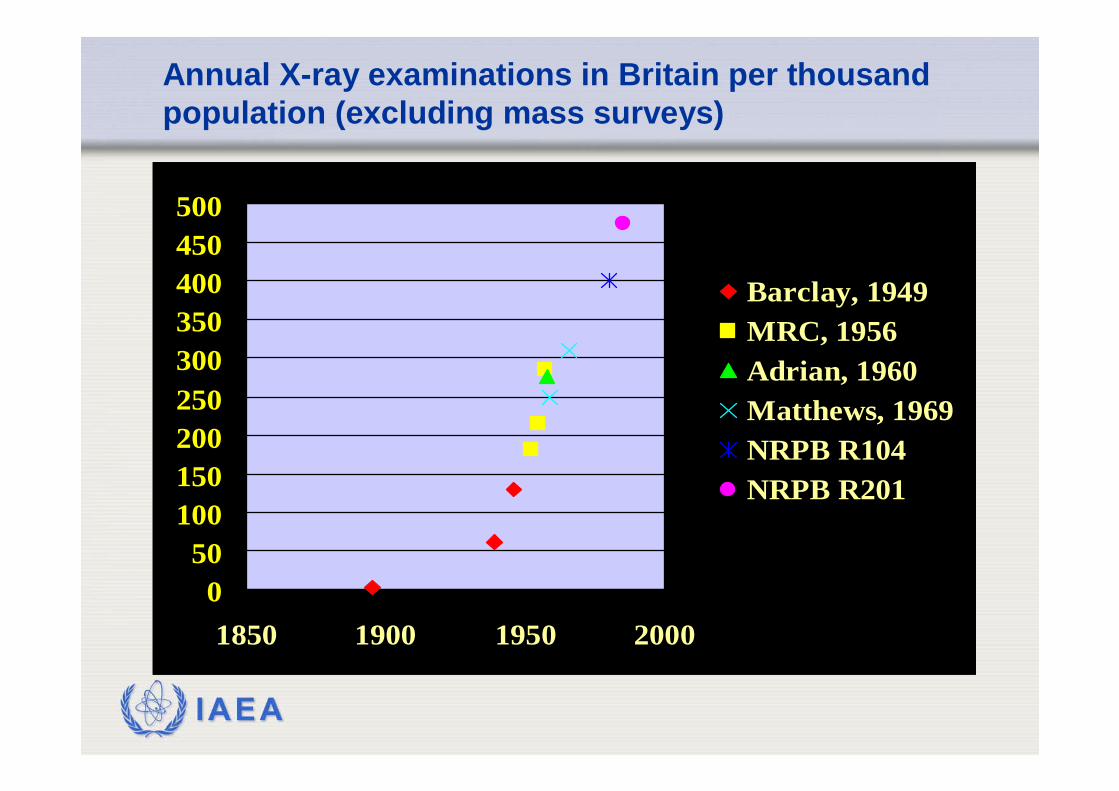
Annual X-ray examinations in Britain per thousand population (excluding mass surveys)
050
100150200250300350400450500
1850 1900 1950 2000
Barclay, 1949MRC, 1956Adrian, 1960Matthews, 1969NRPB R104NRPB R201

IAEA
Global X-ray figures
• In 2000 : 360 examinations / 1000 people• 1991-1995 : 330 exams / 1000 people
• 75% examinations in countries with Healthcare Level I
• 1% examinations in countries with Healthcare Level III & IV

IAEA
Developments in diagnostic radiology
• Screen-film radiography
• Fluoroscopy
• Computed tomography
• Digital fluorography
• Computed radiography
• Direct digital radiography

IAEA
Screen-film radiography
• Projection radiography
• Fixed X-ray beam
• Attenuated through patient
• Detected by screens ⇒ light photons
• Light detected by film ⇒ image (2D)
• Films can be displayed / transported / stored

IAEA

IAEA

IAEA
Developments in diagnostic radiology
• Screen-film radiography
• Fluoroscopy
• Computed tomography
• Digital fluorography
• Computed radiography
• Direct digital radiography

IAEA
Fluoroscopy
• Allows real-time imaging
• Often uses contrast media for functional imaging
• Uses image intensifier
• Displays images on TV screen
• Can take ‘snapshot’ images for storage
• Specialist applications (cardiology, gastrointestinal etc.)

IAEA

IAEA
Developments in diagnostic radiology
• Screen-film radiography
• Fluoroscopy
• Computed tomography
• Digital fluorography
• Computed radiography
• Direct digital radiography

IAEA
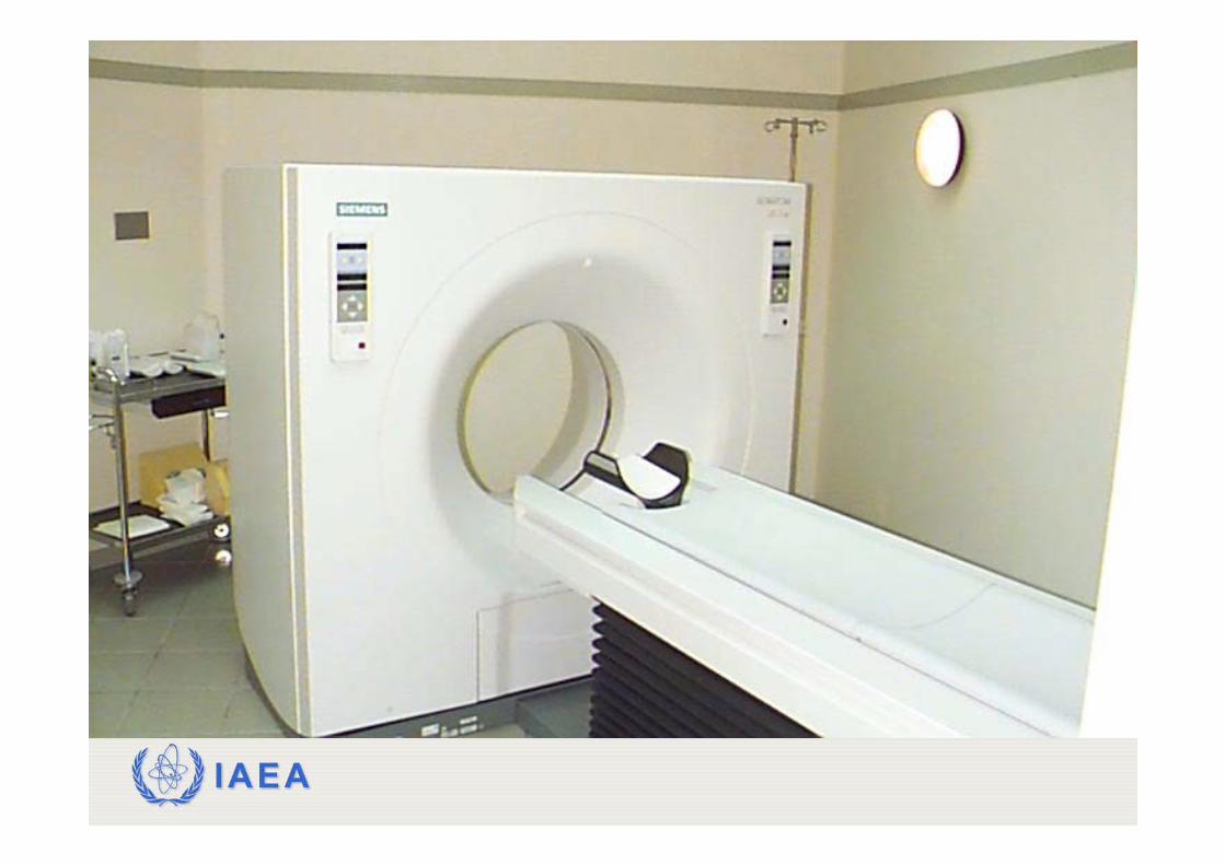
Computed Tomography
• Rotating fan beam of X-rays & detectors
• Computer reconstructs cross-sectional images (3D)
• Single slice ⇒ multi slice
• Axial scanning ⇒ helical scanning
• Real time imaging
IAEA

IAEA

IAEA

IAEA

IAEA
Developments in diagnostic radiology
• Screen-film radiography
• Fluoroscopy
• Computed tomography
• Digital fluorography
• Computed radiography
• Direct digital radiography

IAEA
Digital techniques
• Computed radiography (CR) – film replaced with storage phosphor plate
• Direct digital radiography (DDR) – active matrix detector converts directly to digital signal
• Interventional radiology now possible
• Improved image quality – What’s happened to doses?

IAEA
Radiation effects on humans
• Severe injury and death was an occupational hazard with early radiation workers
• Approx 300 fatalities in early workers(names on Hamburg monument)
• Becquerel developed skin burns and tumours
• Marie Curie died of leukemia at 67
Roentgen ray pioneer Mihran Kassabian (1870-1910)

IAEA
Radiation effects on humans
1. Stochastic effects
• Probability of effect occurring is proportional to dose(no threshold)
• Evidence: Japanese survivors, early radiologists,uranium miners, radium dial painters
• The stochastic effect of interest is carcinogenesis

IAEA
Prob
abili
ty o
f eff
ect
Dose
Stochastic effects
natural incidence
100%
reduced probability becauseof e.g. cell kill

IAEA
Radiation effects on humans
2. Deterministic effects
• No effect below a threshold dose
• Above threshold, severity of effect increases with dose
Carcinogenesis & deterministic effects are somatic effects – they affect the irradiated individual

IAEA
Typical threshold doses for deterministic effects
0.153.5
2.5
0.52.0
0.5
23
TestesTemporary sterilityPermanent sterilityOvaries
SterilityLens
OpacitiesCataractsBone marrow
Depression of haematopoiesisSkin
ErythemaTemporary epilation
Threshold dose (brief exp) GyTissue & Effect

IAEA
Seve
rity
or p
roba
bilit
y of
eff
ect
Dose
Deterministic effects
well-definedthreshold

IAEA
Radiation effects on humans
3. Genetic damage
• chromosome damage – breakage followed by faulty repairno convincing evidence
Problems of genetic risk assessment
• only gonad exposure is relevant• mutations may be recessive• mutations may be unstable

IAEA
Relative contributions to UK population dose
IAEA
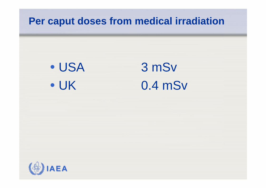
Per caput doses from medical irradiation
• USA 3 mSv• UK 0.4 mSv

IAEA
Relative contribution to UK frequency and collective dose
2015141311862
1.51.511
2.03.30.91.61.32.92.924255.60.925
Computed tomographyLumbar spineBarium enemaBarium mealIntravenous urographyAbdomenPelvisChestLimbs & jointsSkullThoracic spineDental
% Collective dose
% FrequencyExamination
IAEA
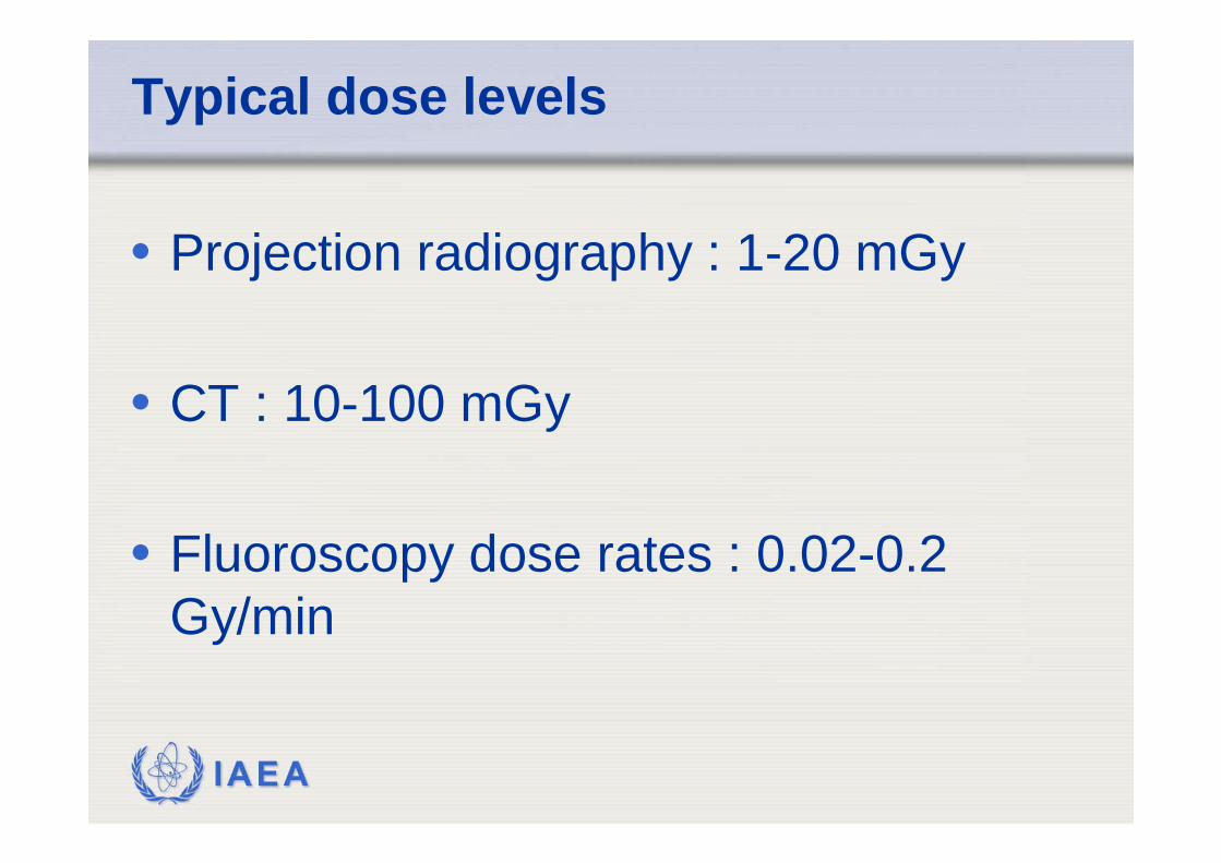
Typical dose levels
• Projection radiography : 1-20 mGy
• CT : 10-100 mGy
• Fluoroscopy dose rates : 0.02-0.2 Gy/min

IAEA
Quality assurance
• Diagnostic image quality required with as low as dose as possible
• Framework provided by WHO & BSS• Includes
• Measurement of physical X-ray parameters• Image quality assessment• Dose assessment
• BSS requires guidance levels for achievable doses
• EU requires diagnostic reference levels

IAEA
Two good reasons for dosimetry in x-ray diagnostics
• A tool for setting up and control of standards of a good practice• Quantities should be easy to measure
• A tool for estimation of radiation detriment and injury• Quantities should have a direct link to potential
risk from the exposure

IAEA
Two pillars of dosimetry in diagnostics
• Clinical needs• Correct diagnosis is the main goal of any x-ray
examination• Need to protect a patient• Balancing between necessary dose and quality
of image (important role of medical physicists)
• International system of measurements• Mechanism for consistency in radiation
dosimetry (role of SSDLs)

IAEA
Historical problems of dosimetry in x-ray diagnostics
• General radiography• Backscattered radiation included or not?• Absorbed dose or air kerma?• Material in which the measured quantity is
specified (air, PMMA, water, tissue)• Same symbols used for different quantities
(ESD)?• Different names used for the same quantity
• Similar problems occur for other modalities

IAEA
ICRU and IAEA documents
• Patient dosimetry for x rays used in medical imaging• ICRU Report 74, published in 2005
• Dosimetry in diagnostic radiology: an international code of practice• IAEA Technical Reports Series No 457, 2007
Recommended