Embed Size (px)
Citation preview

Zurich Open Repository andArchiveUniversity of ZurichMain LibraryStrickhofstrasse 39CH-8057 Zurichwww.zora.uzh.ch
Year: 2015
Hemophagocytosis in cutaneous autoimmune disease
Kerl, Katrin ; Wolf, Ingrid H ; Cerroni, Lorenzo ; Wolf, Peter ; French, Lars E ; Kerl, Helmut
Abstract: BACKGROUND: The significance of the histological visualization of hemophagocytosis in tis-sues depends on the context, varying from a nonspecific phenomenon to a characteristic or diagnosticfeature for certain disease entities. Hemophagocytosis is also one of the key features of macrophageactivation syndrome (MAS) (hemophagocytic syndrome) a potentially life-threatening complication ofunderlying conditions such as infections, malignancy, and autoimmune disorders. Clinical manifestationsof MAS are high fever, pancytopenia, liver dysfunction, and coagulopathy. These clinical symptoms aredue to an abnormal activation of the immune system in a strong association with the cytokine milieu. Thediagnosis of MAS may be easily missed; it is usually detected in the bone marrow, lymph node, liver, andspleen. Only few reports exist in the literature with histological description of cutaneous hemophagocy-tosis as a sign for MAS in patients with lymphoma and infection. In this report, the authors present theclinicopathological and immunohistochemical features of 3 patients with cutaneous hemophagocytosis,specifically erythrophagocytosis, associated with autoimmune disease, and discuss the relevance of thesefindings. OBSERVATION: The authors report 3 patients who developed cutaneous hemophagocytosisduring the course of an underlying autoimmune disorder. One patient suffered from dermatomyositis,the other 2 patients from systemic lupus erythematosus, whereby one of them was a 3-month old girlwith neonatal lupus erythematosus. The patient with dermatomyositis developed MAS according to thecurrent diagnostic criteria. Although the 2 other patients had an acute flare of their autoimmune diseasewith histological signs of cutaneous hemophagocytosis, they did not fulfill the complete criteria for a diag-nosis of MAS. Histiocyte proliferation and activation with increase of cytokines could be demonstrated byimmunohistology. CONCLUSIONS: This report is the first to describe hemophagocytosis in cutaneousbiopsies of patients with autoimmune diseases, associated with a complete or incomplete constellation ofMAS. Key players in this process are histiocytes/macrophages engaged in phagocytosis of erythrocytes.Hemophagocytosis observed in skin biopsies may be a diagnostic clue for MAS and an indicator for apotentially aggressive course of the underlying disease.
DOI: https://doi.org/10.1097/DAD.0000000000000166
Posted at the Zurich Open Repository and Archive, University of ZurichZORA URL: https://doi.org/10.5167/uzh-108685Journal ArticlePublished Version
Originally published at:Kerl, Katrin; Wolf, Ingrid H; Cerroni, Lorenzo; Wolf, Peter; French, Lars E; Kerl, Helmut (2015).Hemophagocytosis in cutaneous autoimmune disease. American Journal of Dermatopathology, 37(7):539-543.DOI: https://doi.org/10.1097/DAD.0000000000000166

ORIGINAL ARTICLE
Hemophagocytosis in Cutaneous Autoimmune Disease
Katrin Kerl, MD,* Ingrid H. Wolf, MD,† Lorenzo Cerroni, MD,† Peter Wolf, MD,† Lars E. French, MD,*
and Helmut Kerl, MD†
Background: The significance of the histological visualization of
hemophagocytosis in tissues depends on the context, varying from
a nonspecific phenomenon to a characteristic or diagnostic feature
for certain disease entities. Hemophagocytosis is also one of the
key features of macrophage activation syndrome (MAS) (hemo-
phagocytic syndrome) a potentially life-threatening complication
of underlying conditions such as infections, malignancy, and
autoimmune disorders. Clinical manifestations of MAS are high
fever, pancytopenia, liver dysfunction, and coagulopathy. These
clinical symptoms are due to an abnormal activation of the immune
system in a strong association with the cytokine milieu. The
diagnosis of MAS may be easily missed; it is usually detected in
the bone marrow, lymph node, liver, and spleen. Only few reports
exist in the literature with histological description of cutaneous
hemophagocytosis as a sign for MAS in patients with lymphoma
and infection. In this report, the authors present the clinicopatho-
logical and immunohistochemical features of 3 patients with
cutaneous hemophagocytosis, specifically erythrophagocytosis,
associated with autoimmune disease, and discuss the relevance of
these findings.
Observation: The authors report 3 patients who developed
cutaneous hemophagocytosis during the course of an underlying
autoimmune disorder. One patient suffered from dermatomyositis,
the other 2 patients from systemic lupus erythematosus, whereby
one of them was a 3-month old girl with neonatal lupus
erythematosus. The patient with dermatomyositis developed
MAS according to the current diagnostic criteria. Although the 2
other patients had an acute flare of their autoimmune disease with
histological signs of cutaneous hemophagocytosis, they did not
fulfill the complete criteria for a diagnosis of MAS. Histiocyte
proliferation and activation with increase of cytokines could be
demonstrated by immunohistology.
Conclusions: This report is the first to describe hemophagocytosis
in cutaneous biopsies of patients with autoimmune diseases,
associated with a complete or incomplete constellation of MAS.
Key players in this process are histiocytes/macrophages engaged in
phagocytosis of erythrocytes. Hemophagocytosis observed in skin
biopsies may be a diagnostic clue for MAS and an indicator for
a potentially aggressive course of the underlying disease.
Key Words: hemophagocytosis, macrophage activation syndrome,
autoimmune disease
(Am J Dermatopathol 2014;0:1–5)
INTRODUCTIONHemophagocytosis is the process of engulfment of blood
cells, in particular erythrocytes, by histiocytes through the cell-surface scavenger receptor CD1631 and is a histological featureof selected diseases, including infections and malignancy. He-mophagocytosis is only rarely observed in the skin, and itssignificance is variable. It may be a characteristic well-known histological feature of certain entities such as primarycutaneous g/d T-cell lymphoma, intravascular large B-celllymphoma, or Rosai–Dorfman disease, but it may also repre-sent a nonspecific phenomenon, for example, in the late stageof hemorrhagic disorders. When hemophagocytosis arises inthe context of systemic symptoms, it may also be a key featureindicative of a severe and potentially life-threatening conditioncharacterized by a hyperactivation of the immune system andexcessive secretion of cytokines. This syndrome has been re-ported under different names in the literature, including hemo-phagocytic syndrome and secondary hemophagocyticlymphohistiocytosis, and it belongs to the spectrum of macro-phage associated histiocytoses2,3; the primary or familial formis a genetic disorder with mutations in the perforin gene or ingenes implicated in the exocytosis of cytotoxic granules fromcytotoxic T lymphocytes.4 The secondary form occurs asa severe complication of an underlying disorder and was firstreported in patients with different types of infection5 andmalignancies, lymphomas in particular.6 Macrophage activa-tion syndrome (MAS) is the term applied for hemophagocyticsyndrome occurring in the context of autoimmune disease. In1991, Wong et al7 reported patients with active systemic lupuserythematosus, whose bone marrow biopsies revealed hemo-phagocytosis. Since then, MAS has been reported in the con-text of several autoimmune diseases including systemic onsetjuvenile rheumatoid arthritis, systemic lupus erythematosus,dermatomyositis, progressive systemic sclerosis, and mixedconnective tissue disease.8–13 It has also been observed in Stilldisease, a disorder now recognized to belong to the group ofauto-inflammatory diseases involving a dysregulation of theinnate immune system.14,15
The clinical symptoms of MAS are due to an uncon-trolled and excessive activation of the immune system witha proliferation of stimulated histiocytes and lymphocytes andenhanced secretion of pro-inflammatory cytokines such tumornecrosis factor alpha (TNF-a), interferon-g (IFN-g),interleukin-6 (IL-6), and IL-1b.16 This cytokine storm leads
From the *Department of Dermatology, University Hospital of Zurich, Zürich,Switzerland; and †Department of Dermatology, Medical University ofGraz, Graz, Austria.
The authors declare no conflicts of interest.Reprints: Katrin Kerl, MD, Department of Dermatology, University Hospital
of Zurich, CH-8091 Zurich, Switzerland (e-mail: [email protected]).© 2014 Lippincott Williams & Wilkins
Am J Dermatopathol � Volume 0, Number 0, Month 2014 www.amjdermatopathology.com | 1
Copyright ª Lippincott Williams & Wilkins. Unauthorized reproduction of this article is prohibited.

to a sepsis-like clinical presentation with high fever, hepatos-plenomegaly, cytopenia, disseminated intravascular coagula-tion, and neurological symptoms. Characteristic laboratoryfindings of MAS include anemia, and thrombocytopenia, hy-pofibrinogenemia, elevation of ferritin, and liver enzymes.Hemophagocytosis in different organs is an important crite-rion for the diagnosis of MAS, although its presence or theproof of its existence is not mandatory for establishing thediagnosis. Specific diagnostic criteria for MAS have beensuggested17,18 and are summarized in Table 1.
We demonstrate for the first time hemophagocytosis(erythrophagocytosis) in skin specimens of 3 patients withautoimmune disease. One patient with dermatomyositisshowed acute and severe aggravation of his illness and fulfilledthe criteria for the diagnosis of MAS. In 2 patients withsystemic lupus erythematosus, hemophagocytosis in the skinwas not associated with the full clinical presentation of MAS.
OBSERVATIONS
Clinical Findings
Patient 1
A 62-year-old patient presented with high fever,lethargy, muscle weakness, excessive edema with violaceousdiscoloration, echymosis, ulceration and crusting of theperiorbital regions (Fig. 1). Gottron papules were obviousover knuckles and interphalangeal joints. Internal investiga-tions and laboratory tests revealed: elevation of muscle andliver enzymes, pancytopenia, splenomegaly, lymphadenopa-thy, elevated ferritine, and hypertriglyceridemia.
Electromyography showed signs of myositis. Bonemarrow biopsy failed to exhibit macrophage proliferationand hemophagocytosis. Based on these findings together withthe skin biopsy (see below), a diagnosis of dermatomysitiscomplicated by MAS was made. The general condition of thepatient deteriorated rapidly, and he died a few days afterhospital admission.
Patient 2A 29-year-old woman with long standing systemic
lupus erythematosus displayed annular and polycycliclesions with scaly borders on the trunk and extensor surfacesof the arms (Fig. 2). In addition, a butterfly rash on the faceand nail fold erythema could be observed. She complainedalso of fatigue and joint pain. Laboratory tests revealedpositive results for antinuclear antibodies, anti-dsDNA
TABLE 1. Diagnostic Guidelines for Macrophage ActivationSystem Complicating Systemic Juvenile Idiopathic Arthritis
Laboratory criteria
Decreased platelet count (#262 · 109/L)
Elevated levels of aspartate aminotransferase (.59 U/L)
Decreased white blood cell count (#4.0 · 109/L)
Hypofibrinogenemia (#2.5 g/L)
Clinical criteria
Central nervous system dysfunction (irritability, disorientation, lethargy,headache, seizures, coma)
Hemorrhages (purpura, easy bruising, mucosal bleeding)
Hepatomegaly ($3 cm below the costal arch)
Histopathological criterion
Evidence of macrophage hemophagocytosis in the bone marrow aspirate
The diagnosis of MAS requires the presence of any 2 or more laboratory criteria or
of any 2 or 3 or more clinical and/or laboratory criteria.
Adapted from Ravelli et al. J Pediatr. 2005.16,17 Adaptations are themselves works
protected by copyright. So in order to publish this adaptation, authorization must be
obtained both from the owner of the copyright in the original work and from the owner
of copyright in the translation or adaptation.
FIGURE 1. Patient 1. Autoimmune hemophagocytic syn-drome. A 62-year-old man with dermatomyositis complicatedby MAS. Excessive swelling, violet color, hemorrhage, andcrusting of the eyelids and the periorbital region.
FIGURE 2. Patient 2. Systemic lupus erythematosus showingannular and polycyclic lesions with a scaly border on the arm.
Kerl et al Am J Dermatopathol � Volume 0, Number 0, Month 2014
2 | www.amjdermatopathology.com � 2014 Lippincott Williams & Wilkins
Copyright ª Lippincott Williams & Wilkins. Unauthorized reproduction of this article is prohibited.
antibodies, anti-Ro/SSA antibodies, proteinuria, and lowserum complement levels.
Patient 3A 3-month-old girl presented with disseminated
reddish-brown lesions on the head and trunk. Periorbitalscaly erythematous confluent patches were also observed(“raccoon eye” appearance). Other relevant findings wereantinuclear antibodies, anti-Ro/SSA, and anti-La/SSB posi-tive; atrioventricular heart block, first degree. Diagnosis wasneonatal lupus erythematosus.
Histological and Immunohistological FindingsSkin biopsy specimens were embedded in paraffin and
Hematoxilin-Eosin-staining was performed. In addition, thefollowing markers used for the assessment of monocyte–mac-rophage activation were studied: CD163 (for M1 and M2,macrophage subpopulations, Leica, clone 10D6 Germany),
TNF-a (R&D Systems, mouse monoclonal, clone 28401,England, United Kingdom), IFN-g (rabbit polyclonal; Abcam,England, United Kingdom), IL-6 (rabbit polyclonal; Abcam),and IL-1b (rabbit polyclonal; Abcam) were performed in theskin biopsies of patients 1 and 2.
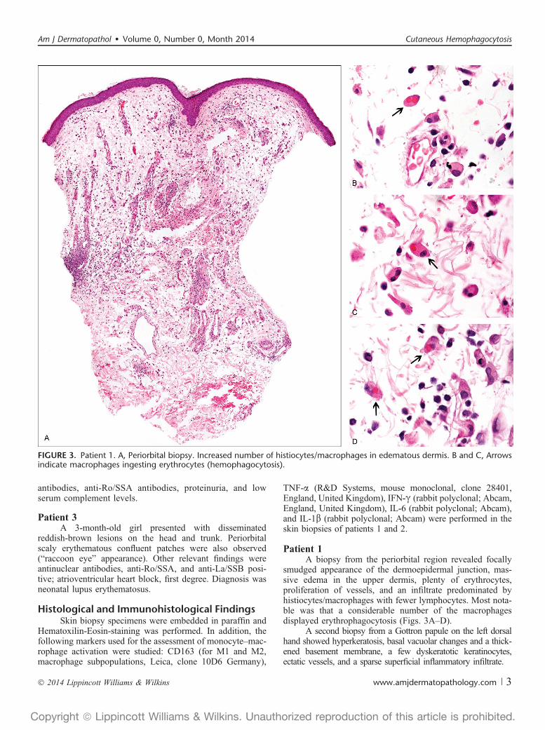
Patient 1A biopsy from the periorbital region revealed focally
smudged appearance of the dermoepidermal junction, mas-sive edema in the upper dermis, plenty of erythrocytes,proliferation of vessels, and an infiltrate predominated byhistiocytes/macrophages with fewer lymphocytes. Most nota-ble was that a considerable number of the macrophagesdisplayed erythrophagocytosis (Figs. 3A–D).
A second biopsy from a Gottron papule on the left dorsalhand showed hyperkeratosis, basal vacuolar changes and a thick-ened basement membrane, a few dyskeratotic keratinocytes,ectatic vessels, and a sparse superficial inflammatory infiltrate.
FIGURE 3. Patient 1. A, Periorbital biopsy. Increased number of histiocytes/macrophages in edematous dermis. B and C, Arrowsindicate macrophages ingesting erythrocytes (hemophagocytosis).
Am J Dermatopathol � Volume 0, Number 0, Month 2014 Cutaneous Hemophagocytosis
� 2014 Lippincott Williams & Wilkins www.amjdermatopathology.com | 3
Copyright ª Lippincott Williams & Wilkins. Unauthorized reproduction of this article is prohibited.

Patient 2A biopsy from the left arm showed characteristic
epidermal changes of lupus erythematosus. Striking featureswere a marked edema in the upper dermis with hemorrhageand numerous macrophages engaged in phagocytosis oferythrocytes (Figs. 4A–C).
Imunohistochemical analysis [performed in patient 1 onthe face biopsy (Fig. 5A), patient 2 on the arm biopsy] revealednumerous CD163-positive macrophages within the upperdermis. In addition, a high expression of IL-6, TNF-a, IFN-g,and IL-1b could be detected in these cells.
Patient 3A biopsy from the trunk featured a thinned epidermis
with scale crusts, basal vacuolar alteration, necrotic kerati-nocytes, dermal edema, erythrocyte extravasation, andlymphohistiocytic infiltrates with neutrophils. Again, mostremarkable was the identification of macrophages containingphagocyted red blood cells.
DISCUSSIONHere, we describe the clinical, histopathological, and
immunohistochemical features of 3 patients with cutaneoushemophagocytosis in the context of underlying autoimmunedisease (systemic lupus erythematosus and dermatomyositis),associated with MAS in 1 case.
Hemophagocytosis is a phenomenon that is only rarelyobserved in the skin, and its significance is dependent on theclinicopathological context. It is a hallmark feature of MAS,
a potentially life-threatening syndrome complicating malig-nancies, infections, and autoimmune disease.
Although hemophagocytosis is typical for MAS, its merepresence is not automatically synonymous with MAS. Hemo-phagocytosis may be absent in MAS and Hemophagocyticsyndrome, especially in the initial stages. However, it mayoccur as a nonspecific phenomenon in various context as bloodtransfusions, infection, autoimmune disease, and other causesof red blood cell destruction. Recently, 2 articles reportedperivascular hemophagocytosis with signs of vasculitis in skinbiopsies of patients without the full clinical presentation ofMAS. The authors discuss the possibility of hemophagocytosisas a sign of incomplete MAS versus it being simply a non-specific sign of leucocytoclastic vasculitis.19,20
In our patients, the skin biopsies were performed at themoment of an acute flare in the activity of their underlyingautoimmune disease. One of the patients was then diagnosedwith MAS; the finding of hemophagocytosis in the skinbiopsy of this patient was an important additional clue to thediagnosis.
The significance of the finding of hemophagocytosis inthe skin biopsies of our 2 patients with systemic lupuserythematosus, who did not have all criteria required for theclassification as MAS is more difficult to evaluate. It might beinterpreted as a sign of disease activity as it was associatedwith a flare. Further observations are required to clarify thesignificance of the cutaneous hemophagocytic process inautoimmune disease without full clinical evidence of MAS.
Interestingly, a proliferation of a significant number ofCD163-positive histiocytes/macrophages could be demonstrated
FIGURE 4. Patient 2. Biopsy fromthe arm. A, Markedly edematouspapillary dermis with proliferation ofhistiocytes and extravasated eryth-rocytes. B and C, Arrows indicatehemophagocytosis. Note appositionof erythrocytes to macrophage inFig. 4C.
Kerl et al Am J Dermatopathol � Volume 0, Number 0, Month 2014
4 | www.amjdermatopathology.com � 2014 Lippincott Williams & Wilkins
Copyright ª Lippincott Williams & Wilkins. Unauthorized reproduction of this article is prohibited.

within the dermis. CD163 is a hemoglobin–haptoglobin scav-enger receptor and a lineage specific marker for histiocytes inMAS. The soluble form of CD163 has been identified as a poten-tial marker for the diagnosis of MAS.1
We also analyzed the pattern of cytokine expression inthe skin biopsy specimens of our patients 1 and 2 and foundoverproduction of cytokins such as TNF-a, IFN-g, IL-6, andIL-1b, showing evidence of activation of histiocytes. Thesecytokines have been demonstrated to play a crucial role in thepathogenesis of MAS16 and can induce hemophagocytosis.
In conclusion, this is the first description of hemopha-gocytosis in skin biopsies of patients with autoimmunediseases, delivering also evidence on the involvement ofcytokine-producing activated macrophages. The diagnosisof MAS is often challenging and may be delayed or evenmissed because of the fact that there may be overlappingclinical features with the underlying autoimmune disorder.The presence of hemophagocytosis, without being specific
or mandatory for the diagnosis of MAS, is a very importantclue and is usually observed in biopsies of lymphoid tissues.However, hemophagocytosis observed in skin biopsies canalso be a sign of MAS. Demonstration of hemophagocytosisin skin biopsies may help to establish the diagnosis of MASand also potentially indicate a more severe course of theunderlying disorder. It should also provide new insights inthe mechanisms of host defense and the pathogenesis ofautoimmune disease.
REFERENCES1. Schaer DJ, Schleiffenbaum B, Kurrer M, et al. Soluble hemoglobin-
haptoglobin scavenger receptor CD163 as a lineage-specific marker inthe reactive hemophagocytic syndrome. Eur J Haematol. 2005;74:6–10.
2. Ramanan AV, Schneider R. Macrophage activation syndrome—what’sin a name! J Rheumatol. 2003;30:2513–2516.
3. Janka GE, Lehmberg K. Hemophagocytic lymphohistiocytosis: patho-genesis and treatment. Hematology Am Soc Hematol Educ Program.2013;2013:605–611.
4. Ueda I, Kurokawa Y, Koike K, et al. Late-onset cases of familial hemo-phagocytic lymphohistiocytosis with missense perforin gene mutations.Am J Hematol. 2007;82:427–432.
5. Risdall RJ, McKenna RW, Nesbit ME, et al. Virus-associated hemopha-gocytic syndrome: a benign histiocytic proliferation distinct from malig-nant histiocytosis. Cancer. 1979;44:993–1002.
6. Kadin ME, Kamoun M, Lamberg J. Erythrophagocytic T gamma lym-phoma: a clinicopathologic entity resembling malignant histiocytosis. NEngl J Med. 1981;304:648–653.
7. Wong KF, Hui PK, Chan JK, et al. The acute lupus hemophagocyticsyndrome. Ann Intern Med. 1991;114:387–390.
8. Sekigawa I, Suzuki J, Nawata M, et al. Hemophagocytosis in autoim-mune disease. Clin Exp Rheumatol. 2001;19:333–338.
9. Stéphan JL, Zeller J, Hubert P, et al. Macrophage activation syndromeand rheumatic disease in childhood: a report of four new cases. Clin ExpRheumatol. 1993;11:451–456.
10. Atteritano M, David A, Bagnato G, et al. Haemophagocytic syndrome inrheumatic patients. A systematic review. Eur Rev Med Pharmacol Sci.2012;16:1414–1424.
11. Kumakura S, Ishikura H, Kondo M, et al. Autoimmune-associated he-mophagocytic syndrome. Mod Rheumatol. 2004;14:205–215.
12. Takahashi K, Kumakura S, Ishikura H, et al Reactive hemophagocytosisin systemic lupus erythematosus. Intern Med. 1998;37:550–553.
13. Deane S, Selmi C, Teuber SS, et al. Macrophage activation syndrome inautoimmune disease. Int Arch Allergy Immunol. 2010;153:109–120.
14. Kumakura S, Ishikura H, Munemasa S, et al. Adult onset Still’s diseaseassociated hemophagocytosis. J Rheumatol. 1997;24:1645–1648.
15. Coffernils M, Soupart A, Pradier O, et al. Hyperferritinemia in adultonset Still’s disease and the hemophagocytic syndrome. J Rheumatol.1992;19:1425–1427.
16. Billiau AD, Roskams T, Van Damme-Lombaerts R, et al. Macrophageactivation syndrome: characteristic findings on liver biopsy illustratingthe key role of activated, IFN-gamma-producing lymphocytes and IL-6-and TNF-alpha-producing macrophages. Blood. 2005;105:1648–1651.
17. Ravelli A, Magni-Manzoni S, Pistorio A, et al. Preliminary diagnosticguidelines for macrophage activation syndrome complicating systemicjuvenile idiopathic arthritis. J Pediatr. 2005;146:598–604.
18. Davì S, Consolaro A, Guseinova D, et al; MAS Study Group. An inter-national consensus survey of diagnostic criteria for macrophage activa-tion syndrome in systemic juvenile idiopathic arthritis. J Rheumatol.2011;38:764–768.
19. Valentín SM, Montalván E, Sánchez JL. Perivascular hemophagocytosis:report of 2 cases and review of the literature. Am J Dermatopathol. 2010;32:716–719.
20. Draper NL, Morgan MB. Dermatologic perivascular hemophagocytosis:a report of two cases. Am J Dermatopathol. 2007;29:467–469.
FIGURE 5. Immunohistological analysis. A, High proportion ofCD163-positive activated histiocytes/macrophages (patient 1).B, Strong expression of IL-1b and TNF-a in histiocytes/macrophages (patient 2).
Am J Dermatopathol � Volume 0, Number 0, Month 2014 Cutaneous Hemophagocytosis
� 2014 Lippincott Williams & Wilkins www.amjdermatopathology.com | 5
Copyright ª Lippincott Williams & Wilkins. Unauthorized reproduction of this article is prohibited.